Fujiki et al. Nanoscale Research Letters 2011, 6:347 doc
Bạn đang xem bản rút gọn của tài liệu. Xem và tải ngay bản đầy đủ của tài liệu tại đây (1.55 MB, 8 trang )
NANO EXPRESS Open Access
STM-induced light emission from thin films of
perylene derivatives on the HOPG and Au
substrates
Aya Fujiki
1*
, Yusuke Miyake
1
, Yasushi Oshikane
1
, Megumi Akai-Kasaya
1
, Akira Saito
1,2
and Yuji Kuwahara
1
Abstract
We have investigated the emission properties of N,N’-diheptyl-3,4,9,10-perylenetetracarboxylic diimide thin films by
the tunneling-electron-induced light emission technique. A fluorescence peak with vibronic progressions with large
Stokes shifts was observed on both highly ordered pyrolytic graphite (HOPG) and Au substrates, indicatin g that the
emission was derived from the isolated-molecule-like film condition with sufficient π-π interaction of the perylene
rings of perylenetetracarb oxylic diimide molecules. The upconversion emission mechanism of the tunneling-
electron-induced emission was discussed in terms of inelastic tunneling including multiexcitation processes. The
wavelength-selective enhanced emission due to a localized tip-induced surface plasmon on the Au substrate was
also obtained.
Introduction
Control of molecular emission from organic materials
has attracted much attention owing to its potential
applications not only in basic molecular science but also
in research on soft material devices such a s organic
light-emitting diodes (OLEDs) and biosensors [1-4].
Scanning-tunneling-microscope-induced light emission
(STM -LE) spectroscopy is highly effective for characte r-
izing the optical and electronic properties of nanoscale
materials such as organic single molecules or thin films
at the atomic scale. However, it involves serious analyti-
cal difficulties in receiving extremely weak signals from
the objective materials. To overcome such difficulties, it
is promising to combine STM-LE spectroscopy wit h
plasmon enhancement on surfaces. Surface plasmons at
the interface between metallic and dielectric media gen-
erate an intense electromagnetic field on the surface,
which provides an efficient enhancement field for some
optical processes such as the fluorescence/phosphores-
cence emission and optical absorption of organic mate-
rials on a metal surface [1]. We have first observed the
fluorescence of Cu phthalocyanine under enhancement
utilizing an STM-tip-induced plasmon (TIP) [5]. For
light emission from single molecules, Qiu et al. [6]
reported light emission from individual Zn(II)-etiopor-
phyrin I molecules adsorbed on Al
2
O
3
/NiAl(110), in
which an o xide buffer layer is used to prevent fluores-
cence quenching and disturbance of pronounced plas-
mon emission [7-9]. They explained that the spectra
were due to the de-excitation of excited anion states
resulting from hot electron injection. The plasmon
enhancement effect is also expected to be applied to the
development of light-emitting diodes [2,10]. Recently,
we have developed a high-efficiency OLED including Au
nanoparticles owing to the enhancement effect of loca-
lized surface plasmons on metal nanostructures [10].
Perylenetetracarboxylic diimide (PTCDI) and its deri-
vatives are n-type semiconductors [11,12], used in var-
ious optoelectronic devices such as thin-film transistors
[13], photovoltaic [14], and light-emitting diodes [15].
PTCDI molecules have been expected as a material of
single-molecule devices [16] because of the high thermal
and photostabilities of PTCDI. In this study, we have
studied the STM-LE from N,N’-dihe ptyl-3,4,9,10-peryle-
netetracarboxylic diimide (PTCDI-C7) thin films on
HOPG and Au substrates. We elucidated the intrinsic
optical properties of PTCDI-C7 in terms of the STM-LE
spectra o n the HOPG substrate comp ared with the
absorption and photoluminescence (PL) spectra, and
demonstrated the wavelength control of enhanced
* Correspondence:
1
Department of Precision Science & Technology, Graduate school of
Engineering, Osaka University, 2-1 Yamada-oka, Suita 565-0871, Japan
Full list of author information is available at the end of the article
Fujiki et al. Nanoscale Research Letters 2011, 6:347
/>© 2011 Fujiki et al; licensee Springer. This is an Open Access article distributed under the terms of the Creative Commons Attribution
License ( enses/by/2.0), which permits unrestricted use, distribution, and reproduc tion in any medium,
provided the origin al work is properly cited.
molecular luminescence, i.e., the selective enhancement
of the resonant wavelength of PTCDI-C7 through TIP
enhancement effects on the Au substrate. We also dis-
cussed the emission mechanism of upconversion
fluorescence.
Experimental
PTCDI-C7 was synthesized by a modification of a pre-
viously reported method [17,18]. A freshly cleaved
HOPG and Au thin films evaporated on mica were
used as the substrates. PTCDI-C7 thin films were pre-
pared by spin-coating 0.4 mg/ml PTCDI-C7 solution
in 1-tetradecene at a spin velocity of 1000 rpm, fol-
lowed by rinsing with the solvent and drying in
vacuum desiccators for 24 h. The film thickness was
about 5-10 nm, which was determined by comparing
the PL intensities o f the PTCDI-C7 thin films fabri-
cated by the spin coating method with those fabricated
by evaporation in vacuum with thicknesses of 5, 10,
15, and 20 nm, which were estimated using a thickness
monitor. STM (Digital Instruments Co. Ltd., USA,
Nanoscope IIIa) measurement was carried out at room
temperature under ambient conditions and a mechani-
cally sharpened Pt/Ir tip was used. The collected
photons were guided to a phot omultiplier tube (Hama-
matsu Photonics, Japan, R-649S) using an optical fiber
to obtain a light intensity map (the dark count was
less than 1 count per second (cps) at 253 K; the wave-
length detection range was 300-850 nm). To acquire
optical spectra, a grating spectrometer (Roper Scienti-
fic, USA, SpectraPro-300i) with a liquid-N
2
-cooled
charge-coupled device camera (Roper Scientific, USA,
Spec-10:100B/LN; the detection range was 200-1100
nm) was employed. The absorption and photolumines-
cence (PL) spectra of PTCDI-C7 were obtained using a
UV-visible/NIR spectrophotometer (Hitachi High-
Technologies Co., Japan, U-3010) and a custom-built
system with an argon-ion laser (Edmond Optics, USA,
Multi-Line 150 mW) at 514 nm, respectively.
Results and discussion
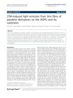
Figure 1a,c shows STM topographic images of the
PTCDI-C7 thin films on the HOPG and Au substrates,
and Figure 1b,d shows photon intensity maps corre-
sponding to the STM images in Figure 1a,c, respectively.
These pairs of topographic and photon images were
obtained simultaneously in the constant-current mode.
The surface roughnesses of the molecular films in
Figure 1a,c were induced by the surface morphologies of
the pristine substrates: The surface of the HOPG sub-
strate was atomically flat and that of the as-deposited
Au substrate showed a relatively large corrugation. In
both the substrates, it was found that the molecules are
not well crystallized but show an amorphous behavior.
Homogeneous emissions were observed from the entire
scanned area in both Figure 1b,d, so that homogeneous
and smooth PTCDI-C7 thin films were formed on both
the substrates, which showed a good correspondence of
the STM topographic images. In the STM-LE measure-
ment in this study, the tip was placed in contact with
the thin film under our high-current condition; as a
result, the tips might have swept molecules during the
scan, in which tunneling electrons directly passed
through the thin film to the substrate without an air gap
between the tip and the sample.
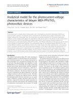
Figure 2 shows the STM-LE spectrum obtained from
the PTCDI-C7 thin film (blue line) on the HOPG sub-
strate. The spectrum shown in b lack represents the
result of a 1-tetradecene (solvent) thin film without
PTCDI-C7 molecules on the HOPG substrate. In both
the spectra, the sample bias voltage, tunneling current,
and accumulation time were fixed at +2.2 V, 20 nA, and
15 min, respectively. Both the spectra were acquired
with the tip scanning 50 × 50 nm
2
of the surface. No
emission was observed from the 1-tetradecene thin film;
in contrast, sufficient emission was observed from the
PTCDI-C7 thin film on the HOPG substrate. To the
best of our knowledge, there are only a few STM-LE
studies of the HOPG substrate, since there is no surface
plas mon mode on the HOPG surface in the visible light
wavelength region and plasmon enhancement cannot b e
effectively used to obtain meaningful STM-LE intensities
from adsorbed molecules. We consid ered that the suff i-
cient intensity of the STM-LE from the PTCDI-C7 thin
film on the HOPG substrate is caused by a high quan-
tum yield of the radiative decay of PTCDI-C7 (93%
[19]). Uehara and Ushioda [20] reported the STM-LE of
asinglemoleculeofrhodamine6Gadsorbedonthe
HOPG surface. In their study, the quantum yield of
light emission via the transition of an electron from the
lowest unoccupied molecular orbital to the highest
occupied molecular orbital was also high (95% [21]).
Note that we obtained no light emission from the
PTCD I-C7 thin film fabricated by deposition in vacuum
on the HOPG surface, suggesting that the morphology
of molecular thin films affected by fabricatio n processes
affects the emission efficiency in STM-LE. A strong visi-
ble light is radiate d by TIP on the metal substrates such
as Au, Ag, and Cu. TIP emission is superimposed on
the emission from the adsorbed molecules, so that it is
difficult to extract the true spectra of target molecules
on metal surfaces. Thus, the STM-LE spectra of
adsorbed molecules on an HOPG substrate with no
plasmon resonance in the visible spectral range can be
used to analyze the intrinsic molecular emission without
any disturbance of TIP emission, although the interac-
tion of the molecules with the HOPG surface must be
taken into account.
Fujiki et al. Nanoscale Research Letters 2011, 6:347
/>Page 2 of 8
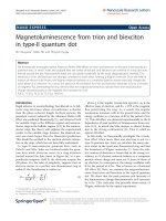
Figure 3 shows the absorption spectra of PTCDI-C7
dissolved in 1-tetradecene (0.4 mg/ml, solid line) and of
the PTCDI-C7 thin film fabricated on an indium-tin
oxide (ITO) substrate using the spin coating method
(dashed line), in which the same method of sample pre-
paration as that for the PTCDI-C7 thin film on the
HOPG substrate was employed. In the spectrum of
PTCDI-C7 solution, we found three distinct peaks at
455, 485, and 520 nm. Thes e peaks are attributed to the
S
1
(0-0) transition (S
1
is a first singlet excited state of
PTCDI-C7, numbers in parentheses denote the vibronic
levels in the initial and final states) and its vibronic pro-
gressions with a n energetic distance betwe en the peaks
of approximately 0.18 eV. The excitation energy from
the ground (S
0
) state to the S
1
state of PTCDI and its
derivatives is 2.36 eV [22], and the energy intervals of
the peaks correspond to the energy of the benzene-ring
stretch oscillation of perylene (0.15 eV [23]). The
obtained absorption spectrum of PTCDI-C7 solution
was in good agreement with those in a pr evious report
on perylene derivatives such as N,N’-dimethyl-P TCDI
and N,N’ -bis(2,6-xylyl)-PT CDI in dilute solutions by
Schouwink et al. [24]. It is considered that the spectrum
of PTCDI-C7 solution in Figu re 3 is governed by mono-
mer absorption and not ascribed to dimers or larger
aggr egates [25], which could be a result of the relatively
long alkane substituents of PTCDI-C7 that prevent their
aggregation through their steric effect. For the PTCDI-
C7 thin film, in contrast, the spectrum became highly
broadened with an additional small peak at 565 nm
Figure 1 STM topographic images and photon integration maps of PTCDI-C7 thin films. STM topographic images on (a) HOPG and (c) Au
substrates, and photon integration maps on (b) HOPG and (d) Au substrates. Pairs of a topographic image and a photon map ((a) and (c), (b)
and (d)) were obtained simultaneously (Vs = +2.2 V, It = 20 nA).
Fujiki et al. Nanoscale Research Letters 2011, 6:347
/>Page 3 of 8
compared with that of PTCDI-C7 solution. The peak
broadening and the emergence of the new peak are
caused by the strong π-π interaction within molecular
aggregates, and by the formation of dimers [22,24] or a
crystal phase [24,25] due to the strong molecular stack-
ing between PTCDI skeletons, respectively.
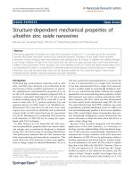
Figure 4 shows the PL spectrum of the PTCDI-C7
thin film on the HOPG substrate (green line). The
STM-LE spectrum of the PTCDI-C7 thin film on the
HOPG substrate is shown in blue in the figure. It was
found that the PL spectrum had a pronounced peak at
680 nm and shoulders at 625 and 750 nm. The obtained
peaks of the PL spectrum were ascribed to the vibronic
progressions related to the S
1
(0-0) transition at 520 nm,
as shown in the absorption spectra in Figure 3, because
theenergyintervalsoftheobservedPLpeakswere
approximately 0.17 eV corresponding to the stretching
energy of perylene rings, as mentioned earlier. The
peaks at 625, 680, and 750 nm were assigned to the S
1
(0-2), S
1
(0-3), and S
1
(0-4) transitions with respect to the
S
1
state, res pectively. The PL spectrum included a large
Stokes shift of approximately 100 nm compared with
the absorption spectra. Note that the PL spectra of the
PTCDI-C7 thin films on the ITO and HOPG substrates
almost coincided with each other in terms of peak
shape and position (data not sho wn), indicating that the
electronic configurations, which are related to the
optical properties of the PTCDI-C7 thin films on the
ITO and HOPG substrates, were similar to each other.
In the STM-LE spectrum of the PTCDI-C7 thin film,
some pronounced pe aks were observed at 550-950 nm.
The peaks of STM-LE were explained by the vibronic
progressions related to the S
1
transition because the
peak positions in the PL and STM-LE spectra almost
coincided with each other. One can see that the STM-
LE spectrum has a broad band up to 900 nm and that
the peaks including higher indexes of progressions (up
to the S
1
(0-5) transition at 860 nm) are more discrimin-
able than those of the PL spectrum. This result would
be derived from our STM-LE condition, e.g., with a
local electric field between the STM tip and the sub-
strate surface or with structural deformation of the
molecules scratched by a scanning STM tip, which
affects the transition probability of electronic excitation
or radiation.
Our interest in both PL and STM-LE spectra was
aroused by our observation of distinct vibronic progres-
sions, simila r to the case of the isolated molecular cond i-
tion, even in the thin-film c onfiguration of PTCDI-C7
where a moderate intermolecular interaction appeared on
the fluorescence spectra in the form of a large (approxi-
mately 100 nm) Stokes shift. We assumed that PTCDI-C7
molecules had a poorly crystalline orientation/distribution
Figure 2 STM-LE spectra of PTCDI-C7 thin film (blue line) and
solvent molecules (black line) on HOPG substrate (Vs = +2.2 V,
It = 20 nA, acquisition time = 15 min). Both spectra are
smoothened by averaging the 10 nearest points of the raw data.
Figure 3 Absorption spectra of PTCDI-C7 dissolved in 1-
tetradecene (0.4 mg/ml) (solid line) and of PTCDI-C7 thin film
fabricated using the spin coating method on ITO (dashed line),
whose intensities are normalized at 520 nm wavelength.
Fujiki et al. Nanoscale Research Letters 2011, 6:347
/>Page 4 of 8
in the thin film fabricated by the spin coating method due
to the steric effect of long alkane substituents, which led
them to h ave a quasi-isolated molecular condition in the
thin film structure in terms of the perylene-ring-stretching
vibration, although proper π-π stacking exhibiting a large
Stokes shift and peak broadening in the spectra of the thin
film structures remained. How to extend the fact that the
electronic configurations of the PTCDI-C7 molecules are
modified by the distribution in the thin film, such as
induction, conjugation, and electrostatic, remains contro-
versial. To evaluate such electronic effects, other experi-
ments, such as photoemission spectroscopy and scanning
tunneling spectroscopy, should be required.
Figure 5 shows the STM-LE spectra of the PTCDI-C7
thin films on the Au (red line) and HOPG (blue line)
substrates. Two spectra were obtained under the same
STM conditions (Vs = +2.2 V, It = 20 nA). The e mis-
sion on the Au substrate originated from the PTCDI-C7
molecules because the peak positions for the Au sub-
strate were consistent with those for the HOPG sub-
strate. It should be noticed that the emission intensities
of the peaks at 750 and 860 nm were significantly
enhanced about fivefold, whereas the peak intensities at
625 and 680 nm were unchanged. Such a selective
enhancement of the emission peaks can be explained by
the resonance matching with the TIP mode on the Au
substrate. In general, the wavelength of the emission by
TIP strongly depends on both the material and shape of
the metal tip/substrate. In our case, the resonance wave-
length of TIP characterized by the Pt/Ir tip and Au sub-
strate was located in the wavelength range of 700-1000
nm [26]. We clearly showed that TIP selectively
enhances emission peak s related to vibronic transitions
that are energy-matched to the resonance wavelength of
TIP. Thus far, photoluminescence measurements of
molecular t hin films related to surface plasmon
enhancement effects have been carried out. They have
shown that molecular fluorescence/phosphorescence
intensities are significantly enhanced on noble-metal
surfaces [27,28]; however, it is difficult to control the
selective enhancement on metal surfaces because the
wavelength of surface plasmons varies over a wide band
owing to the nanoscale and random roughnes s of actual
metal surfaces. For the selective enhancement of mole-
cular emission, the resonance energy for the fluores-
cence/phosphorescence of luminescent layers and their
associated surface plasmon excitation mode should be
adjusted using size- and shape-controlled metal nano-
particles [10,29]. Ino et al. [30] observed STM-LE lumi-
nescence from one of the perylene derivatives (i.e.,
3,4,9,10-peryle netetracarboxylic dianhydride: PTCDA)
deposited on a Ag(111) surface. They found that not
only molecular emission but also plasmon-mediated
emission is quenched in the case of 1 ML PTCDA
adsorption owing to the hybridization of the surface
electronic state and the modification of the dielectric
constant of the STM gap. In the 2 ML PTCDA thin
film, however, they observed one broad structureless
peak of molecular fluorescence. The behavior of the
Figure 4 Photoluminescence spectrum excited by an Ar-Ne
laser at 514 nm (green line) and STM-LE spectrum (blue line)
of PTCDI-C7 thin film on HOPG substrate (Vs = +2.2 V, It = 20
nA, acquisition time = 15 min).
Figure 5 STM-LE spectra of PTCDI-C7 thin films on Au (red
line) and HOPG (blue line) substrates (Vs = +2.2 V, It = 20 nA,
acquisition time = 15 min).
Fujiki et al. Nanoscale Research Letters 2011, 6:347
/>Page 5 of 8
STM-LE of PTCDI-C7 obtained in this study differed
from their results, which mig ht be due to the morphol-
ogy of the thin films used.
To discuss the mechanism of STM-LE emission from
PTCDI-C7 in more detail, we determined the sample
bias voltage dependence of the spectra of PTCDI-C7.
Figure 6a,b shows the variation in the emission spectra
as a function of the sample bias voltages on the HOPG
and Au substrates, respectively. The arrows indicate the
wavelengths of the quantum cutoff energies converted
from the corresponding bias voltages. It was considered
that the emission from the PTCDI-C7 thin film on the
HOPG surface was excited by inelastic tunneling [31]
because no polarity dependence of the STM-LE spectra
was observed (data not shown), indicating that the injec-
tion-type electron-hole recombination mechanism, as in
an OLED, is impossible.
The most surprising result in terms of th e excitation
mechanism in this study was that the sample bias vol-
tage (the energy of tunneling electrons) of all the
observed STM-LE emissions shown in Figure 6 did not
satisfy the excitati on energy of the S
1
(0-0) transition of
2.36 eV. Currently, it is difficult to precisely clarify the
excitation mechanism. To realize the obtained phenom-
ena, a total emission process must contain (i) an upcon-
version process, (ii) a novel excited state (S’
1
)
energetically lower than the S
1
state, and (iii) an initial
S
0
state of molecular excitation consisting of higher
vibrational states of PTCDI-C7 (following the electronic
excitation of S
0
(n) ® S
1
(0)). (i) In the first scheme, mul-
tielectron/multistep excitation processes should be
introduced; however, these multiexcitation processes
must be excluded because of the low quantum efficiency
of inelastic tunneling [ 32], which is also supported by
the sample bias dependence of the STM-LE results (Fig-
ure 6) in which all of the emissions satisfied the cutoff
condition (hν ≤ eVs). The triplet-triplet annihilation
(TTA) mechanism enhanced by TIP (we observed the
TTA fluorescence in Cu phthalocyanine thin films on
the Au substrate [5]) could not be accepted since we
observed sufficient intensity of the emission on the
HOPG substrate and the free-base PTCDI has a low
intersystem crossing probability from the singlet state to
the triplet state. (ii) In the second scheme, the molecules
are excited to the S’
1
state derived from an intermolecu-
lar interaction due to molecular aggregation in the film.
We observed a new peak (565 nm) below the S
1
state in
the absorption spectrum, which was also reported in
previous works [22,33]. Note that the energy difference
between the S
1
and S’
1
states was estimated to be 0.34
eV, which is about twice the energy intervals of vibronic
levels, suggesting that a reassignment of the vibronic
transitions of the observed peaks is required. (iii) The
third scheme of the emission mechanism should
include, e.g., thermally assisted excitation to the S
0
(n)
states and the direct excitation of vibrational levels by
inelastic tunneling. Thermal excitation is easi ly excluded
because the excitation of vibrational levels by heat
requires a high temperature of >1800 K in the nanocav-
ity of the STM system (kT = approximately 0.17 eV),
which is refuted by the result of first-principles calcula-
tions [34] and the observed molecular stability. Recently,
Dong et al. [32] have observed unexpected upconversion
electroluminescence such as S
1
(0) ® S
0
(n) for porphyr-
ine molecules adsorbed on a Au(111) surface and pro-
posed that the considerable popul ation rate of electro ns
moving into higher vibrational states in S
0
state is
induced by plasmon-assisted multistep excitation via
Figure 6 Bias voltage dependences of STM-LE spectra of PTCDI-C7 thin films on (a) HOPG and (b) Au substrat es (It = 20 nA,
acquisition time = 15 min). The arrow indicates the quantum cutoff energy (see text) of each sample voltage. The spectra are smoothened by
averaging the 100 nearest points of the raw data.
Fujiki et al. Nanoscale Research Letters 2011, 6:347
/>Page 6 of 8
virtual electronic all excited states in analogy to surface-
enhanced Raman scattering. In their case, TIP, excited
by both tunneling electrons and plasmon-exciton cou-
pling and acting as a near-field light source, was pump-
ing molecules into higher vibrational excited states of
S
0
. In this study, their proposed mechanism could be
applied to the emission of the PTCDI-C7 thin film on
theAusubstrate.Weobservedastrongsamplebias
dependence of the peak intensity of the PTCDI-C7 thin
films on the Au substrate, i.e., the emission peaks con-
sid erably decreased in intensity upon decreasing samp le
bias voltage in the TIP resonance energy region. How-
ever, the above mechanism was hardly accepted in the
case of the HOPG substrate because of the lack of assis-
tance from TIP in the observed energy range. This sug-
gests that the plasmon-assisted direct vibrational
excitation of the ground state S
0
occurs in the case of
the HOPG substrate, since the surface plasmon energy
of the HOPG surface is approximately 60 meV [35] and
the energy of TIP generated between the H OPG surface
and the Pt/Ir tip covers the excitation energy of vibronic
levels of approximately 0.17 eV. In either case, the over-
all excitation and radiation perspectives remain contro-
versial and theoretical support for the STM-LE
mechanism is highly required.
Conclusion
We have investigated the STM-LE from a PTCDI-C7 thin
film on HOPG and Au substrates fabricated by spin coat-
ing. On the HOPG substrate, we obtained significantly
high-emission intensity from the PTCDI-C7 thin films in
spite of the lack of the TIP enhancement effect. In the
comp arison with those of the absorption and PL spectra,
the peaks of the STM-LE spectra were attributed to vibro-
nic progressions of the S
1
(0-0) transition. Using the Au
substrate, the emission intensities of the higher index o f
vibronic peaks, whose energy matched the energy of TIP,
were selectively enhanced compared with those in the case
of the HOPG substrate. The emission mechanism of the
upconversion STM-LE for the PTCDI-C7 thin films could
be interpreted by the inelastic tunneling including t he
multiexcitation of the S
0
states on both HOPG and A u
substrates. Such a selective enhancement of molecular
emission is quite useful for various applications of OLEDs,
plasmonic devices, ultrasensitive sensors, and other
devices, through the control of radiative transitions via an
intense plasmon enhancement effect.
Abbreviations
cps: count per second; HOPG: highly ordered pyrolytic graphite; ITO: indium-
tin oxide; OLEDs: organic light-emitting diodes; PTCDI:
perylenetetracarboxylic diimide; PL: photoluminescence; STM-LE: scanning-
tunneling-microscope-induced light emission; TIP: tip-induced plasmon; TTA:
triplet-triplet annihilation.
Acknowledgements
This research was partially supported by a Grant-in-Aid for Scientific
Research on Innovative Areas “Emergence in Chemistry” from the Ministry of
Education, Culture, Sports, Science and Technology in Japan. The first author
would like to express her gratitude to “The Center of Excellence Program for
Atomically Controlled Fabrication Technology” for educa tional and financial
support.
Author details
1
Department of Precision Science & Technology, Graduate school of
Engineering, Osaka University, 2-1 Yamada-oka, Suita 565-0871, Japan
2
PRESTO, Japan Science and Technology Agency (JST), 4-1-8 Honcho,
Kawaguchi, Saitama 332-0012, Japan
Authors’ contributions
AF and YK conceived of the idea, designed the study, and drafted the
manuscript. AF carried out the experiments and analyzed the data. YM
synthesized PTCDI-C7 and gave suggestions on the preparation of the
sample. YO participated in the experimental setup. MA-K and AS participated
in the analysis of results. All authors read and approved the final manuscript.
Competing interests
The authors declare that they have no competing interests.
Received: 4 November 2010 Accepted: 19 April 2011
Published: 19 April 2011
References
1. Lakowicz JR: Radiative Decay Engineering: Biophysical and Biomedical
Applications. Anal Biochem 2001, 298:1.
2. Kwon M-K, Kim J-Y, Kim B-H, Park I-K, Cho C-Y, Byeon CC, Park S-J: Surface-
Plasmon-Enhanced Light-Emitting Diodes. Adv Mater 2008, 20:1253.
3. HobsonPA,WedgeS,WaseyJAE,SageI,BarnesWL:Surface Plasmon Mediated
Emission from Organic L ight-Emitting Diode. Adv Mater 2002, 14:1393.
4. Greffet J-J: Nanoantennas for Light Emission. Science 2005, 308:1561.
5. Uemura T, Furumoto M, Nakano T, Akai-Kasaya M, Saito A, Aono M,
Kuwahara Y: Local-plasmon-enhanced up-conversion fluorescence from
copper phthalocyanine. Chem Phys Lett 2007, 448:232.
6. Qiu XH, Nazin GV, Ho W: Vibrationally Resolved Fluorescence Excited with
Submolecular Precision. Science 2003, 299:542.
7. Dong Z-C, Guo X-L, Trifonov AS, Dorozhkin PS, Miki K, Kimura K,
Yokoyama S, Mashiko S: Vibrationally Resolved Fluorescence from
Organic Molecules near Metal Surfaces in a Scanning Tunneling
Microscope. Phys Rev Lett 2004, 92:086801.
8. Cavar E, Blüm MC, Pivetta M, Patthey F, Chergui M, Schneider W-D:
Fluorescence and Phosphorescence from Individual C
60
Molecules
Excited by Local Electron Tunneling. Phys Rev Lett 2005, 95:196102.
9. Berndt R, Gaisch R, Gimzewski JK, Reihl B, Schlittler RR, Schneider WD,
Tschudy M: Photon Emission at Molecular Resolution Induced by a
Scanning Tunneling Microscope. Science 1993, 262:1425.
10. Fujiki A, Uemura T, Zettsu N, Akai-Kasaya M, Saito A, Kuwahara Y: Enhanced
fluorescence by surface plasmon coupling of Au nanoparticles in an
organic electroluminescence diode. Appl Phys Lett 2010, 96:43307.
11. Newman CR, Frisbie CD, da Silva Filho DA, Bredas J-L, Ewbank PC, Mann KR:
Introduction to Organic Thin Film Transistors and Design of n-Channel
Organic Semiconductors. Chem Mater 2004, 16:4436.
12. Xu BQ, Xiao X, Yang X, Zang L, Tao NJ: Large Gate Modulation in the
Current of a Room Temperature Single Molecule Transistor. J Am Chem
Soc 2005, 127:2386.
13. Horowitz G, Kouki F, Spearman P, Fichou D, Nogues C, Pan X, Garnier F:
Evidence for n-Type Conduction in a Perylene Tetracarboxylic Diimide
Derivative. Adv Mater 1996, 8:242.
14. Schmidt-Mende L, Fechtenkötter A, Müllen K, Moons E, Friend RH,
MacKenzie JD: Self-Organized Discotic Liquid Crystals for High-Efficiency
Organic Photovoltaics. Science 2001, 293:1119.
15. Alibert-Fouet S, Dardel S, Bock H, Oukachmih M, Archambeau S, Seguy I,
Jolinat P, Destruel P: Electroluminescent Diodes from Complementary
Discotic Benzoperylenes. Chem Phys Chem 2003, 4:983.
16. Zang L, Liu R, Holman MW, Nguyen KT, Adams DM: A Single-Molecule
Probe Based on Intramolecular Electron Transfer. J Am Chem Soc 2002,
124:10640.
Fujiki et al. Nanoscale Research Letters 2011, 6:347
/>Page 7 of 8
17. Struijk CW, Sieval AB, Dakhorst JEJ, Dijk M, Kimkes P, Koehorst RBM,
Donker H, Schaafsma TJ, Picken SJ, Craats AM, Warman JM, Zuilhof H,
Sudhölter EJR: Liquid Crystalline Perylene Diimides: Architecture and
Charge Carrier Mobilities. J Am Chem Soc 2000, 122:11057.
18. Demmig S, Langhals H: Leichtlösliche, lichtechte Perylen-
Fluoreszenzfarbstoffe. Chem Ber 1988, 121:225.
19. Langhals H, Karolin J, Johansson LBÅ: Spectroscopic properties of new
and convenient standards for measuring fluorescence quantum yields. J
Chem Soc Faraday Trans 1998, 94:2919.
20. Uehara Y, Ushioda S: Single molecule spectrum of rhodamine 6G on
highly oriented pyrolytic graphite. Appl Phys Lett 2005, 86:181905.
21. Kubin RF, Fletcher AN: FLUORESCENCE QUANTUM YIELDS OF SOME
RHODAMINE DYES. J Luminescence 1982, 27:455.
22. Balakrishnan K, Datar A, Naddo T, Huang J, Oitker R, Yen M, Zhao J, Zang L:
Effect of Side-Chain Substituents on Self-Assembly of Perylene Diimide
Molecules: Morphology Control. J Am Chem Soc 2006, 128:7390.
23. Akers K, Aroca R, Hor AM, Loutfy RO: Molecular Organization in
Perylenetetracarboxylic Dianhydride Films. J Phys Chem 1987, 91:2954.
24. Schouwink P, Schäfer AH, Seidel C, Fuchs H: The influence of molecular
aggregation on the device properties of organic light emitting diodes.
Thin Solid Films 2000, 372:163.
25. Lifshiz E, Kaplan A, Ehrenfreund E, Meissner D: Optical and Magnetooptical
Measurements of N,N’-Dimethylperylene-3,4,9,10-tetracarboxylic Acid
Diimide Thin Films. J Phys Chem B 1998, 102:967.
26. Uemura T, Akai-Kasaya M, Saito A, Aono M, Kuwahara Y: Spatially resolved
detection of plasmon-enhanced fluorescence using scanning tunneling
microscopy. Surf Interface Anal 2008, 40:1050.
27. Okamoto K, Niki I, Scherer A, Narukawa Y, Mukai T, Kawakami Y: Surface
plasmon enhanced spontaneous emission rate of InGaN/GaN quantum
wells probed by time-resolved photoluminescence spectroscopy. Appl
Phys Lett 2005, 87:071102.
28. Zhang Y, Aslan K, Previte MJR, Geddes CD: Metal-enhanced fluorescence
from copper substrates. Appl Phys Lett 2007, 90:173116.
29. Pompa PP, Martiradonna L, Torre AD, Sala FD, Manna L, Vittorio MD,
Calabi F, Cingolani R, Rinaldi R: Metal-enhanced fluorescence of colloidal
nanocrystals with nanoscale control. Nat Nanotechnol 2006, 1:126.
30. Ino D, Yamada T, Kawai M: Luminescence from 3,4,9,10-
perylenetetracarboxylic dianhydride on Ag(111) surface excited by
tunneling electrons in scanning tunneling microscopy. J Chem Phys
2008,
129:014701.
31. Sakurai M, Thirstrup C, Aono M: New aspects of light emission from STM.
Appl Phys A 2005, 80:1153.
32. Dong ZC, Zhang XL, Gao HY, Luo Y, Zhang C, Chen LG, Zhang R, Tao X,
Zhang Y, Yang JL, Hou JG: Generation of molecular hot
electroluminescence by resonant nanocavity plasmons. Nat Photon 2010,
4:50.
33. Vertsimakha Y, Lutsyk P, Palewska K, Sworakowski J, Lytvyn O: Optical and
photovoltaic properties of thin films of N,N’-dimethyl-3,4,9,10-
perylenetetracarboxylic acid diimide. Thin Solid Films 2007, 515:7950.
34. Chen YC, Zwolak M, Ventra MD: Local Heating in Nanoscale Conductors.
Nano Lett 2003, 3:1691.
35. Jensen ET, Palmer RE, Allison W, Annett JF: Temperature-Dependent
Plasmon Frequency and Linewidth in a Semimetal. Phys Rev Lett 1991,
66:492.
doi:10.1186/1556-276X-6-347
Cite this article as: Fujiki et al.: STM-ind uced light emission from thin
films of perylene derivatives on the HOPG and Au substrates. Nanoscale
Research Letters 2011 6:347.
Submit your manuscript to a
journal and benefi t from:
7 Convenient online submission
7 Rigorous peer review
7 Immediate publication on acceptance
7 Open access: articles freely available online
7 High visibility within the fi eld
7 Retaining the copyright to your article
Submit your next manuscript at 7 springeropen.com
Fujiki et al. Nanoscale Research Letters 2011, 6:347
/>Page 8 of 8