Guerin et al. Annals of Intensive Care 2011, 1:9 potx
Bạn đang xem bản rút gọn của tài liệu. Xem và tải ngay bản đầy đủ của tài liệu tại đây (205.61 KB, 6 trang )
REVIEW Open Access
Efficacy and safety of recruitment maneuvers in
acute respiratory distress syndrome
Claude Guerin
*
, Sophie Debord, Véronique Leray, Bertrand Delannoy, Frédérique Bayle, Gael Bourdin and
Jean-Christophe Richard
Abstract
Recruitment maneuvers (RM) consist of a ventilatory strategy that increases the transpulmonary pressure transiently
to reopen the recruitable lung units in acute respiratory distress syndrome (ARDS). The rationales to use RM in
ARDS are that there is a massive loss of aerated lung and that once the end-inspiratory pressure surpasses the
regional critical opening pressure of the lung units, those units are likely to reopen. There are different methods to
perform RM when using the conventional ICU ventilator. The three RM methods that are mostly used and
investigated are sighs, sustained inflation, and extended sigh. There is no standardization of any of the above RM.
Meta-analysis recommended not to use RM in routine in stable ARDS patients but to run them in case of life-
threatening hypoxemia. There are some concerns regarding the safety of RM in terms of hemodynamics
preservation and lung injury as well. The rapid rising in pressure can be a factor that explains the potential harmful
effects of the RM. In this review, we describe the balance between the beneficial effects and the harmful
consequences of RM. Recent animal studies are discussed.
Definition
Recruitment maneuvers (RM) can be defined as a volun-
tary strategy to increase the transpulmonary pressure
(P
L
) transiently with the goal to reopen those alveolar
unit s that are not aerated or poo rly aerated but reopen-
able. The consequence of this should be the induction
of lung recruitment. This strategy can be performed
by using the conventional ICU ventilator or the high-
frequency oscillation device in the supine or prone posi-
tions. This review concentrates on the MR performed
with the conventional ICU ventilators in the supine
position.
Rationale
The rationale of usi ng RM in patients with the acute
respiratory distress syndrom e (ARDS) stems from three
considerations.
1. ARDS lung is derecruited and recruitable
The loss o f aerated lung volume is the cardinal feature
of ARDS as demonstrated by numerous studies that
used lung computed tomography (CT) scan [1-3].
Alveolar collapse (i.e., atelectasis) results from increased
interstitial pressure and weight of the lung (sponge the-
ory). It can be enhanced by patient-related factors, such
as obesity, increased intra-abdominal pressure, high
levels of inspired oxygen in unstable alveoli, patient dis-
connection from the ventilator, or tracheal suctioning. It
should be stressed that by definition ARDS is a lung
permeability edema, which means that alveoli are not
collapsed, i.e., airless, but liquid-filled. Alveoli also can
be filled by inflammatory cells or blood.
The lung in ARDS can be reaerated by increasing P
L
,
or more exactly transalveolar pressure (= alveolar pres-
sure minus interstitial pressure). The amount of lung
mass that can be recruited, named the lung recruitabil-
ity, has been found to be quite low, averaging 9% of the
total lung mass, between 5 and 45 cm H
2
O[4].Other
investigators have found, by contrast, that all of the lung
mass can be reopened in early ARDS if a sufficient
amount of P
L
is generated to go over the critical open-
ing pressure (COP) of the lung units [5,6].
2. Concept of COP of the lung units
According to this concept, the closed terminal respira-
tory units should reopen once a minimal amount of
* Correspondence:
Service de Réanimation Médicale, Hôpital de la Croix-Rousse, 103 Grande
Rue de la Croix-Rousse, Lyon, 69004 France
Guerin et al. Annals of Intensive Care 2011, 1:9
/>© 2011 Guerin et al; licensee Springer. This is an Open Acce ss article distributed under the terms of the Creative Commons Attribution
License ( which permits unrestricted use, distribution, and reproduction in any medium,
provided the original work is properly cited.
regional P
L
to maintai n patency of small airways and/or
alveoli has been reached. Depending on the mechanisms
and location of closure of the terminal respirator y units,
the amount of COP should vary from relatively lo w
values, such 10 cm H
2
O, to very high values. In humans,
COP values have been found to follow a Gaussian distri-
bution with a mode of approximatel y 25 cm H
2
O [7] or
a bimodal distribution with a second mode close to 40
cm H
2
O [5]. It must be stressed that the full range of
regional COP was as wide as 0 to 60 cm H
2
O [5,7].
3. Lung recruitment is beneficial
Recruiting the lung is a ventilatory strategy that can pre-
vent ventilator-induc ed lung injury (VILI) [8]. This ben-
efit may result from two mechanisms. The first is the
increase in the aerated lung mass, which contributes to
minimize the lung heterogeneity and to increase the size
ofthebabylung.Thesecondisthepreventionofthe
repeated opening and closure of the terminal respiratory
units.
RMs have probably long been used mostly to improve
oxygenation, which is a good thing if this improvement
results from or is associated with lung recruitment.
However, the global effect of RM is actually a balance
between positive effects (reduction in VILI, improve-
ment in oxygenation) and negative effects (increase in
VILI, hemodynamics impairment). From this balance,
one can expect favorable or poor outcome of the patient
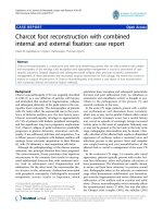
(Figure 1).
Methods to recruit the lung
The RMs are not unique, which is a general limitation
of the tec hnique because it is not standardized as yet.
The earliest RM ever used during mechanical ventila-
tion is probably the sigh [9], which consists of increas-
ing tidal volume or level of positive end-expiratory
pressure (PEEP), depending on the ventilator used, for
one or several breaths. Tidal volume and PEEP level
canbeadjustedtoreachaspecificplateaupressure
(Pplat). Pelosi et al. [10] in ten patients with ARDS
applied three consecutive sighs per minute, each of
them generating Pplat of 45 cm H
2
O, and found that
oxygenation was better, lung static elastance lower,
and functional residual capacity (FRC) greater in the 1-
hour-sigh period than in the no-sigh period. However,
some safety concern could have been raised given that
this schedule would lead to 4,320 occurrences per day
of Pplat 45 cmH
2
O, which is a level well above the 30
cm H
2
O recommended threshold to maintain in ARDS
[11]. The most frequently investigated RM, due to its
apparent simplicity, is the sustained inflation (SI),
which consists of pressurizing the airways at a specific
level and maintaining i t for a given duration. A com-
mon combination is the application of 40 cmH
2
Oair-
waypressurefor40seconds[12-14].Inarandomized
controlled trial involving 30 patients with ARDS, SI of
50 cmH
2
O applied for 30 seconds did not result in
better oxygenation by 30 minutes compared with the
control group free of RM [13]. In that study, SI was
applied after PEEP had been standardized in both
groups similarly. The interaction between pressure and
time is critical in the efficacy and tolerance of RM.
Therefore, some authors introduced the extended sigh
[15], which combines lower pressure level, progressive
rising of airway pressurization, and longer time of
application. High PEEP and pressure-controlled venti-
lation with a fixed driving pressure (= inspiratory pres-
sure minus PEEP) are other ways to perform R M [5].
The RMs we re compared each other in some investi-
gations. It should be stressed that an adequate compari-
son is difficult due t o the pressure-time produ ct, which
should be made identical between the two RMs. For
example, in 19 patients with ARDS, extended sigh was
associated with better oxygenation and higher recruited
volume than single SI 40 cm H
2
O for 40 seconds [16].
Using two or more RMs would have led to different
results. We compared optimal PEEP alone, selected
from a decremental PEEP trial, SI + optimal PEEP and
sighs + optimal PEEP in 12 patients with ARDS in a
cross-over study and found that sighs were associated
with better oxygenation and greater static compliance of
the respiratory system than any other strategy [17].
The meta-analysis of the studies on RMs in A LI/ARDS
byFanetal.[18]concludedthatRMswereneither
recommended nor forbidden and could r ather be used
on a case-by-c ase basis in the most h ypoxemic patients
as a life-saving procedure. Another systematic review did
not recommend the systematic use of RMs in the routine
practice in “ stable” ARDS patients [19]. It should be
mentioned that, apart from severely hypoxemic ARDS
3D2
9,/,
$OYHRODU5HFUXLWPHQW
2YHUGLVWHQVLRQ
9,/,
&DUGLDFRXWSX
W
'D2
,P
S
DFWRQ
S
DWLHQWRXWFRPH
5HVSRQVHWR50 %DODQFH
Figure 1 Balance between benefits (left tray) and risks (right
tray) of the recruitment maneuvers. VILI, ventilator-induced lung
injury; RM, recruitment maneuver; DaO2, oxygen transport.
Guerin et al. Annals of Intensive Care 2011, 1:9
/>Page 2 of 6
patients where RMs could be used to maintain safe oxy-
genation levels, RMs should be applied after tracheal suc-
tioning [20] or patient disconnection. In the early trial,
which introduced the concept of lung protective mechan-
ical ventilation [21], RMs were managed after tracheal
suctioning.
Four lines of considerations cast some doubt about
the routine use of RMs in patients with ARDS.
1. The fact that three randomized, controlled trials
werenotabletodemonstrateabeneficialeffectofRMs
on oxygenation in the routine practice [13,22,23].
2. Some safety concerns [24].
3. The large variability of the oxygenation response
across the patients [23].
4. The relevant end-points in the assessment of RMs
have moved from the oxygenati on improv emen t toward
the VILI prevention.
Factors of the response to RMs
As shown in Table 1, several factors are involved in the
response to RMs in terms of oxyge nation, lung recru it-
ment, or hemodynamics. Some of these effects are dis-
cussed below.
Type of ARDS
This is a major factor because ARDS is highly heteroge-
neous by nature, both within patients and between
patients. The separation between foc al and not focal
ARDS has b een largely accepted. Constantin et al. [25]
separated ARDS patients into focal and not focal mor-
phological patterns from the CT scan and studied the
effect of a single SI applied before and after open lung
ventilation, namely a high PEE P. After the RM, the oxy-
genation remained unchanged in the focal whilst it
improved in the not focal ARDS pattern. Most
importantly, in the focal pattern after the RM, the lung
overdistension markedly increased and was greater than
the lung recruitment elicited by RM. Once RM was
released, the overdistension remained above its level
before RM. In sharp contrast in the not focal ARDS pat-
tern, the recruited volume markedly increased and was
greater than the concomitant overdistension with the
RM. After the RM, the overdistension went back to its
baseline level but the recruited volume remained higher
than its pre-RM level. This result was extended by Grasso
et al. [26] who investigated the effect of a s ingle SI in
three experimental ARDS in pigs: surfactant depletion
with massive derec ruitment and no inflammation; oleic
acid-induced ARDS with massive lung edema and no
inflammation; and hydrochloride acid-induced ARDS
characterized by massive inflammation. The RM did pro-
mote recruitment but also overdistension in the most
anterior parts of the lungs in the three ARDS models,
making the lungs more het erogeneous than before the
RM application. Furthermore, the overdistension, and
hence the lung heterogeneity, was maintained after RM
release. The morphological lung heterogeneity was asso-
ciated with a mark ed functional heterogeneity because
the elastance of the recruited parts of the lungs was sig-
nificantly greater than in the co ntrol animals and than
tha t of the baby lung in each ARDS model. This result is
very important to keep in mind when RM is used.
Another criterion to separate ARDS patients is the
severity of the lung injury. Whereas it is difficult to
accurately and precisely define what severe ARDS is, the
paraquat model of ARDS in rats is useful in this pur-
pose because the lung histomorphometry findings are
different with the dose of paraquat administered. The
intraperitoneal injection of 20 mg/kg paraquat induces
alveolar collapse and interstitial oedema whilst a greater
dose of 25 mg/kg promotes an additional alveolar
oedema. Therefore, low dose of paraquat induced mod-
erate ARDS whilst with high dose of paraquat severe
ARDS would follow. Santiago et al. [27] found that a
single SI induced a significantly greater magnitude of
overdistension, endothelial and epithelial alveolar cells
injury, and apopto sis to the lungs and kidneys in severe
than in moderate paraquat-induced ARDS in rats.
Lung perfusion
Lung perfusion is a critical determinant o f oxygenation.
In a sheep model of surfactant depletion, a single SI
worsened oxygenation in every animal [28]. The
mechanism of this finding was that: 1) the RM did not
recruit the dorsal part of the lungs in which there was a
massive loss of aeration, and 2) redistributed the pul-
monary blood flow toward them. Therefore, the intra-
pulmonary s hunt increased in these depe ndent parts of
the lung leading to oxygenation worsening.
Table 1 Factors potentially involved in the variability of
the response to recruitment maneuvers in ARDS
ARDS-related
Focal vs. nonfocal
Early vs. Late
Severe vs. moderate
Lung recruitability
Associated vasoactive drugs
RM-related
Type of recruitment maneuvers
Distribution of lung perfusion
Transpulmonary pressure
Timing of application
Patient positioning
Post-RM strategy
Post-RM PEEP
ARDS, acute respiratory distress syndrome; RM, recruitment maneuvers; PEEP,
positive ned-expiratory pressure.
Guerin et al. Annals of Intensive Care 2011, 1:9
/>Page 3 of 6
Chest wall elastance
In 22 patients with ARDS, Grasso et al. found [14] that
half was responder in terms of oxygenation after a single
SI and the other half was not. The explanation was that
the chest wall elastance was greater in the non respon-
ders than in responders, and hence, more pressure dissi-
pated into the chest wa ll and less pressure was available
to distend the lung in the non responder than in the
responder group. Therefore, in setting the RM what
counts is not the level of the airway pressure but the
level of P
L
which takes into account the chest wall ela-
stance magnitude.
Post-RM strategy
Lim et al. [15] investigated the eff ects on oxygenation of
three ventilatory strategies in ARDS patients: extended
sigh followed by higher PEEP than or by same PEEP as
before RM, and higher PEEP alone. Oxygenation was
greater in the first stra tegy. The authors extended this
result in a comprehensive experimental study in pigs
[29]. They used three ARDS models (VILI, pneumonia,
oleic acid), three RMs (extended sigh, S I, pressure-con-
trolled ventilation), and three levels of PEEP after the
RM (8, 12, and 16 cm H
2
O).Theyfoundthatthepri-
mary factor of the greater oxygenation was the level of
PEEP after the RM. Because PEEP is an expiratory set-
ting, it should be more rel evant to tailor its level after
having recruited the lung. This consideration is the
background of the decremental PEEP trial, which is an
attractive way to adjust PEEP [30].
Recent advances in RM
Recently, new RMs have been described and a further
assessment of their lung effects was reported that
brought some additional information with clinical impli-
cations. A common feature in these new data is that
they dealt with the role of time and pressure-time pro-
duct during the RMs. Indeed, it has been shown that
almost 80% of the recruited volume after a RM was
obtained within the first 5 seconds, making the remain-
ing 35 seconds of a 40-second RM less useful for the
recruitment but potent ially harmful for the lungs or the
circulation [31].
In the paraquat-induced ARDS model in rats, Rze-
zinski et al. [32] compared a single common SI (40 cm
H
2
O × 40 sec) to a progressive RM in which, starting
from PEEP 15 cm H
2
O the baseline driving pressure of
10 cm H
2
O was increased by three steps of 5 cm H
2
O
lasting 2 minutes each; the end-inspiratory pressure
reached 40 cm H
2
O within 12 minutes and lasted 2
minutes. Lung recruitment and oxygenation were signi f-
icantly greater, whereas static lung elastance, lung
inflammation, alveolar epithelial cells apoptosis, and
alveolar-capillary membrane injury were significantly
lower with progressive RM than with the common SI.
The prolongation of the RM and the pressure.time pro-
duct were likely explanations for the global benefit of
the prolonged RM.
Steimback et al. [33] using again the paraquat-induced
ARDS model in rats, compared 180 sighs per hour, the
same rate as in the early study in humans [10], set to
generate Pplat of 40-cm H
2
O, to 10 sighs per hour at
20- or 40-cm H
2
O targeted Pplat, and to a common SI.
The results, which are summarized in Table 2, are
clearly in favour of a lower rate of sighs and a 40 cm
H
2
O Pplat.
Finally, still by using the paraquat-induced ARDS in
rats, Riva et al. [34] compared a common 40 cm H
2
O×
40-second SI to a RM in which the target pressure of 40
cm H
2
O was reached after 40 seconds as a ramp. Both
were delivered from PEEP 0 or 5 cm H
2
O. The MR gen-
erated as a ramp from 5 cm H
2
O of PEEP reduced over-
distension, alveolar collapse, lung expressi on of mRNA
of procollagen III, and lung static elastance.
Forty patients with ARDS were randomized into S I or
pressure-controlled ventilation adjusted to gene rate the
same pressure-time product [35]. Pressure-controlled
ventilation was associ ated with significantly greater oxy-
genation and with significantly less hemodynamics
derangements as reflected by significantly lower central
venous and pulmonary artery pressures, lower right ven-
tricle work lo ad, and higher cardiac output. The rapid
airway pressure rising duringtheRMcanbeafactor
that explains why RM can promote VILI and may wor-
sen hemodynamics.
Conclusions
Assessing the efficacy of RM on oxygenation only is lar-
gely insufficient and the complete evaluation, as for any
ventilatory strategy in ARDS, must consider the effects
on hemodynamics, lung recruitment, overdistension,
stress and strain [36], and biotrauma [37]. The risks
Table 2 Summary of the comparison of sighs in the study
by Steimback et al. [33]
SI Sighs
180/40
Sighs
10/40
Sighs
10/20
Oxygenation ↑↑ ↓ ↓
Est, L ® ↓↓↑
Alveolar collapse ↓↓ ↓ ↑
Overdistension ® ↑↓®
Alveolar-capillary Membrane injury ↓↑ ↓ ®
Lung apoptosis ↓↑ ↓ ®
mRNA PCIII ↓↑ ↓ ®
SI, sustained inflation; sighs, rate per minute/target plateau pressure; Est, L,
lung static elastance; mRNAPCIII, lung expression of mRAN of procollagen III.
The arrows indicate the direction of change of each variable relative to the
group of injured lungs not receiving recruitment maneuvers.
Guerin et al. Annals of Intensive Care 2011, 1:9
/>Page 4 of 6
associated with RM are both at the lung level (VILI) and
at the systemic level. The systemic risks that may follow
RM are hemodynamics impairment or decompartimen-
talization of the VILI toward distant organs. The RM is
a complex procedure, not standardized as yet. The fac-
tors involved in RM response largely depend on the
underlying lung disease. At the present time, the pre-
vious conservative recommendations of not using RMs
in routine in stable ARDS patients are still valid.
Authors’ contributions
CG wrote the manuscript. SD, VL, BD, FB, GB, and JCR critically reviewed the
manuscript.
Competing interests
The authors declare that they have no competing interests.
Received: 14 March 2011 Accepted: 19 April 2011
Published: 19 April 2011
References
1. Gattinoni L, Caironi P, Pelosi P, Goodman LR: What has computed
tomography taught us about the acute respiratory distress syndrome?
Am J Respir Crit Care Med 2001, 164:1701-1711.
2. Gattinoni L, Caironi P, Valenza F, Carlesso E: The role of CT-scan studies for
the diagnosis and therapy of acute respiratory distress syndrome. Clin
Chest Med 2006, 27:559-570, abstract vii.
3. Puybasset L, Cluzel P, Gusman P, Grenier P, Preteux F, Rouby JJ: Regional
distribution of gas and tissue in acute respiratory distress syndrome. I.
Consequences for lung morphology. CT Scan ARDS Study Group.
Intensive Care Med 2000, 26:857-869.
4. Gattinoni L, Caironi P, Cressoni M, Chiumello D, Ranieri VM, Quintel M,
Russo S, Patroniti N, Cornejo R, Bugedo G: Lung recruitment in patients
with the acute respiratory distress syndrome. N Engl J Med 2006,
354:1775-1786.
5. Borges JB, Carvalho CR, Amato MB: Lung recruitment in patients with
ARDS. N Engl J Med 2006, 355:319-320, author reply 321-312.
6. Borges JB, Okamoto VN, Matos GF, Caramez MP, Arantes PR, Barros F,
Souza CE, Victorino JA, Kacmarek RM, Barbas CS, Carvalho CR, Amato MB:
Reversibility of lung collapse and hypoxemia in early acute respiratory
distress syndrome. Am J Respir Crit Care Med 2006, 174:268-278.
7. Crotti S, Mascheroni D, Caironi P, Pelosi P, Ronzoni G, Mondino M, Marini JJ,
Gattinoni L: Recruitment and derecruitment during acute respiratory
failure: a clinical study. Am J Respir Crit Care Med 2001, 164:131-140.
8. Dreyfuss D, Saumon G: Ventilator-induced lung injury: lessons from
experimental studies. Am J Respir Crit Care Med 1998, 157:294-323.
9. Levine M, Gilbert R, Auchincloss JH Jr: A comparison of the effects of
sighs, large tidal volumes, and positive end expiratory pressure in
assisted ventilation. Scand J Respir Dis 1972, 53:101-108.
10. Pelosi P, Cadringher P, Bottino N, Panigada M, Carrieri F, Riva E, Lissoni A,
Gattinoni L: Sigh in acute respiratory distress syndrome. Am J Respir Crit
Care Med 1999, 159:872-880.
11. ARDSnet: Ventilation with lower tidal volumes as compared with
traditional tidal volumes for acute lung injury and the acute respiratory
distress syndrome. The Acute Respiratory Distress Syndrome Network. N
Engl J Med 2000, 342:1301-1308.
12. Oczenski W, Hormann C, Keller C, Lorenzl N, Kepka A, Schwarz S,
Fitzgerald RD: Recruitment maneuvers during prone positioning in
patients with acute respiratory distress syndrome. Crit Care Med 2005,
33:54-61, quiz 62.
13. Oczenski W, Hormann C, Keller C, Lorenzl N, Kepka A, Schwarz S,
Fitzgerald RD: Recruitment maneuvers after a positive end-expiratory
pressure trial do not induce sustained effects in early adult respiratory
distress syndrome. Anesthesiology 2004, 101:620-625.
14. Grasso S, Mascia L, Del Turco M, Malacarne P, Giunta F, Brochard L,
Slutsky AS, Marco Ranieri V: Effects of recruiting maneuvers in patients
with acute respiratory distress syndrome ventilated with protective
ventilatory strategy. Anesthesiology 2002, 96:795-802.
15.
Lim CM, Jung H, Koh Y, Lee JS, Shim TS, Lee SD, Kim WS, Kim DS, Kim WD:
Effect of alveolar recruitment maneuver in early acute respiratory
distress syndrome according to anti-derecruitment strategy, etiological
category of diffuse lung injury, and body position of the patient. Crit
Care Med 2003, 31:411-418.
16. Constantin JM, Jaber S, Futier E, Cayot-Constantin S, Verny-Pic M, Jung B,
Bailly A, Guerin R, Bazin JE: Respiratory effects of different recruitment
maneuvers in acute respiratory distress syndrome. Crit Care 2008, 12(2):
R50.
17. Badet M, Bayle F, Richard JC, Guerin C: Comparison of optimal positive
end-expiratory pressure and recruitment maneuvers during lung-
protective mechanical ventilation in patients with acute lung injury/
acute respiratory distress syndrome. Respir Care 2009, 54:847-854.
18. Fan E, Wilcox ME, Brower RG, Stewart TE, Mehta S, Lapinsky SE, Meade MO,
Ferguson ND: Recruitment maneuvers for acute lung injury: a systematic
review. Am J Respir Crit Care Med 2008, 178:1156-1163.
19. Hodgson C, Keating JL, Holland AE: Recruitment manoeuvres for adults
with acute lung injury receiving mechanical ventilation. Cochrane
Database Syst Rev 2009, 15.
20. Maggiore SM, Lellouche F, Pigeot J, Taille S, Deye N, Durrmeyer X,
Richard JC, Mancebo J, Lemaire F, Brochard L: Prevention of endotracheal
suctioning-induced alveolar derecruitment in acute lung injury. Am J
Respir Crit Care Med 2003, 167:1215-1224.
21. Amato MB, Barbas CS, Medeiros DM, Magaldi RB, Schettino GP, Lorenzi-
Filho G, Kairalla RA, Deheinzelin D, Munoz C, Oliveira R, Takagaki TY,
Carvalho CR: Effect of a protective-ventilation strategy on mortality in
the acute respiratory distress syndrome. N Engl J Med 1998, 338:347-354.
22. Meade MO, Cook DJ, Griffith LE, Hand LE, Lapinsky SE, Stewart TE, Killian KJ,
Slutsky AS, Guyatt GH: A study of the physiologic responses to a lung
recruitment maneuver in acute lung injury and acute respiratory distress
syndrome. Respir Care 2008, 53:1441-1449.
23. Brower RG, Morris A, MacIntyre N, Matthay MA, Hayden D, Thompson T,
Clemmer T, Lanken PN, Schoenfeld D: Effects of recruitment maneuvers in
patients with acute lung injury and acute respiratory distress syndrome
ventilated with high positive end-expiratory pressure. Crit Care Med 2003,
31:2592-2597.
24. Constantin JM, Cayot-Constantin S, Roszyk L, Futier E, Sapin V, Dastugue B,
Bazin JE, Rouby JJ: Response to recruitment maneuver influences net
alveolar fluid clearance in acute respiratory distress syndrome.
Anesthesiology 2007, 106:944-951.
25. Constantin JM, Grasso S, Chanques G, Aufort S, Futier E, Sebbane M, Jung B,
Gallix B, Bazin JE, Rouby JJ, Jaber S: Lung morphology predicts response
to recruitment maneuver in patients with acute respiratory distress
syndrome. Crit Care Med 2010, 38:1108-1117.
26. Grasso S, Stripoli T, Sacchi M, Trerotoli P, Staffieri F, Franchini D, De
Monte V, Valentini V, Pugliese P, Crovace A, Driessen B, Fiore T:
Inhomogeneity of lung parenchyma during the open lung strategy: a
computed tomography scan study. Am J Respir Crit Care Med 2009,
180:415-423.
27. Santiago VR, Rzezinski AF, Nardelli LM, Silva JD, Garcia CSNB, Maron-
Gutierrez T, Ornellas DS, Morales MM, Capelozzi VL, Marini J, Pelosi P,
Rocco PRM: Recruitment maneuver in experimental acute lung injury:
The role of alveolar collapse and edema. Crit
Care Med 2010,
38:2207-2214.
28. Musch G, Harris RS, Vidal Melo MF, O’Neill KR, Layfield JD, Winkler T,
Venegas JG: Mechanism by which a sustained inflation can worsen
oxygenation in acute lung injury. Anesthesiology 2004, 100:323-330.
29. Lim SC, Adams AB, Simonson D, Dries DJ, Broccard AF, Hotchkiss JR,
Marini JJ: Intercomparison of recruitment maneuver efficacy in three
models of acute lung injury. Crit Care Med 2004, 32:2371-2377.
30. Girgis K, Hamed H, Khater Y, Kacmarek RM: A decremental PEEP trial
identifies the PEEP level that maintains oxygenation after lung
recruitment. Respir Care 2006, 51:1132-1139.
31. Albert SP, DiRocco J, Allen GB, Bates JH, Lafollette R, Kubiak BD, Fischer J,
Maroney S, Nieman GF: The role of time and pressure on alveolar
recruitment. J Appl Physiol 2009, 106:757-765.
32. Rzezinskia AF, Oliveira GP, Santiago VR, Santos RS, Ornellas DS, Morales MM,
Capelozzi VL, Amato MBP, Conde MB, Pelosi P, Rocco PRM: Prolonged
recruitment manoeuvre improves lung function with less ultrastructural
Guerin et al. Annals of Intensive Care 2011, 1:9
/>Page 5 of 6
damage in experimental mild acute lung injury. Respiratory Physiology
and Neurobiology 2009, 169:271-281.
33. Steimback PW, Oliveira GP, Rzezinski AF, Silva PL, Garcia CSNB, Rangel G,
Morales MM, Silva JRL, Capelozzi VL, Pelosi PP, Rocco PRM: Effects of
frequency and inspiratory plateau pressure during recruitment
manoeuvres on lung and distal organs in acute lung injury. Intensive
Care Med 2009, 35:1120-1128.
34. Riva DR, Contador RS, Baez-Garcia CSN, Xisto DG, Cagido VR, Martini SV,
Morales MM, Rocco PRM, Faffe DS, Zin WA: Recruitment maneuver: RAMP
versus CPAP pressure profile in a model of acute lung injury. Respir
Physiol Neurobiol 2009, 169:62-68.
35. Iannuzzi M, De Sio A, De Robertis E, Piazza O, Servillo G, Tufano MA:
Different patterns of lung recruitment manoeuvers in primary acute
respiratory distress syndrome: effects on oxygenation and central
hemodynamics. Minerva Anestesiol 2010, 76:692-698.
36. Chiumello D, Carlesso E, Cadringher P, Caironi P, Valenza F, Polli F,
Tallarini F, Cozzi P, Cressoni M, Colombo A, Marini JJ, Gattinoni L: Lung
stress and strain during mechanical ventilation for acute respiratory
distress syndrome. Am J Respir Crit Care Med 2008, 178:346-355.
37. Tremblay LN, Slutsky AS: Ventilator-induced injury: from barotrauma to
biotrauma. Proc Assoc Am Phys 1998, 110:482-488.
doi:10.1186/2110-5820-1-9
Cite this article as: Guerin et al.: Efficacy and safety of recruitment
maneuvers in acute respiratory distress syndrome. Annals of Intensive
Care 2011 1:9.
Submit your manuscript to a
journal and benefi t from:
7 Convenient online submission
7 Rigorous peer review
7 Immediate publication on acceptance
7 Open access: articles freely available online
7 High visibility within the fi eld
7 Retaining the copyright to your article
Submit your next manuscript at 7 springeropen.com
Guerin et al. Annals of Intensive Care 2011, 1:9
/>Page 6 of 6