Báo cáo hóa học: " Identification and characterization of alkaline serine protease from goat skin surface metagenome" docx
Bạn đang xem bản rút gọn của tài liệu. Xem và tải ngay bản đầy đủ của tài liệu tại đây (1.62 MB, 10 trang )
ORIGINAL ARTICLE Open Access
Identification and characterization of alkaline
serine protease from goat skin surface
metagenome
Paul Lavanya Pushpam, Thangamani Rajesh, Paramasamy Gunasekaran
*
Abstract
Metagenomic DNA isolated from goat skin surface was used to construct plasmid DNA library in Escherichia coli
DH10B. Recombinant clones were screened for functional protease activity on skim milk agar plates. Upon screening
70,000 clones, a clone carrying recombinant plasmid pSP1 exhibited protease activity. In vitro transposon
mutagenesis and sequencing of the insert DNA in this clone revealed an ORF of 1890 bp encoding a protein with
630 amino acids which showed significant sequence homology to the peptidase S8 and S53 subtilisin kexin sedolisin
of Shewanella sp. This ORF was cloned in pET30b and expressed in E. coli BL21 (DE3). Although the cloned Alkaline
Serine protease (AS-protease) was overexpressed, it was inactive as a result of forming inclusion bodies. After
solubilisation, the protease was purified using Ni-NTA chromatography and then refolded properly to retain protease
activity. The purified AS-protease with a molecular mass of ~63 kDa required a divalent cation (Co
2+
or Mn
2+
) for its
improved activity. The pH and temperature optima for this protease were 10.5 and 42°C respectively.
Introduction
Proteases are present in all living forms as they are
involved in various metabolic processes. They are mainly
involved in hydrolysis of the peptide bonds (Gupta et al.
2002). Proteases are classified into six ty pes based on the
functional groups in their active sites. They are aspartic,
cysteine, glutamic, metallo, serine, and threonine pro-
teases. They are also classified as exo-peptidases and
endo-peptidases, based on the position of the peptide
bond cleavage. Proteases find a wide range of applications
in food, pharmaceutical, leather and textile, detergent,
diagnostics industries and also in waste management
(Rao et al. 1998). Thus, they contribute to almost 40% of
enzyme sales in the industrial market. Though proteases
are found in plants and animals, microbial proteases
acco unt for two-third of share in the co mmercially avail-
able proteases (Kumar and Takagi 1999).
Proteases are also classified as acidic, neutral or alkaline
proteases based on their pH optima. The largest share of
the enzyme market is occupie d by detergent proteases,
which are mostly alkaline serine protease and active at
neutral to alkaline pH range. Alkaline serine proteases
have Aspar tate (D) and Histidine (H) residues along with
Serine (S) in their active site forming a catalytic triad
(Gupta et al. 2002). Serine prote ases contribute t o one
third of the share in the enzyme market and are readily
inactivated by Phenyl Methane Sulfonyl Fluoride (PMSF)
(Page a nd Di Cera 2008 ). Based on the sequence and
structural similarities, all the kno wn proteases are classi-
fied into clans and families and are available in the MER-
OPS database (Rawlings and Barrett 1993).
Several microbial proteases from the culturable organ-
isms have been chara cterized. However, very few pro-
teases have been identified through culture independent
metagenomic approach (Schloss and Handelsman 2003).
In metagenomics study, the total nucleic acid content of
the environmental samples is analysed. The DNA may be
isol ated by direct or indirect methods followed by purifi-
cation (Gabor et al. 2003); Rajendhran and Gunas ekaran
2008). Metagenomics approac h has been recently
employed in identifying number of novel genes encoding
biocatalysts or molecules which are of p harmaceutical
and industrial importance. Interestingly, the metage-
nomic libraries were mainly screened for enzymes like
lipases and esterases (Lee et al. 2004; Rhee et al. 2005;
Voget et al. 2003), proteases (Lee et al. 2007), amylases
* Correspondence:
Department of Genetics, Centre for Excellence in Genomic Sciences, School
of Biological Sciences, Madurai Kamaraj University, Madurai, India 625021.
Pushpam et al. AMB Express 2011, 1:3
/>© 2011 Lavanya et al; licensee Springer. This is an Open Access article distributed under the terms of the Creative Commons
Attribution License (http://creativecom mons.org/licenses/by/2.0), which permits unrestricted use, distribution, and reproduction in
any medium, provided the original work is prop erly cited .
(Rondon et al. 2000; Voget et al. 2003), chitinase (Cottrell
et al. 1999) and nitrilases (Robertson et al. 2004). Despite
the success rate, very few attempts were made on the
identification of proteases from metagenomic libraries.
We report here an Alkaline Serine p rotease (AS-pro-
tease), identified from the goat skin metagenomic library,
which showed homology to peptidase S8 and S53 subtili-
sin kexin and sedolisin of Shewanella sp. Surprisingly,
this AS-protease requires Co
2+
or Mn
2+
metal ions for its
improved activity.
Materials and methods
Materials, bacterial strains and culture conditions
Goat skins were obtained from butcheries in and around
Madurai for metagenomic DNA isolatio n. Reagents for
PCR, Taq DNA polymeras e, oligonucleotide primers,
and all biochemicals were from Sigma-Aldrich (St.
Louis, MO, USA). T4 DNA ligase and restriction
enzymes were from MBI Fermentas (Opelstrasse,
Germany). Escherichia coli strains and plasmids used in
this study are listed in Table 1. E. coli DH5a and E. coli
BL21 (DE3) were used for gene cloning and protein
expression studies respectively.
DNA manipulation techniques
Standard procedures for plasmid isolation, restriction
enzyme digestion, ligation, competent cell preparation
and transformation were used as described by
(Sambrook et al. (1989)). Metagenomic DNA was iso-
lated using a modified indirect DN A extraction method
(Gabor et al. 2003). T he goat skin (10 cm × 10 cm) was
suspended in 0.75% (w/v) NaCl and kept under agitation
at 180 rpm for 30 min. The supernatant was collected
and a pellet was obtained by centrifugation (10,000 × g
for 10 min at 4°C). The pelle t was rinsed and suspended
in blending buffer (100 mM Tris-HCl [pH 8.0], 100 mM
sodium EDTA [pH 8.0], 0.1% SDS) and homogenized.
The homogenized mixture was subjected to low-speed
centrifugation (1000 × g for 10 min at 10°C), and th e
supernatant containing bacterial cells was collected,
while the coarse particles and high molecular weight
DNA in the pellet was subjecte d to furth er centrifuga-
tion cycles as described above. Supernatant obtained
from the three rounds of cell extraction were pooled.
The supernatant we re centrifuged at 10,000 × g for
30 min at 4°C and the cell pellet was rinsed with chrom-
bach buffer (0.33 M Tris-HCl, 1 mM EDTA, pH 8).
Then the mixt ure was suspended in lysis buffer
(100 mM Tris-HCl, 100 mM EDTA, 1.5 M NaCl), in
the presence of 0.1 mg of proteinase K and 1 mg of
lysozyme and incubated at 37°C for 30 min. Lysis was
completed by adding 1 ml of 20% SDS and incub ated
for 2 h at 65°C with shaking every 30 min. The superna-
tant was collected by centrifugation at 6000 × g for
10 min at 30°C and the pellets were re-extracted twice
with 1 ml lysis buffer, vortexing for a few seconds, and
incubating at 65°C for 10 min. The supernatant was
extracted with equal volume of chloroform: isoamyl
alcohol (24:1). DNA in the aqueous phase was precipi-
tated by addition of 0.6 volumes of isopropanol and
incubated at -20°C for 1 h. The precipitate was collected
by centrifugation at 10, 000 × g for 15 min at 4°C and
then washed with 70% ethanol. The DNA pellet was
suspended in 200 μl TE buffer (10 mM Tris-HCl, 1 mM
EDTA, pH 8) and stored at -20°C.
Metagenomic DNA was partially digested with HindIII
and the DNA fragments ranging about 3-8 kb were
separated with QIAquick gel extraction kit (Qiagen,
Hilden, Germany) and cloned into pUC19, resulting in
plasmid pSP1 which was transformed into E. coli
DH10B by electroporation (200 Ω,25μFand2.5kV)
using Gene Pulser (Bio-Rad, USA). Transformants were
Table 1 List of bacterial strains and plasmids used in this study
Strains/plasmids Genotype/Description Reference/Source
E. coli DH5a F
-
endA1 glnV44 thi-1 recA1 relA1 gyrA96 deoR nupG F80dlacZΔM15 Δ(lacZYA-argF)
U169, hsdR17(r
K
-
m
K
+
), l-
Invitrogen (CA, USA)
E. coli DH10B F
-
endA1 recA1 galE15 galK16 nupG rpsL ΔlacX74 F80lacZΔM15 araD139 Δ(ara, leu)
7697 mcrA Δ(mrr-hsdRMS-mcrBC) l
-
Invitrogen (CA, USA)
E. coli BL21 (DE3) F
-
ompT gal dcm lon hsdS
B
(r
B
-
m
B
-
) l(DE3 [lacI lacUV5-T7 gene1 ind1 sam7 nin5]) Novagen (CA, USA)
pUC19 Ap
r
; Cloning vector Stratagene (CA, USA)
pTZ57R/T Ap
r
; PCR cloning vector MBI Fermentas (Opelstrasse, Germany)
pET30b Kn
r
; Expression vector; T7 promoter Novagen (CA, USA)
pSP1 pUC19 harbouring the AS-protease ORF; Ap
r
This study
pTSP1 AS- Protease ORF cloned in pTZ57R/T; Ap
r
This study
pETP1 AS- protease ORF cloned in pET30b; Kn
r
This study
Pushpam et al. AMB Express 2011, 1:3
/>Page 2 of 10
selected on LB agar plates supplemented with 100 μgof
ampicillin/ml, X-gal (20 μg/ml) and IPTG (4 0 μg/ml)
and inc ubated at 37°C for overnight. T he white recom-
binant clones were scored and maintained.
Screening the metagenomic library for proteolytic activity
The recombinant clones were screened for proteolytic
activity on LB agar ampicillin plates supplemented with
1% (w/v) skim milk (Lee et al. 2007) and incubated at
37°C for 48 - 72 h. Proteolytic clones were selected
based on the formation of zone of clearance around the
colony.
In vitro transposon mutagenesis and sequencing
The recombinant plasmid was used as template for in vitro
transposon mu tagenesis using Templat e Generation Sys-
tem II kit (TGS, F-702; Finnzyme, Finland). E. coli DH10B
carrying the plasmid pSP1 was transformed with the artifi-
cial Mu transposon by electroporation and the transfor-
mants were selected on LB agar plates containing
ampicillin (100 μg/ml) and kanamycin (30 μg/ml). Further,
the strains carrying the plasmid with the mutated protease
were screened on 1% skim milk-LB agar plate for a nega-
tive activity. The plasmids from the mutants were isolated
and the regions adjacent to the transposons were
sequenced using t ranspos on spec ific primer. Bla stN an d
BlastP analyses were carried out to find sequence identity
and homology (Altschul et al. 1990). Signal peptide of the
protein was predicted using the S ignalP 3.0 server http://
www.cbs.dtu.dk/services/SignalP/ (Bendtsen et al. 2004).
Multiple sequence ali gnment was performed with t he
sequences (MER048892; Shewanella baltica, MER087187;
Shewanella woodyi, MER016525; Pseudoalteromonas sp.
AS-11) in the MEROPS peptidase database http://merops.
sanger.ac.uk (Rawlings and Barrett 1993) to assign the
family for t he identified protease and also in the NCBI
database.
Cloning and expression of protease encoding gene
The complete ORF of the protease was amplified with
the primers MP1F (5’-ATGCATAAGAAACATTTAA-
TAGCA3’)andMP1R(5’CTAGTAGCTTGCACT-
CAGCTGAAC-3’) and cloned into pTZ57R/T vector,
andtheresultantplasmidwasusedtotransformE. coli
DH5a. The cloned protease gene was confirmed by
DNA sequencing using the BigDye Terminator sequen-
cing method and an ABI PRISM 3700 sequencer
(Applied Biosystems, Foster City, CA). The protease
gene was agai n amplified from the re combinant plasmid
with and without the signal peptide using forward
primers P1FS 5’ -GCGC
CATATGCATAAGAAA-
CATTTAATAG-3’ (NdeI site is underlined) and P1FWS
5’-ATTA
CATATGGAATACCAAGCGACTATGG-
TAAG-3’ ( Nd eI site is underlined) and reverse primer
P1RH 5’-TAAT
AAGCTTGTAGCTTGCACTCAGCTG-
3’ (HindIII site is underlined). The PCR product was
digest ed with NdeIandHindIII and liga ted with expres-
sion vector pET30b to obtain another recombinant plas-
mid, in which the protease gene was under the control
of the T7 promoter. This recombinant plasmid was then
used to transform E. coli BL21 (DE3). E. coli BL21
(DE3) carrying recombinant plasmid was grown over-
nightat37°CinLBmediumcontainingkanamycin
(30 μg/ml). Fresh LB medium with kanamycin was
inoculated with 1% (v/v) of overnight culture and incu-
bated at 37°C until the culture reached an absorbance of
0.4 at OD
600
. The culture was then induced with
0.1 mM of isopropy l-b-D-thiogalactopyr anoside (IPTG).
The induced cells were harvested by centrifugation at 4°
C for 10 min at 12,000 × g an d washed with 50 mM
Tris-buffer (pH 7.5). The cells were then disrupted
by sonication (five times for 30 s with 30 s interval)
(Labsonic U, Germany), and centrifuged at 12 000 × g
for 30 min. Both the soluble and pellet fractions were
analysed for protease activity.
SDS-PAGE and Zymogram analysis
The proteins from the insoluble fraction after sonicat ion
were resolved on Sodium dodecyl sulphate-polyacryla-
mide gel electrophoresis (SDS-PAGE) (Laemmli 1970).
The gel was stained with Coomassie brilliant blue
R-250. The molecular mass of protein was determined
by comparison with the mobility of molecular weight
markers (Fermentas, Opelstrasse, Germany). For zymo-
gram analysis, the protein were separated on the SDS-
PAGE with 0.1% (w/v) gelatin in the separating gel
(Bressollier et al. 1999). After electrophoresis, the gel
was incubated with 2.5% (v/v) Triton X-100 at 37°C for
30 min for the removal of SDS followed by another
round of incubation in 50 mM Tris (pH 7.4) for 30 min.
The gel was then incubated in the same buffer at 37°C
for 4 h. Zone of clearance within the gel was checked
after staining with Coomassie brilliant blue R-250.
Purification of protease
The cell pellets was resuspended in 20 mM Tris-HCl
buffer (pH 7.5), disrupted by sonication and centri fuged
at 10,000 × g for 30 min. The insoluble fraction after
sonication, containing the recombinant protein was col-
lected and solubilised in 3 ml of cold 2 M urea contain-
ing 20 mM Tris-HCl buffer, 0.5 M NaCl and 2% Triton
X-100 (pH 8.0) and centrifuged at 10,000 × g for
10 min. The supernatant was discarded and the pellet
fraction was further washed once with the same buffer
and then resuspended in 5 ml of 20 mM Tris-HCl buf-
fer containing 8 M urea, 0.5 M NaCl, 5 mM imidazole,
1 mM 2-mer captoethanol (pH 8.0) , and stirred at room
temperature for 30-60 min to solubilise the recombinant
Pushpam et al. AMB Express 2011, 1:3
/>Page 3 of 10
protein. The solubilised proteins were passed through
Ni-NTA Affinity column (Sigma Chemicals, USA) and
eluted with imidazole following the manufacturer’s
recommendation. The purified protein with urea was
then refolded in 20 mM Tris buffer by drop dilution
method (Howarth et al. 2006). The refolded protein was
used for further characterization.
Enzyme assay
In standard conditions, the reaction mixture contained
480 μlof1%(w⁄ v) azocasein, 2 mM CaCl
2
and appro-
priate dilution of enzyme in 50 mM Tris buffer, pH 7.5
(Radha and Gunasekaran 2007). The reac tion mixture
was incubated at 37°C for 30 min. The reaction was ter-
minated by adding 600 μl of 10% (w/v) trichloroacetic
acid and kept on ice for 15 min followed by centrifuga-
tion at 15,000 × g at 4°C for 10 min. Eight hundred
microlitre of the supernatant were neutralized by adding
200 μl of 1.8 N NaOH, and the absorbance at 420 nm
(A
420
) was measured using a spectrophotometer (Hitachi
U-2000, Japan). The control samples were the extract
from the E. coli BL21 (pET30b) only. One unit of pro-
tease activity was defined as the amount of enzyme
required to yield an increase in absorbance of 0.01 at
A
420
in 30 min at 37°C.
Effect of metal ions, inhibitors, solvents, detergents and
reducing agents
Protease was purified as previously described followed
by extensive dialysis in the presence of 10 mM EDTA in
50 mM Tris buffer (pH 7.5) and then, the enzyme was
assayed under standard conditions in the presence of
different metal ions (Mn
2+
,Ca
2+
,Co
2+
,Ni
2+
,Hg
2+
and
Zn
2+
). The purified protease was pre-incubated with dif-
ferent metal ions (0.1, 1 and 5 mM), inhibito rs (5 mM),
detergents (0.5 - 1%) and reducing agent (b-ME)
(5 mM) for 15 min at 37°C. The residual activity was
measured under standard assay condition.
Physicochemical characterization
The effect of temperature on the acti vity of the p urif ied
AS-protease was determined at the temperature range
of 10°C to 85°C at pH 7.5. Thermal stability of the puri-
fied AS-protease was estimated by incubating the
enzyme in 50 mM Tris buffer at different temperatures
(35°C, 45°C and 55°C) in the presence of 5 mM CoCl
2
.
At different intervals, samples were withdr awn and the
residual activity was measured under standard assay
condition. The optimum pH of AS-protease activity was
measured at 37°C with different buffer: 50 mM Sodium
acetate buffer (pH 4-5.5), 50 mM Tris buffer (pH 6.5-
8.5), 50 mM sodium carbonate buffer (pH 9), and 50
mM glycine-NaOH buffer (pH 10.5-12.5).
Determination of kinetic parameters
The recombinant protease was assayed with 0.1-10 mg/
ml azocasein in 50 mM Tris buffer (pH 7.5) containing
5mMCo
2+
at 42°C for 10 min. Kinetic parameters,
such as K
m
(mg/ml) K
cat
(min
-1
)andV
max
(U/mg-
protein) for substrates were obtained using Line-weaver
Burk plot.
Results
Construction and screening of metagenomic library from
Goat skin
Diverse microbial population (both culturable and non
culturable) with majority of them with proteolytic activ-
ity was found on the goat skin surface (Kayalvizhi and
Gunasekaran, 2008). Therefore, metagenomic DNA
(~5 μg/ml) of the goat skin surface was isolated by an
indirect extraction method as described in materials and
methods. A small-insert metagenomic library in pUC19
was constructed. Analysis of the randomly selected
recombinant clones revealed that the clones had the
insert DNA of an average size of ~3.2 kb.
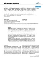
Screening of 70,000 recombinant clones for proteolytic
activity revealed one clone carrying recombinant plas-
mid designated as pSP1 that exhibited a zone of clear-
ance on LB skim milk agar plate after 36 h of
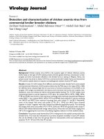
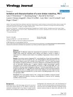
incubation at 37°C (Figure 1). Since insert DNA in this
clone was 3.8 kb (Figure 2), the protease gene could
have been expressed with its own promoter (Figure 3).
Transposon mutagenesis on pSP1 was carried out to
have Tn insertion w ithin t he protease coding region in
the insert DNA (Figure 2). Randomly selected transpo-
son carrying protease negative mutants were seq uenced
and alignment of these sequences lead to the identifica-
tion of the protease open reading frame (ORF).
Analysis of the cloned protease gene
TheORFencodingtheproteasewasamplifiedand
cloned in pTZ57R/T vector and the resultant construct
was designated as pTSP1. Analysis of the insert DNA
sequence as described above, revealed an ORF (1890 bp)
with ATG as start codon and TAG as termination codon.
The deduced amino acid sequence of the protease com-
prises of 630 amino acids and an estimated molecular
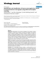
mass of 65,540 Da. Multiple sequence alignment of this
protease was performed with other known protease
sequences in the NCBI database and shown in Figure 4.
The amino acid sequence of this AS-protease displayed
98% sequence similarity with uncharacterized proteases
of various Shew anella sp. in the NCBI database and a
maximum of 85% similarity with S8A secreted peptida-
seA of Shewanella baltica MEROPS database (Rawlings
and Barrett 1993). These results suggested that the
cloned protease belongs to serine family peptidase.
Pushpam et al. AMB Express 2011, 1:3
/>Page 4 of 10
At the N terminus o f this AS-protease sequence, pre-
sence of a signal peptide with 23 amino acids was pre-
dicted using the SignalP program (Bendtsen et al. 2004).
ThePfamanalysisofthisproteaseshowedaconserved
catalytic domain of peptidase S8 family and two pre-
peptidase C-terminal domains. This AS-protease con-
tained active site residues within the catalytic motif
Asp-Thr/Ser-Gly, His-Gly-Thr-His and Gly-Thr-Ser-
Met-Ala-X-Pro, which is characteristic of serine subfam-
ilyS8A.Resultsfromthesequenceanalysisofthis
protease suggested it to be serine protease subfamily S8A.
Expression of AS-protease gene
The protease coding ORF was amplified and cloned
into the expression vector pET30b and resultant
recombinant plasmid was designated as pETP1. Upon
induction, the E. coli BL21 (DE3) harbouring the
recombinant plasmid pETP1 expressed the cloned pro-
tease gene.
Further, proteins in the recombinant cell extract was
resolved on SDS-PAGE revealed an ove r expressed pro-
tein of 66 kDa (Figure 5A) which is in agreement with
the predicted molecular mass for the cloned AS-
protease. The protein was expressed as inclusion bodies,
which was later solubilised with urea as mentioned in
materials and methods. The solubilised protein was pur-
ified on Ni-NTA Affinity Chromatography (Figure 5B)
andthenrefoldedbydropdilution.Thepurified
refolded protein exhibited a maximum activity of
100.2 U ml
-1
(specific activity 83.56 U mg
-1
).
Protease
positive clone
Figure 1 Functional screening of metagenomic library for protease activity on skim mil k agar plate. Metagenomic library consisting of
70,000 clones were screened on skim milk plate for protease activity. The positive clone showing zone of clearance in skim milk agar plate is
indicated by an arrow.
Figure 2 Schematic representation of the insert metagenomic DNA and the position of transposon used for sequenc ing the coding
region. Each inverted triangle represents the individual insertion of transposon in the protease coding gene. Black dotted arrow indicates the
orientation and location of protease gene. 4Fe-4S represents 4Fe-4S ferredoxin iron-sulfur binding domain protein, S8 & S53 - peptidase S8 and
S53 subtilisin kexin sedolisin, sterol - Sterol-binding domain protein, U32 - peptidase U32.
Pushpam et al. AMB Express 2011, 1:3
/>Page 5 of 10
Effect of pH and temperature
The effect of pH on the purified AS-protease was
examined at 37°C. Purified AS-protease exhibited max-
imum activity at pH 10.5 (Figure 6A), confirming it to
be an alkaline protease. This protease exhibited 75 -
85% of activity at a pH range of 7.5 to 9.5. The
proteolytic activity was significantly decreased above
pH 11.5 and below pH 7.0. Proteolytic activity was
found maximum at 42°C (Figure 6B) but exhibited
only 65 and 85% of the maximum activity at the tem-
perature range of 35°C and 55°C respectively. Thermal
stability of the purified AS-protease was estimated at
1 43
Bacilli promoter SD (1) TTGCCGTTCAT TTTCCCAATA
AS-protease SD (1) AGGTAAGCCTTAAGCATTA
E.coli promoter (1) TTCTCGGCGTTGAA TGTGGGGGAAACATCCCCATATACT
44 86
Bacilli promoter SD (22) CAAT AAGGATGACTATTT-TGGTAAAATTCAGAATGTGAG
AS-protease SD (20) AACTGGGCAGGTTGAAAATACCTTCTACATTGGATTATGTCTC
E.coli promoter (44) GACG TACATGTTAATAGATGGCGTGAAGCACAGTCGTGTCAT
87 128
Bacilli promoter SD (61) GAA-TCATCAAATACATATTCAAGAAAGGGAAGAGGAGAATG
AS-protease SD (63) GAAGTCTGTGGAGACATAAA-AAGAAAATGGAGTTCAACATG
E.coli promoter (86) TTACCTGGCGGAAATTAAACTAAGAGAGAGCTCT ATG
-35 region
-10 region
SD
Figure 3 Comparison of AS-protease promoter with o ther promoter sequences. A probable promoter regions (-35, -10 region) and a
Shine-Dalgarno (SD) region is shown by solid lines and is highlighted. Bacilli protease promoter represents, Bacillus stearothermophilus protease
promoter. Protease promoter represents the predicted alkaline serine protease promoter region. E. coli protease promoter represents, E.coli lon
protease promoter.
Figure 4 Multiple sequence alignment of AS-protease gene sequence from metagenome. Proteases used for alignment are S. bal ti ca ,
peptidase S8 and S53 subtilisin kexin sedolisin [Shewanella baltica OS185] (YP_001367387.1); S. violacea, extracellular alkaline serine protease
precursor, putative [Shewanella violacea DSS12] (YP_003556880.1); S. denitrificans, peptidase S8 and S53, subtilisin, kexin, sedolisin [Shewanella
denitrificans OS217] (YP_562027.1). Pseudoalteromonas, extracellular alkaline serine protease 2 [Pseudoalteromonas sp. AS-11]. The AS-protease
sequence identified from metagenome is indicated by arrows in the left. Conserved residues are letters in dark blue background. Catalytic
residues are boxed in red outline.
Pushpam et al. AMB Express 2011, 1:3
/>Page 6 of 10
different temperatures (35°C, 45°C and 55°C) in the
presence of 5 mM CoCl
2
and activity was measured at
42°C. The AS-protease was stable a t 35°C for 60 min.
However, the stability of this protease decreased drasti-
cally between 45°C and 55°C with half-life of 60 and
20 min respectively (Figure 7).
Effects of metal ions and additives
The AS-protease activity was estimated in the presence
of metal ions (5 mM) and different additives. Protease
was purified as p reviously described without metal ions
followed by extensive dialys is in the presence of 10 mM
EDTA. All metal ions at low concentrations (0.1 mM
and1mM)didnotaffectsignificantlytheprotease
activity. Even at 5 mM concentration, Zn
2+
,Hg
2+
and
Ni
2+
did not affe ct the protease activity whereas Fe
2+
significantly inhibited protease activity. However, Co
2+
and Mn
2+
enhanced protease activity by 2.25 and 2 fold
respectively (Table 2) . This improved protease activity
was not affected by the presence of EDTA.
Substrate specificity
The substra te specificity of AS-protease was examined
by usin g different proteins (Casein, Bovine serine albu-
min (BSA) and gelatin [0.1% w/v]) as substrate in the
reaction mixtures. AS-protease exhibited relatively high
activity on casein. But this pr otease exhibited only 55
and 58% activity on BSA and Gelatin substrates
respectively.
Kinetic parameters
Initial velocities of the purified AS-protea se on different
concentrations of azocasein were determined under the
standard assay conditions at pH 10.5 (Figure 8). The
Lineweaver-Burk plot was constructed and the calcu-
lated V
max
, K
m
and k
cat
for azocasein are 366 U/mg,
0.13 mg/ml and 24,156 min
-1
respectively.
Nucleotide sequence accession number
The nucleotide sequence of the AS-protease gene
obtained from metagenome was deposited in the Gen-
Bank database under the accession number HM370566.
Discussion
In this study, an attempt was made to identify a pro-
tease gene from the goat skin surface metagenome. The
eukaryotic DNA concentration was lower in the metage-
nomic DNA prepared using the indirect methods than
the direct method (Gabor et al. 2003). Therefore, we
have used indirect extrac tion method for the isolation of
metagenomic DNA from goat skin surface and we were
able to identify, overexpress, purify and characterize a
protease gene by screening recombinant clones.
We have ea rlier reported that go at skin contains
diverse species of ba cteria including several uncultur-
able bacteria in addition to the culturable proteolytic
bacteria that are predominant and are involved in the
degradation of the skin (Kayalvizhi and Gunasekaran
2008). This does not rule out the possible role of the
Figure 5 SDS-PAGE and zymogram analysis of the purified AS-prot ease. Lane M, molecular weight marker proteins (14.4 to 116 kDa);
Solublised pellet fraction of E. coli BL21 (pET30b) (lane 1) and E. coli BL21 (pETP1) (lane 2); purified AS-protease (lane 3); zymogram of purified
protease (lane 4). An arrow indicates the purified AS- protease.
Pushpam et al. AMB Express 2011, 1:3
/>Page 7 of 10
unculturable bacteria in the degradation of the animal
skin. Therefore, the goat skin surface was selected as
DNA source for the construction of metagenomic
library and to screen for protease gene. Identification
of protease gene from metagenomic library was pre-
viously unsuccessful (Jones et al. 2007; Rondon et al.
2000). However, few other function al metalloproteases
were identified through metagenomic approach (Lee
et al. 2007; Waschkowitz et al. 2009; Gupta et al.
2002). The unsuccessful attempts in identification o f
protease genes from metagenomic library could be
attributed to the problems associated with the expres-
sion of cloned gene in the h eterologous h ost (Handels-
man 2004) and low frequency of target sequence in
the metagenomic library (Henne et al. 1999). T he ser-
ine protease gene identified in the present study
showed maximum similarity with peptidase S8 and S53
subtilisin kexin and sedolisin from S. baltica. Though the
sequence from S. baltica is available in the NCBI database,
there are no reports on the functional characterization of
the peptidase S8 and S53 subtilisin kexin and sedolisin
from S. baltica. MEROPS database search confirmed that
the AS-protease belongs to serine protease S8A family
(Jaton-Ogay et al. 1992; Larsen et al. 2006). Based on the
multiple sequence alignment, it was found that the cataly-
tic amino acids are conserved as a catalytic triad (D165,
H198 and S350) as found in other proteases (Larsen et al.
2006; Rawlings and Barrett 1993).
Themetagenomeinsertsequencewassimilartothe
sequence found in different strains of Shewanella, suggest-
ing that the insert from metagenome could have been
derived from a strain of Shewanella sp. Majority of Shewa-
nella sp. a re of marine origin (Fredrickson et al. 2008),
among which few species are involved in spoilage of fish
under stored conditions (Jorgensen and Huss 1989). Thus
pH
4 5 6 7 8 9 10 11 12
Relative activity (%)
0
20
40
60
80
100
120
Temperature (°C)
0 1020304050607080
Relative activity (%)
0
20
40
60
80
100
120
(A)
(B)
Figure 6 Effect of pH and temperature on the activity of AS-
protease. The AS- protease activity was maximum at pH 10.5 (A)
and at temperature 42°C (B) and these values were taken as 100%
for comparison. Each value represents the mean of triplicate
measurements and varied from the mean by not more than 10%.
Time interval (min)
0 20406080100
Relative activity (%)
10
100
Figure 7 Thermal stability profiles of the purified protease in
the presence of 5 mM Co2+ at 55°C (●), 45°C (▼), 40°C (■) and
35°C (○). Residual activity was measured at standard conditions.
Table 2 Effect of inhibitors, metal ions and solvents on
AS-protease activity
Additives Relative activity (%)
None 100
PMSF (5 mM) 22
EDTA (5 mM) 100
DTT (5 mM) 38
b-ME (5 mM) 38
DMSO (1%) 34
SDS (0.5%) 26
Iso-propanol (1%) 125
MnCl
2
(5 mM) 200
CaCl
2
(5 mM) 138
CoCl
2
(5 mM) 225
NiSO
4
(5 mM) 109
FeSO
4
(5 mM) 27
HgCl
2
(5 mM) 113
ZnCl
2
(5 mM) 94
The purified AS-protease was preincubated with inhibitors, metal ions or
additives/solvents for 15 min at 37°C. The activity of protease measured
without any additive was set as 100%.
Pushpam et al. AMB Express 2011, 1:3
/>Page 8 of 10
it is presumed that members of Shewanella sp. are present
in the microbiome of the goat skin during degradation.
Members of Shewanella sp. are Gram-negative bacteria
belonging to the class Gammaproteobacteria. Sig nificant
similarity between Shewanella and E. coli could be respon-
sible for the possible expre ssion of cloned gene heterolo-
gous system.
Although AS-protease gene was expressed, this pro-
tease was produced as inclusion bodies in E. coli when
it was overexpressed. Similar expression was seen with
subtilisin-like protease gene from Shewanella sp.
(Kulakova et al. 1999). A lipase gene from a metagenome
was also reported to be overexpressed in E. coli (Park et al.
2007 ) and produced as incl usion bodies. In this case, the
lipase activity was detected in zymogram. I n the present
study, the AS- protease in the inclus ion bodies was inac-
tive but was solubilised and purified under denat uring
conditions. The purified AS-protease was then refolded by
drop dilution meth od to recover its activity. Similarly,
cysteine proteinase of E. histolytica was recovered from
the inclusion bodies (Quintas-Granados et al. 2009).
Alkaline proteases find a number of appl ications in
food industry (Neklyudov et al. 2000), leather processing
industry (Va rela et al. 1997), waste management (Dalev
1994), medical applications (Kudrya and Simonenko
1994). Proteases are used i n detergents and clean ing
agent for a long time (Sakiyama et al. 1998; Showell
1999). The purified metagenomic AS-protease showed
maximumactivityatpH10.5suggestingthatitisan
alkaline protease (Larsen et al. 2006; Moreira et al.
2003). The purified protease was inhibited by phenyl
methyl sulfonyl fluride (PMSF), which is a characteristic
nature of serine protease (Gupta et al. 2002; Moreira
et al. 2003; Xiaoqing Zhang et al. 2010). DTT, b-ME
and DMSO were found to inhibit the protease activity,
as observed with property of other proteases (Sierecka
1998). In general, most of the serine proteases show
enhanced activity in the pre sence of Ca
2+
(Dodia et al.
2008; Singh et al. 2001). In our study, Co
2+
and Mn
2+
had improved the AS-protease activity by 2.5 and 2 fold
respectively. The se metal ions may be important cofac-
tors for the proteolytic activity of t he enzyme (Ghorbel
et al. 2003; Kumar and Takagi 1999).
The largest share of the enzyme market is occupied by
deter gent resistant proteases which are active and st able
in the alkaline pH range (Gupta et al. 2002). The Serine
proteases of S8A (subtilisin-like) are generally used in
laundry and detergent industries. Hence, the identified
AS-protease with maximum activity at alkaline pH
range of 1 0.5 will find application in t he detergent and
laundry industries. Also metal ions play an important
role in enhancing the enzyme activity. According to ear-
lier repo rts, Ca
2+
enhanced the protease activity (Dodia
et al. 2008; Singh et al. 2001) and stability. We report
here for the first t ime that Co
2+
enhances the protease
activity. Hence, AS-protease in the presence of Co
2+
can
be used in detergent industries.
In summary, functional screening of the metagenomic
library revealed a protease positive clone. The sequence
analysis and enzyme assay strongly suggested that this
alkaline protease is a member of serine protease family.
This AS-protease is ready for detailed investigation such
as X-ray crystallography and pro tein engineering studies
to understand the molecular mechanism of its activity.
Thus, the functional metagenomics pave the way to dis-
cover novel genes for biotechnological applications.
Acknowledgements
Authors thank Department of Biotechnology, New Delhi, India for the
financial support through a grant (BT/PR- 8346/BCE/08/489/2006). PLP and
TR thank University Grants Commission, New Delhi, India for the research
fellowship under the scheme for meritorious students in Biosciences (F.No.
4-1/2006(BSR)/5-67/2007). The Centre for Advanced studies in Functional
Genomics, Centre for Excellence in Genomic Sciences and Networking
Resource Centre in Biological Sciences are gratefully acknowledged for
support facilities.
Received: 24 December 2010 Accepted: 28 March 2011
Published: 28 March 2011
References
Altschul SF, Gish W, Miller W, Myers EW, Lipman DJ (1990) Basic local alignment
search tool. J Mol Biol 215:403–410
Bendtsen JD, Nielsen H, von Heijne G, Brunak S (2004) Improved prediction of
signal peptides: SignalP 3.0. J Mol Biol 340:783–795
Bressollier P, Letourneau F, Urdaci M, Verneuil B (1999) Purification and
characterization of a keratinolytic serine proteinase from Streptomyces
albidoflavus. Appl Environ Microbiol 65:2570–2576
Cottrell MT, Moore JA, Kirchman DL (1999) Chitinases from uncultured marine
microorganisms. Appl Environ Microbiol 65:2553–2557
Figure 8 Lineweaver-Burk plot of the AS- protease.The
Lineweaver-Burk plot was made from the results of the protease
assay using different concentrations of azocaesin as substrate under
standard conditions. The calculated Vmax and Km for azocasein are
366 U/mg and 0.13 mg/ml respectively.
Pushpam et al. AMB Express 2011, 1:3
/>Page 9 of 10
Dalev PG (1994) Utilization of waste feathers from poultry slaughter for
production of a protein concentrate. Bioresour Technol 48:265–267
Dodia MS, Rawal CM, Bhimani HG, Joshi RH, Khare SK, Singh SP (2008)
Purification and stability characteristics of an alkaline serine protease from a
newly isolated Haloalkaliphilic bacterium sp. AH-6. J Ind Microbiol Biotechnol
35:121–131
Fredrickson JK, Romine MF, Beliaev AS, Auchtung JM, Driscoll ME, Gardner TS,
Nealson KH, Osterman AL, Pinchuk G, Reed JL, Rodionov DA, Rodrigues JL,
Saffarini DA, Serres MH, Spormann AM, Zhulin IB, Tiedje JM (2008) Towards
environmental systems biology of Shewanella. Nat Rev Microbiol 6:592–603
Gabor EM, de Vries EJ, Janssen DB (2003) Efficient recovery of environmental
DNA for expression cloning by indirect extraction methods. FEMS Microbiol
Ecol 44:153–163
Ghorbel B, Sellami-Kamoun A, Nasri M (2003) Stability studies of protease from
Bacillus cereus BG1. Enzyme Microb Technol 32:513–518
Gupta R, Beg QK, Lorenz P (2002) Bacterial alkaline proteases: molecular
approaches and industrial applications. Appl Microbiol Biotechnol 59:15–32
Handelsman J (2004) Metagenomics: application of genomics to uncultured
microorganisms. Microbiol Mol Biol Rev 68:669–685
Henne A, Daniel R, Schmitz RA, Gottschalk G (1999) Construction of
environmental DNA libraries in Escherichia coli and screening for the
presence of genes conferring utilization of 4-hydroxybutyrate. Appl Environ
Microbiol 65:3901–3907
Howarth M, Chinnapen DJ, Gerrow K, Dorrestein PC, Grandy MR, Kelleher NL, El-
Husseini A, Ting AY (2006) A monovalent streptavidin with a single
femtomolar biotin binding site. Nat Methods 3:267–273
Jaton-Ogay K, Suter M, Crameri R, Falchetto R, Fatih A, Monod M (1992)
Nucleotide sequence of a genomic and a cDNA clone encoding an
extracellular alkaline protease of Aspergillus fumigatus. FEMS Microbiol Lett
71:163–168
Jones BV, Sun F, Marchesi JR (2007) Using skimmed milk agar to functionally
screen a gut metagenomic library for proteases may lead to false positives.
Lett Appl Microbiol 45:418–420
Jorgensen BR, Huss HH (1989) Growth and activity of Shewanella putrefaciens
isolated from spoiling fish. Int J Food Microbiol 9:51–62
Kayalvizhi N, Gunasekaran P (2008) Production and characterization of a low-
molecular-weight bacteriocin from Bacillus licheniformis MKU3. Lett Appl
Microbiol 47:600–607
Kudrya VA, Simonenko IA (1994) Alkaline serine proteinase and lectin isolation
from the culture fluid of Bacillus subtilis. Appl Microbiol Biotechnol
41:505–509
Kulakova L, Galkin A, Kurihara T, Yoshimura T, Esaki N (1999) Cold-active serine
alkaline protease from the psychrotrophic bacterium Shewanella strain AC10:
Gene cloning and enzyme purification and characterization. Appl Environ
Microbiol 65:611–617
Kumar CG, Takagi H (1999) Microbial alkaline proteases: from a bioindustrial
viewpoint. Biotechnol Adv 17:561–594
Laemmli UK (1970) Cleavage of structural proteins during the assembly of the
head of bacteriophage T4. Nature 227:680–685
Larsen AN, Moe E, Helland R, Gjellesvik DR, Willassen NP (2006) Characterization
of a recombinantly expressed proteinase K-like enzyme from a
psychrotrophic Serratia sp. FEBS J 273:47–60
Lee DG, Jeon JH, Jang MK, Kim NY, Lee JH, Kim SJ, Kim GD, Lee SH (2007)
Screening and characterization of a novel fibrinolytic metalloprotease from a
metagenomic library. Biotechnol Lett 29:465–
472
Lee SW, Won K, Lim HK, Kim JC, Choi GJ, Cho KY (2004) Screening for novel
lipolytic enzymes from uncultured soil microorganisms. Appl Microbiol
Biotechnol 65:720–726
Moreira KAP, Teixeira TS, MFS Porto, ALF Lima Filho JL (2003) New alkaline
protease from Nocardiopsis sp.: partial purification and characterization.
Process Biochemistry 39:67 – 72
Neklyudov AD, Ivankin AN, Berdutina AV (2000) Properties and uses of protein
hydrolysates. Appl Biochem Microbiol 36:452–459
Page MJ, Di Cera E (2008) Serine peptidases: classification, structure and function.
Cell Mol Life Sci 65:1220–1236
Park HJ, Jeon JH, Kang SG, Lee JH, Lee SA, Kim HK (2007) Functional expression
and refolding of new alkaline esterase, EM2L8 from deep-sea sediment
metagenome. Protein Expr Purif 52:340–347
Quintas-Granados LI, Orozco E, Brieba LG, Arroyo R, Ortega-Lopez J (2009)
Purification, refolding and autoactivation of the recombinant cysteine
proteinase EhCP112 from Entamoeba histolytica. Protein Expr Purif 63:26–32
Radha S, Gunasekaran P (2007) Cloning and expression of keratinase gene in
Bacillus megaterium and optimization of fermentation conditions for the
production of keratinase by recombinant strain. J Appl Microbiol
103:1301–1310
Rao MB, Tanksale AM, Ghatge MS, Deshpande VV (1998) Molecular and
biotechnological aspects of microbial proteases. Microbiol Mol Biol Rev
62:597–635
Rawlings ND, Barrett AJ (1993) Evolutionary families of peptidases. Biochem J
290(Pt 1):205–218
Rhee JK, Ahn DG, Kim YG, Oh JW (2005) New thermophilic and thermostable
esterase with sequence similarity to the hormone-sensitive lipase family,
cloned from a metagenomic library. Appl Environ Microbiol 71:817–825
Robertson DE, Chaplin JA, DeSantis G, Podar M, Madden M, Chi E, Richardson T,
Milan A, Miller M, Weiner DP, Wong K, McQuaid J, Farwell B, Preston LA,
Tan X, Snead MA, Keller M, Mathur E, Kretz PL, Burk MJ, Short JM (2004)
Exploring nitrilase sequence space for enantioselective catalysis. Appl Environ
Microbiol 70:2429–2436
Rondon MR, August PR, Bettermann AD, Brady SF, Grossman TH, Liles MR,
Loiacono KA, Lynch BA, MacNeil IA, Minor C, Tiong CL, Gilman M,
Osburne MS, Clardy J, Handelsman J, Goodman RM (2000) Cloning the soil
metagenome: a strategy for accessing the genetic and functional diversity of
uncultured microorganisms. Appl Environ Microbiol 66:2541–2547
Sakiyama T, Kabayama M, Tomita M, Nakamura J, Mukai H, Tomita Y, Furukawa K
(1998) Distribution of glycoproteins with beta-N-acetylgalactosaminylated N-
linked sugar chains among bovine tissues. Biochim Biophys Acta
1380:268–274
Sambrook J, Fritsch EF, Maniatis T (1989) Molecular cloning. A 622 Laboratory
Manual. Cold Spring Harbor NY: Cold Spring 623 Harbor Laboratory
Schloss PD, Handelsman J (2003) Biotechnological prospects from
metagenomics. Curr Opin Biotechnol 14:303–310
Showell MS (1999) Enzymes, detergent. In: Flickinger MC, Drew SW (eds)
Encyclopedia of bioprocess technology: fermentation, biocatalysis and
bioseparation 2:958–971
Sierecka JK (1998) Purification and partial characterization of a neutral protease
from a virulent strain of Bacillus cereus. Int J Biochem Cell Biol 30:579–595
Singh J, Vohra RM, Sahoo DK (2001) Purification and characterization of two
extracellular alkaline proteases from a newly isolated obligate alkalophilic
Bacillus sphaericus. J Ind Microbiol Biotechnol 26:387 –393
Varela H, Ferrari MD, Belobradjic L, Vazquez A, Loperena ML (1997) Skin
unhairing proteases of Bacillus subtilis: production and partial
characterization. Biotechnol Lett 19:755–758
Voget S, Leggewie C, Uesbeck A, Raasch C, Jaeger KE, Streit WR (2003)
Prospecting for novel biocatalysts in a soil metagenome. Appl Environ
Microbiol 69:6235–6242
Waschkowitz T, Rockstroh S, Daniel R (2009) Isolation and characterization of
metalloproteases with a novel domain structure by construction and
screening of metagenomic libraries. Appl Environ Microbiol 75:2506–2516
Xiaoqing Zhang QL, Zhang Guoqing, Wanga Hexiang, Ng Tzibun (2010)
Purification and molecular cloning of a serine protease from the mushroom
Hypsizigus marmoreus. Process Biochemistry 45:724–730
doi:10.1186/2191-0855-1-3
Cite this article as: Pushpam et al.: Identification and characterization of
alkaline serine protease from goat skin surface metagenome. AMB
Express 2011 1:3.
Submit your manuscript to a
journal and benefi t from:
7 Convenient online submission
7 Rigorous peer review
7 Immediate publication on acceptance
7 Open access: articles freely available online
7 High visibility within the fi eld
7 Retaining the copyright to your article
Submit your next manuscript at 7 springeropen.com
Pushpam et al. AMB Express 2011, 1:3
/>Page 10 of 10