Moncayo EJNMMI Research 2011, 1:9 http://www.ejnmmires.com/content/1/1/9 REVIEW Open doc
Bạn đang xem bản rút gọn của tài liệu. Xem và tải ngay bản đầy đủ của tài liệu tại đây (1.4 MB, 16 trang )
REVIEW Open Access
Reflections on the theory of “silver bullet”
octreotide tracers: implications for ligand-receptor
interactions in the age of peptides, heterodimers,
receptor mosaics, truncated receptors, and
multifractal analysis
Roy Moncayo
1,2
Abstract
The classical attitude of Nuclear Medicine practitioners on matters of peptide-receptor interactions has maintained
an intrinsic monogamic character since many years. New advances in the field of biochemistry and even in clinical
Nuclear Medicine have challenged this type of thinking, which prompted me to work on this review. The central
issue of this paper will be the use of somatostatin analogs, i.e., octreotide, in clinical imaging procedures as well as
in relation to neuroendocirne tumors. Newly described characteristics of G-protein coupled receptors such as the
formation of receptor mosaics will be discussed. A small section will enumerate the regulatory processes found in
the cell membrane. Possible new interpretations, other than tumor detection, based on imaging procedures with
somatostatin analogs will be presented. The readers will be taken to situations such as inflammation, nociception,
mechanosensing, chemosensing, fibrosis, taste, and vascularity where somatostatin is involved. Thyroid-associated
orbitopathy will be used as a model for the development of multi-agent therapeutics. The final graphical summary
depicts the multifactorial properties of ligand binding.
Keywords: Michaelis-Menten, ligand, receptor, GPCR, somatostatin, octreotide, homodimers, heterodimers, receptor
mosaics, multifractal analysis, morphogens, morphostats
The setting
In past issues of the European Journal of Nuclear Medi -
cine and Molecular Imaging, some articles have pointed
out puzzling aspects concerning ligand-receptor interac-
tions. Rolleman et al. have documented the situation of
an apparent positive cooperation between non-labeled
somatostatin (SST) analogs andaradio-labeledcom-
pound in vivo [1]. A similar situation of increased tracer
binding in the presence of 100 μg of cold octreotide had
been shown earlier by Hofland [2]. These data seem to
contradict some views of ligand-receptor interactions
which co nstitute the basis of the biochemical and
pharmaceutical work that is daily applied in Nuclear
Medicine imaging.
The aim of this short review is to assemble recently
available information on the physiological roles of soma-
tostatin and similar substances, on modern concepts on
receptors, and on binding modulators, in order to
attempt to arrive at a new level of interpretation that
will put a new light on scintigraphic and binding data.
Thesedatashouldalsobeaguidingcomplementfor
new peptide tracers being developed [3].
Introduction: the basics of receptor binding and the use
of octreotide
The concepts regarding receptor function have been
accommodated over time to a reductionist model that ide-
ally considers one ligand and one receptor. The basic the-
ories behind were developed between 1900 and 1920
Correspondence:
1
Department of Nuclear Medicine, Medical University of Innsbruck, Innsbruck,
Austria
Full list of author information is available at the end of the article
Moncayo EJNMMI Research 2011, 1:9
/>© 2011 Moncayo; licensee Springer. This is an Open Access article distributed under the terms of the Creative Common s Attribution
License (http://c reativecommons.org/licenses/by/2.0), which permits unrestricted use, distribution, and reproduction in any medium,
provided the original work is properly cited.
[4-7]. In 1956, the concept of the ability of a drug to
induce an effect after binding-efficacy-was introduced by
Stephenson [8]. This way of thinking fits into the meta-
phor of the “silver bullet”, i.e., a straightforward solution
thought to have outmost effectiveness (or efficacy). Based
on the theories of an allosteric receptor model, Thron dis-
cussed in 1973 the interplay between agonists and antago-
nists [9]. These theorems have found acceptance in the
field of Nuclear Medicine [10] and have been the basis for
experimental and clinical work extending into the modern
field of peptide therapy using SST analogs (SSA) such as
octreotide and lanreotide.
The most known theorem regarding ligand interac-
tions is the Michaelis and Menten reaction [6], defined
as v =(Vmax
.
S)/(S + K
m
). This equation has been
revised recently from the stand point of fractal kinetics
[11] in order to attempt to reach a higher level of
understan ding of the biochemical reactions found intra-
cellularly [12]. Aranda et al. state: “ Classical enzyme
kinetics, which assumes th e Michaelis-Menten paradigm
with perfectly mixed reactants and homogeneous media,
is strongly limited for applications including intracellular
enzyme reactions. A major difference between a di luted
enzymatic system and that found inside the cell is t he
high mechanical and rheological complexity of the cyto-
plasmic environment that produces anomalous diffusion
phenomena seriously affecting enzyme kinetics of bio-
chemical pathways” [13]. By a simple process of logical
deduction, we should then expect to have in vivo a
highly complex whole body situation when different
types of tissues are being examined through scintigraphy
with octreotide or other tracers in general.
The basis for the development of SST receptor (SSTR)
imaging can be traced back to the research work done
by Roger Guillemin [14,15]. Somatostatin was first
described in 1973 by Brazeau et al. [16]. The same com-
munication reported the bioactivity of a synthetic repli-
cate. From the industrial point of view, researchers
advanced the development of analogs quite soon after
the discovery of somatostatin. The initial work was
based on peptide chemistry by which the SST sequences
related to peptide binding were identified [17-19]. In
order to validate the binding ability, ligand binding
assays were established [20]. On clinical grounds, one of
the first applications of unlabelled octreotide was the
treatment of acromegaly [21]. In the field of Nuclear
Medicine, radioactive-labeled octreotide tracers have
been in clinical use since the 1990s [22,23] becoming an
established diagnostic procedure [24]. The characteris-
tics of natural and synthetic analogs in relation to recep-
tor internalization, as well as the co nformational
changes due to labeling with Yttrium or Gallium hav e
been recently summarized by Hofland and Lamberts
[25], and by Deshmukh et al. [26], respectively.
It has to be mentioned that some of the in vitro
researc h with SSA in relation to neuroendocrine tumors
(NET) is based on the use of pancreatic carcinoma cell
lines which had been chemically induced by azaserine
[27]. The histological picture of these tumors varies
from “poo rly differentiated solid carcinomas to well-dif-
ferentiated variants which form acini” (rat CA20948
tumors) [27]. Due to the diversit y of forms of NET, one
should be cautious when ext rapolating results from
these in vit ro observations [28]. Unfortunately this cell
line is still in use in 2011.
At the present time, a high level of technological
development has been reached by combining Ga
68
-
labeled SST analogs and whole body PET-CT scanning
[29] which now delivers more information about tracer
distribution. In view of this advanced imaging situation
with a higher level of molecular resolution, it is impor-
tant to review the physiology related to SSTR imaging
in order to come to an adequate interpretation.
Somatostatin receptors in fibrosis, vascularity,
inflammation, and taste
Since the 1990s, one main application of SSTR imaging
is in the diagnosis of neuroendocrine tumors [23]. At
the time these tumors are diagnosed, there is usually
clinically evident hepatic involvement. One yet unex-
plained biological characteristic of carcinoids is that to
tendtodevelopfibrosis[30,31].Itshouldbekeptin
mind that liver fibrosis is accompanied by SSTR expres-
sion [32]. The role of stromal fibrosis in connection
with octreotide uptake has been described by Öhrwall et
al. [33]. Lebtahi et al. have shown the influence of pul-
monary tissue fibrosis on octreotide uptake [34]. It fol-
lows that scintigraphic evidence of octreotide uptake
might be an expression of fibrosis, which unfortunately
cannot be distinguished from tumor. In a disease char-
acterized by fibrosis, i.e., fibrous dysplasia, Chen et al.
documented octreotide uptake which remained
unchanged even after treatment with Sandostatin
®
[35].
Besides fibrosis, another cause for apparent increase of
bindin g sites in a tumor might be in relation with blood
vessels [36]. This same property has been praised as a
therapeutical option of somatostatin, i.e., that of being
anti-angiogenic [37,38].
Experimental studies in macaques conducted by Guo
et al. [39] have demonstrated the pattern of physiologi-
cal deve lopment and expression of SST in the intestinal
tract. The animals showed expression in mucosal crypt s
and as well as in the myenteric n erve plexus. Concomi-
tant to this development the concentration of SST in
the liver declined. Deficiency of SSTR2 can alter the
mesenteric sensitivity of afferent nerves upon distention,
i.e., mechanosensing, or acid exposure, i.e., che mosen-
sing [40]. In a ddition, SST containing neurons can be
found in the enteric mucosa [41]. Under experimental
Moncayo EJNMMI Research 2011, 1:9
/>Page 2 of 16
fasting conditions, the number of SSTR can diminish
and return to normal after refeeding [42].
Recently Gonkowski and Całka [43] have demon-
strated a modulation of SST immunoreactivity in the
nervous structures of the porcine descending colon
under experimental pathological conditions. In situa-
tions of intestinal inflammation, one can find changes in
the concentration of SST as well as of SSTR [44]. Intest-
inal inflammation can be worsened when SSTR are not
present [45]. In other words, these findings imply that a
functioning SST system seems to be important i n the
control of intestinal inflammation [46].
In view of these data, we should ask ourselves: is there
a link between intestinal inflammation and carcinoids?
Recent studies have indeed delivered evidence relating
both processes. West et al. have shown that carcinoids
are 15 times more common in patients with Crohn’ s
disease [47]. In a similar way, Grassia et al. have ana-
lyzed the setting of ulcerative colitis [48] and proposed
that long-standing inflammation could induce changes
towards tumor development. Another interesting link
between inflammati on and carcinoids has been provided
by Sciola et al. [49]. The authors documented that high
levels of chromogranin A, a marker of ne uroendocrine
tumors, can occur in patients with inflammatory bowel
disease. In a more general way, one has to consider the
interactions of the enteric nervous system wit h the
immune system [50] and situations of intestinal inflam-
mation [51,52]. In an attempt to extrapolate experimen-
tal results, Corleto has recently summarized data that
deals with SSTR knockout mice, which he believes
could be of use for a better understanding of gastroin-
testinal tract functions and SST [53].
Keeping in mind the initial descriptions of SST in the
CNS as well as in GI physiology, it should not surprise
us that SST together with other peptides can be
involved in visceral sensations such a s taste [54]. In
addition, homologies between taste receptors and the
sequence of SST and opiate receptors have been
described [55]. The relevance of this homology will be
discussed in the section of heterodimers.
Somatostatin in nociception and its relation to
mechanosensing
A series of studies have documented the relation
between SST and nociception as well as with counter-
regulation in inflammatory situations [56-61]. Changes
in the expression of SSTR can be invo lved in alterations
of chemical sensitivity as well as of mechanosensing in
afferent mesenteric nerves [62]. Modulation of pain
transmission has a complex circuitry which includes
SSTR [63]. During the sensitization of nociceptors it has
been demonstrated that SST interacts with the vanilloid
receptor TRPV1 [64]. The fam ily of vanilloid receptors
is involved in mechanosensory conduction [65-68]. μ-
Opioid receptor activation can modulate thermal hyper-
sensitivity associated with tissue inflammation through
the TRPV1 channels [69]. It is interesting to note that
in experimental pulp inflammation both SST and opioid
levels are found to be locally increased [70]. While these
actions might seem to be unrelated to SSTR, a later sec-
tion dealing with receptor dimerization will bring more
light into this issue. Taking that SSTR expression is
related to inflammation and nociception in the sur-
roundings of a gastrointestinal tumor, one could expect
that some of tracer binding patterns might be re lated to
these processes.
Already in 1990, in the article by Lamberts et al. on
the use of iodine-labeled octreotide [71] an anti-noci-
ceptive action of unlabelled short-acting octreotide was
described. In 1991 a similar property was described for
a long-acting somatostatin analog [72]. A newer SSA,
vapreotide, has also been characterized as being anti-
nociceptive [73].
Octreotide scanning in thyroid-associated orbitopathy-what
can we still learn?
In previous studies at the Medical University of
Innsbruck we have been involved in the use of octreo-
tide scanning for the eval uation of the inflammatory
components of thyroid-associated orbitopathy (TAO)
[74,75]. Recently, we have been able to describ e muscu-
loskeletal components in this disease based on scinti-
graphic data [76]. While SSTR imaging was positive in
TAO patients, the use of cold Sandostatin
®
did not ful-
fill the expectations of clinicians and patients. Based on
the rather disappointing approaches with immune mod-
ulators for the treatment of TAO, we have recently
started to apply a different diagnostic and therapeutic
appr oach. By applying diagnostic concepts of TCM, one
can characterize these patients as being Qi deficient.
Thi s clinical diagnosis coincides with experimental data
from Liu et al. who used the herbal formulation Sijunzi
(containing Panax ginseng, Poria cocos, Atractylodes
macrocephala,andGlycyrrhiza uralensis [77]) in order
to t reat experimental Qi deficiency [78]. This treatment
was able to lower the levels of SST in the colon mucosa.
In TAO, we have started to use a multi-a gent herbal
preparation based on the use of Western herbs [79].
The formulation used for TAO patients includes Ruta
graveolens [80], Anemone pulsatilla, Hypericum perfora-
tum, Serenoa serrulata, Schisandra chinensis [81],
Ophiopogon japonicus, Glycyrrhiza glabra,andZingiber
officinale [79]. Hypericum, first described in 1975 [82],
can affect the sub-cellular localization of the retinoid X
receptor [83] and acts also as antidepressant and anti-
inflammatory [84] also through interaction with the
CRH-1 r eceptor [85]. Due to interactions of Hypericum
with hepatic metabolism of drugs [86], it is not advisable
to administer it together with other pharmaceuticals.
Moncayo EJNMMI Research 2011, 1:9
/>Page 3 of 16
However, an important action of Hypericum is that of
preventing inflammation related fibrosis [87]. Serenoa
has been mostly characterized for its use in benign pros-
tate hypertrophy [88]. Schisandra can positively influ-
ence the glutathione levels and thus achieve anti-
oxidative effects [89] while at thesametimeitprotects
from proteoglycan degradation [90]. The pregnane X
receptor can also b e activated both by Schisandra and
Glycyrrhiza [91]. Ophiopogon has anti-inflammatory
properties [92,93]. Finally, Zingiber can inhibit platelet
aggregation and has anti-inflammatory properties
[94-97]. Translating this approach into treatment terms
we can describe it as a multi-agent multi-target strategy.
The reader might ask now, how can this knowledge be
used in Western medicine? Roth and collaborators have
published several articles dealing the inv estigations of
the receptorome, which is the portion of the proteome
encoding receptors [98]. Based on this principle they
have been able to identify ligands from psychoactive
plants that interact with the receptorome [99]. This is a
multi-agent multi-target environment of real life. Sucher
has recently presented an ext ensive analysis of herbs for
neuroprotective use which were also investigated under
a multi-component multi-target approach [100]. Straube
et al. have recently proposed the use of multitarget ther-
apeutics for treating headache [101].
While the departing point of this article was to under-
stand the characteristics of scintigraphic studies with
SST analogs, we should be aware that medicinal herbs,
and potentially nutrients also, can interact with peptide
hormones in such a way as to increase the endogenous
levels of SST [102-109]. Similar actions, i.e., raising SST
levels, can also be observed for omeprazole [110], a drug
which is also used in the treatment of carcinoids [111].
Time to think over-somatostatin is not alone-urotensin,
cortistatin, and somatostatin
In 1995, two independent research groups discovered a
new putative neuropeptide receptor called SENR
[55,112] . This was followed by the identi fication of uro-
tensin as the endogenous ligand for SENR (GPR14)
[113]. Quite recently, an interaction of urotensin II and
of urotensin II-related peptide with SSTR 2 and 5 has
been described [114]. Recent data h as also confirmed
the relationship between SSTR gen es and those of UII/
URP which are now viewed as a super family [115].
Truncated SSTR have been recently described in
rodents [116]. Neuronostatin, a peptide contained in the
SST gene, has been also described quite recently [117].
New physiological relations can be expected to emerge
for SST and corstistatin due to similar distribution pat-
terns in tumors [118,119]. Another aspect of SST-cortis-
tatin receptors is the association of the cortistatin
MrgX2 r eceptor to nociception [120,121], thus comple-
menting the fun ctions of SST which w ere described
above. Cortistatin has not only similarities with the
receptor binding sequence of SST but this also applies
to the SST analogs octreot ide and lanreotide [122]. The
initial descriptions of cortistatin were made by de Lecea
et al. [123] followed by Fukusumi et al. [124]
The modern language of receptors: mosaics and dimers,
RAMPS, and arrestins
Our medical and biochemical training has told us that
one correct ligand interacts with one correct receptor.
While it might be correct for in vitro situations where
purified receptors are being used, the biological environ-
ment contains dynamic structur es. Early publications on
receptor cooperativity were centered on receptor sys-
tems such as the cardiac muscarinic receptors [125]. In
this setting Wreggett and Welss identified receptor moi-
eties with an apparent molecular ma ss of 60-75 kDa, as
well as 190 and 240 kDa. These la st two species were
interpreted as homotrimers and homotetramers, respec-
tively. In addition the eluted receptors were accompa-
nied by a mix ture of guanyl nucleotide-binding proteins
(G-proteins) [125].
The SSTR belongs to the group of G-protein coupled
receptors (GPCR). In GPCR, activation of G proteins is
induced by receptor-effector coupling [126]. In 1998,
Gouldson et al. presented theoretical and experimental
data regarding the hypotheses of receptor dimerization
based on work with the models of the b eta2-adrenergic
receptor [127]. They concluded that two processes were
important in GPCR activation namely dimerization and
domain swapping. Non-ligand receptor activation, how-
ever, can also be achieved through receptor-independent
activators of G-protein signaling [128].
Another nomenclature for GPCR is that of the seven-
transmembrane heli cal receptors (7TM). Several articles
have described characteristics of thi s family of receptors
[126,129,130]. The five main types of families can be
summarized by the term GRAFS which includes Gluta-
mate, Rhodopsin, Adhesion, Frizzled/taste2, and Secretin
recept ors [131,132]. GPCR have the property of forming
dimers, either homo- or heterodimers [133,134]. This
type of association has been also termed receptor
mosaics [135-139]. Keeping these fa cts in mind, the
reader of this review was already introduced to the con-
cept of fractal analysis of ligand-receptor interactions
[13]. These ideas have been already included in modern
models that look at ligand binding in the “ ag e o f
dimers” as we should acknowledge [140,141]. Further
steps in these models are the evaluation of dimer sym-
metry [142] as well as the struct ural form of these
receptor mosaics which is important for signaling, traf-
ficking, and oligomer intercommunication [143]. Besides
these GPCR models, structural genomics have been
used for protein expression, purification, and c rystallo-
graphy [144].
Moncayo EJNMMI Research 2011, 1:9
/>Page 4 of 16
In the field of SSTR, the year 2000 marked the start-
ing point for new knowledge regarding dimer formation.
Rocheville et al. described the formation of functional
homo- and hetero-dimers [145]. This experiment
unveiled novel biochemical properties of the ligand-
receptor interac tion in the sense of molecular cross-talk
among the receptor subtypes. Hukovic et al. have shown
an agonist-dependent regulation of cloned human
SSTR1 [146]. In vivo studies have shown a positive effect
of pre-exposure of SST on the expression of its own
subtype 2 receptors in the arcuate nucleus [147]. In the
following years, more information has been gathered.
Differences in the dimerization of SSTR subtypes have
been investigated by the research groups of Pfeiffer,
Grant, and Jacobs [148-15 0]. These studies revealed dif-
ferences in re ceptor kinetics depending on the type of
dimers. Durán-Prado and collaborators have recently
summarized data on heterodimer formation in relation
to SST signal ing and control [151]. Besides the situation
of dimerizat ion induced by an agonist, Reubi et al. have
recently described the effect of a DOTA chelator that
changes the action profile of the tracer, i.e., from
antagonist to agonist [152].
While we are accustomed to think exclusively one way
on SST and SSTR while looking at SSTR imaging, real-
life biochemistry might be different. Besides the property
of SSTR dimer fo rmation, heterodimers involving other
receptors types are now know n or have been developed
experimentally. One type of heterodimer is related to
dopamine and SSTR which was originally described by
Rocheville [153]. These hetero-oligomers of dopamine
and SSTR had enhanced fun ctional activi ty. The second
type of heterodimer includes opioid and SSTR, which
were originally described by Pfeiffer et al. [154,155]. In
this context, it is important to mention other work done
on opioid receptors. Using an in vitro system Gomes et
al. [156] have observed that rather low doses of some
delta-selective ligands can lead to a significant increase
in the binding of a m u receptor agonist. It i s important
to keep in mind these heterologous interactions since
patients with NET might be treated at some time with
these types of pharmacological agents. Analysis of the
GPRC genome shows interesting relationships of the
SST and opioid receptors [157]. The MCHR2 and
NPBWR2 genes are found at the roots of the SST and
opioid receptors branch. GPR32 and GPR33 are under
the SST and opioid receptor cluster. Among opioid
receptors heterodimers of mu and delta receptors c an
be found [156]. A physiological meaning of this type of
heterodimers might be related to nociception.
New data on receptor functioning requ ires also a new
language. Taking an example of receptor dimerization in
relation to SST [153] Kenakin [158] describes new inter-
actions on the SSTR5-D2 heterodimer, termed conduit,
having SST 14 as a ligand, termed guest. The dopamine
receptor D2 agonist quinpirole increases the binding
affinity of somatostatin-14 while the dopamine receptor
D2 antagonist sulpirid e decreases the binding. These
last two are called modulators. O’ Toole et al. have
described the co expression of SSTR2 and D2 receptors
in GEP tumors [159]. The authors concluded that bi-
specific agonists such as SST(2)/SST(5) or SST(2)/D(2)
could be tested in these tumors. This type of reasoning
has stimulated research work on the side of the ligands
leading to the synthesis o f ligands such as BIM23A357
and BIM23A770 which can bind both SST and dopa-
mine receptors [160]. Recently, Arvigo et al. [161] have
described somatostatin and dopamine receptor interac-
tions in cell lines (prostate and lung cancer). Synergistic
stimulation had effects on the inhibition of cell
proliferation.
Following ligand binding on the cell membrane,
further mechanisms have to control the signaling of the
ligands. One of these mechanisms involves arrestins.
Receptors can be classified according to their ability to
bind arrestin [162]: “ Class A receptors (ß2 adrenergic
receptor, mu opioid receptor, endothelin type A recep-
tor, dopamine D1A receptor, and a 1b adrenergic recep-
tor) can bind ß-arrestin2 with higher affinity than ß-
arrestin1 and do not interact with visual arrestin. In
contrast, class B receptors (angiotensin II type 1A recep-
tor, neurotensin receptor 1, vasopressin V2 receptor,
thyrotropin-releasing hormone receptor, and substance
P receptor) bound both ß arrestin isoforms with similar
high affinities and also interacted with visual arrestin”
[162]. The arrestins, ß-1 and ß-2, are negative regulators
of GPCR signaling which translocate to the cell mem-
brane. Here, they bind the occupied receptors. This is
followed by uncoupling of the receptors from G-pro-
teins, leading finally to internalization, and by this,
desensitation occurs [163]. Further actions of arrestin
on histone acetylation and gene transcription have been
described [164]. In a similar way as it happens with
GPCR, the arres tins can also dimerize [165]. SSTR regu-
lation by arre stins has also been demonstrated in recent
years [166-168]. Finally the so-called RAMPS function
as accessory proteins that are needed f or the adequate
placing and function of certain GPCRs [169].
GPCR, caveolae, lipid rafts, and oxidative stress
The cellular localization of the receptors implies that
they have to interact with the membrane and this is a
function that depends on its physical properties. I will
mention few structures that are relevant in this context.
The caveolae membrane system describes a functional
complex related to the delivery of molecules to specific
locations in the cell [170]. Subunits of G proteins can
bin d to caveolae so that their function is also related to
these elements [171-173]. Finally, the structure of a
Moncayo EJNMMI Research 2011, 1:9
/>Page 5 of 16
recept or is connected to cholester ol [174-176]. A reduc-
tion of cholesterol can lead to an increase in ligand
binding, however, the level of intracellular signaling
might be reduced [177]. Not only cholesterol (and
maybe cholesterol modifying therapies) but also micro-
nutrientscanplayaroleonmembranefluidity[178].
Membrane rigidity depends also on lipid peroxidation
[179]. Receptor density and membrane fluidity can be
influenced by oxidativ e stress [180,181] . While oxidative
stress can be sought within the diseased organ, one
should also consider potential side effects of medical
actions. We have recently described the negative influ-
ence of radiation exposure during peptide receptor
radionuclide therapy on Se levels [182]. By decreasing
Se levels, several protective selenoproteins will be com-
promised resulting in impaired protection against oxida-
tive st ress [183]. It follows that nutrition, anti-oxidants,
and lipid-modifying therapies have to be included in our
vision of receptor function [184] and possibly modula-
tion. Among the caveolins [185], Caveolin-1 is curren tly
being investigated in the context of tumo r development
[186]. Regulating mechanisms that maintain its expres-
sion could turn to be a potential tumor regulator due to
tumor suppressor functions.
The total environment in the light of PET/CT imaging-
images and postulates
When we carry out SSTR imaging for NET diagnosis,
we are conditioned apriorito consider octreotide
uptake as tumor expression. We should realize that SST
is only one player among others in a complex system
[187-192]. When we do SSTR imaging, we have to rea-
lize that modern imaging techniques have the potential
of delivering new evidence on the distribution of SSTR.
Based on the use of modern PET/CT imaging with a
Ga
68
-labeled octreotide tracer, it is now possible to
detect tracer uptake in bodily structures that have not
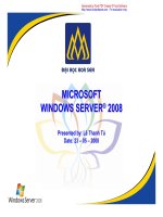
been considered before. Figure 1 presents the uptake of
99mTc-labeled HYNIC-TOC in a fibrot ic abdominal
surgical scar, showing that tissue repair involves SSTR.
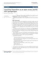
An “ ignored ” finding in NET patients can be seen in
Figure 2 w here tracer uptake within a small intestinal
loop is demonstrated. On theoretical grounds, one can
suspect t hat an inflammatory process is present. Inflam-
matory gut diseases and NET have been discussed
above.
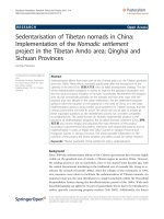
Figures 3 and 4 depict octreotide localized uptake in
peri-muscular structures of the thigh both in a male and
a female patient. The images have been taken from
PET/CT studies of patients with NET investigated with
68Ga-DOTA-TOC. The distribution pattern delineates
muscular fasciae [193]. For a general description of the
distribution of superficial and deep fasciae of the body,
the readers are referred to Gerlach [194]. Such fascia
structures have a longitudinal distribution between the
muscles and are considered to be involved in epimuscu-
lar myofascial force transmission (e.g., Figure nine in
[195]). Conventional three-view reconstruction algo-
rithms in Nuclear Medicine do not produce a suitable
Figure 1 99mTc
99
-HYNIC-TOC uptake in fibrotic scar tissue
after median laparotomy.
Moncayo EJNMMI Research 2011, 1:9
/>Page 6 of 16
image of this t ype of aligned structures. An adequate
approach to achieve this would be to apply the princi-
ples of diffusion spectrum MRI tractography [196]. The
applicability of this method in the investigation of myo-
cardial, i.e., muscular structures, has been recently
demonstrated [197,198].
Betwee n the fasciae and connective tissue in the body,
tensile forces are active. Standerwick and Roberts have
described these relations for craniofacial growth [199]. I
propose that the punctual uptake is in relation to
mechanosensing, since distension of the muscles, e.g.,
through eccentric exercise, will act on these structures
[200-202]. This is the basic assumption that we have
included in the musculoskeletal model of thyroid-asso-
ciated orbitopathy [76].
The careful observer of figures will also recognize tra-
cer uptake in the subcutaneous fatty structures. While it
is not possible to deliver an exact anatomical correlate
of this binding site, the distribution pattern might be
involved in processes of metabolic signaling such as
lipolysis [203,204]. In humans, changes of both SST
secretion and SSTR expression has bee n described in
conditions of infection and inflammation of adipose tis-
sue [205].
Recent data on SSTR in the early years of the new century
In the process of editing the final version of this manu-
script, I came across some recent data on SSTR which
should be mentioned here. The data presented above
has in a way a historical character. New developments,
however, change the face of science.
One of the most relevant aspects on SSTR is that of
truncated somatostatin receptors. The group of Cór-
doba-Chacón et al. have described a series of new spli-
cing variants of the SSTR-5 molecule in pituitary
tumors as well as in rodents [ 206,207]. The result of
these alterations is the appearance of SSTR that have
different numbers of trans-membrane domains (TMD).
In their recent publication [208], they describe some
characteristics of the human spliced variants of the SST
receptor subtype 5. Among t hese is the novel evidence
that demonstrate that an SST can react in different ways
to very si milar analogs of SST. One clinically important
Figure 2 PET/CT Ga
68
-TOC uptake in intestinal structures, in abdominal muscles, and abdominal fatty tissue. The image was taken from
a 56-year-old female patient with a NET.
Moncayo EJNMMI Research 2011, 1:9
/>Page 7 of 16
issue is the finding of SST analog-resistant somatotropi-
nomas where the variant called hsst5TMD4 was found
[209]. In their review on the topic of truncated somatos-
tatin receptors, Córdoba-Chacón et al. discuss the issues
that led to a novel reasoning for the interpretation of
SST-knocko ut models [208]. The data discussed origi-
nated new explanations for the different effects of SST
and corti statin at different levels. This finding is of out-
most importance in the clinical management of pat ients
with NET, since sole tracer binding will not be able to
identify the existence of this truncated receptor variant,
while at the same time therapeutic efforts with either
unlabelled or labeled SSTA might remain unsuccessful.
Implementation of these methods in cases of NET is
badly needed!
Besides changes in the receptor peptide structure, a
further factor that influences binding is the carbohydrate
component of the moiety [210]. In a recent article,
Møller et al. discuss the influence of the carbohydrate
component on SSTR (section 2.3.2. in [211]). Glycosyla-
tion moieties are present in SSTR5 [212].
An ample description and discussion on somatostatin
receptors based on the experience with patients with
acromegaly has been recently presented by Colao et al.
[213]. The clear advantage in the field of acromegaly it
that it is possible to investigate the efficacy of different
therapeutic approaches by determining the targets, e.g.,
growth hormone, IGF-I, as well as the characteristics of
the pituitary tumor. This is not always the case in cases
with NET since the decision to initiate treatment might
simply come from scintigraphic results [214] even when
the patients have no significant endocrinological altera-
tions. One important aspect in Colao’ s paper is the
description of SSA resistant cases. This situation might
also occur in NET.
Man is not alone in nature. Developmental aspects o f
receptors have been presented by other authors. Gahete
et al. have summarized data related to the development
of somatostatin receptors from fish to mammals [215].
EmphasishasbeenputonthesectiononnewSST
receptors. In a similar way Vaudry et al. have described
the evolution of urotensin receptors [216]. These early
Figure 3 A 61-year-old male patient presenting intense uptake in muscular structures and in fatty tissues.LowlevelsofSewere
documented in this case.
Moncayo EJNMMI Research 2011, 1:9
/>Page 8 of 16
forms of SSTRs have the potential of being more easily
accessed for scientific research.
Conclusions: the fiction in science [217,218]
Science is an art. In doing medical research, we attempt
to recognize the elements involved in this art frame and
await confirmation of hypotheses by going through
empirical and evidence-based paths [219-226]. In view
of the biochemical complexity of receptor interaction
outlined in this review, I believe that the most suitable
graphi cal and operational representation is that of coali-
tionchessasproposedbyArnoldSchoenberginthe
1920s [227]. Coalition che ss involves four players with
each player moving different ches s fi gures. It allows the
possibility to form coalitions between the players-as ago-
nists or antagonists. Using this chess variant as a con-
cept, one can imagine “the players” being involved in
receptor interaction. Modern methods like functional
proteomics and genomics [228], functional nutrige-
nomics [229], receptorome mining [98], chemogenomics
[230], and metabolomics [231] will surely gain relevance
in the field of Nuclear Medicine in order to decipher
these m ultiple interactions. Multiagent multitarget pro-
cedures, similar to the herbal combination described
herein, could be analyzed by nutriome methods [232].
For NM, one could envision the use of multivalent tra-
cers or tracer mixtures in therapeutic situations. In addi-
tion, we can expect that a new terminology will be
proposed for receptor forms and interactions [233,234].
The influence of morphogens and morphostats on NET
is also added as a r esearch direction to be kept in mind
[190,191,235-240]. Finally, basic research will start to get
involved with spliceosome dynamics [241] in order to
provide answers to the truncated forms of SSTR.
I present a graphi cal summary of this review in Figure
5. Upon seeing the figure, the r eader might recall the
words of William Wordsworth: “ My heart leaps up
when I behold a rainbow in the sky” .Iproposethat
when we look at octreotide scintigraphy, while still
strictly sticking to the procedure guidelines [242,243],
we should think on inflammation, nociception, mechan-
osensing,chemosensing,fibrosis,taste,vascularity,and
also tumor. An increase of any of these single processes
Figure 4 A 46-year-old female patient with axial mis-al ignment, the mid-line is shifted to the left. The uptake intensity is greater as in
the previous case. Axial mis-alignment could influence mechanotension of the muscular structures and produce enhanced SSTR expression.
Moncayo EJNMMI Research 2011, 1:9
/>Page 9 of 16
might result in increased tracer uptake making up a
rainbow in our imaging sky. While people tend to
believe w hat they see, and Nuclear Medicine is an ima-
ging specialty indeed, we should not forget the basic
principle of uncertainty in science [244]. Quoting Cas-
tillo while he refers to Feynman [245] one reads: “He
goesontosaythatifwearefreeofdoubtandignor-
ance, we will not get any new ideas and make no
progress”.
Author information
Roy Moncayo is trained in Internal Medicine, Endocri-
nology, Nuclear Medicine, Chinese Acupunture, and
Western Herbal Therapies and holds a MAS in Health
and Fitness. He is Deputy Head of the Department of
Nuclear Medicine at the Medical University in
Innsbruck. His clinical experience with octreotide is
centered on thyroid-associated orbitopathy. He carries
out complementary work on musculoskeletal disorders
at WOMED, Innsbruck.
Abbreviations
68Ga-TOC:
68
Ga-DOTA
0
-Tyr
3
octreotide; 7TM: seven7 -transmembrane helical
receptors; CNS: central nervous system; GI: gastrointestinal; GPCR: G-protein
coupled receptors; MRI: magnetic resonance imaging; NET: neuroendocrine
tumors; RAMPS: receptor-activity-modifying proteins; Se: selenium; SSA:
somatostatin analogs; SST: somatostatin; SSTR: somatostatin receptors; TAO:
thyroid- associated orbitopathy; TCM: traditional Chinese medicine; TMD:
transmembrane domains.
Acknowledgements
The Nuclear Medicine images have been produced at the Department of
Nuclear Medicine of the Medical University of Innsbruck. Financial support
for literature search and ordering was provided by WOMED.
The first version of this paper was presented at the: “3rd INTERNATIONAL
CONFERENCE ON RADIOPHARMACEUTICAL THERAPY (ICRT-2009)” in
Cartagena, Colombia, on November 2009. The article was originally
conceived as an editorial but come time come a larger scope.
The author wants to express sincere thanks to the reviewers who made
several precious and constructive suggestions.
Author details
1
Department of Nuclear Medicine, Medical University of Innsbruck, Innsbruck,
Austria
2
WOMED, Karl-Kapferer-Strasse 5, 6020 Innsbruck, Austria
Authors’ contributions
RM conceived the idea, wrote the manuscript, and drew the graphics.
Competing interests
The author declares that he has no competing interests.
Received: 10 May 2011 Accepted: 26 July 2011 Published: 26 July 2011
References
1. Rolleman EJ, Kooij PP, De Herder WW, Valkema R, Krenning EP, de Jong M:
Somatostatin receptor subtype 2-mediated uptake of radiolabelled
somatostatin analogues in the human kidney. Eur J Nucl Med Mol
Imaging 2007, 34:1854-1860.
2. Hofland LJ, Lamberts SW, van Hagen PM, Reubi JC, Schaeffer J, Waaijers M,
et al: Crucial role for somatostatin receptor subtype 2 in determining the
uptake of [111In-DTPA-D-Phe1]octreotide in somatostatin receptor-
positive organs. J Nucl Med 2003, 44:1315-1321.
Figure 5 Graphical summary of this review. This graphical summary of this review presents several real-time players that make up the total
environment behind octreotide scintigraphy. (1) Nutrition: sufficient macro- and micronutrients of good quality are the starting point. Intestinal
function will have an influence on absorption. (2) Endogenous ligands: somatostatin, corticostatin, urotensin, neuronostatin. (3) Pharmacological
agents: octreotide, DOTA-chelators in tracers, lanreotide, omeprazole, lipid modifiers. (4) Ability to cope with oxidative status and inflammatory
conditions. (5) Characteristics of the GPCR system: homo- and heterodimers, truncated receptors, RAMPS, arrestins, etc. (6) Morphostats, and cell
regulators in malignancy.
Moncayo EJNMMI Research 2011, 1:9
/>Page 10 of 16
3. Mansi L, Virgolini I: Diagnosis and therapy are walking together on
radiopeptides’ avenue. Eur J Nucl Med Mol Imaging 2011, 38:605-612.
4. Henri V: Lois générales de l’action des diastases. Paris , 1 1903.
5. Langley JN: On the reaction of cells and of nerve-endings to certain
poisons, chiefly as regards the reaction of striated muscle to nicotine
and to curare. J Physiol 1905, 33:374-413.
6. Michaelis L, Menten M: Die Kinetik der Invertinwirkung. Biochem Z 1913,
49:333-369.
7. Clark AJ: The reaction between acetyl choline and muscle cells. J Physiol
1926, 61:530-546.
8. Stephenson RP: A modification of receptor theory. Br J Pharmacol
Chemother 1956, 11:379-393.
9. Thron CD: On the analysis of pharmacological experiments in terms of
an allosteric receptor model. Mol Pharmacol 1973, 9:1-9.
10. Kilbourn MR, Zalutsky MR: Research and clinical potential of receptor
based radiopharmaceuticals. J Nucl Med 1985, 26:655-662.
11. Savageau MA: Michaelis-Menten mechanism reconsidered: implications
of fractal kinetics. J Theor Biol 1995, 176:115-124.
12. Grima R, Schnell S: A systematic investigation of the rate laws valid in
intracellular environments. Biophys Chem 2006, 124:1-10.
13. Aranda JS, Salgado E, Muñoz-Diosdado A: Multifractality in intracellular
enzymatic reactions. J Theor Biol 2006, 240:209-217.
14. Guillemin R: Hypothalamic hormones a.k.a. hypothalamic releasing
factors. J Endocrinol 2005, 184:11-28.
15. Guillemin R: Somatostatin: The beginnings, 1972. Mol Cell Endocrinol 2008,
286:3-4.
16. Brazeau P, Vale W, Burgus R, Ling N, Butcher M, Rivier J, et al: Hypothalamic
polypeptide that inhibits the secretion of immunoreactive pituitary
growth hormone. Science 1973, 179:77-79.
17. Marbach P, Bauer W, Briner U, Dopfner W, Petcher T, Pless J: Structure-
function relationships of somatostatin analogs.
Horm Res 1988, 29:54-58.
18.
Pless J: The history of somatostatin analogs. J Endocrinol Invest 2005,
28:1-4.
19. Weckbecker G, Lewis I, Albert R, Schmid HA, Hoyer D, Bruns C:
Opportunities in somatostatin research: biological, chemical and
therapeutic aspects. Nat Rev Drug Discov 2003, 2:999-1017.
20. Porstad OP, Schonbrunn A, Martin JB: Somatostatin radioreceptor assay:
development and application to the measurement of somatostatinlike
activity in the rat central nervous system. Can J Biochem Cell Biol 1983,
61:532-537.
21. Weckbecker G, Raulf F, Stolz B, Bruns C: Somatostatin analogs for
diagnosis and treatment of cancer. Pharmacol Ther 1993, 60:245-264.
22. Bakker WH, Krenning EP, Breeman WA, Koper JW, Kooij PP, Reubi JC, et al:
Receptor scintigraphy with a radioiodinated somatostatin analogue:
radiolabeling, purification, biologic activity, and in vivo application in
animals. J Nucl Med 1990, 31:1501-1509.
23. Krenning EP, Bakker WH, Kooij PP, Breeman WA, Oei HY, de Jong M, et al:
Somatostatin receptor scintigraphy with indium-111-DTPA-D-Phe-1-
octreotide in man: metabolism, dosimetry and comparison with iodine-
123-Tyr-3-octreotide. J Nucl Med 1992, 33:652-658.
24. Gabriel M, Muehllechner P, Decristoforo C, von Guggenberg E, Kendler D,
Prommegger R, et al: 99mTc-EDDA/HYNIC-Tyr(3)-octreotide for staging
and follow-up of patients with neuroendocrine gastro-entero-pancreatic
tumors. Q J Nucl Med Mol Imaging 2005, 49:237-244.
25. Hofland LJ, Lamberts SW: The pathophysiological consequences of
somatostatin receptor internalization and resistance. Endocr Rev 2003,
24:28-47.
26. Deshmukh MV, Voll G, Kühlewein A, Mäcke H, Schmitt J, Kessler H, et al:
NMR studies reveal structural differences between the gallium and
yttrium complexes of DOTA-D-Phe1-Tyr3-octreotide. J Med Chem 2005,
48:1506-1514.
27. Longnecker DS, Lilja HS, French J, Kuhlmann E, Noll W: Transplantation of
azaserine-induced carcinomas of pancreas in rats. Cancer Lett 1979,
7:197-202.
28. Melis M, Forrer F, Capello A, Bijster M, Bernard BF, Reubi JC, et al: Up-
regulation of somatostatin receptor density on rat CA20948 tumors
escaped from low dose [177Lu-DOTA0,Tyr3]octreotate therapy. Q J Nucl
Med Mol Imaging 2007, 51:324-333.
29. Gabriel M, Decristoforo C, Kendler D, Dobrozemsky G, Heute D, Uprimny C,
et al: 68Ga-DOTA-Tyr3-octreotide PET in neuroendocrine tumors:
comparison with somatostatin receptor scintigraphy and CT. J Nucl Med
2007, 48:508-518.
30. Modlin IM, Shapiro MD, Kidd M: Carcinoid tumors and fibrosis: an
association with no explanation. Am
J Gastroenterol 2004, 99:2466-2478.
31. Druce M, Rockall A, Grossman AB: Fibrosis and carcinoid syndrome: from
causation to future therapy. Nat Rev Endocrinol 2009, 5:276-283.
32. Pan Q, Li DG, Lu HM, Lu LY, You HN, Xu QF: Relationship between
somatostatin receptors and activation of hepatic stellate cells. Chin Med
J (Engl) 2004, 117:1665-1669.
33. Öhrvall U, Westlin JE, Nilsson S, Wilander E, Juhlin C, Rastad J, et al: Human
biodistribution of [111In]diethylenetriaminepentaacetic acid-(DTPA)-D-
[Phe1]-octreotide and peroperative detection of endocrine tumors.
Cancer Res 1995, 55:5794s-5800s.
34. Lebtahi R, Moreau S, Marchand-Adam S, Debray MP, Brauner M, Soler P,
et al: Increased uptake of 111In-octreotide in idiopathic pulmonary
fibrosis. J Nucl Med 2006, 47:1281-1287.
35. Chen CC, Czerwiec FS, Feuillan PP: Visualization of fibrous dysplasia
during somatostatin receptor scintigraphy. J Nucl Med 1998, 39:238-240.
36. Fjallskog ML, Ludvigsen E, Stridsberg M, Oberg K, Eriksson B, Janson ET:
Expression of somatostatin receptor subtypes 1 to 5 in tumor tissue and
intratumoral vessels in malignant endocrine pancreatic tumors. Med
Oncol 2003, 20:59-67.
37. Danesi R, Agen C, Benelli U, Paolo AD, Nardini D, Bocci G, et al: Inhibition
of experimental angiogenesis by the somatostatin analogue octreotide
acetate (SMS 201-995). Clin Cancer Res 1997, 3:265-272.
38. Woltering EA, Barrie R, O’dorisio TM, Arce D, Ure T, Cramer A, et al:
Somatostatin analogues inhibit angiogenesis in the chick chorioallantoic
membrane. J Surg Res 1991, 50:245-251.
39. Guo MM, Tan QH, Fan H, Huang MH, Wang CH, Qiu XQ, et al: [Changes of
somatostatin and expression of somatostatin receptor in small intestine
and liver tissues during macaque development]. Acta Physiol Sin 2005,
57:719-724.
40. Rong W, Winchester WJ, Grundy D: Spontaneous hypersensitivity in
mesenteric afferent nerves of mice deficient in the sst2 subtype of
somatostatin receptor. J Physiol 2007, 581:779-786.
41. Hens J, Vanderwinden JM, De Laet MH, Scheuermann DW, Timmermans JP:
Morphological and neurochemical identification of enteric neurones
with mucosal projections in the human small intestine. J Neurochem
2001, 76:464-471.
42. Roca B, Fernandez-Valencia R, Arilla E: Effects of fasting and refeeding on
somatostatin concentration and binding to cytosol from rabbit gastric
mucosa. Gut
1988, 29:642-646.
43.
Gonkowski S, Calka J: Changes in the somatostatin (SOM)-like
immunoreactivity within nervous structures of the porci ne desce nding
colon under various pathological factors. Exp Mol Pathol 2010,
88:416-423.
44. Koch TR, Carney JA, Morris VA, Go VL: Somatostatin in the idiopathic
inflammatory bowel diseases. Dis Colon Rectum 1988, 31:198-203.
45. Rong W, Winchester WJ, Grundy D: Spontaneous hypersensitivity in
mesenteric afferent nerves of mice deficient in the sst2 subtype of
somatostatin receptor. J Physiol 2007, 581:779-786.
46. Reubi JC, Laissue J, Waser B, Horisberger U, Schaer JC: Expression of
somatostatin receptors in normal, inflamed, and neoplastic human
gastrointestinal tissues. Ann N Y Acad Sci 1994, 733:122-137.
47. West NE, Wise PE, Herline AJ, Muldoon RL, Chopp WV, Schwartz DA:
Carcinoid tumors are 15 times more common in patients with Crohn’s
disease. Inflamm Bowel Dis 2007, 13:1129-1134.
48. Grassia R, Bodini P, Dizioli P, Staiano T, Iiritano E, Bianchi G, et al:
Neuroendocrine carcinomas arising in ulcerative colitis: coincidences or
possible correlations? World J Gastroenterol 2009, 15:4193-4195.
49. Sciola V, Massironi S, Conte D, Caprioli F, Ferrero S, Ciafardini C, et al:
Plasma chromogranin a in patients with inflammatory bowel disease.
Inflamm Bowel Dis 2009, 15:867-871.
50. Genton L, Kudsk KA: Interactions between the enteric nervous system
and the immune system: role of neuropeptides and nutrition. Am J Surg
2003, 186:253-258.
51. Van Op dB, van Nassauw L, Lantermann K, van Marck E, Timmermans JP:
Effect of intestinal inflammation on the cell-specific expression of
somatostatin receptor subtypes in the murine ileum. Neurogastroenterol
Motil 2007, 19:596-606.
Moncayo EJNMMI Research 2011, 1:9
/>Page 11 of 16
52. Van Op dB, Lantermann K, Torfs P, van Marck E, van Nassauw L,
Timmermans JP: Distribution and expression levels of somatostatin and
somatostatin receptors in the ileum of normal and acutely Schistosoma
mansoni-infected SSTR2 knockout/lacZ knockin mice. Neurogastroenterol
Motil 2008, 20:798-807.
53. Corleto VD: Somatostatin and the gastrointestinal tract. Curr Opin
Endocrinol Diabetes Obes 2010, 17:63-68.
54. Mantyh PW, Hunt SP: Neuropeptides are present in projection neurones
at all levels in visceral and taste pathways: from periphery to sensory
cortex. Brain Res 1984, 299:297-312.
55. Tal M, Ammar DA, Karpuj M, Krizhanovsky V, Naim M, Thompson DA: A
novel putative neuropeptide receptor expressed in neural tissue,
including sensory epithelia. Biochem Biophys Res Commun 1995,
209:752-759.
56. Carlton SM, Du J, Zhou S, Coggeshall RE: Tonic control of peripheral
cutaneous nociceptors by somatostatin receptors. J Neurosci 2001,
21:4042-4049.
57. Carlton SM, Zhou S, Kraemer B, Coggeshall RE: A role for peripheral
somatostatin receptors in counter-irritation-induced analgesia.
Neuroscience 2003, 120:499-508.
58. Bär KJ, Schurigt U, Scholze A, Segond von Banchet G, Stopfel N, Bräuer R,
et al: The expression and localization of somatostatin receptors in dorsal
root ganglion neurons of normal and monoarthritic rats. Neuroscience
2004, 127:197-206.
59. Abd El-Aleem SA, Morales-Aza BM, McQueen DS, Donaldson LF:
Inflammation alters somatostatin mRNA expression in sensory neurons
in the rat. Eur J Neurosci 2005, 21:135-141.
60. Dong ZQ, Xie H, Ma F, Li WM, Wang YQ, Wu GC: Effects of
electroacupuncture on expression of somatostatin and
preprosomatostatin mRNA in dorsal root ganglions and spinal dorsal
horn in neuropathic pain rats. Neurosci Lett 2005, 385:189-194.
61. Pintér E, Helyes Z, Szolcsányi J: Inhibitory effect of somatostatin on
inflammation and nociception. Pharmacol Ther 2006, 112:440-456.
62. Rong W, Winchester WJ, Grundy D: Spontaneous hypersensitivity in
mesenteric afferent nerves of mice deficient in the sst2 subtype of
somatostatin receptor. J Physiol 2007, 581:779-786.
63. Pan HL, Wu ZZ, Zhou HY, Chen SR, Zhang HM, Li DP: Modulation of pain
transmission by G-protein-coupled receptors. Pharmacol Ther 2008,
117:141-161.
64. Carlton SM, Zhou S, Du J, Hargett GL, Ji G, Coggeshall RE: Somatostatin
modulates the transient receptor potential vanilloid 1 (TRPV1) ion
channel. Pain 2004, 110:616-627.
65. Gillespie PG, Walker RG: Molecular basis of mechanosensory transduction.
Nature 2001, 413:194-202.
66. Huang CL: The
transient receptor potential superfamily of ion channels. J
Am Soc Nephrol 2004, 15:1690-1699.
67. O’Neil RG, Heller S: The mechanosensitive nature of TRPV channels.
Pflugers Arch 2005, 451:193-203.
68. Liedtke W: Transient receptor potential vanilloid channels functioning in
transduction of osmotic stimuli. J Endocrinol 2006, 191:515-523.
69. Endres-Becker J, Heppenstall PA, Mousa SA, Labuz D, Oksche A, Schäfer M,
et al: Mu-opioid receptor activation modulates transient receptor
potential vanilloid 1 (TRPV1) currents in sensory neurons in a model of
inflammatory pain. Mol Pharmacol 2007, 71:12-18.
70. Mudie AS, Holland GR: Local opioids in the inflamed dental pulp. J Endod
2006, 32:319-323.
71. Lamberts SW, Bakker WH, Reubi JC, Krenning EP: Somatostatin-receptor
imaging in the localization of endocrine tumors. N Engl J Med 1990,
323:1246-1249.
72. Eschalier A, Aumaître O, Ardid D, Fialip J, Duchêne-Marullaz P: Long-lasting
antinociceptive effect of RC-160, a somatostatin analog, in mice and
rats. Eur J Pharmacol 1991, 199:119-121.
73. Betoin F, Ardid D, Herbet A, Aumaitre O, Kemeny JL, Duchene-Marullaz P,
et al: Evidence for a central long-lasting antinociceptive effect of
vapreotide, an analog of somatostatin, involving an opioidergic
mechanism. J Pharmacol Exp Ther 1994, 269:7-14.
74. Moncayo R, Baldissera I, Decristoforo C, Kendler D, Donnemiller E:
Evaluation of immunological mechanisms mediating thyroid-associated
ophthalmopathy by radionuclide imaging using the somatostatin analog
111In-octreotide. Thyroid 1997, 7:21-29.
75. Kainz H, Bale R, Donnemiller E, Gabriel M, Kovacs P, Decristoforo C, et al:
Image fusion analysis of (99 m)Tc-HYNIC-octreotide scintigraphy and CT/
MRI in patients with thyroid-associated orbitopathy: the importance of
the lacrimal gland. Eur J Nucl Med Mol Imaging 2003, 30:1155-1159.
76. Moncayo R, Moncayo H: A musculoskeletal model of low grade
connective tissue inflammation in patients with thyroid associated
ophthalmopathy (TAO): the WOMED concept of lateral tension and its
general implications in disease. BMC Musculoskelet Disord 2007, 8:17.
77. Liu Y, Yang J, Cai Z: Chemical investigation on Sijunzi decoction and its
two major herbs Panax ginseng and Glycyrrhiza uralensis by LC/MS/MS.
J Pharm Biomed Anal 2006, 41:1642-1647.
78. Liu Q, Cai G: [Content of somatostatin and cholecystokinin-8 in
hypothalamus and colons in a rat model of spleen-deficiency
syndrome]. Zhong Xi Yi Jie He Xue Bao 2007, 5:555-558.
79. Ross J: Combining western herbs and chinese medicine. Principles, practice &
materia medica. 1 edition. Seattle: Greenfields Press; 2003.
80. Bohlmann J, Lins T, Martin W, Eilert U:
Anthranilate synthase from Ruta
graveolens.
Duplicated AS alpha genes encode tryptophan-sensitive and
tryptophan-insensitive isoenzymes specific to amino acid and alkaloid
biosynthesis. Plant Physiol 1996, 111:507-514.
81. Panossian A, Wagner H: Stimulating effect of adaptogens: an overview
with particular reference to their efficacy following single dose
administration. Phytother Res 2005, 19:819-838.
82. Bystrov NS, Dobrynin VN, Kolosov MN, Chernov BK, Chervin II: [Structure of
the chromophoric part of hyperforin]. Dokl Akad Nauk SSSR 1975,
225:1327-1328.
83. Zeng JZ, Sun DF, Wang L, Cao X, Qi JB, Yang T, et al: Hypericum sampsonii
induces apoptosis and nuclear export of retinoid X receptor-alpha.
Carcinogenesis 2006, 27:1991-2000.
84. Medina MA, Martinez-Poveda B, Amores-Sanchez MI, Quesada AR:
Hyperforin: more than an antidepressant bioactive compound? Life Sci
2006, 79:105-111.
85. Wirz A, Simmen U, Heilmann J, Calis I, Meier B, Sticher O: Bisanthraquinone
glycosides of Hypericum perforatum with binding inhibition to CRH-1
receptors. Phytochemistry 2000, 55:941-947.
86. Beerhues L: Hyperforin. Phytochemistry 2006, 67:2201-2207.
87. Dell’Aica I, Caniato R, Biggin S, Garbisa S: Matrix proteases, green tea, and
St. John’s wort: biomedical research catches up with folk medicine. Clin
Chim Acta 2007, 381:69-77.
88. Yarnell E: Botanical medicines for the urinary tract. World J Urol 2002,
20:285-293.
89. Chiu PY, Ko KM: Schisandrin B-induced increase in cellular glutathione
level and protection against oxidant injury are mediated by the
enhancement of glutathione synthesis and regeneration in AML12 and
H9c2 cells. Biofactors 2006, 26:221-230.
90. Choi SI, Park SR, Heo TR: Matrix degradation inhibitory effect of
Schisandra fructus on human articular cartilage and chondrocytes. J
Ethnopharmacol 2006, 106:279-284.
91. Mu Y, Zhang J, Zhang S, Zhou HH, Toma D, Ren S, et al: Traditional
Chinese medicines Wu Wei Zi (Schisandra chinensis Baill) and Gan
Cao (Glycyrrhiza uralensis Fisch) activate pregnane X receptor and
increase warfarin clearance in rats. J Pharmacol Exp Ther 2006,
316:1369-1377.
92. Kou J, Sun Y, Lin Y, Cheng Z, Zheng W, Yu B, et al: Anti-inflammatory
activities of aqueous extract from Radix Ophiopogon japonicus and its
two constituents. Biol Pharm Bull 2005,
28:1234-1238.
93.
Ko KM, Leung HY: Enhancement of ATP generation capacity, antioxidant
activity and immunomodulatory activities by Chinese Yang and Yin
tonifying herbs. Chin Med 2007, 2:3.
94. Nurtjahja-Tjendraputra E, Ammit AJ, Roufogalis BD, Tran VH, Duke CC:
Effective anti-platelet and COX-1 enzyme inhibitors from pungent
constituents of ginger. Thromb Res 2003, 111:259-265.
95. Chrubasik S, Pittler MH, Roufogalis BD: Zingiberis rhizoma: a
comprehensive review on the ginger effect and efficacy profiles.
Phytomedicine 2005, 12:684-701.
96. Grzanna R, Lindmark L, Frondoza CG: Ginger–an herbal medicinal product
with broad anti-inflammatory actions. J Med Food 2005, 8:125-132.
97. Lantz RC, Chen GJ, Sarihan M, Solyom AM, Jolad SD, Timmermann BN: The
effect of extracts from ginger rhizome on inflammatory mediator
production. Phytomedicine 2007, 14:123-128.
Moncayo EJNMMI Research 2011, 1:9
/>Page 12 of 16
98. Roth BL: Receptor systems: will mining the receptorome yield novel
targets for pharmacotherapy? Pharmacol Ther 2005, 108:59-64.
99. Roth BL, Lopez E, Beischel S, Westkaemper RB, Evans JM: Screening the
receptorome to discover the molecular targets for plant-derived
psychoactive compounds: a novel approach for CNS drug discovery.
Pharmacol Ther 2004, 102:99-110.
100. Sucher NJ: Insights from molecular investigations of traditional Chinese
herbal stroke medicines: implications for neuroprotective epilepsy
therapy. Epilepsy Behav 2006, 8:350-362.
101. Straube A, Aicher B, Fiebich BL, Haag G: Combined analgesics in
(headache) pain therapy: shotgun approach or precise multi-target
therapeutics? BMC Neurol 2011, 11:43.
102. Naito T, Itoh H, Yasunaga F, Takeyama M: Rikkunshi-to raises levels of
somatostatin and gastrin in human plasma. Biol Pharm Bull 2001,
24:841-843.
103. Hu J, Wang Q, Liang W: [Effects of bushen yizhi recipe on somatostatin-
like immunopositive and somatostatin messenger ribonucleic acid
expressed-positive neurons in Alzheimer’s disease model rats]. Zhongguo
Zhong Xi Yi Jie He Za Zhi 2000, 20:533-535.
104. Naito T, Itoh H, Yasunaga F, Takeyama M: Hange-shashin-to raises levels of
somatostatin, motilin, and gastrin in the plasma of healthy subjects. Biol
Pharm Bull 2002, 25:327-331.
105. Gong Z, Yuan Y, Lou K, Tu S, Zhai Z, Xu J: Mechanisms of Chinese herb
emodin and somatostatin analogs on pancreatic regeneration in acute
pancreatitis in rats. Pancreas 2002, 25:154-160.
106. Katagiri F, Itoh H, Takeyama M: Effect of Sho-hange-ka-bukuryo-to on
gastrointestinal peptide concentrations in the plasma of healthy human
subjects. Biol Pharm Bull 2004, 27:1674-1678.
107. Naito T, Itoh H, Nagano T, Takeyama M: Effects of Ninjin-to on levels of
brain-gut peptides (motilin, vasoactive intestinal peptide, gastrin, and
somatostatin) in human plasma. Biol Pharm Bull 2001, 24:194-196.
108. Naito T, Itoh H, Takeyama M: Effects of Hange-koboku-to (Banxia-houpo-
tang) on neuropeptide levels in human plasma and saliva. Biol Pharm
Bull 2003, 26:1609-1613.
109. Hiruma-Lima CA, Calvo TR, Rodrigues CM, Andrade FD, Vilegas W, Brito AR:
Antiulcerogenic activity of Alchornea castaneaefolia: effects on
somatostatin, gastrin and prostaglandin. J Ethnopharmacol 2006,
104:215-224.
110. Katagiri F, Inoue S, Itoh H, Takeyama M: Omeprazole raises somatostatin
and motilin in human plasma. Biol Pharm Bull 2005, 28:370-373.
111. Pelley RJ, Bukowski RM: Recent advances in diagnosis and therapy of
neuroendocrine tumors of the gastrointestinal tract. Curr
Opin Oncol
1997, 9:68-74.
112. Marchese A, Heiber M, Nguyen T, Heng HH, Saldivia VR, Cheng R, et al:
Cloning and chromosomal mapping of three novel genes, GPR9, GPR10,
and GPR14, encoding receptors related to interleukin 8, neuropeptide Y,
and somatostatin receptors. Genomics 1995, 29:335-344.
113. Mori M, Sugo T, Abe M, Shimomura Y, Kurihara M, Kitada C, et al: Urotensin
II is the endogenous ligand of a G-protein-coupled orphan receptor,
SENR (GPR14). Biochem Biophys Res Commun 1999, 265:123-129.
114. Malagon MM, Molina M, Gahete MD, Duran-Prado M, Martinez-Fuentes AJ,
Alcain FJ, et al: Urotensin II and urotensin II-related peptide activate
somatostatin receptor subtypes 2 and 5. Peptides 2008, 29:711-720.
115. Tostivint H, Lihrmann I, Vaudry H: New insight into the molecular
evolution of the somatostatin family. Mol Cell Endocrinol 2008, 286:5-17.
116. Cordoba-Chacon J, Gahete MD, Durán-Prado M, Pozo-Salas AI,
Malagón MM, Gracia-Navarro F, et al: Identification and characterization of
new functional truncated variants of somatostatin receptor subtype 5 in
rodents. Cell Mol Life Sci 2010, 67:1147-1163.
117. Samson WK, Zhang JV, Avsian-Kretchmer O, Cui K, Yosten GL, Klein C, et al:
Neuronostatin encoded by the somatostatin gene regulates neuronal,
cardiovascular, and metabolic functions. J Biol Chem 2008,
283:31949-31959.
118. Allia E, Tarabra E, Volante M, Cerrato M, Ghigo E, Muccioli G, et al:
Expression of cortistatin and MrgX2, a specific cortistatin receptor, in
human neuroendocrine tissues and related tumours. J Pathol 2005,
207:336-345.
119. Volante M, Rosas R, Allia E, Granata R, Baragli A, Muccioli G, et al:
Somatostatin, cortistatin and their receptors in tumours. Mol Cell
Endocrinol 2008, 286:219-229.
120. Yang S, Liu Y, Lin AA, Cavalli-Sforza LL, Zhao Z, Su B: Adaptive evolution of
MRGX2, a human sensory neuron specific gene involved in nociception.
Gene 2005, 352:30-35.
121. Robas N, Mead E, Fidock M: MrgX2 is a high potency cortistatin receptor
expressed in dorsal root ganglion. J Biol Chem 2003, 278:44400-44404.
122. Ferone D, Boschetti M, Resmini E, Giusti M, Albanese V, Goglia U, et al:
Neuroendocrine-immune interactions: the role of cortistatin/
somatostatin system. Ann N Y Acad Sci 2006, 1069:129-144.
123. de Lecea L, Criado JR, Prospero-Garcia O, Gautvik KM, Schweitzer P,
Danielson PE, et al: A cortical neuropeptide with neuronal depressant
and sleep-modulating properties.
Nature 1996, 381:242-245.
124.
Fukusumi S, Kitada C, Takekawa S, Kizawa H, Sakamoto J, Miyamoto M, et al:
Identification and characterization of a novel human cortistatin-like
peptide. Biochem Biophys Res Commun 1997, 232:157-163.
125. Wreggett KA, Wells JW: Cooperativity manifest in the binding properties
of purified cardiac muscarinic receptors. J Biol Chem 1995,
270:22488-22499.
126. Birnbaumer L, Abramowitz J, Brown AM: Receptor-effector coupling by G
proteins. Biochim Biophys Acta 1990, 1031:163-224.
127. Gouldson PR, Snell CR, Bywater RP, Higgs C, Reynolds CA: Domain
swapping in G-protein coupled receptor dimers. Protein Eng 1998,
11:1181-1193.
128. Takesono A, Cismowski MJ, Ribas C, Bernard M, Chung P, Hazard S III, et al:
Receptor-independent activators of heterotrimeric G-protein signaling
pathways. J Biol Chem 1999, 274:33202-33205.
129. Lefkowitz RJ: The superfamily of heptahelical receptors. Nat Cell Biol 2000,
2:E133-E136.
130. Rios CD, Jordan BA, Gomes I, Devi LA: G-protein-coupled receptor
dimerization: modulation of receptor function. Pharmacol Ther 2001,
92:71-87.
131. Fredriksson R, Lagerström MC, Lundin LG, Schiöth HB: The G-protein-
coupled receptors in the human genome form five main families.
Phylogenetic analysis, paralogon groups, and fingerprints. Mol Pharmacol
2003, 63:1256-1272.
132. Fredriksson R, Schiöth HB: The repertoire of G-protein-coupled receptors
in fully sequenced genomes. Mol Pharmacol 2005, 67:1414-1425.
133. Javitch JA: The ants go marching two by two: oligomeric structure of G-
protein-coupled receptors. Mol Pharmacol 2004, 66:1077-1082.
134. Skrabanek L, Murcia M, Bouvier M, Devi L, George SR, Lohse MJ, et al:
Requirements and ontology for a G protein-coupled receptor
oligomerization knowledge base. BMC Bioinformatics 2007, 8:177.
135. Agnati LF, Fuxe K, Zini I, Lenzi P, Hökfelt T: Aspects on receptor regulation
and isoreceptor identification. Med Biol 1980, 58:182-187.
136. Reggio PH: Computational methods in drug design: modeling G protein-
coupled receptor monomers, dimers, and oligomers. AAPS J 2006, 8:
E322-E336.
137. Agnati LF, Fuxe KG, Goncharova LB, Tarakanov AO: Receptor mosaics of
neural and immune communication: Possible implications for basal
ganglia
functions. Brain Res Rev 2007, 58:400-414.
138. Fuxe K, Canals M, Torvinen M, Marcellino D, Terasmaa A, Genedani S, et al:
Intramembrane receptor-receptor interactions: a novel principle in
molecular medicine. J Neural Transm 2007, 114:49-75.
139. Fuxe K, Marcellino D, Rivera A, Diaz-Cabiale Z, Filip M, Gago B, et al:
Receptor-receptor interactions within receptor mosaics. Impact on
neuropsychopharmacology. Brain Res Rev 2008, 58:415-452.
140. Durroux T: Principles: a model for the allosteric interactions between
ligand binding sites within a dimeric GPCR. Trends Pharmacol Sci 2005,
26:376-384.
141. Giraldo J: On the fitting of binding data when receptor dimerization is
suspected. Br J Pharmacol 2008, 155:17-23.
142. Rovira X, Pin JP, Giraldo J: The asymmetric/symmetric activation of GPCR
dimers as a possible mechanistic rationale for multiple signalling
pathways. Trends Pharmacol Sci 2010, 31:15-21.
143. Salahpour A, Angers S, Bouvier M: Functional significance of
oligomerization of G-protein-coupled receptors. Trends Endocrinol Metab
2000, 11:163-168.
144. Lundstrom K: Structural genomics of GPCRs. Trends Biotechnol 2005,
23:103-108.
145. Rocheville M, Lange DC, Kumar U, Sasi R, Patel RC, Patel YC: Subtypes of
the somatostatin receptor assemble as functional homo- and
heterodimers. J Biol Chem 2000, 275:7862-7869.
Moncayo EJNMMI Research 2011, 1:9
/>Page 13 of 16
146. Hukovic N, Rocheville M, Kumar U, Sasi R, Khare S, Patel YC: Agonist-
dependent up-regulation of human somatostatin receptor type 1
requires molecular signals in the cytoplasmic C-tail. J Biol Chem 1999,
274:24550-24558.
147. Tannenbaum GS, Turner J, Guo F, Videau C, Epelbaum J, Beaudet A:
Homologous upregulation of sst2 somatostatin receptor expression in
the rat arcuate nucleus in vivo. Neuroendocrinology 2001, 74:33-42.
148. Pfeiffer M, Koch T, Schröder H, Klutzny M, Kirscht S, Kreienkamp HJ, et al:
Homo- and heterodimerization of somatostatin receptor subtypes.
Inactivation of sst(3) receptor function by heterodimerization with sst
(2A). J Biol Chem 2001, 276:14027-14036.
149. Grant M, Collier B, Kumar U: Agonist-dependent dissociation of human
somatostatin receptor 2 dimers: a role in receptor trafficking. J Biol Chem
2004, 279:36179-36183.
150. Jacobs S, Schulz S: Intracellular trafficking of somatostatin receptors. Mol
Cell Endocrinol 2007, 286:58-62.
151. Durán-Prado M, Malagón MM, Gracia-Navarro F, Castaño JP: Dimerization
of G protein-coupled receptors: New avenues for somatostatin receptor
signalling, control and functioning. Mol Cell Endocrinol 2008, 286:63-68.
152. Reubi JC, Erchegyi J, Cescato R, Waser B, Rivier JE: Switch from antagonist
to agonist after addition of a DOTA chelator to a somatostatin analog.
Eur J Nucl Med Mol Imaging 2010, 37:1551-1558.
153. Rocheville M, Lange DC, Kumar U, Patel SC, Patel RC, Patel YC: Receptors
for dopamine and somatostatin: formation of hetero-oligomers with
enhanced functional activity. Science 2000, 288:154-157.
154. Pfeiffer M, Koch T, Schröder H, Laugsch M, Höllt V, Schulz S:
Heterodimerization of somatostatin and opioid receptors cross-
modulates phosphorylation, internalization, and desensitization. J Biol
Chem 2002, 277:19762-19772.
155. Pfeiffer M, Kirscht S, Stumm R, Koch T, Wu D, Laugsch M, et al:
Heterodimerization of substance P and mu-opioid receptors regulates
receptor trafficking and resensitization. J Biol Chem 2003,
278:51630-51637.
156. Gomes I, Jordan BA, Gupta A, Trapaidze N, Nagy V, Devi LA:
Heterodimerization of mu and delta opioid receptors: A role in opiate
synergy. J Neurosci 2000, 20:RC110.
157. Gloriam DE, Fredriksson R, Schiöth HB: The G protein-coupled receptor
subset of the rat genome. BMC Genomics 2007, 8:338.
158. Kenakin TP: ’7TM receptor allostery: putting numbers to shapeshifting
proteins. Trends Pharmacol Sci 2009, 30:460-469.
159. O’Toole D, Saveanu A, Couvelard A, Gunz G, Enjalbert A, Jaquet P, et al: The
analysis of quantitative expression of somatostatin and dopamine
receptors
in gastro-entero-pancreatic tumours opens new therapeutic
strategies. Eur J Endocrinol 2006, 155:849-857.
160. Ferone D, Saveanu A, Culler MD, Arvigo M, Rebora A, Gatto F, et al: Novel
chimeric somatostatin analogs: facts and perspectives. Eur J Endocrinol
2007, 156(Suppl 1):S23-S28.
161. Arvigo M, Gatto F, Ruscica M, Ameri P, Dozio E, Albertelli M, et al:
Somatostatin and dopamine receptor interaction in prostate and lung
cancer cell lines. J Endocrinol 2010, 207:309-317.
162. Oakley RH, Laporte SA, Holt JA, Caron MG, Barak LS: Differential affinities
of visual arrestin, beta arrestin1, and beta arrestin2 for G protein-
coupled receptors delineate two major classes of receptors. J Biol Chem
2000, 275:17201-17210.
163. Ma L, Pei G: Beta-arrestin signaling and regulation of transcription. J Cell
Sci 2007, 120:213-218.
164. Kang J, Shi Y, Xiang B, Qu B, Su W, Zhu M, et al: A nuclear function of
beta-arrestin1 in GPCR signaling: regulation of histone acetylation and
gene transcription. Cell 2005, 123:833-847.
165. Storez H, Scott MG, Issafras H, Burtey A, Benmerah A, Muntaner O, et al:
Homo- and hetero-oligomerization of beta-arrestins in living cells. J Biol
Chem 2005, 280:40210-40215.
166. Tulipano G, Stumm R, Pfeiffer M, Kreienkamp HJ, Höllt V, Schulz S:
Differential beta-arrestin trafficking and endosomal sorting of
somatostatin receptor subtypes. J Biol Chem 2004, 279:21374-21382.
167. Tulipano G, Schulz S: Novel insights in somatostatin receptor physiology.
Eur J Endocrinol 2007, 156(Suppl 1):S3-S11.
168. Ramirez JL, Watt HL, Rocheville M, Kumar U: Agonist-induced up-
regulation of human somatostatin receptor type 1 is regulated by beta-
arrestin-1 and requires an essential serine residue in the receptor C-tail.
Biochim Biophys Acta 2005, 1669:182-192.
169. Parameswaran N, Spielman WS: RAMPs: The past, present and future.
Trends Biochem Sci 2006, 31:631-638.
170. Anderson RG: The caveolae membrane system. Annu Rev Biochem 1998,
67:199-225.
171. Chini B, Parenti M: G-protein coupled receptors in lipid rafts and
caveolae: how, when and why do they go there? J Mol Endocrinol 2004,
32:325-338.
172. Oh P, Schnitzer JE: Segregation of heterotrimeric G proteins in cell
surface microdomains. G(q) binds caveolin to concentrate in caveolae,
whereas G(i) and G(s) target lipid rafts by default. Mol Biol Cell 2001,
12:685-698.
173.
Ostrom RS, Insel PA: The evolving role of lipid rafts and caveolae in G
protein-coupled receptor signaling: implications for molecular
pharmacology. Br J Pharmacol 2004, 143:235-245.
174. Xu W, Yoon SI, Huang P, Wang Y, Chen C, Chong PL, et al: Localization of
the kappa opioid receptor in lipid rafts. J Pharmacol Exp Ther 2006,
317:1295-1306.
175. Harikumar KG, Puri V, Singh RD, Hanada K, Pagano RE, Miller LJ: Differential
effects of modification of membrane cholesterol and sphingolipids on
the conformation, function, and trafficking of the G protein-coupled
cholecystokinin receptor. J Biol Chem 2005, 280:2176-2185.
176. Gouldson PR, Dean MK, Snell CR, Bywater RP, Gkoutos G, Reynolds CA:
Lipid-facing correlated mutations and dimerization in G-protein coupled
receptors. Protein Eng 2001, 14:759-767.
177. Sjögren B, Hamblin MW, Svenningsson P: Cholesterol depletion reduces
serotonin binding and signaling via human 5-HT7(a) receptors. Eur J
Pharmacol 2006, 552:1-10.
178. Garcia JJ, Martinez-Ballarin E, Millan-Plano S, Allue JL, Albendea C, Fuentes L,
et al: Effects of trace elements on membrane fluidity. J Trace Elem Med
Biol 2005, 19:19-22.
179. Dobretsov GE, Borschevskaya TA, Petrov VA, Vladimirov YA: The increase of
phospholipid bilayer rigidity after lipid peroxidation. FEBS Lett 1977,
84:125-128.
180. Van der Vliet A, Bast A: Effect of oxidative stress on receptors and signal
transmission. Chem Biol Interact 1992, 85:95-116.
181. Borst JW, Visser NV, Kouptsova O, Visser AJ: Oxidation of unsaturated
phospholipids in membrane bilayer mixtures is accompanied by
membrane fluidity changes. Biochim Biophys Acta 2000, 1487:61-73.
182. Moncayo R, Traeger A: Decreased selenium levels after Peptide Receptor
Radionuclide Therapy (PRRT) in patients with neuroendocrine tumors:
Implications for the antioxidant status. Eur J Nucl Med Mol Imaging 2011,
38:1580-1581.
183. Michiels C, Raes M, Toussaint O, Remacle J: Importance of Se-glutathione
peroxidase, catalase, and Cu/Zn-SOD for cell survival against oxidative
stress. Free Radic Biol Med 1994, 17:235-248.
184. Diplock AT, Charleux JL, Crozier-Willi G, Kok FJ, Rice-Evans C, Roberfroid M,
et al: Functional food science and defence against reactive oxidative
species. Br J Nutr 1998, 80(Suppl 1):S77-112.
185. Patel HH, Murray F, Insel PA: Caveolae as organizers of pharmacologically
relevant signal transduction molecules. Annu Rev Pharmacol Toxicol 2008,
48:359-391.
186. Goetz JG, Lajoie P, Wiseman SM, Nabi IR: Caveolin-1
in tumor progression:
the good, the bad and the ugly. Cancer Metastasis Rev 2008, 27:715-735.
187. Nakakura EK, Sriuranpong VR, Kunnimalaiyaan M, Hsiao EC, Schuebel KE,
Borges MW, et al: Regulation of neuroendocrine differentiation in
gastrointestinal carcinoid tumor cells by notch signaling. J Clin Endocrinol
Metab 2005, 90:4350-4356.
188. Kunnimalaiyaan M, Vaccaro AM, Ndiaye MA, Chen H: Inactivation of
glycogen synthase kinase-3beta, a downstream target of the raf-1
pathway, is associated with growth suppression in medullary thyroid
cancer cells. Mol Cancer Ther 2007, 6:1151-1158.
189. Pinchot SN, Pitt SC, Sippel RS, Kunnimalaiyaan M, Chen H: Novel targets for
the treatment and palliation of gastrointestinal neuroendocrine tumors.
Curr Opin Investig Drugs 2008, 9:576-582.
190. Potter JD: Morphostats: a missing concept in cancer biology. Cancer
Epidemiol Biomarkers Prev 2001, 10:161-170.
191. Potter JD: Morphogens, morphostats, microarchitecture and malignancy.
Nat Rev Cancer 2007, 7:464-474.
192. Baker SG, Soto AM, Sonnenschein C, Cappuccio A, Potter JD, Kramer BS:
Plausibility of stromal initiation of epithelial cancers without a mutation
Moncayo EJNMMI Research 2011, 1:9
/>Page 14 of 16
in the epithelium: a computer simulation of morphostats. BMC Cancer
2009, 9:89.
193. Benjamin M: The fascia of the limbs and back–a review. J Anat 2009,
214:1-18.
194. Gerlach UJ, Lierse W: Functional construction of the superficial and deep
fascia system of the lower limb in man. Acta Anat (Basel) 1990, 139:11-25.
195. Huijing PA: Epimuscular myofascial force transmission: a historical review
and implications for new research. International Society of Biomechanics
Muybridge Award Lecture, Taipei, 2007. J Biomech 2009, 42:9-21.
196. Wedeen VJ, Wang RP, Schmahmann JD, Benner T, Tseng WY, Dai G, et al:
Diffusion spectrum magnetic resonance imaging (DSI) tractography of
crossing fibers. Neuroimage 2008, 41:1267-1277.
197. Sosnovik DE, Wang R, Dai G, Wang T, Aikawa E, Novikov M, et al : Diffusion
spectrum MRI tractography reveals the presence of a complex network
of residual myofibers in infarcted myocardium. Circ Cardiovasc Imaging
2009, 2:206-212.
198. Sosnovik DE, Wang R, Dai G, Reese TG, Wedeen VJ: Diffusion MR
tractography of the heart. J Cardiovasc Magn Reson 2009, 11:47.
199. Standerwick RG, Roberts WE: The aponeurotic tension model of
craniofacial growth in man. Open Dent J 2009, 3:100-113.
200. Becker RF: The meaning of fascia and fascial continuity. Osteopath Ann
1975, 3:8-32.
201. Cooper GJ: Some clinical considerations on fascia in diagnosis and
treatment. J Am Osteopath Assoc 1979, 78:336-347.
202. Vazquez Botet M, Sanchez JL: The fascia in systemic scleroderma. JAm
Acad Dermatol 1980, 3:36-42.
203. Strosser MT, Scala-Guenot D, Koch B, Mialhe P: Inhibitory effect and mode
of action of somatostatin on lipolysis in chicken adipocytes. Biochim
Biophys Acta 1983, 763:191-196.
204. Simon MA, Romero B, Calle C: Characterization of somatostatin binding
sites in isolated rat adipocytes. Regul Pept 1988, 23:261-270.
205. Seboek D, Linscheid P, Zulewski H, Langer I, Christ-Crain M, Keller U, et al:
Somatostatin is expressed and secreted by human adipose tissue upon
infection and inflammation. J Clin Endocrinol Metab 2004, 89
:4833-4839.
206.
Durán-Prado M, Gahete MD, Martínez-Fuentes AJ, Luque RM, Quintero A,
Webb SM, et al: Identification and characterization of two novel
truncated but functional isoforms of the somatostatin receptor subtype
5 differentially present in pituitary tumors. J Clin Endocrinol Metab 2009,
94:2634-2643.
207. Córdoba-Chacón J, Gahete MD, Durán-Prado M, Pozo-Salas AI,
Malagón MM, Gracia-Navarro F, et al: Identification and characterization of
new functional truncated variants of somatostatin receptor subtype 5 in
rodents. Cell Mol Life Sci 2010, 67:1147-1163.
208. Córdoba-Chacón J, Gahete MD, Durán-Prado M, Luque RM, Castaño JP:
Truncated somatostatin receptors as new players in somatostatin-
cortistatin pathophysiology. Ann N Y Acad Sci 2011, 1220:6-15.
209. Durán-Prado M, Saveanu A, Luque RM, Gahete MD, Gracia-Navarro F,
Jaquet P, et al: A potential inhibitory role for the new truncated variant
of somatostatin receptor 5, sst5TMD4, in pituitary adenomas poorly
responsive to somatostatin analogs. J Clin Endocrinol Metab 2010,
95:2497-2502.
210. Rens-Domiano S, Reisine T: Structural analysis and functional role of the
carbohydrate component of somatostatin receptors. J Biol Chem 1991,
266:20094-20102.
211. Møller LN, Stidsen CE, Hartmann B, Holst JJ: Somatostatin receptors.
Biochim Biophys Acta 2003, 1616:1-84.
212. Helboe L, Møller M, Nørregaard L, Schiødt M, Stidsen CE: Development of
selective antibodies against the human somatostatin receptor subtypes
sst1-sst5. Brain Res Mol Brain Res 1997, 49:82-88.
213. Colao A, Auriemma RS, Lombardi G, Pivonello R: Resistance to
somatostatin analogs in acromegaly. Endocr Rev 2011, 32:247-271.
214. Ur E, Bomanji J, Mather SJ, Britton KE, Wass JA, Grossman AB, et al:
Localization of neuroendocrine tumours and insulinomas using
radiolabelled somatostatin analogues, 123I-Tyr3-octreotide and 111In-
pentatreotide. Clin Endocrinol 1993, 38:501-506.
215. Gahete MD, Córdoba-Chacón J, Durán-Prado M, Malagón MM, Martínez-
Fuentes AJ, Gracia-Navarro F, et al: Somatostatin and its receptors from
fish to mammals. Ann N Y Acad Sci 2010, 1200:43-52.
216. Vaudry H, Do Rego JC, Le Mevel JC, Chatenet D, Tostivint H, Fournier A,
et al: Urotensin II, from fish to human. Ann N Y Acad Sci 2010, 1200:53-66.
217. Djerassi C: Chemical birth of the pill. 1992. Am J Obstet Gynecol 2006,
194:290-298.
218. Erren TC, Falaturi P: Research insights and insides:"Science-in-Fiction
” as
a
contribution to the Third Culture Concepts. Med Hypotheses 2009,
72:487-490.
219. Freezer LW: Theories concerning the causation of disease. Am J Public
Health 1921, 11:908-912.
220. Rodin AE: A historical survey of disease concepts. Can Med Assoc J 1962,
87:124-128.
221. Hill AB: The environment and disease: association or causation? Proc R
Soc Med 1965, 58 :295-300.
222. Bessinger CD Jr: Doctoring: the philosophic milieu. South Med J 1988,
81:1558-1562.
223. Rizzi DA: Causal reasoning and the diagnostic process. Theor Med 1994,
15:315-333.
224. Black N: Why we need observational studies to evaluate the
effectiveness of health care. BMJ 1996, 312:1215-1218.
225. Atkins D, Best D, Briss PA, Eccles M, Falck-Ytter Y, Flottorp S, et al: Grading
quality of evidence and strength of recommendations. BMJ 2004,
328:1490.
226. Grimes DA, Schulz KF: Compared to what? Finding controls for case-
control studies. Lancet 2005, 365:1429-1433.
227. Coalition chess by Arnold Schoenberg. 2010, Ref Type: Internet
Communication.
228. Haberkorn U, Altmann A, Mier W, Eisenhut M: Impact of functional
genomics and proteomics on radionuclide imaging. Semin Nucl Med
2004, 34:4-22.
229. Fuchs D, Winkelmann I, Johnson IT, Mariman E, Wenzel U, Daniel H:
Proteomics in nutrition research: principles, technologies and
applications. Br J Nutr 2005, 94:302-314.
230. van der HE, Peironcely JE, Ijzerman AP, Beukers MW, Lane JR, van
Vlijmen HW, et al: A novel chemogenomics analysis of G protein-coupled
receptors (GPCRs) and their ligands: a potential strategy for receptor de-
orphanization. BMC Bioinformatics 2010, 11:316.
231. Kell DB: Theodor Bucher Lecture. Metabolomics, modelling and machine
learning in systems biology-towards an understanding of the languages
of cells. Delivered on 3 July 2005 at the 30th FEBS Congress and the 9th
IUBMB conference in Budapest. FEBS J 2006, 273:873-894.
232. Fenech MF: Nutriomes and nutrient arrays-the key to personalised
nutrition for DNA damage prevention and cancer growth control.
Genome Integr 2010,
1:11.
233.
Agnati LF, Guidolin D, Vilardaga JP, Ciruela F, Fuxe K: On the expanding
terminology in the GPCR field: the meaning of receptor mosaics and
receptor heteromers. J Recept Signal Transduct Res 2010, 30:287-303.
234. Fuxe K, Marcellino D, Borroto-Escuela DO, Frankowska M, Ferraro L,
Guidolin D, et al: The changing world of G protein-coupled receptors:
from monomers to dimers and receptor mosaics with allosteric
receptor-receptor interactions. J Recept Signal Transduct Res 2010,
30:272-283.
235. Helm CL, Fleury ME, Zisch AH, Boschetti F, Swartz MA: Synergy between
interstitial flow and VEGF directs capillary morphogenesis in vitro
through a gradient amplification mechanism. Proc Natl Acad Sci USA
2005, 102:15779-15784.
236. Hofsli E, Thommesen L, Yadetie F, Langaas M, Kusnierczyk W, Falkmer U,
et al: Identification of novel growth factor-responsive genes in
neuroendocrine gastrointestinal tumour cells. Br J Cancer 2005,
92:1506-1516.
237. Shraiman BI: Mechanical feedback as a possible regulator of tissue
growth. Proc Natl Acad Sci USA 2005, 102:3318-3323.
238. Izquierdo-Kulich E, Nieto-Villar JM: Morphogenesis of the tumor patterns.
Math Biosci Eng 2008, 5:299-313.
239. Wartlick O, Kicheva A, González-Gaitán M: Morphogen gradient formation.
Cold Spring Harb Perspect Biol 2009, 1:a001255.
240. Aulehla A, Pourquié O: Signaling gradients during paraxial mesoderm
development. Cold Spring Harb Perspect Biol 2010, 2:a000869.
241. Smith DJ, Query CC, Konarska MM: “Nought may endure but mutability":
spliceosome dynamics and the regulation of splicing. Mol Cell 2008,
30:657-666.
242. Bombardieri E, Ambrosini V, Aktolun C, Baum RP, Bishof-Delaloye A, Del
Vecchio S, et al: 111In-pentetreotide scintigraphy: procedure guidelines
for tumour imaging. Eur J Nucl Med Mol Imaging 2010, 37:1441-1448.
Moncayo EJNMMI Research 2011, 1:9
/>Page 15 of 16
243. Virgolini I, Ambrosini V, Bomanji JB, Baum RP, Fanti S, Gabriel M, et al:
Procedure guidelines for PET/CT tumour imaging with 68Ga-DOTA-
conjugated peptides: 68Ga-DOTA-TOC, 68Ga-DOTA-NOC, 68Ga-DOTA-
TATE. Eur J Nucl Med Mol Imaging 2010, 37:2004-2010.
244. Castillo M: The Uncertainty of Science and the Science of Uncertainty.
AJNR Am J Neuroradiol 2010, 31:1767-1768.
245. Feynman RP: The Uncertainty of Science. 2010 [ />lehre/pmo/eng/Feynman-Uncertainty.pdf], Ref Type: Internet
Communication.
doi:10.1186/2191-219X-1-9
Cite this article as: Moncayo: Reflections on the theory of “silver bullet”
octreotide tracers: implications for ligand-receptor interactions in the
age of peptides, heterodimers, receptor mosaics, truncated receptors,
and multifractal analysis. EJNMMI Research 2011 1:9.
Submit your manuscript to a
journal and benefi t from:
7 Convenient online submission
7 Rigorous peer review
7 Immediate publication on acceptance
7 Open access: articles freely available online
7 High visibility within the fi eld
7 Retaining the copyright to your article
Submit your next manuscript at 7 springeropen.com
Moncayo EJNMMI Research 2011, 1:9
/>Page 16 of 16