Moerman et al. EJNMMI Research 2011, 1:12 http://www.ejnmmires.com/content/1/1/12 PRELIMINARY pdf
Bạn đang xem bản rút gọn của tài liệu. Xem và tải ngay bản đầy đủ của tài liệu tại đây (1.65 MB, 9 trang )
PRELIMINARY RESEARCH Open Access
P-glycoprotein at the blood-brain barrier: kinetic
modeling of
11
C-desmethylloperamide in mice
using a
18
F-FDG μPET scan to determine the
input function
Lieselotte Moerman
1*
, Dieter De Naeyer
2
, Paul Boon
3
and Filip De Vos
1
Abstract
Purpose: The objective of this study is the implementation of a kinetic model for
11
C-desmethylloperamide (
11
C-
dLop) and the determination of a typical parameter for P-glycoprotein (P-gp) functionality in mice. Since arterial
blood sampling in mice is difficult, an alternative method to obtain the arterial plasma input curve used in the
kinetic model is proposed.
Methods: Wild-type (WT) mice (pre-injected with saline or cyclosporine) and P-gp knock-out (KO) mice were
injected with 20 MBq of
11
C-dLop, and a dynamic μPET scan was initiated. Afterwards, 18.5 MBq of
18
F-FDG was
injected, and a static μPET scan was started. An arterial input and brain tissue curve was obtained by delineation of
an ROI on the left heart ventricle and the brain, respectively based on the
18
F-FDG scan.
Results: A comparison between the arterial input curves obtained by the alternative and the blood sampling
method showed an acceptable agreement. The one-tissue compartment model gives the best results for the brain.
In WT mice, the K
1
/k
2
ratio was 0.4 ± 0.1, while in KO mice and cyclosporine-pretreated mice the ratio was much
higher (2.0 ± 0.4 and 1.9 ± 0.2, respectively). K
1
can be considered as a pseudo value K
1
, representing a
combination of passive influx of
11
C-desmethylloperamide and a rapid washout by P-glycoprotein, while k
2
corresponds to slow passive efflux out of the brain.
Conclusions: An easy to implement kinetic modeling for imaging P-glycoprotein function is presented in mice
without arterial blood sampling. The ratio of K
1
/k
2
obtained from a one-tissue compartment model can be
considered as a good value for P-glycoprotein functionality.
Background
Multidrug transporters, with P-glycoprotein (P-gp) as
most investigated, are a large family of ATP-binding
cassette membrane proteins, which appear to have been
developed as a mechanism to protect the body from
harmful substances [1]. In the blood-brain barrier (BBB),
P-gp are responsible for pumping toxic compounds out
of the brain, resulting in low concentrations of endogen-
ous and exogenous compounds in the brain. Moreover
P-gp overexpression has been observed in brain tissues,
obtained after surgery in some epileptic patients [2-4],
and could also play a role in other neurological diseases.
Since these studies are invasive, it would be useful to
have a noninvasiv e method to predict if P-gp is upregu-
lated in patients.
P-gp function can be studied in vivo with radiolabelled
substrates. Desmethylloperamide is a metabolite of
loperamide, a lice nsed antidiarrheal agent without cen-
tral nervous system side effects because P-gp excludes it
from the brain [5].
11
C-desmethylloperamide (
11
C-dLop)
is believed to be the most p romising tracer to evaluate
P-gp function in the brain [6]. One of the standard
methods to investigate the P-gp function in particular, is
the use of P-gp knock-out mice. The combination with
* Correspondence:
1
Laboratory of Radiopharmacy, Faculty of Pharmaceutical Sciences, Ghent
University, Ghent, Belgium
Full list of author information is available at the end of the article
Moerman et al. EJNMMI Research 2011, 1:12
/>© 2011 Moerman et al; licensee Springer. This is an Open Access article distributed under the terms of the Creative Commons
Attribution License ( which permits unrestricted use, distribution, and reproduction in
any medium, provided the original work is properly cited.
P-gp blocking studies will give an unquestionable indica-
tion of the P-gp function [7].
The objective of this study is the implementation of a
kinetic model for
11
C-dLop and the determination of a
typical parameter for P-gp functio nality in mice. To set
up a kinetic model, it is essential to obtain an arterial
input curve, especially if there is no reference region
available. Since arterial blood sampling, the gold stan-
dard to obtain arterial input curves is very difficult in
mice because of the small size and fragility of the mouse
blood arteries; an alte rnative method to acquire the
arterial plasma input curve for the kinetic mode l is
proposed.
Methods
Animals
Male P-gp knock-out (KO) (Mdr1a (-/-)) mice were pur-
chased from Taconic (Hudson, NY, USA) and male
wild-type (WT) mice (FV B) were purchased f rom
Charles River Laboratories (Brussels, Belgium) or Ele-
vage Janvier (Le Genest Saint Isle, France). The study
was approved by the Ghent University local ethical com-
mittee, and all procedures were performed in accor-
dance with the regulations of the Belgian law. All mice
had access to food and water ad libitum before the start
of the study.
During the entire scan procedure, the animals were
kept under anesthesia with 1.5% isoflurane (Medini N.
V., Oostkamp, Belgium) administered through a mask
and were placed on a heating pad (37°C).
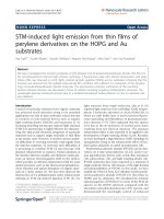
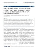
Radiosynthesis
The synthesis of
11
C-dLop was performed by the methy-
lation of the precursor didesmethylloperamide with
11
C-
iodomethane (Figure 1) as reported earlier by our insti-
tution [8]. Didesmethylloperamide was kindly provided
by Janssen Pharmaceutica (Beerse, Belgium), while tetra-
butylammoniumhydroxide, N,N-dimethylformamide and
dimethylsulfoxide were purchased from Sigma-Aldrich
(Bornem, Belgium).
Comparison of
11
C-dLop left heart ventricle time-activity
curve and blood counter measurement time-activity
curve
WT mice (n =3)wereanesthetizedwithisoflurane
(1.5%) and cannulated with a polyethylene catheter (60
cm, PE10), filled with heparinised saline (0.9%). One end
of the catheter was inserted in the carotid artery of the
mice by a precise operation, and at the other end, a syr-
inge needle was inserted. The animals were fixed on the
μPET scanner, the catheter was inserted inside the
detector and the withdrawing syringe was placed on the
main pumping unit as described by Convert et al. [9].
Both the μPET scanner (LabPet8; resolution, 1.5 mm)
and microvolumetric blood counter (Gamma Medica-
Ideas, Quebec, Canada) acquisitions were started in syn-
chronization and subsequent 20-MBq
11
C-dLop, dis-
solved in 100 to 150 μl saline/ethanol mixture (9/1, v/v)
was injected intravenously (i.v.). Blood was collected at a
constant rate of 10 μl/min for the entire 30-min acquisi-
tion time, and the blood time-activity curve was dis-
played in real time by the software of the
microvolumetric blood counter. Immediately after the
end of the
11
C-dLop scan, the mice were injected with
18.5 MBq of
18
F-FDG in a tail vein. Twenty minutes
after
18
F-FDG injection, a static μPET scan was started
for 20 min.
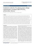
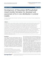
Dynamic
11
C-dLop PET data were sorted into frame
sequence s of 5 s (n =12),10s(n =6),1min(n =4),2
min (n =2),5min(n = 2 ), 10 min (n =1).Aregionof
interest (ROI) was drawn manually a round the left ven-
tricle of the heart (Figure 2A) on the
18
F-FDG scan
images. Since the position of the mice w as unaffected
between the
11
C-dLop and the
18
F-FDG scan, the ROI
of the left heart ventricle on the
18
F-FDG scan could be
pasted on the
11
C-scan images (Figure 2B) to derive an
Figure 1 Radiosynthesis of
11
C-desmethylloperamide. Didesmethylloperamide is methylated with
11
CH
3
I to obtain
11
C-desmethylloperamide
in the presence of tetrabutylammoniumhydroxide, dimethylsulfoxide, and dimethylformamide.
Moerman et al. EJNMMI Research 2011, 1:12
/>Page 2 of 9
arterial blood input function. Data from the blood coun-
ter were corrected for dispersion with the following for-
mula: C
a
(t)=g(t)+τ
disp
×(dg/dt), where C
a
(t)isthe
real whole blood activity curve in mice, g(t)themea-
sured data and dg/dt the derivative of g. τ
disp
, the disper-
sion factor was calculated according to Convert et al.
[9].
The estimated input function (
18
F-FDG-derived) and
the measured input function (blood counter) were com-
pared by a direct and indirect method. The direct
method, as described by Fa ng and Muzic [10], evaluated
the input functions by calculating the area under the
curve (AUC) difference. Indirect comparison examined
the impact of the estimated
18
F-FDG-derived input
function on an estimated kinetic parameter from the
kinetic model, like the K
1
/k
2
ratio, as described later on
(see PET data analysis and kinetic modeling of
11
C-
dLop). The AUC difference was calculated as absolute
values of (AUC
PET
-AUC
bloodcounter
)/AUC
bloodcounter
×
100 and the error percentage of K
1
/k
2
ratio as absolute
values of (K
1
/k
2PET
- K
1
/k
2bloodcounter
)/(K
1
/k
2bloodcounter
)
× 100.
Kinetic model for
11
C-dLop
PET experiments
Before positioning the anesthetized mice on the scanner,
WT mice (n = 3) were injected i.v. 30 min before the
tracer injection with saline (100 μl, controls, n =3)or
50 mg cyclosporine/kilogram body weight (n =3)
(Novartis, Vilvoorde, Belgium). Approximately 2 0 MBq
of
11
C-dLop, dissolved in 100 to 250 μl saline/ethanol
mixture (9/1, v/v) was administered via a tail vein, and
the dynamic μPET scan was initiated. After the
11
C-
dLop scan, the mice were injected with approximately
18.5 MBq of
18
F-FDG in a tail vein (100 μl). Twenty
minutes a fter the
18
F-FDG injection, a static μPET scan
was started for 20 min. KO mice (n = 3) were handled
inthesamewayastheWTmice,withexceptionofthe
pretreatment procedure.
Determination of percent parent compound in plasma and
plasma-whole blood ratio of
11
C-dLop
The determination of percent parent compound (
11
C-
dLop) in plasma over time w as performed in WT (pre-
treated with saline or 50 mg cyclosporine/kilogram body
weight, n = 3 per group and per time point) and KO
mice (n = 3 per time point) using a high-performance
liquid chromatography (HPLC) assay. Thirty minutes
after pretreatment, the mice were injected with 22.2 to
30 MBq of
11
C-dLop (300 μl) and were kill ed at 1, 10,
and 30 min postinjection (p.i.). Blood was collected by
cardiac puncture, and the brain was excised. Plasma
(200 μl) was obtained after centrifugation (3,000 g, 6
min). Subsequently, 800 μl and 1 ml of acetonitri le
(Chem-Lab N.V., Zedelgem, Belgium) were added to the
brain and plasma, respectively. Both samples were vor-
texed (1 min), centrifuged (3,000 g, 3 min) , and counted
for radioactivity. A supernatant was isolated and ana-
lyzed with an HPLC system (Grace Econosphere C18,
10 μm, 10 × 250 mm, eluted with acetonitrile/20 mM
sodium acetate (70/30, v/v) as mobile phase at 7 ml/
min). Elution fractions of30swerecollectedand
counted for radioactivity. Percent parent compound was
calculated as the sum of the counts determined in the
fractions containing
11
C-desmethylloperamide (deter-
mined by co-injection with cold desmethylloperamide
and UV detection at 220 nm) divided by the total
counts of all collected fractions.
To determine the plasma-whole blood ratio, the mice
(n = 3) were injected with 4.80 to 5.55 MBq of
11
C-
dLop (300 μl) and were killed at 0.5, 1, 2, 3, 5, and 10
min p.i Blood was collected from the heart by cardiac
puncture, counted for radioactivity, and c entrifuged for
10 min (3,000 g). Plasma and blood pelle t were sepa-
rated, weighted, and counted for radioactivity. To obtain
the plasma-to-whole blood ratio, counts from plasma
and blood pellet were averaged for weight.
PET data analysis and kinetic modeling of
11
C-dLop
Dynamic
11
C-dLop PET data were sorted into frame
sequences as mentioned above. The arterial blood input
curve obtained from the μPET was corrected for
plasma-whole blood ratio and metabolites. An ROI was
signed around the whole brain on the
18
F-FDG scan
images and was used to determine the
11
C-dLop brain
time-activity curve (Figure 3). All data were loaded and
analyzed with the PMOD software package (version 3.1.,
PMOD Technologies Ltd., Zurich, Switzerland).
Standardized uptake values (SUVs) were calculated
using the following equation: A /(ID/BW), where A is the
decay-corrected radioactivity concentration in the brain
Figure 2 Transversal image of the mice after injection with
11
C-desmethylloperamide and
18
F-FDG. The ROI delineates the
left heart ventricle on the (A)
18
F-scan and (B)
11
C-scan images
(color scale: black, lowest radioactivity uptake; red, highest
radioactivity uptake).
Moerman et al. EJNMMI Research 2011, 1:12
/>Page 3 of 9
(measured in kilobecquerels per cubic centimeter), ID is
the injected dose of
11
C-dLop (measured in kilobecquer-
els), and BW is the mice body weight (measured in
grams), resulting in SUVs expressed as grams per millili-
ter. To a ccount for mice differences in t he blood con-
centrations, which are the drivingforceforthebrain
concentrations, the brain-to-blood ratio was calculated
using the SUVs in the blood and in the brain. A one-tis-
sue compartment model was investigated, in which the
rate constant s K
1
and k
2
represent, respectively, the rate
of transport from plasma to brain and the rate of out-
flow from the brain to the plasma. A two-tissue com-
partment model (with or without k4 fixed to 0) was also
considered, since interaction of
11
C-dLop in the brain
might occur. The volume of vasculature was set as a
variable in the compartment model.
Statistical analysis
All calculated outcome parameters, differences between
WT mice with and without cyclosporine, and KO mice
were investigated with ANOVA and Bonferroni post hoc
testing. The level of statistical significance was set to 5%.
Results
Radiosynthesis
Based on
11
CH
3
I,
11
C-dLop was prepared with a radio-
chemical yield of 32% (decay-corrected) and with a
radiochemical purity of >95%. The sp ecific activity aver-
aged around 70 ± 2 GBq/μmol.
Comparison of
11
C-dLop left heart ventricle time-activity
curve and blood counter measurement time-activity
curve
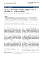
The data from the blood counter were corrected for dis-
persion with τ
disp
calculated as 28 s. A comparison
between the left heart ventricle time-activity curves and
blood counter dispersion corrected time-activity c urves
showed acceptable agreement by graphical inspection
(Figure 4). The AUC difference was 3.5% ± 4.2%, and
the error percentage of the K
1
/k
2
ratio was 6.5% ± 3.2%.
Kinetic model for
11
C-dLop
Determination of percent parent compound in plasma and
plasma-whole blood ratio of
11
C-dLop
The percent parent compound
11
C-dLop at different
time points p.i. in mice are summarized in Table 1. Sta-
tistical differences were observed either between WT
and KO (P < 0.001) and between saline and cyclosporine
pretreated WT mice (P < 0.001).
Within the first half minute after
11
C-dLop injection,
the average ratio of tracer (
11
C-dLop and
11
C-metabo-
lites) plasma concentration to tracer (
11
C-dLop and
11
C-
metabolites) whole blood concentration was 0.67 ± 0.04.
At 1 min after the tracer injection, the value dropped
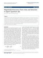
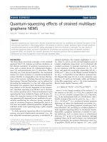
Figure 3 An overview of the proposed method t o determine a kinetic model of
11
C-desmethylloperamide in mice.A
18
F-FDG static
μPET scan is used to obtain the input function and the brain time-activity curve by drawing an ROI around the left heart ventricle and the brain.
Moerman et al. EJNMMI Research 2011, 1:12
/>Page 4 of 9
slightly to 0.49 ± 0.06, whil e at 3 min the ratio w as
restabilized to 0.67 ± 0.07. A mean ratio for all time
points (0.64 ± 0.09) was further used as correction fac-
tor between blood and plasma.
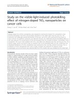
PET data analysis and kinetic modeling of
11
C-dLop
Differences in brain uptake of
11
C-dLop were clearly
observed (Figure 5). The b rain SUVs calculated for KO,
WT, and WT mice pretreated with cyclosporine were
displayed in Figure 6A. In wild-type mice without pre-
treatment of cyclosporine, the average brain SUVs were
0.250, while in pretreated and KO mice SUVs were sig-
nificantly higher (0.693 and 0.526, respectively).
Although cyclosporine pretreatment of wild-type mice
showed higher SUVs in the brain compared to knock-
out mice, no statistical difference was observed (P >
0.05), probably due to larger standard deviations in
knock-out mice. To exclude variation for the blood con-
centration over time between the different mice strains,
SUVs were determined in the left heart ventricle. No
statistical differences in the left heart ventricle SUVs
(Figure 6B) were obtained. The b rain-to-plasma SUVs
are significant different between wild-type mice and KO
mice and cyclosporine pretreated wild-type mice (Figu re
6C).
The two-tissue compartment model (with or without
k4 fixed to 0) did not provide a significantly better fit
than the one-tissue compartment model (Figure 7)
(Akaike crit erion values were in the same range). More-
over, the two-tissue compartment model estimated the
kinetic parameters K
1
and k
2
with poorer identifiabil ity
than the one-tissue compartment model based on per-
cent covariance values. Hence, Table 2 provides a sum-
mary of parameters estimated from the one-tissue
compartment model with the noninvasive (left heart
ventricle-based) method used to determine the input
curve. K
1
in WT mice is statistically smaller than K
1
in
Figure 4 Comparison of standard and new
11
C-
desmethylloperamide TAC. Comparison of
11
C-
desmethylloperamide left heart ventricle time-activity curve (TAC)
and blood counter dispersion corrected time-activity curve in a
mouse.
Table 1 Percentage of the parent compound (
11
C-dLop)
in plasma
Mouse strain and pretreatment %
11
C-desmethylloperamide in
plasma
1 min p.i. 10 min p.i. 30 min p.i.
WT, saline 95 ± 1 72 ± 5 53 ± 3
WT, 50 mg cyclosporine/kg 95 ± 1 54 ± 5 23 ± 9
KO 98 ± 1 51 ± 1 34 ± 9
Percentage of the parent compound (
11
C-dLop) in plasma at 1, 10, and 30
min p.i. in different mouse strains and after different pretreatments. Results
are expressed as percent of total radioactivity ± standard deviation. WT, wild-
type mice; KO, knock-out mice.
Figure 5 Sagittal images of mice after intravenous
administration of
11
C-dLop. Sagittal images of knock-out mice (A),
wild-type mice without cyclosporine pretreatment (B) and wild-type
mice with cyclosporine pretreatment (50 mg/kg body weight, 30
min before tracer injection) (C), after intravenous administration of
20.0 ± 2.0 MBq of
11
C-dLop. In each mice, the brains are indicated;
the difference in tracer brain uptake between wild-type (no
pretreatment), knock-out, and with cyclosporine-pretreated wild-
type mice is clearly visible (color scale: black, lowest radioactivity
uptake; red, highest radioactivity uptake).
Moerman et al. EJNMMI Research 2011, 1:12
/>Page 5 of 9
knock-out mice (P = 0.008) and in cyclosporine-pre-
treated mice (P = 0.025), wh ile the k
2
is in the same
range in a ll mice (P > 0.050). The differences between
WT and knock-out mice or between saline and cyclos-
porine pretreatment in WT mice are also reflected in
the K
1
/k
2
ratio (P = 0.001).
Discussion
Biochemical process steps of a tracer in a tissue can be
described by an appropriate tracer kinetic model. The
behavior of a tracer is usually simplified and described
by some mathematical kinetic compartments [11]. This
model should be able to estimate the amount of radio-
activity in each compartment, and the rate of exchange
between these compartments. In PET imaging, these
rate constants directly provide information on physiolo-
gical parameters characterizing the behavior of the tra-
cer in the tissue of interest. In case there is no reference
region available, an arterial input curve is necessary to
set up a kinetic model. Manual or automatic blood sam-
pling is generally accepted as the gold standard to deter-
mine the arterial input curve. Nevertheless, in mice
arterial sampling is technically difficult because of the
relatively small diameters and fragility of the mouse
blood arteries [12]. In addition, the t otal blood volume
of a mouse is very limited (1.7 ml), making repeated
blood sampling impossible without affecting the home-
ostasis of the mice [13]. Alter native methods to obtain
an arterial input function are the use of a population
database, based on a high number of mice or an arterial
input function derived from PET images [14]. Attempts
to determine the arterial input function in small animals
from PET images were not convincing. Difficult delinea-
tion of the left heart ventricle on the PET scan in m ice
[15] or background signals from surrounding tissues in
rats [16] were the main problems. Due to blurred
11
C-
dLop imag es on early as well as late time frames, it was
impossible to delineate the left heart ventricle accu-
rately. We therefore propose a new image-derived
method, using a
18
F-FDG scan after a
11
C-dLop scan.
Unlike
11
C-dLop,
18
F-FDG shows a selective uptake in
the myocardium [17-19], making the determination of
the left ventricle easy without the problem of spill-in of
activity fro m the surrounding lungs. A comparison
between the left heart ventricle time-activity curves
Figure 6 Standard uptake values of
11
C-desmethylloperamide. SUVs of
11
C-desmethylloperamide in wild-type mice with saline (1) or 50 mg/
kg cyclosporine (2) pretreatment and in knock-out mice (3), expressed in grams per milliliter in function of time in brain (A) and in the left heart
ventricle (LV) (B). The ratio of SUV
brain
/SUV
LV
is depicted in graph (C).
Figure 7 One- and two-compartment model fittings for mice (n
= 3), which were injected with
11
C-dLop. Circles represent
observed μPET data taken from a region of interest drawn on the
brain.
Table 2 Summary of kinetic parameters
K
1
(ml/cc/min) k
2
(1/min) K
1
/k
2
WT 1 0.054 0.190 0.28
WT 2 0.042 0.120 0.35
WT 3 0.027 0.059 0.46
KO 1 0.190 0.120 1.58
KO 2 0.230 0.095 2.42
KO 3 0.190 0.100 1.90
CYCLO 1 0.250 0.150 1.66
CYCLO 2 0.120 0.063 1.90
CYCLO 3 0.180 0.086 2.09
Summary of kinetic parameters estimated from the one-tissue compartment
model for
11
C-desmethylloperamide for all mice studied, using the
noninvasive (left heart ventricle-based) method to determine the input curve.
CYCLO, wild-type mice pretreated with 50 mg/kg cyclosporine, 30 min before
tracer injection; KO, knock-out mice; WT, wild-type mice.
Moerman et al. EJNMMI Research 2011, 1:12
/>Page 6 of 9
(alternative method) and blood counter time-activity
curves (corrected for dispersion) showed acceptable gra-
phical agreement. A small AUC difference (3.5%) was
observed compared to Green et al. (18%) [20], who did
not use a
18
F-FDG scan to delineate the left heart ven-
tricle, but instead a small ROI based on the highest
activity in the aorta area on the earliest time frames.
Also, the comparison of the K
1
/k
2
ratio showed analog
correlations (6.5%) between standard blood sampling
and our proposed method. However, one must rea lize
that the usefulne ss of our method must be validated for
each radioligand because determination of the arterial
input function based on the left ventricle could lead to a
poor resemblance with the blood sampling input curve
especially for radioligands with high myocardial uptake.
Both in wild-type and in P-gp knock-out mice, the
percent of parent compound was investigated, resulting
in variations probably due to the influence of cyclospor-
ine or to an adaptation of the body to the absence of P-
gp efflux transporters. These differences are not an
obstacle concerning our experiment because the latter
correction was introduced to take these differences into
consideration.
Variations in
11
C-dLop brain uptake between wild-
type and knock-out/cyclosporine-pretreated mice were
clearly observed in μPET images and SUVs. Moreover,
differences in
11
C-dLop uptake in the intestines were
observed and could be explained by the absence of P-gp
in KO mice resulting in a lower tracer uptake, while in
WT mice P-gp located in the intestines pumps the tra-
cer out of the blood into the intestines, resulting in
higher uptake. The higher radioactivity in the abdomen
of WT mice, as observed in Figure 5, could also be
explained as higher uptake in the liver, which is in
accordance with results obtained in humans [21]. Never-
theless, kinetic parameters obtained from a compart-
ment model will provide useful mathematical
information about the behavior of the tracer. Since no
statistical difference in model fittings between the one-
and two-compartment model was observed, the simplest
model, meaning the one-tissue compartment model, was
preferred. This is in accordance to the results mentioned
by Kreisl et al. [22]. In a one-tissue compartment model,
the tracer behaves in a straightforward manner
explained by an uptake in the brain with a speed, repre-
sented by the kinetic parameter K
1
, and efflux out of the
brain described by k
2
. Binding with any receptors in the
brainormetabolisationofthetracerinthebrainwill
not occur in this model. Lazarova et al. [6] already men-
tioned that
11
C-dLop showed no clinical relevant inter-
action with the opiate receptors in the brain.
The kinetic parameters K
1
and k
2
obtained from a
one-tissue compartment mo del of
11
C-dLop were eval-
uated in WT, KO, and WT mice pretreated with
cyclosporine. One should expect that K
1
, which repre-
sents the passive influx of the tracer in the brain,
should not change betwe en the different groups. k
2
,
which represents the efflux out of the brain by P-gp
transport, was supposed to be lower in KO mice and
in the WT mice pretreated with cyclosporine. Our
data showed that the K
1
was statistically lower in WT
mice compared to KO or cyclosporine-pretreated WT
mice, while the k
2
was very similar in all tested mice.
Kreisl et al. [22] reported the same result after block-
age of the P-gp with tariquidar and suggested that tari-
quidar increased brain uptake of
11
C-dLop by
increasing its entry (K
1
) rather than by decreasing its
efflux (k
2
). The substrate is captured in the endothelial
cells, before it enters the intracellular compartme nt.
Therefore, if P-gp captures all of the substrate while in
transit through the membrane, its effect is entirely on
K
1
. If some of the substrate escapes and has time to
interact with the intracellular milieu, and if there is an
efflux from the cell, P-gp will both decrease K
1
and
increase k
2
[23]. Nevertheless, we think that also a
time influence of the P-gp transport should be taken
into consideration. The course of the brain SUV curve
(Figure 6A) in WT mice demonstrates a fast uptake in
thebrain,followedbyarapidwashoutofthebrain,
resulting in an SUV of 0.25 already 1 min after the tra-
cer injection, while i n KO and pretreated WT mice
also a fast uptake was observed, followed by an accu-
mulation in the brain of the tracer combined with a
slow efflux. The observed different course of the brain
curve between WT and KO mice, even as between
cyclosporine pretreated WT mice suggests that the
duration of the scan could play an important role on
the determination of the kinetic parameters in the
kinetic model.
This hypothesis was substantiated by the results of K
1
and k
2
obtainedinaone-tissuecompartmentmodel
with incorporation of only the first 2 min of dynamic
scanning. These results showed a statistically higher k
2
in WT mi ce (8.0 ± 0. 1) compared to KO (2.3 ± 0.9; P =
0.070)andcomparedtocyclosporinepretreatedWT
mice (1.5 ± 0.5; P = 0.002), while K
1
was statistically not
different between the different groups (P >0.05).This
means that during the first 2 min after administration of
11
C-dLop, efflux out of the brain is dominated by efflux
transporters, while at later time points passive diffusion
is more important. The K
1
/k
2
ratio of WT obtained with
the 2-min scan data were statistically different compared
to the ratios in KO and compared to cyclosporine-pre-
treated WT mice. So, we propose K
1
as a pseudo value,
representing a combination of passive influx of
11
C-
dLop through the BBB and a rapid energy dependent
output by P-gp, while k
2
corresponds to slow passive
efflux out of the brain (Figure 8).
Moerman et al. EJNMMI Research 2011, 1:12
/>Page 7 of 9
Conclusion
The use of an easy to implement
11
C-desmethyllopera-
mide kinetic model in mice for imaging P-gp function
is presented without arterial blood sampling. The
method to determine the input function is based on
the delineation of an ROI on the
18
F-FDG scan i mages
and using this ROI on images obtained from a
dynamic scan with
11
C-dLop. The K
1
or K
1
/k
2
ratio
obtained from the
11
C-dLop tracer kinetic model is a
good parameter for the active P-gp rate and can be
applied in future experiments to evaluate the role of
the upregulation of P-gp in psychotropic drug resis-
tance, such as refractory epilepsy and in tumor resis-
tance to therapy.
Abbreviations
AED: antiepileptic drugs; AUC: area under the curve; BBB: blood-brain barrier;
BW: mice body weight;
11
C-dLop:
11
C-desmethylloperamide; DMF:
dimethylformamide; DMSO: dimethylsulfoxide; ID: injected dose; i.v.:
intravenously; KO: P-glycoprotein knock-out mice; P-gp: P-glycoprotein; p.i.:
post injection; SUVs: standardized uptake values; TBAH:
tetrabutylammoniumhydroxide; WT: wild-type mice.
Acknowledgements
We are grateful to the cyclotron team for their support during the synthesis
of the tracer. We would like to thank Philippe Joye for the animal
manipulation before and during the scans and Steven Deleye for the
reconstructions of the scans. Janssen Pharmaceutica is acknowledged for the
donation of desmethylloperamide and didesmethylloperamide. We also like
to thank FWO-Vlaanderen for funding and Prof. Pascal Verdonck for the
scientific support.
Author details
1
Laboratory of Radiopharmacy, Faculty of Pharmaceutical Sciences, Ghent
University, Ghent, Belgium
2
Department of Civil Engineering, Institute
Biomedical Technology, Ghent University, Ghent, Belgium
3
Laboratory for
Clinical and Experimental Neurophysiology (LCEN), Department of
Neurology, Ghent University Hospital, Ghent, Belgium
Authors’ contributions
LM designed and carried out the experimental studies and has written the
manuscript. DD has investigated and corrected the blood plasma curve for
dispersion. PB and FD participated in the design of the study and helped to
draft the manuscript. The manuscript has been seen and approved by all
authors.
Competing interests
This work was supported and funded by a Ph.D. grant of the Institute for
the Promotion of Innovation through Science and Technology in Flanders
(IWT-Vlaanderen). Research work of Dieter De Naeyer was also funded by
FWO-Vlaanderen. Prof. Paul Boon has received fees for presentations and
Figure 8 Schematic representatio n of different methods to determine the rate constants obtained from one-tissue compartment
model. (A) Rate constants K
1
and k
2
, obtained from one-tissue compartment model with all scan data incorporated. K
1
represents the passive
influx, while k
2
is a combination of active and passive efflux. (B) shows rate constants obtained from a one-tissue compartment model with only
the first 2 min of the scan data. Pseudo K
1
is defined as a combination of the passive influx and active efflux, but k
2
only represents passive
efflux. C, concentration of
11
C-dLop.
Moerman et al. EJNMMI Research 2011, 1:12
/>Page 8 of 9
travel grants from UCB Pharma and Janssen-Cilag. The remaining authors
have no conflicts of interest.
Received: 23 February 2011 Accepted: 29 July 2011
Published: 29 July 2011
References
1. Löscher W, Potschka H: Role of multidrug transporters in
pharmacoresistance to antiepileptic drugs. J Pharmacol Exp Ther 2002,
301:7-14.
2. Sisodiya SM, Lin WR, Harding BN, Squier MV, Thom M: Drug resistance in
epilepsy: expression of drug resistance proteins in common causes of
refractory epilepsy. Brain 2002, 125:22-31.
3. Marchi NM, Hallene KL, Kight KM, Cucullo L, Moddel G, Bingaman W,
Dini G, Vezzani A, Janigro D: Significance of MDR1 and multiple drug
resistance in refractory human epileptic brain. BMC Med 2004, 2:1-10.
4. Tishler DM, Weinberg KI, Hinton DR, Barbaro N, Annette GM, Raffel C: MDR1
gene expression in brain of patients with medically intractable epilepsy.
Epilepsia 1995, 36:1-6.
5. Sadeque AJM, Wandel C, He HB, Shah S, Wood AJJ: Increased drug
delivery to the brain by P-glycoprotein inhibition. Clin Pharmacol Ther
2000, 68:231-237.
6. Lazarova N, Zoghbi SS, Hong J, Seneca N, Tuan E, Gladding RL, Liow JS,
Taku A, Innis RB, Pike VW: Synthesis and evaluation of (N-methyl-11C)N-
desmethyl-loperamide as a new and improved PET radiotracer for
imaging P-gp function. J Med Chem 2008, 51:6034-6043.
7. Zhang Y, Bachmeier C, Miller DW: In vitro and in vivo models for assessing
drug efflux transporter activity. Adv Drug Deliver Rev 2003, 55:31-51.
8. Moerman L, Wyffels L, Slaets D, Raedt R, Boon P, De Vos F: Antiepileptic
drugs modulate P-glycoproteins in the brain: a mice study with
11
C-
desmethylloperamide. Epilepsy Res 2011, 94:18-25.
9. Convert L, Morin-Brassard G, Cadorette J, Archambault M, Bentourkia M,
Lecomte R: A new tool for molecular imaging: the microvolumetric β
blood counter. J Nucl Med 2007, 48:1197-1206.
10. Fang YD, Muzic RF: Spillover and partial-volume correction for image-
derived input functions for small-animal
18
F-FDG PET studies. J Nucl Med
2008, 49:606-614.
11. Carson RE: Tracer Kinetic modeling in PET. In Positron emission
tomography: Basic Science and clinical practice. Volume 1 1 edition. Edited
by: Valk PE, Bailey DL, Townsend DW, Maisey MN. London: Springer;
2003:147-179.
12. Green LA, Gambhir SS, Srinivasan A, Banerjee PK, Hoh CK, Cherry SR,
Sharfstein S, Barrio JR, Herschman HR, Phelps ME: Noninvasive methods for
quantitating blood time-activity curves from mouse PET images
obtained with fluorine-18-fluorodeoxyglucose. J Nucl Med 1998,
39:729-734.
13. Davies B, Morris T: Physiological parameters in laboratory animals and
humans. Pharm Res 1993, 10:1093-1095.
14. Bentourkia M, Zaidi H: Tracer kinetic modeling in nuclear medicine:
Theory and application. In Quantitative analysis in nuclear medicine
imaging. Volume 1 1 edition. Edited by: Zaidi H. New York: Springer;
2006:391-414.
15. Choi SJ, Kim SY, Kim SJ, Lee JS, Lee SJ, Park SA, Lee SJ, Yun SC, Im KC,
Oh SJ, Kim SW, Kim JS, Ryu JS, Moon DH: Reproducibility of the kinetic
analysis of 3’-deoxy-3’(
18
F)fluorothymidine positron emission
tomography in mouse tumor models. Nucl Med Biol 2009, 36:711-719.
16. Pain F, Lanièce P, Mastrippolito R, Gervais P, Hantraye P, Besret L: Arterial
input function measurement without blood sampling using a β-
microprobe in rats. J Nucl Med 2004, 45:1577-1582.
17. Phelps ME, Hoffman ED, Selin C, Huang SC, Robinson G, MacDonald N,
Schelbert H, Kuhl DE: Investigation of (18F)2-fluoro-2-deoxyglucose for
the measure of myocardial glucose metabolism. J Nucl Med 1978,
19:1311-1319.
18. Landoni C, Bettinardi V, Lucignani G, Gilardi MC, Striano S, Fazio F: A
procedure for wall detection in (
18
F)FDG positron emission tomography
heart studies. Eur J Nucl Med 1996, 23:18-24.
19. Kim J, Herrero P, Sharp T, Laforest R, Rowland DJ, Tai YC, Lewis JS,
Welch MJ: Minimally invasive method of determining blood input
function from PET images in rodents. J Nucl Med 2006, 47:330-336.
20. Green LA, Nguyen K, Berenji B, Iyer M, Bauer E, Barrio J, Namavari M,
Satyamurthy N, Gambhir SS: A tracer kinetic model for
18
F-FHBG for
quantitating herpes simplex virus type 1 thymidine kinase reporter gene
expression in living animals using PET. J Nucl Med 2004, 45:1560-1570.
21. Seneca N, Zoghbi SS, Liow JS, Kreisl W, Herscovitch P, Jencko K,
Gladding RL, Taku A, Pike VW, Inins RB: Human brain imaging and
radiation dosimetry of
11
C-N-desmethyl-loperamide, a PET radiotracer to
measure the function of P-glycoprotein. J Nucl Med 2009, 50:807-813.
22. Kreisl WC, Liow JS, Kimura N, Seneca N, Zoghbi SS, Morse CL, Herscovitch P,
Pike VW, Innis RB: P-glycoprotein function at the blood-brain barrier in
humans can be quantified with the substrate radiotracer
11
C-N-
desmethyl-loperamide. J Nucl Med 2010, 51:559-566.
23. Kannan P, Zoghbi SS, Halldin C, Gottesman MM, Innis RB, Hall MD: Imaging
the function of P-glycoprotein with radiotracers: pharmacokinetics and
in vivo applications. Clin Pharmacol Ther 2009, 86:368-377.
doi:10.1186/2191-219X-1-12
Cite this article as: Moerman et al.: P-glycoprotein at the blood-brain
barrier: kinetic modeling of
11
C-desmethylloperamide in mice using a
18
F-FDG μPET scan to determine the input function. EJNMMI Research
2011 1:12.
Submit your manuscript to a
journal and benefi t from:
7 Convenient online submission
7 Rigorous peer review
7 Immediate publication on acceptance
7 Open access: articles freely available online
7 High visibility within the fi eld
7 Retaining the copyright to your article
Submit your next manuscript at 7 springeropen.com
Moerman et al. EJNMMI Research 2011, 1:12
/>Page 9 of 9