Chan et al. EJNMMI Research 2011, 1:15 http://www.ejnmmires.com/content/1/1/15 ORIGINAL doc
Bạn đang xem bản rút gọn của tài liệu. Xem và tải ngay bản đầy đủ của tài liệu tại đây (1.04 MB, 11 trang )
ORIGINAL RESEARCH Open Access
A comparison of
111
In- or
64
Cu-DOTA-trastuzumab
Fab fragments for imaging subcutaneous HER2-
positive tumor xenografts in athymic mice using
microSPECT/CT or microPET/CT
Conrad Chan
1
, Deborah A Scollard
1
, Kristin McLarty
1
, Serena Smith
1
and Raymond M Reilly
1,2,3*
Abstract
Background: Our objective was to compare
111
In- or
64
Cu-DOTA-trastuzumab Fab fragments for imaging small or
large s.c. tumor xenografts in athymic mice that display a wide range of human epidermal growth factor receptor-
2 (HER2) expression using microSPECT/CT or microPET/CT.
Methods: Trastuzumab Fab were labeled with
111
In or
64
Cu by conjugation to 1,4,7,10-tetraazacyclododecane N, N’,
N’’,N’’’-tetraacetic acid (DOTA). The purity of
111
In- and
64
Cu-DOTA-trastuzumab Fab was measured by SDS-PAGE
and HPLC. HER2 binding affinity was determined in saturation radioligand binding assays using SKBR-3 cells (1.3 ×
10
6
HER2/cell). MicroSPECT/CT and microPET/CT were performed in athymic mice bearing s.c. BT-20 and MDA-MB-
231 xenografts with low (0.5 to 1.6 × 10
5
receptors/cell), MDA-MB-361 tumors with intermediate (5.1 × 10
5
receptors/cell) or SKOV-3 xenografts with high HER2 expression (1.2 × 10
6
receptors/cell) at 24 h p.i. of 70 MBq (10
μg) of
111
In-DOTA-trastuzumab Fab or 22 MBq (10 μg) of
64
Cu-DOTA-trastuzumab Fab or irrelevant
111
In- or
64
Cu-
DOTA-rituximab Fab. Tumor and normal tis sue uptake were quantified in biodistribution studies.
Results:
111
In- and
64
Cu-DOTA-trastuzumab were > 98% radiochemically pure and bound HER2 with high affinity
(K
d
= 20.4 ± 2.5 nM and 40.8 ± 3.5 nM, respectively). MDA-MB-361 and SKOV-3 tumors were most clearly imaged
using
111
In- and
64
Cu-DOTA-trastuzumab Fab. Significantly higher tumor/blood (T/B) ratios were found for
111
In-
DOTA-trastuzumab Fab than
111
In-DOTA-rituximab Fab for BT-20, MDA-MB-231 and MDA-MB-361 xenografts, and
there was a direct association between T/B ratios and HER2 expression. In contrast, tumor uptake of
64
Cu-DOTA-
trastuzumab Fab was significantly higher than
64
Cu-DOTA-rituximab Fab in MDA-MB-361 tumors but no direct
association with HER2 expression was found. Both
111
In- and
64
Cu-DOTA-trastuzumab Fab imaged small (5 to 10
mm) or larger (10 to 15 mm) MDA-MB-361 tumors. Higher blood, liver, and spleen radioactivity were observed for
64
Cu-DOTA-trastuzumab Fab than
111
In-DOTA-trastuzumab Fab.
Conclusions: We conclude that
111
In-DOTA-trastuzumab Fab was more specific than
64
Cu-DOTA-trastuzumab Fab
for imaging HER2-positive tumors, especially those with low receptor density. This was due to higher levels of
circulating radioactivity for
64
Cu-DOTA-trastuzumab Fab which disrupted the relationship between HER2 density
and T /B ratios. Use of alternative chelators that more stably bind
64
Cu may improve the association between T/B
ratios and HER2 density for
64
Cu-labeled trastuzumab Fab.
Keywords: indium-111, copper-64, HER2, MicroSPECT, MicroPET, DOTA, trastuzumab Fab, breast cancer, ovarian
cancer
* Correspondence:
1
Department of Pharmaceutical Sciences, University of Toronto, Toronto,
M5S 3M2, ON, Canada
Full list of author information is available at the end of the article
Chan et al. EJNMMI Research 2011, 1:15
/>© 2011 Chan et al; licensee Springer. This is an Open Access article distributed under the terms of the Creative Commons Attribution
License (http://creativ ecommons.org/licenses/by/2.0), which permits unrestricted use, distribution, and repr oduction in any medium,
provided the original work is properly cited.
Background
The human epidermal growth factor receptor-2 (HER2) is
overexpressed in 20% to 25% of breast cancers (BC) and is
the target for treatment with trastuzumab (Herceptin), a
humanized IgG
1
monoclonal antibody (mAb) [1,2]. HER2
amplification is normally assessed ex vivo in a primary
tumor biopsy by immunohistochemical (IHC) staining for
HER2 protein or by fluoresecence in situ hybridization to
detect increased HER2 gene copy number [3]. However,
discordance in HER2 expression between primary and
metastatic BC has been found in 20% to 30% of cases [4,5]
and thus, it would be useful to have an imaging technique
to assess HER2 phenotype in situ in BC lesions. Several
investigators have shown that HER2 e xpression can be
imaged in human BC xenografts in athymic mice by single
photon emission computed tomography (SPECT) using
trastuzumab or its Fab fragments labeled with
111
In or
99
m
Tc [6-9]. These studies have been exte nded to imaging
HER2-positive BC in patients using
111
In-labeled trastuzu-
mab IgG [2,10]. More recently, positron-emission tomo-
graphy (PET) using trastuzumab labeled with
89
Zr has
shown promise for imaging HER2 expression in tumor
xenograft mouse models and in patients with metastatic
BC [11,12]. Imaging also offers an opportunity to detect
response to HER2-targeted therapies in BC [13]. We pre-
viously reported that SPECT with
111
In-labeled pertuzu-
mab (anti-HER2) detected early response to treatment
with trastuzumab (Herceptin) in athymic mice bearing s.c.
MDA-MB-361 BC xenografts [14]. Smith-Jones et al.
demonstrated that PET with
68
Ga-labeled trastuzumab F
(ab’)
2
fragments identified response of HER2-positive BT-
474 human BC tumors in mice to treatment with heat
shock protein (Hsp90) inhibitors [15].
PET offers several potential advantages compared to
SPECT for imaging tumors because it has higher intrinsic
sensitivity, is more easily quantified, and in some
instances offers higher spatial resolution. Despite these
apparent benefits, few studies have reported a compari-
son of PET and SPECT for imaging HER2-positive
tumors using the same agent labeled with a single
photon-emitter or positron-emitter. Dijkers et al. com-
pared
89
Zr- and
111
In-labeled trastuzumab in mice bear-
ing s.c. SK-OV-3 human ovarian cancer xenografts and
reported no significant differences in tumor and normal
tissue uptake [12]. MicroPET with
89
Zr-labeled trastuzu-
mab visualized these t umors, but the corresponding
microSPECT images with
111
In-labeled trastuzumab were
not presented.
In this study, we compared microSPECT/CT and
microPET/CT for imaging s.c. human tumor xenografts
expressing a wide range of HER2 density in a thymic
mice using trastuzumab Fab fra gments modified with
1,4,7,10-tetraazacyclododecane N, N’,N″,N’″-tetraacetic
acid (DOTA) for co mplexing
111
In or
64
Cu.
64
Cu decays
with a half-life of 12.7 h by positron emission [Eb
+
=
0.65 MeV (17.4%)], b
-
emission [E b = 0 .58 MeV (39% )]
and electron capture (43.6%).
111
In decays by electron
capture with a half-life of 2.8 days emitting Auger elec-
trons and two g-photons [Eg = 171 keV (90%) and 245
keV (94%)]. DOTA was selected as a chelator because
both
111
In and
64
Cu form thermodynamically stable
complexes with DOTA (K
d
=10
24
and 10
23
M
-1
, respec-
tively) [16,17].
64
Cu complexed to DOTA and linked to
mAbs and peptides has been widely studied for PET
imaging of tumors [15,18-23]. Fab fragments were
selected for these studies because their pharmacokinetics
of tumor uptake and elimination from the blood and
normal tissues is compatible with the half-lives of
64
Cu
and
111
In [24].
Materials and methods
Preparation of Fab fragments
Trastuzumab (Herceptin) and rituximab (anti-CD20;
Rituxan) are humanized IgG
1
mAbs and were obtained
from Roche Pharmaceuticals Ltd. (Mississauga, ON,
Canada). Fab fragments were prepared by digestion with
immobilized papain (Pierce Chemical Co., Rockford, IL,
USA) and purified as reported [7,25]. Fab purity was
assessed by sodium dodecyl sulfate polyacrylamide gel
electrophoresis (SDS-PAGE) on a 4% to 20% Tris HCl gra-
dient mini-gel (BioRad, Mississauga, ON, Canada) and by
size-exclusion high performance liquid chromatography
(HPLC). For SDS-PAGE, Fab (10 μg) were electrophoresed
under non-reducing and reducing [dithiothreitol (DTT)]
conditions. The gel was stained with Coomassie R-250
brilliant blue (Bio-Rad). Size-exclusion HPLC was per-
formed on a BioSep SEC-2000 column (Phenomenex,
Torrance, CA, USA) eluted with 100 mM NaH
2
PO
4
buffer
(pH 7.0) at a flow rate of 0.8 mL/min in line with a diode
array detector (PerkinElmer, Wellesley, MA, USA) moni-
toring at 280 n m. Fab fragments were concentrated and
buffer-exchanged into 50 mM NaHCO
3
buffer (pH 7.5)
on an Amicon Ultracel 30 K device (M
r
cut-off = 30 kDa;
Millipore Corp., Billerica, MA, USA). Trace metals were
removed from all buffers using Chelex-100 cation-
exchange resin (BioRad). The final Fab fragments concen-
tration was measured spectrophotometric ally [E
280 nm
=
1.45 (mg/mL)
-1
cm
-1
] [7] and was adjusted to 5 mg/mL
with 50 mM NaHCO
3
buffer, pH 7.5.
DOTA conjugation and radiolabeling of Fab fragments
Trastuzumab or rituximab Fab fragments were modified
with DOTA for complexing
111
In or
64
Cu by reaction of
1.5 mg of Fab in 300 μLofNaHCO
3
buffer (pH 7.5)
with a 60- or 90-fold excess, respectively, of the N-
hydroxysuccinimidyl ester of 1,4,7,10-tetraazacyclodode-
cane tetraacetic acid (NHS-DOTA; Macrocyclics, Dallas,
TX, USA). The conjugation reaction was performed at
Chan et al. EJNMMI Research 2011, 1:15
/>Page 2 of 11
4°C for 18 h. DOTA-conjugated Fab were purified from
excess DOTA by transferring to an Amicon Ultracel 30
K device, diluting to 12.0 mL with 1 M CH
3
COONH
4
buffer, pH 6.0 and centrifuging at 4,000 × g for 15 min.
This purification step was repeate d six times. Finally,
purified DOTA-Fab were recove red and the concentra-
tion determined spectrophotometrically [E
280 nm
=1.45
(mg/mL)
-1
cm
-1
]. The final concentration was adjusted
to 5 mg/mL with 1 M CH
3
COONH
4
buffer, pH 6.0.
Radiolabeling was performed by incubating 50 μgof
DOTA-Fab in 10 μLofCH
3
COONH
4
buffer, pH 6.0 with
360 MBq of
111
InCl
3
(> 7 GBq/mL; MDS-Nordion,
Kanata, ON, Canada) or 216 MBq of
64
CuCl
2
(> 4 GBq/
mL; MDS-Nordion) for 3 h at 46°C.
111
In- or
64
Cu-labeled
DOTA-Fab were purified on an Amicon Ultracel 30 K
device. The final radiochemical purity was measured by
instant thin layer-silica gel chromatography (ITLC-SG;
Pall Life Sciences, Ann Arbor, MI, USA) developed in 100
mM sodium citrate, pH 5.0 or by size-exclusion HPLC
using a flow-through radioactivity detector (FSA; PerkinEl-
mer). The R
f
values for
111
In- or
64
Cu-DOTA-Fab on
ITLC were 0.0 and those for
111
In- or
64
Cu-DOTA or free
radionuclides were 1.0. The DOTA substitution level of
the Fab fragments (chelators/molecule) was measured by
labeling a 10 μL aliquot of the unpurified conjugation
reaction with
111
In, then determining the proportion of
111
In-DOTA-Fab vs.free
111
In-DOTA by ITLC-SG and
multiplying this fraction by the molar ratio used in the
reaction [26].
HER2 binding affinity of
111
In- and
64
Cu-DOTA-
trastuzumab Fab
The HER2 binding affinity of
111
In- and
64
Cu-DOTA-
trastuzumab Fab was determined by direct (saturation)
radioligand binding assays using SKBR-3 human BC
cells (1.3 × 10
6
HER2/cell) [9]. Briefly, increasing con-
centrations (0 to 600 nmol/L) of
111
In- or
64
Cu-DOTA-
trastuzumab Fab were incubated with 1 × 10
5
cells in
24-well plates at 4°C for 3 h. Unbound radioactivity was
removed and the dishes wer e rinsed two times with
phosphate-buffered saline. The cells were dissolved in
100 mM NaOH, recovered, and the total cell-bound
radioactivity (TB) was measured in a g-counter (Perki-
nElmer Wizard 3). The assay was repeated in the pre-
sence of 16 μmol/L of unlabeled trastuzumab IgG to
measure non-specific binding (NSB). Specific binding
(SB; nanomoles per liter) was calculated by subtracting
NSBfromTBandwasplottedvs. the concentration of
111
In- or
64
Cu-DOTA-trastuzumab Fab (nanomoles per
liter) added. The resulting curve was fitted by non-linear
regression to a one-site receptor-binding model by
Prism Ver. 4. 0 software (Graph Pad, San Diego, CA,
USA). The dissociation constant (K
d
) and maximum
number of receptors per cell (B
max
) were calculated and
compared for
111
In- and
64
Cu-DOTA-trastuzumab Fab.
Tumor and normal tissue distribution studies
The tumor and normal tissue distribution of
111
In- or
64
Cu-DOTA-trastuzumab Fab were determined at 24-h
post-intravenous (tail vein) injection (p.i.) in athymic mice
with s.c. human tumor xenografts with a wide range of
HER2 density. This time point was selected due to the
short physical half-l ife of
64
Cu (12.7 h) and because high
tumor uptake [> 5 percent injected dose per gram (% i.d./
g)] and tumor/blood (T/B)ratios(>4:1)werepreviously
found for
111
In-DTPA-trastuzumab Fab at 24 h p.i. [7].
Tumors were established in female athymic (CD1-nud e)
mice by s.c. inoculation of 1 × 10
7
MDA-MB-231, BT-20,
or MDA-MB-361 BC cells expressing 5.4 × 10
4
, 1.6 × 10
5
,
or 5.1 × 10
5
HER2/cell, respectively, or with SK-OV- 3
ovarian c a ncer cells displaying 1.2 × 10
6
HER2/cell [27]. At
4 to 7 weeks post-inoculation, when tumors were well
established (5 to 15 mm in diameter), groups of mice (n =
4) were injected i.v. (tail vein) with 12 MBq (10 μg) of
111
In-DOTA-trastuzumab Fab or 18 MBq (10 μg) of
64
Cu-
DOTA-trastuzumab Fab. To determine if tumor uptake
was specific, control groups of mice (n =4)withMDA-
MB-231, BT-20, or MDA-MB-361 xenografts were injected
i.v. with 12 MBq (10 μg) of irrelevant
111
In-DOTA-rituxi-
mab F ab or 18 MBq ( 10 μg) of
64
Cu-DOTA-rituximab F ab.
Mice were euthanized by cervical dislocation u nder general
anaesthesia. Tumor a n d normal tissue uptake of ra dioactiv-
ity was measured in a g-scintillation counter (Wizard 3,
PerkinElmer, Waltham, MA) was expressed as percent
injected dose per gram and as tumor/normal tis sue (T/NT)
ratios. The relationship between tumor/blood (T/B) ratios
and HER2 density was examined. The uptake of
111
In- or
64
Cu-DOTA-trastuzumab Fab fragments in small (5 to 10
mm diameter) vs. larger (10 to 15 mm diameter) tumor
xenografts was compared.
MicroSPECT and microPET imaging
MicroSPECT was performedat24hp.i.of70MBq(10
μg) of
111
In-DOTA-trastuzumab Fab or
111
In-DOTA-
rituximab Fab in athymic mice with s.c. HER2-positive
tumor xenografts. Anaesthesia was induced and main-
tained by inhalation of 2% isoflurane in O
2
. MicroSPECT
was performed on a NanoSPECT/CT tomograph (Bioscan,
Washington, DC, USA) equipped with four NaI scintilla-
tion detectors fitted with 1.4-mm multi-pinhole collima-
tors [full-width half-maximum (FWHM) = 1.2 mm]. A
total of 24 projections were acquired in a 256 × 256 matrix
with a minimum of 80,000 counts per projection. Micro-
SPECT image acquisition time was 85 to 120 mins. Micro-
SPECT images were reconstructed using an ordered-
subset expectation maximization (OSEM) algorithm (nine
Chan et al. EJNMMI Research 2011, 1:15
/>Page 3 of 11
iterations). Prior to microSPECT imaging, cone-beam CT
images were acquired (180 projections, 1 s/projection, 45
kVp) on the NanoSPECT/CT system. Co-registration of
microSPECT and CT images was performed using Invivo-
Scope software (Bioscan).
MicroPET was performed at 24 h p.i. of 22 MBq (10 μg)
of
64
Cu-DOTA-trastuzumab Fab or
64
Cu-DOTA-rituxi-
mab Fab on a Focus 220 microPET tomograph (Siemens
Preclinical Solutions, Knoxville, TN, USA). Images were
acquired for 20 mins and reconstructed using OSEM, fol-
lowed by a maximum a posteriori probability reconstruc-
tion algorithm with no correction for attenuation or
partial-volume effects. The FWHM resolution of the
microPET tomograph was 1.6 mm. Immediately after ima-
ging, CT was performed on an eXplore Locus Ultra Precli-
nical CT scanner (GE Healthcare, Mississauga, ON,
Canada) with routine acquisition parameters (80 kVp,
70 mA, and voxel size of 150 × 150 × 150 mm). MicroPET
and CT images were coregistered using Inveon Research
Workplace software (Siemens). All animal studies were
conducted under a protocol (no. 989.9) approved by the
Animal Use Committee at the University Health Network
following Canadian Council on Animal Care guidelines.
Statistical analyses
Statistical significance of comparisons were assessed by
Student’s t test (P < 0.05).
Results
Preparation of
111
In and
64
Cu-labeled DOTA-Fab
fragments
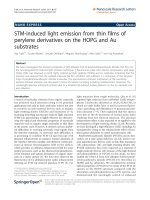
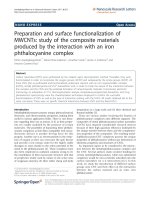
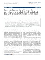
SDS-PAGE (Figure 1a) and size-exclusion HPLC (Fig-
ure 1b,c) demonstrated that pure (> 98%) Fab frag-
ments of trastuzumab and rituximab were obtained by
digestion of intact IgG
1
with immobilized papain using
a previously reported method [7,25]. Reaction of tras-
tuzumab and rituximab Fab with a 60- or 90-fold
excess of NHS-DOTA for 18 h at 4°C resulted in sub-
stitution of 3.7 ± 0.2 and 2.5 ± 0.3 DOTA chelators
per molecule, respectively. The pre-purification label-
ing efficiencies for
111
In-DOTA-trastuzumab Fab,
111
In-DOTA-rituximab Fab,
64
Cu-DOTA-trastuzumab
Fab, and
64
Cu-DOTA-rituximab Fab were 75.5 ± 5.4%,
76.8 ± 1.5%, 65.9 ± 4.9%, and 67.9 ± 5.7%, respectively.
Following purification, the radiochemical purity was >
98% for all radioimmunoconjugates by ITLC (not
shown) and size-exclusion HPLC (Figure 1b,c). The
specific activities of
111
In- and
64
Cu-DOTA-tras tuzu-
mab Fab fragments used in microSPECT and micro-
PET and biodistribution studies were 3.6 to 4.9 MBq/
μgand1.3to4.7MBq/μg, respectively. The specific
activities of
111
In- and
64
Cu-DOTA-rituximab Fab
were 1.3 to 5.8 and 1.9 to 2.8 MBq/μg.
HER2 binding affinity of
111
In- and
64
Cu-DOTA-
trastuzumab Fab
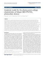
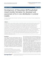
Direct (saturation) radioligand binding assays showed
that
111
In- and
64
Cu-DOTA-trastuzumab Fab bound
specifically to HER2 on SKBR-3 cells (Figure 2a,b). The
K
d
values for
111
In- and
64
Cu-DOTA-trastuzumab Fab
were 20.4 ± 2.5 nM and 40.8 ± 3.5 nM (P <0.01),
respectively. These values were similar to the K
d
for
111
In-DTPA-trastuzumab Fab binding to SKBR-3 c ells
previously reported by our group (K
d
= 48 nM) [25].
There was no specific binding of
111
In-DOTA-rituximab
to SKBR-3 cells (not shown). The B
max
values for
111
In-
and
64
Cu-DOTA-trastuzumab Fab on SKBR-3 cells were
1.4 ± 0.1 × 10
6
receptors/cell and 2.3 ± 0.1 × 10
6
recep-
tors/cell, respectively (P < 0.001).
Tumor and normal tissue distribution studies
The tumor and normal tissue uptake of
111
In- and
64
Cu-
DOTA-trastuzumab Fab at 24 h p.i. in athymic mice
bearing s.c. MDA-MB-361 human BC xenografts (5.1 ×
10
5
HER2/cell) are shown i n Table 1. Blood levels were
threefold significantly higher for
64
Cu- than
111
In-
DOTA-trastuzumab Fab (1.40 ± 0.16% vs. 0.42 ± 0.08%
i.d./g; P < 0.0001). Similarly, liver uptake was threefold
significantly greater for
64
Cu- than
111
In-DOTA-trastu-
zumab Fab (8.52 ± 0.81% vs. 3.13 ± 0.15% i.d./g; P <
0.0001). Radioactivity concentrations were higher in
heart, lungs, stomach, intestines, and spleen for
64
Cu-
than
111
In-DOTA-trastuzumab Fab (Table 1). However,
kidney uptake was not significantly different between
64
Cu- and
111
In-DOTA-trastuzumab Fab (57.00 ± 7.09%
vs. 62.85 ± 6.45% i.d./g; P = 0.268). There was no signifi-
cant difference in tumor accumulation for
111
In- and
64
Cu-DOTA-trastuzumab Fab (4.00 ± 0.90% vs.5.00±
1.2% i.d./g; P = 0.228 ). Due to the higher blood and
liver radioactivity, the T/B and tumor/liver (T/L)ratios
were three- and twofold significantly lower, respectively
for
64
Cu- than
111
In-DOTA-trastuzumab Fab (3.56 ±
0.62 vs. 9.73 ± 2.46; P = 0.003 and 0.59 ± 0.16 vs. 1.27 ±
0.26 vs.; P = 0.004, respectively; Table 2). T/NT ratios
for
64
Cu-DOTA-trastuzumab Fab were significantly
lower than
111
In-DOTA-trastuzumab Fab for all tissues
except kidneys and muscle (Table 2). There was no sig-
nificant d ifference in the uptake of
111
In- or
64
Cu-
DOTA-trastuzumab Fab in small (5 to 10 mm diameter)
vs. larger (10 to 15 mm) MDA-MB-361 tumor xeno-
grafts (4.00 ± 0.91% vs. 6.12 ± 0.84% i.d./g; P = 0.138
and 5.01 ± 1.20% vs. 7.12 ± 1.67% i.d./g, P = 0.342,
respectively). Absolute tumor uptake was not informa-
tive on the relationship between tumor localization of
the radioimmunoconjugates and HER2 expression.
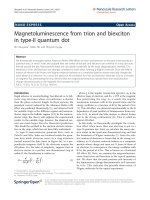
Tumor uptake of
111
In-DOTA-trastuzumab in MDA-
MB-231, BT-20, MDA-MB-361, or SKOV-3 xenografts
Chan et al. EJNMMI Research 2011, 1:15
/>Page 4 of 11
with increasing HER2 density was 4.7 ± 0.6%, 5.5 ±
0.7%, 4.0 ± 0.9%, and 5.4 ± 0.4% i.d./g. Tumor uptake of
64
Cu-DOTA-trastuzumab in MDA-MB-231, BT-20,
MDA-MB-361, or SKOV-3 xenografts was 4.4 ± 1.6%,
2.6 ± 1.8%, 5.0 ± 1.2%, and 5.0 ± 3.0% i.d./g. However,
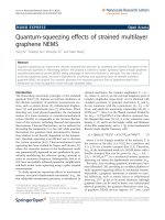
there was a strong direct relationship between T/B
ratios for
111
In-DOTA-trastuzumab Fab and tumor
HER2 density (Figure 3a). Moreover, the T/B ratios for
111
In-DOTA-trastuzumab Fab were significantly greater
than irrelevant
111
In-DOTA-rituximab Fab for MDA-
MB-231, BT-20, and MDA-MB-361 xenografts, demon-
strating specific localization. The T/B ratios for
64
Cu-
DOTA-trastuzumab Fab were significantly greater than
64
Cu-DOTA-rituximab Fab for MDA-MB-361 tumors
with high HER2 density (P < 0.001), but not for MDA-
MB-231 or BT-20 xenograft s with low HER2 expression
(P = 0.0709 and 0.528, respe ctively; Figure 3b). Th e
localization of
111
In- or
64
Cu-DOTA-rituximab Fab in
SK-OV-3 tumors was not determined. No relationship
between the T/B ratios for
64
Cu-DOTA-trastuzumab
Fab and HER2 density was established (Figure 3b).
MicroSPECT/CT and microPET/CT imaging
Representative microSPECT and microPET images of
athy mic mice with s.c. tumor xenografts with increasing
HER2 density at 24 h p.i. of
111
In- or
64
Cu-DOTA-tras-
tuzuma b Fab, respectively are shown in Figures 4 and 5.
MicroSPEC T/CT and mi croPET/CT imag es were
displayed as coronal slices with the plane selected to
optimal ly display the tumor uptake of
111
In-DOTA-tras-
tuzumab Fab or
64
Cu-DOTA-trastuzumab Fab. MDA-
MB-231 tumors with low HER2 expression (5.4 × 10
4
receptors/cell; Figures 4a and 5a) were least intensely
imaged while SK-OV-3 tumors with high HER2 density
a
50
150
25
MW 1 2 3 4 5 6
c
0 1.0 2. 0
3.0 4. 0 5. 0 6.0 7.0 8.0
9.0 10. 0 11.0 12.0 13.0 14.0 15.0
16.0 17.0
0
200
400
600
800
1000
1200
1400
1600
1800
2000
2200
2400
0
.
0
-125.50
-125.00
-124.50
-124.00
-123.50
-123.00
-122.50
-122.00
-121.50
-121.00
-120.50
-120.00
-119.50
-119.00
-118.50
-118.00
-117.50
-117.00
0.0
Abs
280
radioactivity
0 5.0 10.0 15.0
Radioactivity or Absorbance at 280 nm
(
abrbitrary units)
Time
(
mins
)
b
0 1.0 2.0 3.0 4.0 5.0 6.0 7.0
8.0 9.0 10.0 11. 0 12.0 13.0 14. 0 15.0 16.0
17.0
0
500
1000
1500
2000
2500
3000
3500
4000
4500
5000
5500
6000
6500
7000
7500
8000
0.0
-125.00
-124.50
-124.00
-123.50
-123.00
-122.50
-122.00
-121.50
-121.00
-120.50
-120.00
-119.50
-119.00
0.0
Abs
280
radioactivity
0 5.0 10.0 15.0
Radioactivity or Absorbance at 280 nm
(
abrbitrary units)
Time (mins)
Figure 1 SDS-PAGE and size-exclusion HPLC. (a) SDS-PAG E analysis on a 4% to 20% Tris HCl gradient mini-gel stained with Coomassie
Brilliant Blue of trastuzumab (lane 1), trastuzumab Fab (lane 2), and DOTA-trastuzumab Fab (lane 3) under non-reducing conditions or these
same proteins under reducing conditions (lanes 4 to 6, respectively). Molecular weight markers are shown (lane MW). The positions of 150, 50,
and 25 kDa markers are indicated. Size-exclusion HPLC analyses of (b)
111
In-DOTA-trastuzumab Fab (top panel) and (c)
64
Cu-DOTA-trastuzumab
Fab (bottom panel) with detection of absorbance at 280 nm or radioactivity.
Chan et al. EJNMMI Research 2011, 1:15
/>Page 5 of 11
(1.2 × 10
6
receptors/cell) were most clearly seen (Figures
4c and 5c) with
111
In- o r
64
Cu-DOTA-trastuzumab. An
intermediate tumor signal was found for MDA-MB-361
xenografts with 5.1 × 10
5
HER2/cell (Figures 4b and
5b). The specificity of tumor localization of
111
In- or
64
Cu-DOTA-trastuzumab Fab was shown by the lower
accumulation of
111
In- or
64
Cu-DOTA-rituximab Fab on
images of mice bearing MDA-MB-361 xenografts (Fig-
ures 4d and 5d). There was no difference in the ability
of
111
In- or
64
Cu-DOTA-trastuzumab Fab to image
small (5 to 10 mm; Figure 6a,c) or larger (10 to 15 mm;
Figure 6b, d) MDA-MB-361 tumors. The kidneys were
most prominent on microSPECT images of
111
In-
DOTA-trastuzumab Fab, while microPET with
64
Cu-
DOTA-trastuzumab Fab showed high liver and kidney
uptake.
Discussion
Our results revealed that both microSPECT/CT and
microPET/CT with
111
In- or
64
Cu-DOTA-trastuzumab
Fab fragments were able to image s.c. human tumor
xenografts in mice with low, intermediate, or high HER2
a
b
0 100 200 300 400 500 600 70
0
0
20000
40000
60000
80000
100000
120000
TB
NSB
SB
64
Cu-DOTA-trastuzumab Fab added (nM)
64
Cu-DOTA-trastuzumab Fab Bound (cpm)
0 100 200 300 400 500 600 700
0
100000
200000
300000
400000
500000
600000
TB
NSB
SB
111
In-DOTA-trastuzumab Fab added (nM)
111
In-DOTA-trastuzumab Fab Bound (cpm)
Figure 2 Dir ect (saturation) radioligand binding assays. Direct (satu ration) radioligand bindin g to SKBR-3 human breast cancer cells of (a)
111
In-DOTA-trastuzumab Fab or (b)
64
Cu-DOTA-trastuzumab Fab, in the absence (total binding; TB) or presence (non-specific binding; NSB) of
excess (16 μM) unlabeled trastuzumab IgG. Specific binding (SB) was calculated by subtraction of NSB from TB. Curves were fitted to a 1-site
receptor-binding model using Prism Ver. 4.0 software (GraphPad).
Table 1 Tumor and normal tissue distribution at 24 h
post-injection of
111
In- or
64
Cu-DOTA-trastuzumab Fab
Percent injected dose/g
a, b, c
Tissue
111
In-DOTA-trastuzumab
Fab
64
Cu-DOTA-trastuzumab
Fab
Blood 0.42 ± 0.08 1.40 ± 0.16
Heart 0.92 ± 0.06 2.42 ± 0.39
Lungs 0.80 ± 0.14 4.86 ± 0.57
Liver 3.13 ± 0.16 8.52 ± 0.81
Kidneys 62.85 ± 6.45 57.00 ± 7.09
Stomach 0.58 ± 0.05 3.41 ± 0.22
Intestines 0.61 ± 0.06 4.76 ± 0.41
Spleen 2.25 ± 0.13 5.00 ± 0.27
Muscle 0.78 ± 0.31 0.75 ± 0.06
Tumor 4.00 ± 0.91 5.01 ± 1.20
111
In- or
64
Cu-DOTA-trastuzumab Fab in athymic mice bearing subcutaneous
MDA-MB-361 human breast cancer xenografts.
a
Values shown are mean ± SD
(n = 4).
b
Significantly different for
111
In- and
64
Cu-DOTA-trastuzumab Fab for
blood, heart, lungs, liver, stomach, intestines, and spleen (all P < 0.001).
c
Not
significantly different for
111
In- and
64
Cu-DOTA-trastuzumab Fab for kidneys,
muscle, or tumor (P > 0.05).
Table 2 Tumor/normal tissue (T/NT) ratios at 24 h post-
injection of
111
In- or
64
Cu-DOTA-trastuzumab Fab
T/NT Ratio
a, b, c
Tissue
111
In-DOTA-trastuzumab
Fab
64
Cu-DOTA-trastuzumab
Fab
Blood 9.73 ± 2.46 3.56 ± 0.62
Heart 4.31 ± 0.83 2.09 ± 0.56
Lungs 5.06 ± 1.12 1.03 ± 0.19
Liver 1.27 ± 0.26 0.59 ± 0.16
Kidneys 0.06 ± 0.02 0.09 ± 0.02
Stomach 6.99 ± 2.05 1.48 ± 0.37
Intestines 6.61 ± 1.78 1.05 ± 0.22
Spleen 1.79 ± 0.48 1.00 ± 0.20
Muscle 6.12 ± 3.33 6.78 ± 1.90
111
In- or
64
Cu-DOTA-trastuzumab Fab in athymic mice bearing subcutaneous
MDA-MB-361 human breast cancer xenografts.
a
Values shown are mean ± SD
(n = 4).
b
Significantly different for
111
In- and
64
Cu-DOTA-trastuzumab Fab for
lungs and intestines (both P < 0.001), blood, heart, liver, stomach (all P < 0.01)
and spleen (P < 0.05).
c
Not significantly different for
111
In- and
64
Cu-DOTA-
trastuzumab Fab for kidneys and muscle (P > 0.05).
Chan et al. EJNMMI Research 2011, 1:15
/>Page 6 of 11
expression. The range of HER2 expression examined
(5.4 × 10
4
to 1.2 × 10
6
receptors/cell) corresponded to
HER2 scores of 0 to 3+ assessed clinically in BC speci-
mens by IHC staining [9]. There was no apparent
increased ability of microPET/CT with
64
Cu-DOTA-
trastuzumab Fab compared to microSPECT/CT with
111
In-DOTA-trastuzumab Fab to visualize MDA-MB-
231 tumors with low HER2 density (1.6 × 10
5
receptors/
cell; Figures 4a and 5a). In addition, there was no
increased ability of microPET/CT using
64
Cu-DOTA-
trastuzumab Fab to image small (5 to 10 mm diameter)
or larger (10 to 15 mm diameter) MDA-MB-361 tumors
with intermediate HER2 expression (5.1 × 10
5
HER2/
cell; Figure 6). The intensity of the tumor signal was
dependent on HER2 expression with tumors with inter-
mediate (MDA-MB-361) or high (SK-OV-3) HER2 den-
sity most readily imaged by microSPECT/CT (Figure 4)
or microPET/CT (Figure 5). However, a threefold higher
dose of radioactivity was administered for microSPECT/
CT than microPET/CT (70 vs.22MBq)andimage
acquisition times were up to sixfold longer for micro-
SPECT/CT (85 to 20 vs. 20 min, respectively). Thus, the
photon detection efficiency (i.e., intrinsic sensitivity) was
much higher for microPET/CT than microSPECT/CT.
Nonetheless, our results revealed that provided that the
administered dose of radioactivity was sufficient and
image acquisitio n times were long enough to yield good
counting statistics, microSPECT/CT with
111
In-DOTA-
trastuzumab Fab was able to ima ge tumors with the
similar HER2 density and size as microPET/CT with
64
Cu-DOTA-trastuzumab. These results agree with
those reported by C heng et al. w ho noted that s.c.
HER2-positiv e SUM190 t umor xenografts were imaged
by either microSPECT or microPET using trastuzumab
conjugated to biotinylated
99 m
Tc- or
18
F-labeled phospho-
diamidate morpholinos (MORFs) through a streptavidin
linker [28]. The doses of
99 m
Tc or
18
F used in their study
were 13 and 0.22 MBq, respectively. Phantom s tudies
revealed that microPET was 15-fold more sensitive in
terms o f photon detection, but the spatial resolution
of microSPECT was superior to that of microPET (1.2 vs.
2.4 mm). The results are also in concordance with those
reported by Wong et al. [29], who showed that s.c. epider-
mal growth factor receptor-positive LS174T human colon
cancer xenografts could be imaged using panitumomab F
(ab’)
2
fragments labeled with
111
In or
86
Y. However, they
compared low resolution planar g-camera imaging with
microPET. In our study, we used similar quality high reso-
lution and high sensitivity small animal imaging technolo-
gies, namely microSPECT/CT (NanoSPECT; Bioscan) and
a
b
M
DA-MB-231
BT-20
M
DA-MB-361
SKOV-3
0
5
10
15
20
25
111
In-DOTA-rituximab Fab
111
In-DOTA-trastuzumab Fab
n.d.
T/B Ratio
*
*
*
MDA-MB-231
BT-20
MDA-MB-361
SKOV-3
0
2
4
6
n.d.
64
Cu-DOTA-trastuzumab Fab
64
Cu-DOTA-rituximab Fab
T/B Ratio
*
Figure 3 Tumor/blood (T/B) ratios. T/B ratios at 24 h p.i. of (a)
111
In-DOTA -trastuzumab Fab and
111
In-DOTA-rituximab Fab or (b)
64
Cu-DOTA-
trastuzumab Fab and
64
Cu-DOTA-rituximab Fab, in s.c. human tumor xenografts in athymic mice with increasing HER2 expression. Values shown
are mean ± SD (n = 4). The HER2 density (receptors/cell) was 5.4 × 10
4
(MDA-MB-231), 1.6 × 10
5
(BT-20), 5.1 × 10
5
(MDA-MB-361) and 1.2 × 10
6
(SK-OV-3). Significant differences (P < 0.05) between
111
In- or
64
Cu-labeled trastuzumab Fab and rituximab Fab are indicated by asterisks. n.d., not
determined.
Chan et al. EJNMMI Research 2011, 1:15
/>Page 7 of 11
microPET (Siemens Focus 220) systems for these
comparisons.
T/B ratios were used to compare the tumor localization
of
111
In- and
64
Cu-DOTA-trastuzumab Fab vs.HER2den-
sity because we previously found that there are differences
in perfusion between different tumor xenografts which
can affect the uptake of radioimmunoconjugates [9]. Use
of T/B ratios minimizes these effects by normalizing for
blood concentrations which then reveals the relationships
between HER2 density and tumor accumulation. More-
over, the T/B ratios are important for discriminating
tumors that have different HER2 expression on the
images. There was a strong and direct association between
the T/B rat ios for
111
In-DOTA-trastuzumab and tumor
HER2 density ( Figure 3a). In addition, the T/B ratios for
111
In-DOTA-trastuzumab Fab were significantly greater
than those of irrelevent control
111
In-DOTA-rituximab
Fab for MDA-MB-231, BT-20, and MDA-MB-361 xeno-
grafts, demonstrating specific localization in tumors with
low or intermediate HER2 expression. In contrast, specific
uptake of
64
Cu-DOTA-trastuzumab Fab was shown in
MDA-MB-361 tumors with intermediate HER2 density
but not for t umors with lower HER2 expression (Figure
3b). Tumor uptake was not significantly different for
64
Cu- and
111
In-DOTA-trastuzumab Fab, but blood
radioa ctivity was threefo ld lower for
111
In-DOTA-trastu-
zumab Fab (Table 1). Thus, T/B ratios were threefold
lower for
64
Cu- than
111
In-DOTA-trastuzumab (3.6:1 vs.
10:1; Table 2) in mice with MDA-MB-361 tumors. The
increased circulating radioactivity for
64
Cu-DOTA-trastu-
zumab Fab may be due to kinetic instability of the
64
Cu-
DOTA complex with transchelation of released
64
Cu to
coppe r binding proteins (e.g., albumin, ceruloplasmin, or
superoxide dismutase) [26]. These
64
Cu-labeled proteins
may non-specifically localize in tumors, disrupting the
association between T/B ratios and HER2 density, espe-
cially for tumors with low HER2 expression (i.e., MDA-
MB-231 and BT-20).
DOTA forms thermodynamically stable complexes with
copper (K
d
=10
23
M
-1
) but kinetic instability of
64
Cu-
DOTA complexes in vivo can lead to loss of radiometal
resulting in high blood radioactivity and liver and spleen
uptake [17]. In addition to the higher levels of blood radio-
activity, we found that the liver and spleen uptake for
64
Cu-DOTA-trastuzumab Fab were three- and twofold
greater, respectively, than
111
In-DOTA-trastuzumab Fab
(Table 1). In order to improve the kinetic stability of
64
Cu
complexes, more thermodynamically stable cross-bridged
(CB-DO2A) or sarcophagine (SarAr) chelators have been
synthesi zed [30,31]. A comparison of
64
Cu complexed to
a
b
cd
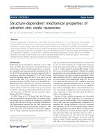
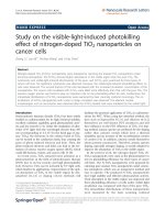
Figure 4 Coronal slice microSPECT/CT images. Images of athymic mice with s.c. human tumor xenografts (solid arrows) with increasing HER2
expression at 24 h p.i. of 70 MBq (10 μg) of
111
In-DOTA-trastuzumab [panels (a), (b), (c)] or
111
In-DOTA-rituximab Fab [panel (d)]. (a) Mouse with
MDA-MB-231 (left panel) and BT-20 (right panel) xenografts with low HER2 density (5.4 × 10
4
and 1.6 × 10
5
receptors/cell, respectively). (b)
Mouse with MDA-MB-361 xenograft with intermediate HER2 density (5.1 × 10
5
receptors/cell). (c) Mouse with 15 to 18 mm diameter (left flank)
and 5 to 10 mm diameter (right flank) SK-OV-3 xenografts with high HER2 density (1.2 × 10
6
receptors/cell). (d) Mouse with MDA-MB-361
xenograft with intermediate HER2 density (5.1 × 10
5
receptors/cell). Bladder radioactivity in panel (c) is indicated by broken arrow. Image
acquisition time was 85 to 120 min and anaesthesia was induced and maintained by inhalation of 2% isoflurane in oxygen. Images were
adjusted to approximately equal intensity.
Chan et al. EJNMMI Research 2011, 1:15
/>Page 8 of 11
ab
cd
Figure 5 Coronal slice microPET/CT images. Images of athymic mice with s.c. human tumor xenografts (arro ws) with increasing HER2
expression at 24 h p.i. of 22 MBq (10 μg)
64
Cu-DOTA-trastuzumab [panels (a), (b), (c)] or
64
Cu-DOTA-rituximab Fab [panel (d)]. (a) Mouse with
MDA-MB-231 and BT-20 xenografts implanted on the left and right flanks, respectively with low HER2 density (5.4 × 10
4
and 1.6 × 10
5
receptors/
cell, respectively). (b) Mouse with MDA-MB-361 xenograft with intermediate HER2 density (5.1 × 10
5
receptors/cell). (c) Mouse with 15 to 18 mm
diameter (left) and 5 to 10 mm diameter (right) SK-OV-3 xenografts with high HER2 density (1.2 × 10
6
receptors/cell). (d) Mouse with MDA-MB-
361 xenograft with intermediate HER2 density (5.1 × 10
5
receptors/cell). Bladder radioactivity seen in one mouse is indicated by a broken arrow.
Image acquisition time was 20 min and anaesthesia was induced and maintained by inhalation of 2% isoflurane in oxygen. Images were
adjusted to approximately equal intensity.
ab
cd
Figure 6 Coronal slice microSPECT/CT [(a) and (b)] or microPET/CT images [(c) and (d)]. Athymic mice with s.c. MDA-MB-361 tumor
xenografts (solid arrows) with intermediate HER2 density (5.1 × 10
5
receptors/cell) at 24 h p.i. of 70 MBq (10 μg) of
111
In-DOTA-trastuzumab Fab
or 22 MBq (10 μg) of
64
Cu-DOTA-trastuzumab Fab, respectively. (a) and (c) Mouse with 5 to 10 mm diameter tumor. (b) and (d) Mouse with 10
to 15 mm diameter tumor. Image acquisition time was 85 to 120 min for microSPECT and 20 min for microPET. Anaesthesia was induced and
maintained by inhalation of 2% isoflurane in oxygen. Images were adjusted to approximately equal intensity within each modality.
Chan et al. EJNMMI Research 2011, 1:15
/>Page 9 of 11
DOTA or CB-DO2A (but not conjugated to mAbs)
showed fourfold lower radioactivity in the blood and two-
fold lower liver accumulation at 24 h p.i. in rats [30]. Voss
et al. noted that ch14.18 mAbs labeled with
64
Cu through
the extremely stable SarAr chelator for PET imaging o f
neuroblastoma or melanoma xenografts in mice exhibited
low liver uptake (5% to 10% i.d./g) but no comparison
with other chelators was provided [31]. Dearling et al.
recently compared the tumor and normal tissue distribu-
tion of these same
64
Cu-labeled ch14.18 m Abs using a
variety of chelators including DOTA and SarAr in mice
bearing M21 melanoma xenografts [17]. Unexpectedly, no
significant differences in tumor or liver uptake were found
for ch14.18 labeled with
64
Cu using DOTA or the much
more stable SarAr chelator. They suggested that in addi-
tion to
64
Cu-chelator stability, factors such as the net
chargeonthechelatorsmayplayanimportantrolein
sequestration of radioactivity by tissues. In our study,
tumor uptake was not significantly different between
111
In- and
64
Cu-DOTA-trastuzumab Fab in mice with
MDA-MB-361 tumors, despite the apparent instability of
64
Cu-DOTA-trastuzumab Fab as eviden ced by higher
levels of radioactivity in the blood, liver, and spleen (Table
1). The use of more stable chelators such as CB-DO2A or
SarAr may diminish blood radioactivity and improve the
association between tumor HER2 density and T/B ratios
for
64
Cu-labeled trastuzumab Fab. The CB-DO2A and
SarAr chelators are unfortunately not yet commercially
avail able in a chemically reactive form for conjugation to
mAbs for
64
Cu labeling.
Conclusion
Provided that administered doses of radioactivity and
acquisition times were sufficient to yield good counting
statistics, we conclude that either microSPECT/CT with
111
In-DOTA-trastuzumab Fab or microPET/CT with
64
Cu-DOTA-trastuzumab Fab visualized small (5 to
10 mm diameter) or larger (10 to 15 mm diameter) s.c.
tumor xenografts with low, intermediate, or high HER2
expression in athymic mice. However, due to the higher
levels of circulating radioactivity for
64
Cu-DOTA-trastuzu-
mab Fab, no association between HER2 density and T/B
ratios was established. In contrast, there was a stro ng
direct association between T/B ratios and HER2 density of
these tumors for
111
In-DOTA-trastuzumab Fab. Thus,
111
In-DOTA-trastuzumab Fab was more specific than
64
Cu-DOTA-trastuzumab Fab for imaging HER2-positive
tumors with low HER2 density. The use of more stable
CB-DO2A or SarAr chelators for
64
Cu may potentially
diminish blood radioactivity, provide a stronger associa-
tion between T/B ratios and tumor HER2 density, and
improve the specificity of imaging with
64
Cu-labeled tras-
tuzumab Fab.
Acknowledgements
This study was supported by a grant from the Ontario Institute for Cancer
Research (1 mm Challenge) with funds from the Province of Ontario. Parts
of this study were presented at the European Association of Nuclear
Medicine Congress, Barcelona, Spain, October 9 to 13, 2009.
Author details
1
Department of Pharmaceutical Sciences, University of Toronto, Toronto,
M5S 3M2, ON, Canada
2
Department of Medical Imaging, University of
Toronto, Toronto, M5S 3E2, ON, Canada
3
Toronto General Research Institute,
University Health Network, Toronto, M5G 2M9, ON, Canada
Authors’ contributions
CC and SS synthesized the
111
In- and
64
Cu-DOTA-trastuzumab Fab fragments
and performed characterization studies. KM and DAS performed microSPECT
and microPET imaging studies. RMR wrote the manuscript with the
assistance of all authors.
Competing interests
The authors declare that they have no competing interests.
Received: 6 July 2011 Accepted: 17 August 2011
Published: 17 August 2011
References
1. Revillion F, Bonneterre J, Peyrat JP: ERBB2 oncogene in human breast
cancer and its clinical significance. Eur J Cancer 1998, 34:791-808.
2. Behr TM, Béhé M, Wörmann B: Trastuzumab and breast cancer. N Engl J
Med 2001, 345:995-996.
3. Owens MA, Horten BC, Da Silva MM: HER2 amplification ratios by
fluorescence in situ hybridization and correlation with
immunohistochemistry in a cohort of 6556 breast cancer tissues. Clin
Breast Cancer 2004, 5:63-69.
4. Munzone E, Nolé F, Goldhirsch A, Botteri E, Esposito A, Zorzino L,
Curigliano G, Minchella I, Adamoli L, Cassatella MC, Casadio C, Sandri MT:
Changes in HER2 status in circulating tumor cells compared with the
primary tumor during treatment for advanced breast cancer. Clin Breast
Cancer 2010, 10:392-397.
5. Pestrin M, Bessi S, Gallardi F, Truglia M, Biggeri A, Biagioni C, Cappadona S,
Biganzoli L, Giannini A, Di Leo A: Correlation of HER2 status between
primary tumors and corresponding circulating tumor cells in advanced
breast cancer patients. Breast Cancer Res and Treatment 2009, 118:523-530.
6. Lub-de Hooge MN, Kosterink JG, Perik PJ, Nijnuis H, Tran L, Bart J,
Suurmeijer AJH, de Jong S, Jager PL, de Vries EGE: Preclinical characterisation
of
111
In-DTPA-trastuzumab. Br J Pharmacol 2004, 143:99-106.
7. Tang Y, Wang J, Scollard DA, Mondal H, Holloway C, Kahn HJ, Reilly RM:
Imaging of HER2/neu-positive BT-474 human breast cancer xenografts
in athymic mice using
111
In-trastuzumab (Herceptin) Fab fragments. Nucl
Med Biol 2005, 32:51-58.
8. Tang Y, Scollard D, Chen P, Wang J, Holloway C, Reilly RM: Imaging of
HER2/neu expression in BT-474 human breast cancer xenografts in
athymic mice using
99 m
Tc-HYNIC-trastuzumab (Herceptin) Fab
fragments. Nucl Med Commun 2005, 26:427-432.
9. McLarty K, Cornelissen B, Scollard DA, Done SJ, Chun K, Reilly RM:
Associations between the uptake of
111
In-DTPA-trastuzumab, HER2
density and response to trastuzumab (Herceptin) in athymic mice
bearing subcutaneous human tumour xenografts. Eur J Nucl Med Mol
Imaging 2009, 36:81-93.
10. Perik PJ, Lub-de Hooge MN, Gietema JA, van der Graaf WTA, de Korte MA,
Jonkman S, Kosterink JG, van Veldhuisen DJ, Sleijfer DT, Jager PL, de
Vries EGE: Indium-111-labeled trastuzumab scintigraphy in patients with
human epidermal growth factor receptor 2-positive metastatic breast
cancer. J Clin Oncol 2006, 24:2276-2282.
11. Dijkers EC, Oude Munnink TH, Kosterink JG, Brouwers AH, Jager PL de
Jong R, van Dongen GA, Lub-de Hooge MN, de Vries EGE: Biodistribution
of
89
Zr-trastuzumab and PET imaging of HER2-positive lesions in
patients with metastatic breast cancer. Clin Pharmacol Ther 2010,
87:586-592.
12. Dijkers ECF, Kosterink JGW, Rademaker AP, Perk LR, van Dongen GA, Bart J,
de Jong R, de Vries EGE, Lub-de Hooge MN: Development and
Chan et al. EJNMMI Research 2011, 1:15
/>Page 10 of 11
characterization of clinical-grade
89
Zr-trastuzumab for HER2/neu
immunoPET imaging. J Nucl Med 2009, 50:974-981.
13. McLarty K, Reilly RM: Molecular imaging as a tool for personalized and
targeted anticancer therapy. Clin Pharmacol Ther 2007, 81:420-424.
14. McLarty K, Cornelissen B, Scollard D, Reilly RM: Micro-SPECT/CT with
111
In-
DTPA-pertuzumab sensitively detects trastuzumab-mediated HER2
downregulation and tumor response in athymic mice bearing MDA-MB-
361 human breast cancer xenografts. J Nucl Med 2009, 50:1340-1348.
15. Smith-Jones PM, Solit DB, Akhurst T, Afroze F, Rosen N, Larson SM: Imaging
the pharmacodynamics of HER-2 degradation in response to Hsp90
inhibitors. Nat Biotechnol 2004, 22:701-706.
16. Garcia R, Kubicek V, Drahos B, Gano L, Santos IC, Campello P, Paulo A,
Toth E, Santos I: Synthesis, characterization and biological evaluation of
In(III) complexes anchored by DOTA-like chelators bearing a quinazoline
moiety. Metallomics 2010, 2:571-580.
17. Dearling JLJ, Voss SD, Dunning P, Snay E, Fahey F, Smith SV, Huston JS,
Meares CF, Treves ST, Packard AB: Imaging cancer using PET - the effect
of the bifunctional chelator on the biodistribution of a
64
Cu-labeled
antibody. Nucl Med Biol 2011, 38:29-38.
18. Olafsen T, Kenanova VE, Sundaresan G, Anderson AL, Crow D, Smith SV,
Huston JS, Meares CF, Treves ST, Packard AB: Optimizing radiolabeled
engineered anti-p185HER2 antibody fragments for in vivo imaging.
Cancer Res 2005, 65:5907-5916.
19. Cai W, Chen K, He L, Cao Q, Koong A, Chen X: Quantitative PET of EGFR
expression in xenograft-bearing mice using
64
Cu-labeled cetuximab, a
chimeric anti-EGFR monoclonal antibody. Eur J Nucl Med Mol Imaging
2007, 34:850-858.
20. Niu G, LI Z, Cao Q, Chen X: Monitoring therapeutic response of human
ovarian cancer to 17-DMAG by non-invasive PET imaging with
64
Cu-
DOTA-trastuzumab. Eur J Nucl Med Mol Imaging 2009, 36:1510-1519.
21. Anderson CJ, Dehdashti F, Cutler PD, Schwartz SW, LaForest R, Bass LA,
Lewis JS, McCarthy DW:
64
Cu-TETA-octreotide as a PET imaging agent for
patients with neuroendocrine tumors. J Nucl Med 2001, 42:213-221.
22. Rogers BE, Bigott HM, McCarthy DW, Manna DD, Kim J, Sharp TL, Welch MJ:
MicroPET imaging of a gastrin-releasing peptide receptor-positive tumor
in a mouse model of human prostate cancer using a
64
Cu-labeled
bombesin analogue. Bioconjug Chem 2003, 14:756-763.
23. Thakur ML, Aruva MR, Gariepy J, Acton P, Rattan S, Prasad S, Wickstrom E,
Alavi A: PET imaging of oncogene overexpression using
64
Cu-vasoactve
intestinal peptide (VIP) analog: comparison with
99 m
Tc-VIP analog. J
Nucl Med 2004, 45:1381-1389.
24. Reilly RM, Sandhu J, Alvarez-Diez TM, Gallinger S, Kirsh J, Stern H: Problems
of delivery of monoclonal antibodies. Pharmaceutical and
pharmacokinetic solutions. Clin Pharmacokinet 1995, 28:126-142.
25. Scollard DA, Chan C, Holloway CMB, Reilly RM: A kit to prepare
111
In-
DTPA-trastuzumab (Herceptin) Fab fragments injection under GMP
conditions for imaging or radioimmunoguided surgery of HER-2 positive
breast cancer. Nucl Med Biol 2011, 38:129-136.
26. Reilly RM: The radiochemistry of monoclonal antibodies and peptides. In
Monoclonal Antibody and Peptide-Targeted Radiotherapy of Cancer. Edited by:
Reilly RM. Hoboken, NJ: John Wiley 2010:39-100.
27. Orlova A, Tolmachev V, Pehrson R, Lindborg M, Tran T, Sandström M,
Nilsson FY, Wennborg A, Abrahmsén L, Feldwisch J: Synthetic affibody
molecules: a novel class of affinity ligands for molecular imaging of
HER2-expressing malignant tumors. Cancer Res 2007, 67:2178-2186.
28. Cheng D, Wang Y, Liu X, Pretorius PH, Liang M, Rusckowski M,
Hnatowich DJ: Comparison of
18
F PET and
99 m
Tc SPECT imaging in
phantoms and in tumored mice. Bioconjug Chem 2010, 21:1565-1570.
29. Wong KJ, Baidoo KE, Nayak TK, Garmestan K, Brechbiel MW, Milenic DE: In
vitro and in vivo pre-clinical analysis of a F(ab’)
2
fragment of
panitumumab for molecular imaging and therapy of HER1-positive
cancers. EJNMMI Research 2011, 1:1-15.
30. Boswell CA, Sun X, Niu W, Wesiman GR, Wong EH, Rheingold AL,
Anderson CJ: Comparative in vivo stability of copper-64-labeled cross-
bridged and conventional tetraazamacrocyclic complexes. J Med Chem
2004, 47:1465-1474.
31. Voss SD, Smith SV, DiBartolo N, McIntosh LJ, Cyr EM, Bonab AA, Dearling JL,
Carter EA, Fischman AJ, Treves ST, Gillies SD, Sargeson AM, Huston JS,
Packard AB: Positron emission tomography (PET) imaging of
neuroblastoma and melanoma with
64
Cu-SarAr immunoconjugates. Proc
Natl Acad Sci USA 2007, 104:17489-17493.
doi:10.1186/2191-219X-1-15
Cite this article as: Chan et al.: A comparison of
111
In- or
64
Cu-DOTA-
trastuzumab Fab fragments for imaging subcutaneous HER2-positive
tumor xenografts in athymic mice using microSPECT/CT or microPET/
CT. EJNMMI Research 2011 1:15.
Submit your manuscript to a
journal and benefi t from:
7 Convenient online submission
7 Rigorous peer review
7 Immediate publication on acceptance
7 Open access: articles freely available online
7 High visibility within the fi eld
7 Retaining the copyright to your article
Submit your next manuscript at 7 springeropen.com
Chan et al. EJNMMI Research 2011, 1:15
/>Page 11 of 11