Biomimetics learning from nature Part 2 pot
Bạn đang xem bản rút gọn của tài liệu. Xem và tải ngay bản đầy đủ của tài liệu tại đây (7.89 MB, 30 trang )
Immobilizedredoxproteins:
mimickingbasicfeaturesofphysiologicalmembranesandinterfaces 23
electrodes represents a powerful alternative, allowing application of direct electrochemistry
and surface-enhanced vibrational spectroelectrochemical techniques. These methods permit
determination of kinetic and thermodynamic parameters of the heterogeneous ET in a
protein that is exposed to physiologically relevant electric fields. Furthermore, ET steps can
be controlled in terms of directionality, distance, and driving force. In addition,
spectroelectrochemical methods can simultaneously probe the active site structure and
conformational dynamics concomitant to the ET.
In this chapter we will present an overview of recent developments in the field of
biocompatible immobilization of membrane-bound and soluble redox proteins on metal
electrodes, and of the spectroelectrochemical techniques used for the in situ characterization
of the structure, thermodynamics and reaction dynamics of the immobilized proteins.
After a brief description of biological ET chains and their constituting complexes (Section 2),
we will introduce some of the strategies for protein immobilization (Section 3), with special
emphasis on self-assembled monolayers (SAMs) of functionalized alkanethiols as versatile
biocompatible coatings that can be tailored according to the specific requirements. In Section
4 we will describe the basic principles of stationary and time-resolved surface-enhanced
vibrational spectroscopies (SERR and SEIRA) as valuable tools for studying specifically the
redox centres or the immobilized metalloproteins. The contents of the first 3 sections are
integrated in Section 5, where recent progress in the immobilization and SERR/SEIRA
characterization of different components of ET respiratory chains, mainly oxygen reductases
and cytochromes will be discussed. We will conclude with a brief outlook (Section 6).
2. Redox proteins under physiological conditions
In this section we will provide a brief introduction to the complex ET chains involved in the
energetics of organisms, i.e. respiratory and photosynthetic chains. In spite of obvious
differences, these two types of systems share a number of common features that must be
taken into account when investigating them using biomimetic approaches. First, both types
of chains consist of a series of membrane-integrated redox active protein complexes that
communicate through hydrophilic (e.g. cytochromes) and hydrophobic (e.g. quinones)
electron shuttles. Second, the energy provided by the sequence of exergonic ET events is
utilized by some of the constituting membrane proteins for translocating protons across the
membrane against an electrochemical gradient. This gradient is, for example, utilized for
driving ATP synthesis. Common to components of both ET chains are the specific reaction
conditions that deviate substantially from redox processes of proteins in solution.
Characteristic features are the restricted mobility of the membrane integral and peripheral
proteins and the potential distribution across the membrane that displays drastic changes in
the vicinity of the lipid head groups, giving origin to strong local electric fields.
2.1 Electron transfer chains
Membranes are essential in cells for defining structural and functional features, controlling
intracellular conditions and responding to the environment. They permit maintaining the non-
equilibrium state that keeps cells alive. Phospholipids are the main components of cell
membranes, responsible for the membrane shape and flexibility. They are self-assembled in
such a manner that non-polar acyl chains driven by hydrophobic interactions orient
themselves towards the center of the membrane, while the polar groups remain exposed to the
solution phase, e.g., the cytoplasm and periplasm. The constituent phospholipids, which are
typically asymmetrically distributed along the membrane, differ between cellular and
mitochondrial membranes. Similar to smectic liquid crystals, membranes present continuous,
ordered and oriented, but inhomogeneous structures (Gennis, 1989; Hianik, 2008).
A large variety of proteins are incorporated into or associated to membranes, including
enzymes, transporters, receptors and structural proteins. Enzymes are the most abundant of
all membrane proteins. Together with water soluble proteins and lipophilic compounds,
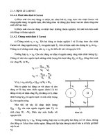
membrane-bound enzymes compose ET chains. In eukaryotic organisms the oxidation of
nutrients such as glucose and fatty acids produces reduced metabolites, namely NADH and
succinate, which upon oxidation deliver electrons though ET chains to molecular oxygen. ET
occurs through a series of sequential redox reactions between multisubunit transmembrane
complexes (Figure 1), situated in the inner mitochondrial membrane of non-photosynthetic
eucaryotic cells, or in the cytoplasmatic (cell) membrane of bacteria and archaea. The
complexes involved in a canonical respiratory chain are:
- Complex I (NADH : ubiquinone oxidoreductase or NADH dehydrogenase): catalyzes two-
electron transfer from NADH to quinone. It is composed of 46 subunits in eukaryotic
complexes, but only of 13 to 14 subunits in bacteria, which ensure the minimal functional
unit. Electrons enter the enzyme trough a non-covalently bound FMN primary acceptor and
are then passed to the quinone molecules via several iron-sulfur clusters.
- Complex II (succinate : ubiquinone oxidoreductase or succinate dehydrogenase): couples
two electron oxidation of succinate to fumarate with reduction of quinone to quinol, by
transferring electrons from a covalently bound FAD, via iron-sulfur clusters to heme
group(s) located in the transmembrane part of the complex, and ultimately to the quinones.
- Complex III (ubiquinol : Cyt-c oxidoreductase or bc
1
complex): catalyzes the transfer of two
electrons from ubiquinol to two Cyt-c molecules. It is composed of 10 to 11 subunits in
mitochondria and 3 subunits in cell membranes of bacteria and archaea, which bear all
prosthetic groups: two low-spin hemes b, a Rieskie type iron-sulfur cluster and a heme c
1
.
The last redox center is located near the docking site of the electron acceptor Cyt-c.
- Complex IV (Cyt-c : oxygen oxidoreductase or Cyt-c oxidase): catalyzes reduction of
oxygen to water by utilizing four electrons received from four molecules of Cyt-c, or
alternative electron donors present in some bacteria and archaea (see below).
Fig. 1. Schematic representation of the mitochondrial respiratory electron transfer chain. The
four complexes (I to IV), and their respective electron transfer reactions are depicted,
together with proton fluxes and ATP synthase.
Biomimetics,LearningfromNature24
The ET reactions through complexes I, III and IV are coupled to proton translocation across
the membrane, contributing to generation and maintenance of a transmembrane
electrochemical potential. Protons move back into the mitochondrial matrix (or cytoplasm)
through the ATP synthase via an energetically downhill process that provides the energy for
the synthesis of ATP.
The eukaryotic photosynthetic ET chain is analogous to the respiratory chain, but
structurally and functionally more complex. It is composed of: three multisubunit
transmembrane complexes, namely photosystem I, photosystem II and the cytochrome b
6
f
complex, several soluble electron carriers (e.g. plastocyanin and ferredoxin), lipophilic
hydrogen carrier plastoquinone, and light harvesting complexes. The trapping of the light
by the two reaction centers (photosystem I and II) results in a charge separation across the
stroma (thylakoid) membrane and furthermore in oxidation of water to oxygen by
photosystem II. The energy produced by this process serves as the driving force for ET
which is, as in respiration, coupled to proton translocation across the membrane and, thus,
to the synthesis of ATP. In addition to respiratory and photosynthetic redox enzymes,
membrane–bound ET chains also include i) cytochrome P450 containing microsomal and
ii) mitochondrial adrenal gland cytochrome P450 systems, that carry out catabolic and
anabolic reactions, with fatty acid desaturase and cytochromes P450, respectively, as
terminal enzymes (Gennis, 1989).
Bacteria and archaea tend to have simpler ET complexes and more versatile respiratory
chains in terms of electron donors and terminal electron acceptors that allow for alternative
ET pathways and, therefore, ensure adaptation to different external conditions (Pereira and
Teixeira, 2004). The gram negative bacterium E. coli, for example, lacks complex III. Instead,
the terminal oxygen reductase in its respiratory ET chain is a quinol : oxygen
oxidoreductase. Moreover, when growing under aerobic conditions, E. coli can express
different quinol oxidases to accommodate to the external conditions. In addition to terminal
oxygen reductases, it can also employ a wide range of terminal electron acceptors besides
oxygen, such as nitrite, nitrate, fumarate or DMSO and express other terminal reductases,
accordingly. Similarly, soil bacterium Paracoccus denitrificans can fine-tune the expression of
the appropriate oxygen reductase (aa
3
, cbb
3
or ba
3
), depending on the oxygen pressure levels
in the surrounding media. Bacteria and archaea also show a high level of diversity in
electron carriers, water soluble proteins (Cyt-c, HiPIP, and Cu proteins like sulfocyanin,
plastocyanin and amicyanin) and structurally different lipophilic quinones.
The intricate complexity of ET chains implies that understanding their functioning on a
molecular level and identification of the factors that govern electro-ionic energy transduction
is virtually impossible, unless simplified biomimetic model systems are utilized. The zero-
order approximation usually consists of purification of the individual proteins and their
characterization by spectroscopic, electrochemical and other experimental methods (Xavier,
2004; Pitcher and Watmough, 2004). This task can be relatively simple for small soluble
proteins but significantly more challenging in the case of membrane complexes, due to the
typically quite large number of cofactors. The main concern towards studying the membrane
components of the redox chains in solution are related to difficulties in reproducing
characteristics of the natural reaction environment, governed by the structural and electrical
properties of membranes. First, mobility of the proteins is strongly restricted. Integral membrane
proteins are embedded into the lipid bilayer and stabilized by hydrophobic interactions. Their
soluble redox partners either bind to the membrane surface or to the solvent exposed part of
the reaction partner. Second, the transition from the non-polar core to the polar surface of the
lipid bilayer implies a substantial variation of dielectric constants, which imposes specific
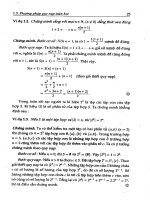
boundary conditions for the movement and translocation of charges. Third, different ion
concentrations on the two sides of the membrane generate transmembrane potential (),
which together with the surface (
s
) and the dipole (
d
) potentials contributes to a complex
potential profile across the membrane with particularly sharp changes and thus very high
electric field strengths (up to 10
9
V/m) in the region of charged lipid head groups (Clarke, 2001)
(Figure 2). Electric fields of such magnitude are expected to affect the dynamics of the charge
transfer processes and the structures of the proteins, thereby resulting in reaction mechanisms
that may differ from those observed in solution.
Fig. 2. Schematic representation of the interfacial potential distribution in a lipid bilayer
(left) and at a SAM-coated electrode (right).
3. Biocompatible protein immobilization
Immobilization of proteins on solid supports such as electrodes may account for two distinct
processes: (i) physical entrapment and (ii) attachment of proteins (Cass, 2007). The former
process refers to a thin layer of protein solution trapped by a membrane or a three-
dimensional polymer matrix on the solid support, resulting in non-organized and non-
oriented protein deposition as, for instance, in sol-gel enzyme electrodes (Gupta and
Chaudhury, 2007). The term attachment refers to covalent binding or non-covalent
adsorption of the enzyme to the solid surface such as tin, indium and titanium oxide,
chemically and electrochemically modified noble metal or carbon electrodes. Adsorption of
proteins on bare solid supports often leads to conformational changes or even denaturation.
Thus, successful immobilization relies almost exclusively on coated electrodes. Surface
coating needs to be well defined in terms of chemical functionalities and physical properties.
Self assembled monolayers (SAMs) of alkanethiols are among the most popular
biocompatible coatings employed in studies of interfacial interactions for addressing
fundamental aspects of heterogeneous ET, but also molecular recognition and cell growth
processes, heterogeneous nucleation and crystallization, biomaterial interfaces, etc
(Ulman, 2000).
Immobilizedredoxproteins:
mimickingbasicfeaturesofphysiologicalmembranesandinterfaces 25
The ET reactions through complexes I, III and IV are coupled to proton translocation across
the membrane, contributing to generation and maintenance of a transmembrane
electrochemical potential. Protons move back into the mitochondrial matrix (or cytoplasm)
through the ATP synthase via an energetically downhill process that provides the energy for
the synthesis of ATP.
The eukaryotic photosynthetic ET chain is analogous to the respiratory chain, but
structurally and functionally more complex. It is composed of: three multisubunit
transmembrane complexes, namely photosystem I, photosystem II and the cytochrome b
6
f
complex, several soluble electron carriers (e.g. plastocyanin and ferredoxin), lipophilic
hydrogen carrier plastoquinone, and light harvesting complexes. The trapping of the light
by the two reaction centers (photosystem I and II) results in a charge separation across the
stroma (thylakoid) membrane and furthermore in oxidation of water to oxygen by
photosystem II. The energy produced by this process serves as the driving force for ET
which is, as in respiration, coupled to proton translocation across the membrane and, thus,
to the synthesis of ATP. In addition to respiratory and photosynthetic redox enzymes,
membrane–bound ET chains also include i) cytochrome P450 containing microsomal and
ii) mitochondrial adrenal gland cytochrome P450 systems, that carry out catabolic and
anabolic reactions, with fatty acid desaturase and cytochromes P450, respectively, as
terminal enzymes (Gennis, 1989).
Bacteria and archaea tend to have simpler ET complexes and more versatile respiratory
chains in terms of electron donors and terminal electron acceptors that allow for alternative
ET pathways and, therefore, ensure adaptation to different external conditions (Pereira and
Teixeira, 2004). The gram negative bacterium E. coli, for example, lacks complex III. Instead,
the terminal oxygen reductase in its respiratory ET chain is a quinol : oxygen
oxidoreductase. Moreover, when growing under aerobic conditions, E. coli can express
different quinol oxidases to accommodate to the external conditions. In addition to terminal
oxygen reductases, it can also employ a wide range of terminal electron acceptors besides
oxygen, such as nitrite, nitrate, fumarate or DMSO and express other terminal reductases,
accordingly. Similarly, soil bacterium Paracoccus denitrificans can fine-tune the expression of
the appropriate oxygen reductase (aa
3
, cbb
3
or ba
3
), depending on the oxygen pressure levels
in the surrounding media. Bacteria and archaea also show a high level of diversity in
electron carriers, water soluble proteins (Cyt-c, HiPIP, and Cu proteins like sulfocyanin,
plastocyanin and amicyanin) and structurally different lipophilic quinones.
The intricate complexity of ET chains implies that understanding their functioning on a
molecular level and identification of the factors that govern electro-ionic energy transduction
is virtually impossible, unless simplified biomimetic model systems are utilized. The zero-
order approximation usually consists of purification of the individual proteins and their
characterization by spectroscopic, electrochemical and other experimental methods (Xavier,
2004; Pitcher and Watmough, 2004). This task can be relatively simple for small soluble
proteins but significantly more challenging in the case of membrane complexes, due to the
typically quite large number of cofactors. The main concern towards studying the membrane
components of the redox chains in solution are related to difficulties in reproducing
characteristics of the natural reaction environment, governed by the structural and electrical
properties of membranes. First, mobility of the proteins is strongly restricted. Integral membrane
proteins are embedded into the lipid bilayer and stabilized by hydrophobic interactions. Their
soluble redox partners either bind to the membrane surface or to the solvent exposed part of
the reaction partner. Second, the transition from the non-polar core to the polar surface of the
lipid bilayer implies a substantial variation of dielectric constants, which imposes specific
boundary conditions for the movement and translocation of charges. Third, different ion
concentrations on the two sides of the membrane generate transmembrane potential (),
which together with the surface (
s
) and the dipole (
d
) potentials contributes to a complex
potential profile across the membrane with particularly sharp changes and thus very high
electric field strengths (up to 10
9
V/m) in the region of charged lipid head groups (Clarke, 2001)
(Figure 2). Electric fields of such magnitude are expected to affect the dynamics of the charge
transfer processes and the structures of the proteins, thereby resulting in reaction mechanisms
that may differ from those observed in solution.
Fig. 2. Schematic representation of the interfacial potential distribution in a lipid bilayer
(left) and at a SAM-coated electrode (right).
3. Biocompatible protein immobilization
Immobilization of proteins on solid supports such as electrodes may account for two distinct
processes: (i) physical entrapment and (ii) attachment of proteins (Cass, 2007). The former
process refers to a thin layer of protein solution trapped by a membrane or a three-
dimensional polymer matrix on the solid support, resulting in non-organized and non-
oriented protein deposition as, for instance, in sol-gel enzyme electrodes (Gupta and
Chaudhury, 2007). The term attachment refers to covalent binding or non-covalent
adsorption of the enzyme to the solid surface such as tin, indium and titanium oxide,
chemically and electrochemically modified noble metal or carbon electrodes. Adsorption of
proteins on bare solid supports often leads to conformational changes or even denaturation.
Thus, successful immobilization relies almost exclusively on coated electrodes. Surface
coating needs to be well defined in terms of chemical functionalities and physical properties.
Self assembled monolayers (SAMs) of alkanethiols are among the most popular
biocompatible coatings employed in studies of interfacial interactions for addressing
fundamental aspects of heterogeneous ET, but also molecular recognition and cell growth
processes, heterogeneous nucleation and crystallization, biomaterial interfaces, etc
(Ulman, 2000).
Biomimetics,LearningfromNature26
The adsorption of proteins on the conducting, coated surface may be non-specific and non-
covalent, i.e. promoted by electrostatic or van der Waals interactions between the surface
functional groups of the modified electrode and amino acid residues of the protein. Non-
covalent but specific interactions, based on molecular recognition, involve affinity coupling
between two proteins such as antibody/antigene. This is the most commonly exploited
immobilization strategy in the growing field of protein microarrays (Hodneland et al., 2002).
Non-covalent and specific interactions also include adsorption of a protein that possesses
well defined charged (or hydrophobic) surface patches on a solid surface with opposite
charge (or hydrophobic). Covalent binding of the protein typically accounts for cross-linking
between functional groups of the protein and the surface, using carboxylate, amino or thiol
side chains of amino acids on the proteins surface. Specifically, for thiol-based attachements
not only natural surface cysteine side chains can be used, but Cys residues can also be
introduced at a certain position on the protein surface, in order to control or to modify the
attachment site.
Tailoring of novel biocompatible coatings and linkers has been a subject of intense research
over the last three decades owing to the importance of protein immobilization under
preservation of the native state structure for fundamental and applied purposes. Aiming to
the same goal, parallel efforts have been made in the rational design of proteins. Due to the
possibility of manipulating DNA sequencies and the availability of bacterial expression
systems for producing engineered proteins from modified genes, it is now feasible to
modify their surface properties in order to promote a particular immobilization strategy
(Gilardi, 2004). Such protein modifications may involve introducing of an additional
sequence such as a histidine tag, or deleting hydrophobic membrane anchors to produce
soluble protein variants.
3.1 Self-assembled monolayers (SAMs) of alkanethiols
Due to the high affinity of thiol groups for noble metals, ω-functionalized alkanethiols
spontaneously self-assemble on metal surfaces, forming densely packed monolayers. They
are commercially available in a wide variety of functional head groups and chain lengths,
allowing fine tailoring of the metal coating by simple immersion of the metal support into a
solution of the alkanethiols. A number of physicochemical techniques for surface analysis
and spectroscopic characterization of SAMs, such as: Raman spectroscopy, reflectance
absorption IR spectroscopy, X-ray photoelectron spectroscopy, high-resolution electron
energy loss spectroscopy, near-edge EXAFS, X-ray diffraction, contact-angle goniometry,
elipsometry, surface plasmon resonance, surface scanning microscopy, STM and AFM, as
well as electrochemical methods, are nowadays routinely used for probing monolayer
assembly, structural properties and stability of SAMs (Love et al., 2005). Several factors
influence the stability and structure of SAMs, such as solvent, temperature, immersion time,
the purity and chain length of the alkanethiols, as well as the purity and the type of the
metal. The fast initial adsorption of the alkanethiol molecules, the kinetics of which is
governed by surface-headgroup interactions, is followed by a slower rearrangement process
driven by inter-chain interactions. Long alkanethiol molecules (n > 10) tend to form more
robust SAMs, owing to both, kinetic and thermodynamic factors. The pKa values of acidic or
basic ω-functional groups of SAMs differ significantly from those of the amphiphiles in
solution. For SAMs with carboxylic head groups the pKa decreases with decreasing chain
length. SAMs are electrochemically stable only within a certain range of potentials, which
depends on the chemical composition of the SAM and the type of metal support. Reductive
desorption typically occurs at potentials of - 1.00 ± 0.25 V (vs. Ag/AgCl). For a more
detailed account on the preparation, tailoring, and characterisation of SAM coatings, the
reader is referred to specialised reviews (Ulman, 1996; Love et al., 2005).
3.2 Immobilization of soluble proteins
SAMs of alkanethiols provide a biocompatible interface for the immobilization of proteins
on metal electrodes allowing for an electrochemical characterization of the protein under
preservation of its native structure. These simple systems can be regarded as biomimetic in
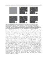
the sense that they reproduce some basic features of biological interfaces. The appropriate
choice of the alkanthiol head group allows in some cases for specific binding of proteins,
Figure 3.
Alkanethiols with pyridinyl head groups may replace the axial Met-80 ligand of the heme in
mitochondrial Cyt-c to establish a direct link between the redox site and the electrode (Wei
et al., 2002; Murgida et al., 2004b; Murgida and Hildebrandt, 2008). Similarly, apo-glucose
oxidase (GOx) was successfully immobilized on a flavin (FAD)-modified metal (Xiao et al.,
2003). The carboxyl-terminated SAMs can be activated by carbodiimide derivatives for
covalent binding of proteins via the NH
2
groups of Lys surface residues. Several enzymes,
like GOx, xanthine oxidase, horse-reddish-peroxidase (HRP), were linked to modified
carbon electrodes through formation of amide bond. In each case, the amperometric
response of these simple bioelectronic devices could be measured upon detection of glucose,
xanthine and hydrogen peroxide, respectively (Willner and Katz, 2000). Carboxylate
headgroups can also provide negatively charged surfaces for the electrostatic
immobilization of proteins with positively charged surface patches, as it is the case of Cyt-c
that possesses a ring-shaped arrangement of positively charged lysine residues, naturally
designed for interaction with the redox partners (Murgida and Hildebrandt, 2008). By
changing the SAM chain length ET rates can be probed as a function of distance (Murgida
and Hildebrandt, 2004a; Todorovic et al., 2006). Furthermore, SAMs permit systematic
control of the strength of the interfacial electric field. The potential drop across the
electrode/SAM/protein interface, and thus the electric field strength experienced by the
immobilized protein, can be described based on a simple electrostatic model (Figure 2) as a
function of experimentally accessible parameters. Within this model, the electric field
strength E
F
at the protein binding site can be described in terms of the charge densities at the
SAM surface (
c
) and at the redox site (
RC
) as well as of the potential drop at the redox site
(E
RC
= E
0
ads
– E
0
sol
), which increases with the SAM thickness d
c
(Equation 1) (Murgida and
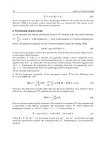
Hildebrandt, 2001a):
C
RCCRCS
CF
E
dE
0
0
)(
(1)
where E
0
ads
and E
0
sol
are the apparent standard reduction potentials of the protein in the
adsorbed state and in solution, respectively, is the inverse Debye length, and
s
and
c
denote the dielectric constants of the solution and the SAM, respectively. For carboxylate-
terminated SAMs, the electric field strength at the Cyt-c binding site is in the order of 10
9
V
m
-1
, which is comparable to the upper values estimated for biological membranes in the
Immobilizedredoxproteins:
mimickingbasicfeaturesofphysiologicalmembranesandinterfaces 27
The adsorption of proteins on the conducting, coated surface may be non-specific and non-
covalent, i.e. promoted by electrostatic or van der Waals interactions between the surface
functional groups of the modified electrode and amino acid residues of the protein. Non-
covalent but specific interactions, based on molecular recognition, involve affinity coupling
between two proteins such as antibody/antigene. This is the most commonly exploited
immobilization strategy in the growing field of protein microarrays (Hodneland et al., 2002).
Non-covalent and specific interactions also include adsorption of a protein that possesses
well defined charged (or hydrophobic) surface patches on a solid surface with opposite
charge (or hydrophobic). Covalent binding of the protein typically accounts for cross-linking
between functional groups of the protein and the surface, using carboxylate, amino or thiol
side chains of amino acids on the proteins surface. Specifically, for thiol-based attachements
not only natural surface cysteine side chains can be used, but Cys residues can also be
introduced at a certain position on the protein surface, in order to control or to modify the
attachment site.
Tailoring of novel biocompatible coatings and linkers has been a subject of intense research
over the last three decades owing to the importance of protein immobilization under
preservation of the native state structure for fundamental and applied purposes. Aiming to
the same goal, parallel efforts have been made in the rational design of proteins. Due to the
possibility of manipulating DNA sequencies and the availability of bacterial expression
systems for producing engineered proteins from modified genes, it is now feasible to
modify their surface properties in order to promote a particular immobilization strategy
(Gilardi, 2004). Such protein modifications may involve introducing of an additional
sequence such as a histidine tag, or deleting hydrophobic membrane anchors to produce
soluble protein variants.
3.1 Self-assembled monolayers (SAMs) of alkanethiols
Due to the high affinity of thiol groups for noble metals, ω-functionalized alkanethiols
spontaneously self-assemble on metal surfaces, forming densely packed monolayers. They
are commercially available in a wide variety of functional head groups and chain lengths,
allowing fine tailoring of the metal coating by simple immersion of the metal support into a
solution of the alkanethiols. A number of physicochemical techniques for surface analysis
and spectroscopic characterization of SAMs, such as: Raman spectroscopy, reflectance
absorption IR spectroscopy, X-ray photoelectron spectroscopy, high-resolution electron
energy loss spectroscopy, near-edge EXAFS, X-ray diffraction, contact-angle goniometry,
elipsometry, surface plasmon resonance, surface scanning microscopy, STM and AFM, as
well as electrochemical methods, are nowadays routinely used for probing monolayer
assembly, structural properties and stability of SAMs (Love et al., 2005). Several factors
influence the stability and structure of SAMs, such as solvent, temperature, immersion time,
the purity and chain length of the alkanethiols, as well as the purity and the type of the
metal. The fast initial adsorption of the alkanethiol molecules, the kinetics of which is
governed by surface-headgroup interactions, is followed by a slower rearrangement process
driven by inter-chain interactions. Long alkanethiol molecules (n > 10) tend to form more
robust SAMs, owing to both, kinetic and thermodynamic factors. The pKa values of acidic or
basic ω-functional groups of SAMs differ significantly from those of the amphiphiles in
solution. For SAMs with carboxylic head groups the pKa decreases with decreasing chain
length. SAMs are electrochemically stable only within a certain range of potentials, which
depends on the chemical composition of the SAM and the type of metal support. Reductive
desorption typically occurs at potentials of - 1.00 ± 0.25 V (vs. Ag/AgCl). For a more
detailed account on the preparation, tailoring, and characterisation of SAM coatings, the
reader is referred to specialised reviews (Ulman, 1996; Love et al., 2005).
3.2 Immobilization of soluble proteins
SAMs of alkanethiols provide a biocompatible interface for the immobilization of proteins
on metal electrodes allowing for an electrochemical characterization of the protein under
preservation of its native structure. These simple systems can be regarded as biomimetic in
the sense that they reproduce some basic features of biological interfaces. The appropriate
choice of the alkanthiol head group allows in some cases for specific binding of proteins,
Figure 3.
Alkanethiols with pyridinyl head groups may replace the axial Met-80 ligand of the heme in
mitochondrial Cyt-c to establish a direct link between the redox site and the electrode (Wei
et al., 2002; Murgida et al., 2004b; Murgida and Hildebrandt, 2008). Similarly, apo-glucose
oxidase (GOx) was successfully immobilized on a flavin (FAD)-modified metal (Xiao et al.,
2003). The carboxyl-terminated SAMs can be activated by carbodiimide derivatives for
covalent binding of proteins via the NH
2
groups of Lys surface residues. Several enzymes,
like GOx, xanthine oxidase, horse-reddish-peroxidase (HRP), were linked to modified
carbon electrodes through formation of amide bond. In each case, the amperometric
response of these simple bioelectronic devices could be measured upon detection of glucose,
xanthine and hydrogen peroxide, respectively (Willner and Katz, 2000). Carboxylate
headgroups can also provide negatively charged surfaces for the electrostatic
immobilization of proteins with positively charged surface patches, as it is the case of Cyt-c
that possesses a ring-shaped arrangement of positively charged lysine residues, naturally
designed for interaction with the redox partners (Murgida and Hildebrandt, 2008). By
changing the SAM chain length ET rates can be probed as a function of distance (Murgida
and Hildebrandt, 2004a; Todorovic et al., 2006). Furthermore, SAMs permit systematic
control of the strength of the interfacial electric field. The potential drop across the
electrode/SAM/protein interface, and thus the electric field strength experienced by the
immobilized protein, can be described based on a simple electrostatic model (Figure 2) as a
function of experimentally accessible parameters. Within this model, the electric field
strength E
F
at the protein binding site can be described in terms of the charge densities at the
SAM surface (
c
) and at the redox site (
RC
) as well as of the potential drop at the redox site
(E
RC
= E
0
ads
– E
0
sol
), which increases with the SAM thickness d
c
(Equation 1) (Murgida and
Hildebrandt, 2001a):
C
RCCRCS
CF
E
dE
0
0
)(
(1)
where E
0
ads
and E
0
sol
are the apparent standard reduction potentials of the protein in the
adsorbed state and in solution, respectively, is the inverse Debye length, and
s
and
c
denote the dielectric constants of the solution and the SAM, respectively. For carboxylate-
terminated SAMs, the electric field strength at the Cyt-c binding site is in the order of 10
9
V
m
-1
, which is comparable to the upper values estimated for biological membranes in the
Biomimetics,LearningfromNature28
vicinity of charged lipid head groups. Higher field strengths are predicted for phosphonate-
terminated SAMs and sulfate monolayers for which |
C
| is distinctly larger. The charge
density of the SAM is defined by the pK
a
of the acidic head groups in the assembly, which
increases with the number of methylene groups, and by the pH of the solution. Thus, the
electric field strength at the protein binding site can be varied within the range ca. 10
8
-10
9
V
m
-1
by changing the length of the alkanethiols without modifying any other parameter. The
strength of the E
F
can also be controlled via the electrode potential and the nature of the
SAM head group, as well as via the pH and ionic strength of the solution (Murgida and
Hildebrandt, 2001a; Murgida and Hildebrandt, 2001b; Murgida and Hildebrandt, 2002;
Murgida and Hildebrandt, 2008).
Fig. 3. Schematic representation of some strategies for biocompatible protein binding to
metal electrodes: A) electrostatic binding of Cyt-c to a COOH-terminated SAM; B)
coordinative binding of Cyt-c to a Py-terminated SAM; C) specific binding of a His-tagged
CcO to a Ni-NTA coated electrode.
SAMs can also be formed by hydroxyl-, amino- and methyl-terminated alkanethiols.
Hydroxyl-terminated alkanethiols favour polar interactions but may also allow for covalent
immobilization (via chlorotriazines and Tyr or Lys amino acid residues) as shown for GOx,
ferritin and urease (Willner et al., 2000). Amino–terminated alkanethiols can provide
positively charged surfaces for electrostatic binding of proteins rich in surface exposed
carboxylic side chains of Asp and Glu, or for cross-linking upon activation of carboxylic
groups of the protein (Willner et al., 2000). Methyl-terminated alkanethiols are suitable for
immobilization of proteins via hydrophobic interactions (Rivas et al., 2002; Murgida and
Hildebrandt, 2008). ´Mixed´ monolayers prepared from alkanethiols with different head-
groups in variable molar ratios, provide a surface engineered with gradients of charge,
capable of accommodating proteins with less well defined (or ´diluted´) surface charge
distribution via the interplay of different interactions. Mixed SAMs of carboxyl and methyl-
terminated alkanethiols were used for HRP immobilization (Hasunuma et al., 2004), while
hydroxyl/methyl-terminated SAMs provided the best coating for immobilization of
genetically manipulated soluble subunits of caa
3
, cbb
3
, and ba
3
oxygen reductases, as well as
some soluble heme proteins (Ledesma et al., 2007; Kranich et al., 2009). Moreover, the use of
mixture of alkanethiols of different chain lengths (and headgroups) may fulfil specific steric
requirements of the adsorbate. This strategy has been successfully employed for
characterizing the interfacial enzymatic reaction of cutinase by electrochemical methods
(Nayak et al., 2007). Other possibilities include mixed SAMs composed of glycol-terminated
and biological-ligand-terminated alkanethiols, which appear to be a surface of choice for
immobilization of a variety of biomolecules including DNA, carbohydrates, antibodies, and
whole bacterial cells that are particularly important for the design and construction of
affinity immunosensors (Clarke, 2001; Love et al., 2005; Collier and Mrksich, 2006).
3.3 Immobilization of membrane proteins
Membrane proteins are partially or fully integrated into the lipid bilayer, requiring,
therefore, a hydrophobic environment to maintain the native structure and avoid
aggregation upon isolation. Besides, they are large, typically composed of several subunits
that are often prone to dissociation during the purification process. The structural and
functional integrity of the proteins in the solubilized form sensitively depends on the type of
detergent used to provide a hydrophobic environment in vitro.
Several models for physiological membranes that display different levels of complexity have
been developed, including Langmuir-Blodget (LB) lipid monolayer films (He et al., 1999),
bilayer lipid films and liposomes (Hianik, 2008). Protein containing lipid monolayer films
formed on solid supports are frequently used for the construction of biosensors.
Phospholipid bilayers can be produced in a controllable manner, with tunable thickness,
surface tension, specific and electrical capacity. They are the most suitable systems for
studies of membrane pores and channels. Liposomes are closed bilayer systems that can be
formed spontaneously either from bacterial cell (or mitochondrial) membrane fractions
containing the incorporated proteins, or from phospholipids subsequently modified by
proteins. They are considered to be good model membranes in studies of transmembrane
enzymes involved in coupled reactions on opposite sides of the membrane, as well as
proteins involved in solute transport or substrate channeling (Gennis, 1989).
Immobilization strategies for ET membrane proteins have been developed particularly in
studies of terminal oxygen reductases. In the simplest approach a detergent-solubilized
protein is spontaneously adsorbed on a metal surface. Most likely, immobilization takes
place via interactions of the detergent molecules with the layer of specifically adsorbed
anions that the metal surface carries above the potential of zero-charge. In fact, the detergent
n-dodecyl-β-D-maltoside, commonly used for solubilization of membrane proteins, has been
shown to adsorb to these surfaces, providing a biocompatible interface for subsequent
protein adsorption under preservation of its structural and functional integrity (Todorovic et
al., 2005). This finding is in contrast to the behavior observed for soluble proteins for which
the direct adsorption on a bare metal, in the absence of detergent, may cause a (partial)
degradation (Murgida and Hildebrandt, 2005). Mixed SAMs composed of CH
3
and OH
terminated alkanethiols were shown to be a promising choice for immobilization of
Immobilizedredoxproteins:
mimickingbasicfeaturesofphysiologicalmembranesandinterfaces 29
vicinity of charged lipid head groups. Higher field strengths are predicted for phosphonate-
terminated SAMs and sulfate monolayers for which |
C
| is distinctly larger. The charge
density of the SAM is defined by the pK
a
of the acidic head groups in the assembly, which
increases with the number of methylene groups, and by the pH of the solution. Thus, the
electric field strength at the protein binding site can be varied within the range ca. 10
8
-10
9
V
m
-1
by changing the length of the alkanethiols without modifying any other parameter. The
strength of the E
F
can also be controlled via the electrode potential and the nature of the
SAM head group, as well as via the pH and ionic strength of the solution (Murgida and
Hildebrandt, 2001a; Murgida and Hildebrandt, 2001b; Murgida and Hildebrandt, 2002;
Murgida and Hildebrandt, 2008).
Fig. 3. Schematic representation of some strategies for biocompatible protein binding to
metal electrodes: A) electrostatic binding of Cyt-c to a COOH-terminated SAM; B)
coordinative binding of Cyt-c to a Py-terminated SAM; C) specific binding of a His-tagged
CcO to a Ni-NTA coated electrode.
SAMs can also be formed by hydroxyl-, amino- and methyl-terminated alkanethiols.
Hydroxyl-terminated alkanethiols favour polar interactions but may also allow for covalent
immobilization (via chlorotriazines and Tyr or Lys amino acid residues) as shown for GOx,
ferritin and urease (Willner et al., 2000). Amino–terminated alkanethiols can provide
positively charged surfaces for electrostatic binding of proteins rich in surface exposed
carboxylic side chains of Asp and Glu, or for cross-linking upon activation of carboxylic
groups of the protein (Willner et al., 2000). Methyl-terminated alkanethiols are suitable for
immobilization of proteins via hydrophobic interactions (Rivas et al., 2002; Murgida and
Hildebrandt, 2008). ´Mixed´ monolayers prepared from alkanethiols with different head-
groups in variable molar ratios, provide a surface engineered with gradients of charge,
capable of accommodating proteins with less well defined (or ´diluted´) surface charge
distribution via the interplay of different interactions. Mixed SAMs of carboxyl and methyl-
terminated alkanethiols were used for HRP immobilization (Hasunuma et al., 2004), while
hydroxyl/methyl-terminated SAMs provided the best coating for immobilization of
genetically manipulated soluble subunits of caa
3
, cbb
3
, and ba
3
oxygen reductases, as well as
some soluble heme proteins (Ledesma et al., 2007; Kranich et al., 2009). Moreover, the use of
mixture of alkanethiols of different chain lengths (and headgroups) may fulfil specific steric
requirements of the adsorbate. This strategy has been successfully employed for
characterizing the interfacial enzymatic reaction of cutinase by electrochemical methods
(Nayak et al., 2007). Other possibilities include mixed SAMs composed of glycol-terminated
and biological-ligand-terminated alkanethiols, which appear to be a surface of choice for
immobilization of a variety of biomolecules including DNA, carbohydrates, antibodies, and
whole bacterial cells that are particularly important for the design and construction of
affinity immunosensors (Clarke, 2001; Love et al., 2005; Collier and Mrksich, 2006).
3.3 Immobilization of membrane proteins
Membrane proteins are partially or fully integrated into the lipid bilayer, requiring,
therefore, a hydrophobic environment to maintain the native structure and avoid
aggregation upon isolation. Besides, they are large, typically composed of several subunits
that are often prone to dissociation during the purification process. The structural and
functional integrity of the proteins in the solubilized form sensitively depends on the type of
detergent used to provide a hydrophobic environment in vitro.
Several models for physiological membranes that display different levels of complexity have
been developed, including Langmuir-Blodget (LB) lipid monolayer films (He et al., 1999),
bilayer lipid films and liposomes (Hianik, 2008). Protein containing lipid monolayer films
formed on solid supports are frequently used for the construction of biosensors.
Phospholipid bilayers can be produced in a controllable manner, with tunable thickness,
surface tension, specific and electrical capacity. They are the most suitable systems for
studies of membrane pores and channels. Liposomes are closed bilayer systems that can be
formed spontaneously either from bacterial cell (or mitochondrial) membrane fractions
containing the incorporated proteins, or from phospholipids subsequently modified by
proteins. They are considered to be good model membranes in studies of transmembrane
enzymes involved in coupled reactions on opposite sides of the membrane, as well as
proteins involved in solute transport or substrate channeling (Gennis, 1989).
Immobilization strategies for ET membrane proteins have been developed particularly in
studies of terminal oxygen reductases. In the simplest approach a detergent-solubilized
protein is spontaneously adsorbed on a metal surface. Most likely, immobilization takes
place via interactions of the detergent molecules with the layer of specifically adsorbed
anions that the metal surface carries above the potential of zero-charge. In fact, the detergent
n-dodecyl-β-D-maltoside, commonly used for solubilization of membrane proteins, has been
shown to adsorb to these surfaces, providing a biocompatible interface for subsequent
protein adsorption under preservation of its structural and functional integrity (Todorovic et
al., 2005). This finding is in contrast to the behavior observed for soluble proteins for which
the direct adsorption on a bare metal, in the absence of detergent, may cause a (partial)
degradation (Murgida and Hildebrandt, 2005). Mixed SAMs composed of CH
3
and OH
terminated alkanethiols were shown to be a promising choice for immobilization of
Biomimetics,LearningfromNature30
detergent-solubilized membrane proteins, such as complex II from R. marinus (unpublished
data). Direct adsorption of solubilized membrane proteins, however, cannot guarantee a
uniform orientation of the immobilized enzyme. In an attempt to overcome this problem, a
preformed detergent solubilized Cyt-c/CcO complex was immobilized on Au electrodes
coated with hydroxyl-terminated alkanetiols at low ionic strength. It was studied by
electrochemical methods, which however, do no permit unambiguous conclusions
regarding the enzyme structure and orientation in the immobilized state (Haas et al., 2001).
A similar approach was applied to a fumarate reductase immobilized on Au electrode with
hydrophobic coating (Kinnear and Monbouquette, 1993).
An alternative immobilization method has been developed for proteins that contain a
genetically introduced His tag (Friedrich et al., 2004; Ataka et al., 2004; Giess et al., 2004;
Hrabakova et al., 2006; Todorovic et al., 2008). After functionalizing the solid support with
Ni (or Zn) NTA (3,3´-dithiobis[N-(5amino-5-carboxy-pentyl)propionamide-N, N´-diacetic
acid)] dihydrochloride) monolayer, the protein can be attached via His coordination to the
Ni center, Figure 3C. The high affinity of the His tag, inserted into the protein sequence
either at N or C terminus, towards Ni-NTA assures large surface coverage of uniformly
oriented protein molecules even at relatively high, physiologically relevant ionic strengths.
The last immobilization step is the reconstitution of a lipid bilayer from 1,2-diphytanoyl-sn-
glycero-3-phosphocholine and the removal of the detergent using biobeads. This method
was recently employed for immobilization of several oxygen reductases on Au and Ag
electrodes. Different steps of the assembly were demonstrated by SEIRA spectroscopy and
atomic-force microscopy, providing the evidence for the formation of the lipid bilayer.
Moreover, separations of the redox centers from the metal surface in the final biomimetic
construct are yet not too large for applying surface enhanced vibrational spectroscopies
(Friedrich et al., 2004).
4. Methods for probing the structure and dynamics of immobilized proteins:
vibrational spectroscopy
It is clear that the development of novel protein-based bioelectronic devices for basic and
applied purposes heavily relies upon design of new biomimetic or biocompatible materials.
However, it also requires appropriate experimental approaches capable of monitoring in situ
the structure and reaction dynamics of the immobilized enzymes under working conditions.
These information are crucial for understanding and eventually improving the performance
of protein-based devices.
Here we will describe basic principles of SERR and SEIRA spectroelectrochemical
techniques, which are among the most powerful approaches for characterization of
thermodynamic, kinetic and structural aspects of immobilized redox proteins.
4.1 (Resonance) Raman and infrared spectroscopies
Raman and IR spectroscopies probe vibrational levels of a molecule, providing information
on molecular structures. A vibrational mode of a molecule will be Raman active only if the
incident light causes a change of its polarizability, while IR active modes require a change in
dipole moment upon absorption of light. For molecules of high symmetry, these selection
rules allow grouping the vibrational modes into Raman- or / and IR-active or -forbidden
modes. Water gives rise to strong IR bands including the stretching and bending modes at
ca. 3400 and 1630 cm
-1
, respectively. The bending mode represents a major difficulty in
studying biological samples due to overlapping with the amide I band in the spectra of
proteins (see below). In IR transmission measurements, therefore, cuvettes of very small
optical paths (a few micrometers) and very high protein concentrations have to be
employed. The attenuated total reflection (ATR) technique allows bypassing the problems
associated with water, facilitating the studies of protein/substrate or protein/ligand
interactions, and enhancing the overall sensitivity. In Raman spectroscopy water is not an
obstacle at room temperature, although ice lattice modes become visible in the low
frequency region in croygenic measurements. A severe drawback of Raman spectroscopy is
its low sensitivity, due to the low quantum yield of the scattering process (< 10
-9
). This
disadvantage can be overcome for molecules that possess chromophoric cofactors, such as
metalloproteins. When the energy of the incident laser light is in resonance with an
electronic transition of the chromophore, the quantum yield of the scattering process
becomes several orders of magnitude higher for the vibrational modes originating from the
chromophore. Thus, the sensitivity and the selectivity of Raman spectroscopy (i.e.,
resonance Raman – RR) are strongly increased and the resultant spectra display only the
vibrational modes of the cofactor, regardless of the size of the protein matrix (Siebert and
Hildebrandt, 2008).
In the last decades RR spectroscopy was proved to be indispensable in the studies of heme
proteins. RR spectra obtained upon excitation into the Soret band of the porphyrin display
´so-called´ core-size marker bands sensitive to the redox and spin state and coordination
pattern of the heme iron in the 1300 – 1700 cm
-1
region (Hu et al., 1993; Spiro and
Czernuszewicz, 1995; Siebert and Hildebrandt, 2008). For instance, transition from a ferric to
a ferrous heme is associated with a ca. 10 cm
-1
downshift of most of the marker bands
(particularly
3
and
4
). The conversion from a six-coordinated low spin (6cLS) heme to a
five-cordinated high spin (5cHS) heme also causes a downshift of some bands (
3
and
2
).
These and further empirical relationships derived from a large experimental data basis
provide valuable tools for elucidating structural details of the heme site and for monitoring
ET and enzymatic processes, as shown for a variety of heme proteins including hemoglobin,
myoglobin, cytochromes, peroxidases and oxygen reductases (Spiro and Czernuszewicz,
1995; Siebert and Hildebrandt, 2008).
IR spectra provide information on the secondary structure of proteins based on the analysis
of the amide I (1600 – 1700 cm
-1
) and amide II (1480 – 1580 cm
-1
) bands. The sensitivity and
selectivity of IR spectroscopy can be greatly improved upon operating in difference mode.
Difference IR spectra obtained from two states of a protein only display those bands that
undergo a change upon transition from one state to the other, thereby substantially
simplifying the analysis (Ataka and Heberle, 2007). IR difference spectroscopy is a sensitive
method for investigating structural changes of proteins that (i) accompany the redox
reaction, (ii) are induced by substrate binding during the catalytic cycle, (iii) occur during
protein folding and unfolding, or (iv) accompany photo-induced processes (Siebert and
Hildebrandt, 2008).
Immobilizedredoxproteins:
mimickingbasicfeaturesofphysiologicalmembranesandinterfaces 31
detergent-solubilized membrane proteins, such as complex II from R. marinus (unpublished
data). Direct adsorption of solubilized membrane proteins, however, cannot guarantee a
uniform orientation of the immobilized enzyme. In an attempt to overcome this problem, a
preformed detergent solubilized Cyt-c/CcO complex was immobilized on Au electrodes
coated with hydroxyl-terminated alkanetiols at low ionic strength. It was studied by
electrochemical methods, which however, do no permit unambiguous conclusions
regarding the enzyme structure and orientation in the immobilized state (Haas et al., 2001).
A similar approach was applied to a fumarate reductase immobilized on Au electrode with
hydrophobic coating (Kinnear and Monbouquette, 1993).
An alternative immobilization method has been developed for proteins that contain a
genetically introduced His tag (Friedrich et al., 2004; Ataka et al., 2004; Giess et al., 2004;
Hrabakova et al., 2006; Todorovic et al., 2008). After functionalizing the solid support with
Ni (or Zn) NTA (3,3´-dithiobis[N-(5amino-5-carboxy-pentyl)propionamide-N, N´-diacetic
acid)] dihydrochloride) monolayer, the protein can be attached via His coordination to the
Ni center, Figure 3C. The high affinity of the His tag, inserted into the protein sequence
either at N or C terminus, towards Ni-NTA assures large surface coverage of uniformly
oriented protein molecules even at relatively high, physiologically relevant ionic strengths.
The last immobilization step is the reconstitution of a lipid bilayer from 1,2-diphytanoyl-sn-
glycero-3-phosphocholine and the removal of the detergent using biobeads. This method
was recently employed for immobilization of several oxygen reductases on Au and Ag
electrodes. Different steps of the assembly were demonstrated by SEIRA spectroscopy and
atomic-force microscopy, providing the evidence for the formation of the lipid bilayer.
Moreover, separations of the redox centers from the metal surface in the final biomimetic
construct are yet not too large for applying surface enhanced vibrational spectroscopies
(Friedrich et al., 2004).
4. Methods for probing the structure and dynamics of immobilized proteins:
vibrational spectroscopy
It is clear that the development of novel protein-based bioelectronic devices for basic and
applied purposes heavily relies upon design of new biomimetic or biocompatible materials.
However, it also requires appropriate experimental approaches capable of monitoring in situ
the structure and reaction dynamics of the immobilized enzymes under working conditions.
These information are crucial for understanding and eventually improving the performance
of protein-based devices.
Here we will describe basic principles of SERR and SEIRA spectroelectrochemical
techniques, which are among the most powerful approaches for characterization of
thermodynamic, kinetic and structural aspects of immobilized redox proteins.
4.1 (Resonance) Raman and infrared spectroscopies
Raman and IR spectroscopies probe vibrational levels of a molecule, providing information
on molecular structures. A vibrational mode of a molecule will be Raman active only if the
incident light causes a change of its polarizability, while IR active modes require a change in
dipole moment upon absorption of light. For molecules of high symmetry, these selection
rules allow grouping the vibrational modes into Raman- or / and IR-active or -forbidden
modes. Water gives rise to strong IR bands including the stretching and bending modes at
ca. 3400 and 1630 cm
-1
, respectively. The bending mode represents a major difficulty in
studying biological samples due to overlapping with the amide I band in the spectra of
proteins (see below). In IR transmission measurements, therefore, cuvettes of very small
optical paths (a few micrometers) and very high protein concentrations have to be
employed. The attenuated total reflection (ATR) technique allows bypassing the problems
associated with water, facilitating the studies of protein/substrate or protein/ligand
interactions, and enhancing the overall sensitivity. In Raman spectroscopy water is not an
obstacle at room temperature, although ice lattice modes become visible in the low
frequency region in croygenic measurements. A severe drawback of Raman spectroscopy is
its low sensitivity, due to the low quantum yield of the scattering process (< 10
-9
). This
disadvantage can be overcome for molecules that possess chromophoric cofactors, such as
metalloproteins. When the energy of the incident laser light is in resonance with an
electronic transition of the chromophore, the quantum yield of the scattering process
becomes several orders of magnitude higher for the vibrational modes originating from the
chromophore. Thus, the sensitivity and the selectivity of Raman spectroscopy (i.e.,
resonance Raman – RR) are strongly increased and the resultant spectra display only the
vibrational modes of the cofactor, regardless of the size of the protein matrix (Siebert and
Hildebrandt, 2008).
In the last decades RR spectroscopy was proved to be indispensable in the studies of heme
proteins. RR spectra obtained upon excitation into the Soret band of the porphyrin display
´so-called´ core-size marker bands sensitive to the redox and spin state and coordination
pattern of the heme iron in the 1300 – 1700 cm
-1
region (Hu et al., 1993; Spiro and
Czernuszewicz, 1995; Siebert and Hildebrandt, 2008). For instance, transition from a ferric to
a ferrous heme is associated with a ca. 10 cm
-1
downshift of most of the marker bands
(particularly
3
and
4
). The conversion from a six-coordinated low spin (6cLS) heme to a
five-cordinated high spin (5cHS) heme also causes a downshift of some bands (
3
and
2
).
These and further empirical relationships derived from a large experimental data basis
provide valuable tools for elucidating structural details of the heme site and for monitoring
ET and enzymatic processes, as shown for a variety of heme proteins including hemoglobin,
myoglobin, cytochromes, peroxidases and oxygen reductases (Spiro and Czernuszewicz,
1995; Siebert and Hildebrandt, 2008).
IR spectra provide information on the secondary structure of proteins based on the analysis
of the amide I (1600 – 1700 cm
-1
) and amide II (1480 – 1580 cm
-1
) bands. The sensitivity and
selectivity of IR spectroscopy can be greatly improved upon operating in difference mode.
Difference IR spectra obtained from two states of a protein only display those bands that
undergo a change upon transition from one state to the other, thereby substantially
simplifying the analysis (Ataka and Heberle, 2007). IR difference spectroscopy is a sensitive
method for investigating structural changes of proteins that (i) accompany the redox
reaction, (ii) are induced by substrate binding during the catalytic cycle, (iii) occur during
protein folding and unfolding, or (iv) accompany photo-induced processes (Siebert and
Hildebrandt, 2008).
Biomimetics,LearningfromNature32
4.2 Surface Enhanced resonance Raman (SERR) and surface enhanced IR
(SEIRA) spectroscopy
Surface enhanced Raman (SER) spectroscopy is based on the increase of the signal intensity
associated with vibrational transitions of molecules situated in close proximity to
nanoscopic metal structures. Two distinct enhancement mechanisms have been identified.
The chemical mechanism originates from charge transfer interactions between the metal
substrate and the adsorbate, and provides a weak enhancement solely for the molecules in
direct contact with the metal. The electromagnetic mechanism is based on the amplified
electromagnetic fields generated upon excitation of the localized surface plasmons of
nanostructured metals. It does not require specific substrate/adsorbate contacts and
provides the main contribution to the overall enhancement. Among different metals tested
as SER substrates, Ag affords the strongest electromagnetic enhancements, due to surface
plasmon resonance in a wide spectral range from the near UV to the IR region. A drawback,
however, is that Ag nanostructures are less stable and chemically less inert than their Au
counterparts. In addition, the low oxidation potential of Ag narrows the range of applicable
potentials in SER-based spectro-electrochemical experiments. For these reasons most efforts
in recent years have been devoted to the development of Au SER substrates, including SER-
active electrodes. The attractiveness of the unsurpassed sensitivity of Ag has also driven
significant efforts towards use of this metal and hybrid Ag/Au structures. To this end, a
large number of highly regular and reproducible Au and Ag SER substrates have been
reported, making use of spheres, tubes, rods, thorns, cavities and wires as building blocks
(Mahajan et al., 2007; Murgida and Hildebrandt, 2008; Lal et al., 2008; Banholzer et al., 2008;
Brown and Milton, 2008; Feng et al., 2008a; Feng et al., 2009).
If the excitation laser is in resonance not only with the energy of surface plasmons of the
metal but also with the electronic transition of the immobilized molecule, the SER and RR
effects combine. The resulting SERR spectra display exclusively the vibrational bands of the
chromophore of the adsorbed species. The use of Ag as SER-active substrate is particularly
suited for studying porphyrins and heme proteins since these molecules exhibit a strong
electronic transition at ca. 410 nm (Soret band) and a weaker one at ca. 550 nm which both
coincide with Ag (but not with Au) surface plasmon resonances. SERR spectra of heme
proteins reveal the same information as RR spectra, such as the oxidation, spin, and
coordination states of the heme group, and in addition their changes as a consequence of
variations of the electrode potential (see bellow) (Siebert and Hildebrandt, 2008).
Molecules adsorbed in the vicinity of nanostructured metal surfaces, such as Ag or Au
islands deposited on inert ATR crystals, experience enhanced absorption of IR radiation,
which is the basis for (ATR) SEIRA spectroscopy. SEIRA spectroscopy has been successfully
employed to probe the structure of immobilized biomolecules including redox proteins and
enzymes (Ataka and Heberle, 2007). The enhancement of the IR bands does not exceed two
orders of magnitude and therefore is smaller than the enhancement of the SERR bands
which may be larger than 10
5
. The distance-dependent decay of the enhancement factor is
less pronounced for SEIRA than for SERR spectroscopy, and both techniques can
successfully probe molecules separated from the surface by up to 5 nm.
The nanostructured metal substrate that amplifies the signals can also serve as a working
electrode in spectroelectrochemical studies. Indeed, potentiometric titrations followed by
SERR and SEIRA have provided important insights into the mechanism of functioning of
several heme proteins immobilized on biocompatible metal electrodes (Murgida and
Hildebrandt, 2004a; Murgida and Hildebrandt, 2005; Murgida and Hildebrandt, 2008).
Both SERR and SEIRA can be employed in the time resolved (TR) mode that enables
probing of dynamics of immobilized proteins. The method requires a synchronization of a
perturbation event with the spectroscopic detection at variable delay times. For TR-SEIRA
spectroscopy acquisition is usually performed in the rapid or step scan mode for probing
events in time windows longer or shorter than 10 ms, respectively. For the study of potential
dependent processes of immobilized redox proteins by TR-SERR, the equilibrium of the
immobilized species is perturbed by a rapid potential jump, and the subsequent relaxation
process is then monitored at different delay times. A prerequisite for applying of TR-SERR is
that the underlying ET processes are fully reversible. The time resolution depends on the
charge reorganization of the double layer of the working electrode and is typically on a
microsecond scale (Murgida and Hildebrandt, 2004a; Murgida and Hildebrandt, 2005;
Murgida and Hildebrandt 2008).
5. Recent developments in the characterization of immobilized redox proteins
In this section we will focus on selected examples of surface enhanced
spectroelectrochemical characterization of ET proteins immobilized on nanostructured
electrodes coated with biomimetic films. The first part is dedicated to membrane oxygen
reductases whose structural, functional and spectroscopic complexity imposes some serious
limits to other experimental approaches. In the second part we will describe recent studies
on soluble electron carrier proteins, mainly cytochromes.
5.1 Membrane proteins: oxygen reductases
Terminal oxygen reductases are the final complexes in aerobic respiratory chains that couple
the four-electron reduction of molecular oxygen to water with proton translocation across
the membrane (vide supra). Intense research efforts have been made in the past decades to
elucidate the mechanism of the molecular functioning of these enzymes. Although
substantial progress has been made, for instance, in determining their three-dimensional
structures, the coupling between the redox processes and proton translocation is not yet
well understood (Garcia-Horsman et al., 1994; Pereira and Teixeira, 2004). Most of the
terminal oxidases are members of the heme - copper superfamily that can be classified into
several families, based on amino acid sequences and intraprotein proton channels. The
members of the family A are mitochondrial–like, possessing amino acid residues that
compose D and K channels, the B-type enzymes have an alternative K channel, while
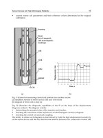
members of the C family possess only a part of the alternative K channel. Oxygen reductases
from bacteria and archaea reveal different subunit and heme-type compositions (Figure 4);
they are simpler than the eukaryotic ones while maintaining the same functionality and
efficiency. The mitochondrial Cyt-c oxidase (CcO) possesses 13 subunits, while the bacterial
heme - copper oxidases, that are also efficient and functional proton pumps, contain three to
four (Gennis, 1989; Garcia-Horsman et al., 1994; Pereira and Teixeira, 2004). Investigating the
catalytic reaction of bacterial complexes is therefore fundamental as the obtained insights
can be extrapolated to the eukaryotic ones. A prerequisite for understanding the mechanism
of functioning of these enzymes that contain multiple redox centers is determination of the
individual midpoint redox potentials of the cofactors under conditions that reproduce some
Immobilizedredoxproteins:
mimickingbasicfeaturesofphysiologicalmembranesandinterfaces 33
4.2 Surface Enhanced resonance Raman (SERR) and surface enhanced IR
(SEIRA) spectroscopy
Surface enhanced Raman (SER) spectroscopy is based on the increase of the signal intensity
associated with vibrational transitions of molecules situated in close proximity to
nanoscopic metal structures. Two distinct enhancement mechanisms have been identified.
The chemical mechanism originates from charge transfer interactions between the metal
substrate and the adsorbate, and provides a weak enhancement solely for the molecules in
direct contact with the metal. The electromagnetic mechanism is based on the amplified
electromagnetic fields generated upon excitation of the localized surface plasmons of
nanostructured metals. It does not require specific substrate/adsorbate contacts and
provides the main contribution to the overall enhancement. Among different metals tested
as SER substrates, Ag affords the strongest electromagnetic enhancements, due to surface
plasmon resonance in a wide spectral range from the near UV to the IR region. A drawback,
however, is that Ag nanostructures are less stable and chemically less inert than their Au
counterparts. In addition, the low oxidation potential of Ag narrows the range of applicable
potentials in SER-based spectro-electrochemical experiments. For these reasons most efforts
in recent years have been devoted to the development of Au SER substrates, including SER-
active electrodes. The attractiveness of the unsurpassed sensitivity of Ag has also driven
significant efforts towards use of this metal and hybrid Ag/Au structures. To this end, a
large number of highly regular and reproducible Au and Ag SER substrates have been
reported, making use of spheres, tubes, rods, thorns, cavities and wires as building blocks
(Mahajan et al., 2007; Murgida and Hildebrandt, 2008; Lal et al., 2008; Banholzer et al., 2008;
Brown and Milton, 2008; Feng et al., 2008a; Feng et al., 2009).
If the excitation laser is in resonance not only with the energy of surface plasmons of the
metal but also with the electronic transition of the immobilized molecule, the SER and RR
effects combine. The resulting SERR spectra display exclusively the vibrational bands of the
chromophore of the adsorbed species. The use of Ag as SER-active substrate is particularly
suited for studying porphyrins and heme proteins since these molecules exhibit a strong
electronic transition at ca. 410 nm (Soret band) and a weaker one at ca. 550 nm which both
coincide with Ag (but not with Au) surface plasmon resonances. SERR spectra of heme
proteins reveal the same information as RR spectra, such as the oxidation, spin, and
coordination states of the heme group, and in addition their changes as a consequence of
variations of the electrode potential (see bellow) (Siebert and Hildebrandt, 2008).
Molecules adsorbed in the vicinity of nanostructured metal surfaces, such as Ag or Au
islands deposited on inert ATR crystals, experience enhanced absorption of IR radiation,
which is the basis for (ATR) SEIRA spectroscopy. SEIRA spectroscopy has been successfully
employed to probe the structure of immobilized biomolecules including redox proteins and
enzymes (Ataka and Heberle, 2007). The enhancement of the IR bands does not exceed two
orders of magnitude and therefore is smaller than the enhancement of the SERR bands
which may be larger than 10
5
. The distance-dependent decay of the enhancement factor is
less pronounced for SEIRA than for SERR spectroscopy, and both techniques can
successfully probe molecules separated from the surface by up to 5 nm.
The nanostructured metal substrate that amplifies the signals can also serve as a working
electrode in spectroelectrochemical studies. Indeed, potentiometric titrations followed by
SERR and SEIRA have provided important insights into the mechanism of functioning of
several heme proteins immobilized on biocompatible metal electrodes (Murgida and
Hildebrandt, 2004a; Murgida and Hildebrandt, 2005; Murgida and Hildebrandt, 2008).
Both SERR and SEIRA can be employed in the time resolved (TR) mode that enables
probing of dynamics of immobilized proteins. The method requires a synchronization of a
perturbation event with the spectroscopic detection at variable delay times. For TR-SEIRA
spectroscopy acquisition is usually performed in the rapid or step scan mode for probing
events in time windows longer or shorter than 10 ms, respectively. For the study of potential
dependent processes of immobilized redox proteins by TR-SERR, the equilibrium of the
immobilized species is perturbed by a rapid potential jump, and the subsequent relaxation
process is then monitored at different delay times. A prerequisite for applying of TR-SERR is
that the underlying ET processes are fully reversible. The time resolution depends on the
charge reorganization of the double layer of the working electrode and is typically on a
microsecond scale (Murgida and Hildebrandt, 2004a; Murgida and Hildebrandt, 2005;
Murgida and Hildebrandt 2008).
5. Recent developments in the characterization of immobilized redox proteins
In this section we will focus on selected examples of surface enhanced
spectroelectrochemical characterization of ET proteins immobilized on nanostructured
electrodes coated with biomimetic films. The first part is dedicated to membrane oxygen
reductases whose structural, functional and spectroscopic complexity imposes some serious
limits to other experimental approaches. In the second part we will describe recent studies
on soluble electron carrier proteins, mainly cytochromes.
5.1 Membrane proteins: oxygen reductases
Terminal oxygen reductases are the final complexes in aerobic respiratory chains that couple
the four-electron reduction of molecular oxygen to water with proton translocation across
the membrane (vide supra). Intense research efforts have been made in the past decades to
elucidate the mechanism of the molecular functioning of these enzymes. Although
substantial progress has been made, for instance, in determining their three-dimensional
structures, the coupling between the redox processes and proton translocation is not yet
well understood (Garcia-Horsman et al., 1994; Pereira and Teixeira, 2004). Most of the
terminal oxidases are members of the heme - copper superfamily that can be classified into
several families, based on amino acid sequences and intraprotein proton channels. The
members of the family A are mitochondrial–like, possessing amino acid residues that
compose D and K channels, the B-type enzymes have an alternative K channel, while
members of the C family possess only a part of the alternative K channel. Oxygen reductases
from bacteria and archaea reveal different subunit and heme-type compositions (Figure 4);
they are simpler than the eukaryotic ones while maintaining the same functionality and
efficiency. The mitochondrial Cyt-c oxidase (CcO) possesses 13 subunits, while the bacterial
heme - copper oxidases, that are also efficient and functional proton pumps, contain three to
four (Gennis, 1989; Garcia-Horsman et al., 1994; Pereira and Teixeira, 2004). Investigating the
catalytic reaction of bacterial complexes is therefore fundamental as the obtained insights
can be extrapolated to the eukaryotic ones. A prerequisite for understanding the mechanism
of functioning of these enzymes that contain multiple redox centers is determination of the
individual midpoint redox potentials of the cofactors under conditions that reproduce some
Biomimetics,LearningfromNature34
basic features of the physiological environment. Published redox properties of oxygen
reductases are typically determined from solution studies and are often contradictory
regarding both the values and the interpretation (Todorovic et al., 2005; Veríssimo et al.,
2007). In this respect, the use of SERR spectroelectrochemical titrations has been proven to
be a valuable tool.
Fig. 4. Schematic representation of several oxygen reductases. A) aa
3
quinol oxidase; B) aa
3
cytochrome c oxidase; C) cbb
3
oxygen reductase. The LS hemes in the respective catalytic
subunits are depicted in orange (heme a) and pink (heme b), the HS hemes are in red (a
3
)
and purple (b
3
), the LS hemes c in FixO and FixP subunits of the cbb
3
are shown in green,
and copper centers (dinuclear Cu
A
and Cu
B
), in blue.
aa
3
quinol oxidase (QO): The aa
3
oxygen reductase from the thermophylic archaeon Acidianus
ambivalens receives electrons directly from the membrane quinone pool, being therefore a
quinol oxidase. It is a type B oxygen reductase that in the catalytic subunit houses two heme
groups, the low-spin (LS) heme a, and the high spin (HS) heme a
3
coupled to Cu
B
in the
catalytic (oxygen binding) center (Figure 4A). The two hemes display different RR spectral
fingerprints. Detergent solubilized QO was directly adsorbed on a bare nanostructured Ag
electrode and investigated by potential-dependent SERR (Todorovic et al., 2005). Adsorption
of the protein to the hydrophilic surface of the phosphate-coated electrode occurred without
displacement of the detergent molecules, which therefore provided a biocompatible
interface. This conclusion was supported by the detection of a SER signal at 2950 cm
-1
from
the Ag electrode immersed into the protein-free buffer solution, that was attributed to the C-
H stretching mode of dodecyl-maltoside. The protein retained its native structure upon
immobilization as confirmed by the comparison of the RR and SERR spectra of QO in
solution and in the adsorbed state (Figure 5A). Namely, all vibrational bands present in the
RR spectra that were assigned to skeletal vibrations and stretching modes of the vinyl and
formyl substituents of hemes a and a
3
were identified in the SERR spectra of the
immobilized QO. Variations of the band intensities between the SERR and RR spectra
originate from the orientation-dependence of the SERR effect, which causes different
enhancements of vibrational modes of HS vs. LS hemes, but also of modes of the same heme
that have different symmetry (see 5.2). The spectra of immobilized QO, measured at a series
of electrode potentials, were subsequently subjected to component analysis. At intermediate
potentials, over forty modes could be identified in the high frequency region of the spectra,
originating from the two heme groups in two redox states. In order to simplify the analysis
and to avoid uncertainties caused by the overlapping of some modes, the quantitative
spectral analysis was based on two modes, the ν
3
and ν
C=O
that are unambiguous indicators
of the redox and spin states of the two hemes. Simplified component spectra, based only on
these modes of each heme group in each redox state, were constructed and used in a global
fit to all experimental SERR spectra by varying the relative contributions of the individual
component spectra (Figure 5B). After conversion of spectral contributions into relative
concentrations, the redox potentials of the two heme sites in QO were determined. The
corresponding Nernst plots display a linear behavior that reveals one-electron transfer
processes, indicating, furthermore that the two hemes can be treated as independent redox
couples with no significant interaction potential.
The results of the study point to a substantially different mechanistic scheme for the energy
transduction in QO. The two redox centers, hemes a and a
3
are uncoupled and exhibit
reversed midpoint potentials with respect to the type A enzymes. In both cases the free
energy, provided by downhill ET reactions, is utilized for vectorial proton transport.
However, for the type A enzymes the exergonicity of the ET cascade requires a sophisticated
network of cooperativities. In contrast, downhill ET in QO is already guaranteed by the
inversion of the intrinsic midpoint potentials of hemes a and a
3
such that a modulation by
cooperativity effects is not required. Moreover, SERR experiments indicate that redox-
linked, electric-field-modulated conformational transitions of the heme a
3
that are relevant
for proton translocation, were blocked in the immobilized, but not in solubilized QO (Das et
al., 1999), suggesting furthermore that, when the membrane potential generated by the
proton pumping activity of QO becomes sufficiently large, the resultant electric field is
capable of blocking the elementary steps of proton translocation. This finding has been
interpreted in terms of a self-regulation mechanism of the proton pumping activity of the
QO (Todorovic et al., 2005).
Fig. 5. RR and SERR spectra of the aa
3
QO. A) high frequency region spectra of reduced QO:
RR of solubilized (upper trace) and SERR of immobilized (lower trace) protein; B) SERR
spectra of the QO at - 3 mV (upper trace) and at + 297 mV (lower trace). Vibrational modes
of the heme a are indicated in green (ferrous) and blue (ferric); modes of the heme a
3
are
shown in red (ferrous) and yellow (ferric); dotted line represents the envelope that includes
all non-assigned bands, black line shows experimental and overall simulated spectra.
Immobilizedredoxproteins:
mimickingbasicfeaturesofphysiologicalmembranesandinterfaces 35
basic features of the physiological environment. Published redox properties of oxygen
reductases are typically determined from solution studies and are often contradictory
regarding both the values and the interpretation (Todorovic et al., 2005; Veríssimo et al.,
2007). In this respect, the use of SERR spectroelectrochemical titrations has been proven to
be a valuable tool.
Fig. 4. Schematic representation of several oxygen reductases. A) aa
3
quinol oxidase; B) aa
3
cytochrome c oxidase; C) cbb
3
oxygen reductase. The LS hemes in the respective catalytic
subunits are depicted in orange (heme a) and pink (heme b), the HS hemes are in red (a
3
)
and purple (b
3
), the LS hemes c in FixO and FixP subunits of the cbb
3
are shown in green,
and copper centers (dinuclear Cu
A
and Cu
B
), in blue.
aa
3
quinol oxidase (QO): The aa
3
oxygen reductase from the thermophylic archaeon Acidianus
ambivalens receives electrons directly from the membrane quinone pool, being therefore a
quinol oxidase. It is a type B oxygen reductase that in the catalytic subunit houses two heme
groups, the low-spin (LS) heme a, and the high spin (HS) heme a
3
coupled to Cu
B
in the
catalytic (oxygen binding) center (Figure 4A). The two hemes display different RR spectral
fingerprints. Detergent solubilized QO was directly adsorbed on a bare nanostructured Ag
electrode and investigated by potential-dependent SERR (Todorovic et al., 2005). Adsorption
of the protein to the hydrophilic surface of the phosphate-coated electrode occurred without
displacement of the detergent molecules, which therefore provided a biocompatible
interface. This conclusion was supported by the detection of a SER signal at 2950 cm
-1
from
the Ag electrode immersed into the protein-free buffer solution, that was attributed to the C-
H stretching mode of dodecyl-maltoside. The protein retained its native structure upon
immobilization as confirmed by the comparison of the RR and SERR spectra of QO in
solution and in the adsorbed state (Figure 5A). Namely, all vibrational bands present in the
RR spectra that were assigned to skeletal vibrations and stretching modes of the vinyl and
formyl substituents of hemes a and a
3
were identified in the SERR spectra of the
immobilized QO. Variations of the band intensities between the SERR and RR spectra
originate from the orientation-dependence of the SERR effect, which causes different
enhancements of vibrational modes of HS vs. LS hemes, but also of modes of the same heme
that have different symmetry (see 5.2). The spectra of immobilized QO, measured at a series
of electrode potentials, were subsequently subjected to component analysis. At intermediate
potentials, over forty modes could be identified in the high frequency region of the spectra,
originating from the two heme groups in two redox states. In order to simplify the analysis
and to avoid uncertainties caused by the overlapping of some modes, the quantitative
spectral analysis was based on two modes, the ν
3
and ν
C=O
that are unambiguous indicators
of the redox and spin states of the two hemes. Simplified component spectra, based only on
these modes of each heme group in each redox state, were constructed and used in a global
fit to all experimental SERR spectra by varying the relative contributions of the individual
component spectra (Figure 5B). After conversion of spectral contributions into relative
concentrations, the redox potentials of the two heme sites in QO were determined. The
corresponding Nernst plots display a linear behavior that reveals one-electron transfer
processes, indicating, furthermore that the two hemes can be treated as independent redox
couples with no significant interaction potential.
The results of the study point to a substantially different mechanistic scheme for the energy
transduction in QO. The two redox centers, hemes a and a
3
are uncoupled and exhibit
reversed midpoint potentials with respect to the type A enzymes. In both cases the free
energy, provided by downhill ET reactions, is utilized for vectorial proton transport.
However, for the type A enzymes the exergonicity of the ET cascade requires a sophisticated
network of cooperativities. In contrast, downhill ET in QO is already guaranteed by the
inversion of the intrinsic midpoint potentials of hemes a and a
3
such that a modulation by
cooperativity effects is not required. Moreover, SERR experiments indicate that redox-
linked, electric-field-modulated conformational transitions of the heme a
3
that are relevant
for proton translocation, were blocked in the immobilized, but not in solubilized QO (Das et
al., 1999), suggesting furthermore that, when the membrane potential generated by the
proton pumping activity of QO becomes sufficiently large, the resultant electric field is
capable of blocking the elementary steps of proton translocation. This finding has been
interpreted in terms of a self-regulation mechanism of the proton pumping activity of the
QO (Todorovic et al., 2005).
Fig. 5. RR and SERR spectra of the aa
3
QO. A) high frequency region spectra of reduced QO:
RR of solubilized (upper trace) and SERR of immobilized (lower trace) protein; B) SERR
spectra of the QO at - 3 mV (upper trace) and at + 297 mV (lower trace). Vibrational modes
of the heme a are indicated in green (ferrous) and blue (ferric); modes of the heme a
3
are
shown in red (ferrous) and yellow (ferric); dotted line represents the envelope that includes
all non-assigned bands, black line shows experimental and overall simulated spectra.
Biomimetics,LearningfromNature36
aa
3
cytochrome c oxidase: The CcO from Rhodobacter sphaeroides is a member of the type A
family of heme copper oxygen reductases that houses three redox centers in the catalytic
subunit (subunit I) and a dinuclear copper Cu
A
in the subunit II (Figure 4B). It is purified
from an organism that is capable of growing heterotrophically via fermentation and aerobic
and anaerobic respiration, with a genetically introduced His-tag, allowing immobilization
of the CcO on a metal electrode via Ni-NTA SAMs, Figure 3C (Friedrich et al., 2004; Ataka et
al., 2004; Giess et al., 2004; Hrabakova et al., 2006; Todorovic et al., 2008). The protein was
specifically attached, uniformly oriented and catalytically active in the biomimetic construct.
The orientation of the attached protein could be controlled since the His-tag was introduced
into the amino acid sequence of R. sphaeroides enzyme either on the C-terminus of subunit I
or on the C-terminus of subunit II. Therefore, the domain that interacts with the
physiological electron donor, Cyt-c, identified to be composed of residues Glu148, Glu157,
Asp195, and Asp214 in subunit II, was either exposed to the solution, or was facing the
metal surface (Ataka et al., 2004). Catalytic currents could be measured under aerobic
conditions when the Cyt-c / CcO complex was allowed to form. Proton pumping activity
was also functional in the construct, as suggested by electrochemical impedance
spectroscopy. SERR spectroscopic studies revealed heterogeneous ET to the heme a, which
was selectively reduced while the heme a
3
remained oxidized, even at the most negative
electrode potentials. The ET between the two hemes is fast in solution, indicating some
alterations of the intramolecular ET in immobilized CcO, possibly due to electric field
dependent perturbation of internal proton translocation steps (Hrabakova et al., 2006).
cbb
3
oxygen reductase: The Bradyrhizobium japonicum cbb
3
oxidase is a type C oxygen reductase
that contains three major subunits: a membrane integral subunit I (FixN), which houses a LS
heme b and the catalytic center (HS heme b
3
- Cu
B
), and subunits II (FixO) and III (FixP),
containing one (His-Met coordinated) and two (bis His and His-Met coordinated) LS hemes
c, respectively (Figure 4C). The cbb
3
oxygen reductases are expressed in various bacteria
under microaerobic conditions and exhibit several unique characteristics (Sharma et al.,
2006). Phylogenetically, they are the most distant and the least understood members of the
heme-copper oxygen reductase superfamily (Pereira and Teixeira, 2004; Pitcher and
Watmough, 2004; Sharma et al., 2006). The cbb
3
oxygen reductases lack the Cu
A
electron
entry site (Garcia-Horsman et al., 1994) and the highly conserved tyrosine residue covalently
bound to the histidyl Cu
B
ligand. Furthermore, many of the amino acid residues involved in
proton conduction through the D- and K- channels of the A-type enzymes are absent in cbb
3
oxygen reductases. These enzymes exhibit the highest NO reductase activity among the
members of the superfamily (Forte et al., 2001; Pitcher and Watmough, 2004; Veríssimo et al.,
2007). The cbb
3
oxygen reductase from B. japonicum possesses a genetically introduced His
tag on the C-terminus of subunit I, i.e. on the cytoplasmic side. As in the previous example,
it was immobilized on Ag (and Au) electrode coated with a (Ni-NTA) SAM, embedded into
a reconstituted phospholipid bilayer, Figure 3C, and studied by surface-enhanced
vibrational spectroscopy and cyclic voltammetry (Figure 6) (Todorovic et al., 2008).
Fig. 6. Immobilized cbb
3
oxygen reductase. A) SEIRA spectra of the cbb
3
immobilized via His-
tag/Ni-NTA (dashed line) and detergent coated electrode (solid line); B) cyclic voltammetry
of the cbb
3
embedded into biomimetic construct in the presence (dashed line) and absence
(dotted line) of electron donor.
SEIRA spectra of the immobilized cbb
3
are dominated by the amide I and II modes (Figure
6A). For membrane proteins with a high content of preferentially parallel helices such as the
subunit I of cbb
3
(Zufferey et al., 1998; Pitcher and Watmough, 2004), SEIRA spectra are
sensitive to the orientation of the helices with respect to the electrode surface, which is
reflected in the intensity ratio of amide I and amide II bands. The amide I mode, that is
mainly composed by the C=O stretching coordinates of the peptide bonds, is associated with
dipole moment changes parallel to the axis of the helices, such that it gains surface
enhancement when the C=O groups, and thus the helices, are oriented perpendicular to the
surface. Conversely, the dipole moment changes of the amide II mode that is mainly
composed of N-H in-plane bending and C-N stretching coordinates, are perpendicular to
the helix axis and therefore gain a weaker enhancement for helices oriented in an upright
position (Marsh et al., 2000). In the SEIRA spectrum of cbb
3
the amide I is observed at 1658
cm
-1
, a characteristic position for a largely α-helical peptide. Its intensity is distinctly higher
than that of the amide II (1548 cm
-1
), which is consistent with a largely perpendicular
orientation of the helices with respect to the electrode surface. A more random orientation of
the enzyme is obtained upon non-specific adsorption of the solubilized cbb
3
on a detergent-
coated electrode as reflected by a ca. two times weaker amide I band and a 1.5 times lower
amide I / amide II intensity ratio, as compared with the His-tag bound cbb
3
(Figure 6A)
(Todorovic et al., 2008). The oxygen reductase catalytic activity of the immobilized cbb
3
was
controlled in situ by cyclic voltammetry. As shown in Figure 6B, large electrocatalytic
currents are observed under aerobic conditions in the presence of the electron donor, while
only capacitive currents were observed in its absence.
Unlike the aa
3
QO and CcO, the cbb
3
oxygen reductase possesses five heme groups, three of
which are 6cLS c-type hemes that are spectroscopically indistinguishable. Moreover, four
out of five hemes are LS, displaying higher Raman cross sections, and therefore partially
obscuring the spectroscopic features of catalytic HS heme b
3
. Reliable component analysis of
the SERR spectra was further aggravated by the high photoreducibility of the enzyme.
Therefore, in order to facilitate the assignment of individual redox transitions to each heme
group of the pentahemic cbb
3
, the individual FixO and FixP subunits (Figure 4C) were
Immobilizedredoxproteins:
mimickingbasicfeaturesofphysiologicalmembranesandinterfaces 37
aa
3
cytochrome c oxidase: The CcO from Rhodobacter sphaeroides is a member of the type A
family of heme copper oxygen reductases that houses three redox centers in the catalytic
subunit (subunit I) and a dinuclear copper Cu
A
in the subunit II (Figure 4B). It is purified
from an organism that is capable of growing heterotrophically via fermentation and aerobic
and anaerobic respiration, with a genetically introduced His-tag, allowing immobilization
of the CcO on a metal electrode via Ni-NTA SAMs, Figure 3C (Friedrich et al., 2004; Ataka et
al., 2004; Giess et al., 2004; Hrabakova et al., 2006; Todorovic et al., 2008). The protein was
specifically attached, uniformly oriented and catalytically active in the biomimetic construct.
The orientation of the attached protein could be controlled since the His-tag was introduced
into the amino acid sequence of R. sphaeroides enzyme either on the C-terminus of subunit I
or on the C-terminus of subunit II. Therefore, the domain that interacts with the
physiological electron donor, Cyt-c, identified to be composed of residues Glu148, Glu157,
Asp195, and Asp214 in subunit II, was either exposed to the solution, or was facing the
metal surface (Ataka et al., 2004). Catalytic currents could be measured under aerobic
conditions when the Cyt-c / CcO complex was allowed to form. Proton pumping activity
was also functional in the construct, as suggested by electrochemical impedance
spectroscopy. SERR spectroscopic studies revealed heterogeneous ET to the heme a, which
was selectively reduced while the heme a
3
remained oxidized, even at the most negative
electrode potentials. The ET between the two hemes is fast in solution, indicating some
alterations of the intramolecular ET in immobilized CcO, possibly due to electric field
dependent perturbation of internal proton translocation steps (Hrabakova et al., 2006).
cbb
3
oxygen reductase: The Bradyrhizobium japonicum cbb
3
oxidase is a type C oxygen reductase
that contains three major subunits: a membrane integral subunit I (FixN), which houses a LS
heme b and the catalytic center (HS heme b
3
- Cu
B
), and subunits II (FixO) and III (FixP),
containing one (His-Met coordinated) and two (bis His and His-Met coordinated) LS hemes
c, respectively (Figure 4C). The cbb
3
oxygen reductases are expressed in various bacteria
under microaerobic conditions and exhibit several unique characteristics (Sharma et al.,
2006). Phylogenetically, they are the most distant and the least understood members of the
heme-copper oxygen reductase superfamily (Pereira and Teixeira, 2004; Pitcher and
Watmough, 2004; Sharma et al., 2006). The cbb
3
oxygen reductases lack the Cu
A
electron
entry site (Garcia-Horsman et al., 1994) and the highly conserved tyrosine residue covalently
bound to the histidyl Cu
B
ligand. Furthermore, many of the amino acid residues involved in
proton conduction through the D- and K- channels of the A-type enzymes are absent in cbb
3
oxygen reductases. These enzymes exhibit the highest NO reductase activity among the
members of the superfamily (Forte et al., 2001; Pitcher and Watmough, 2004; Veríssimo et al.,
2007). The cbb
3
oxygen reductase from B. japonicum possesses a genetically introduced His
tag on the C-terminus of subunit I, i.e. on the cytoplasmic side. As in the previous example,
it was immobilized on Ag (and Au) electrode coated with a (Ni-NTA) SAM, embedded into
a reconstituted phospholipid bilayer, Figure 3C, and studied by surface-enhanced
vibrational spectroscopy and cyclic voltammetry (Figure 6) (Todorovic et al., 2008).
Fig. 6. Immobilized cbb
3
oxygen reductase. A) SEIRA spectra of the cbb
3
immobilized via His-
tag/Ni-NTA (dashed line) and detergent coated electrode (solid line); B) cyclic voltammetry
of the cbb
3
embedded into biomimetic construct in the presence (dashed line) and absence
(dotted line) of electron donor.
SEIRA spectra of the immobilized cbb
3
are dominated by the amide I and II modes (Figure
6A). For membrane proteins with a high content of preferentially parallel helices such as the
subunit I of cbb
3
(Zufferey et al., 1998; Pitcher and Watmough, 2004), SEIRA spectra are
sensitive to the orientation of the helices with respect to the electrode surface, which is
reflected in the intensity ratio of amide I and amide II bands. The amide I mode, that is
mainly composed by the C=O stretching coordinates of the peptide bonds, is associated with
dipole moment changes parallel to the axis of the helices, such that it gains surface
enhancement when the C=O groups, and thus the helices, are oriented perpendicular to the
surface. Conversely, the dipole moment changes of the amide II mode that is mainly
composed of N-H in-plane bending and C-N stretching coordinates, are perpendicular to
the helix axis and therefore gain a weaker enhancement for helices oriented in an upright
position (Marsh et al., 2000). In the SEIRA spectrum of cbb
3
the amide I is observed at 1658
cm
-1
, a characteristic position for a largely α-helical peptide. Its intensity is distinctly higher
than that of the amide II (1548 cm
-1
), which is consistent with a largely perpendicular
orientation of the helices with respect to the electrode surface. A more random orientation of
the enzyme is obtained upon non-specific adsorption of the solubilized cbb
3
on a detergent-
coated electrode as reflected by a ca. two times weaker amide I band and a 1.5 times lower
amide I / amide II intensity ratio, as compared with the His-tag bound cbb
3
(Figure 6A)
(Todorovic et al., 2008). The oxygen reductase catalytic activity of the immobilized cbb
3
was
controlled in situ by cyclic voltammetry. As shown in Figure 6B, large electrocatalytic
currents are observed under aerobic conditions in the presence of the electron donor, while
only capacitive currents were observed in its absence.
Unlike the aa
3
QO and CcO, the cbb
3
oxygen reductase possesses five heme groups, three of
which are 6cLS c-type hemes that are spectroscopically indistinguishable. Moreover, four
out of five hemes are LS, displaying higher Raman cross sections, and therefore partially
obscuring the spectroscopic features of catalytic HS heme b
3
. Reliable component analysis of
the SERR spectra was further aggravated by the high photoreducibility of the enzyme.
Therefore, in order to facilitate the assignment of individual redox transitions to each heme
group of the pentahemic cbb
3
, the individual FixO and FixP subunits (Figure 4C) were
Biomimetics,LearningfromNature38
overexpressed in E. coli with their transmembrane domain truncated, purified in soluble
form, and characterized by SERR spectroelectrochemistry. Upon combining the SERR-
spectroelectrochemical data for the subunits and for the integral enzyme, it was possible to
provide a consistent analysis of redox potentials of the individual cofactors in the cbb
3
oxidase. On the basis of these findings, a sequence of ET events in the cbb
3
enzyme was
postulated. The Met/His heme c of either the FixO or FixP subunit, which exhibit the lowest
redox potentials, serves as the electron entry site of the complex. According to the order of
redox potentials, the subsequent electron acceptor was identified as the bis-His heme c in
the FixP or/and the HS heme b in the catalytic subunit, and the final one is the heme b
3
(Todorovic, et al., 2008). The obtained data also shed light on a controversially discussed role
of the FixP subunit in cbb
3
oxygen reductases. The values of redox potentials obtained by
SERR potentiometric titration reveal that the presence of FixP can be considered as
redundant in the ET pathway. Rather, it can be associated with oxygen sensing properties,
as also suggested for the cbb
3
enzyme from P. stutzeri (Pitcher and Watmough, 2004b).
5.2 Soluble proteins
As discussed in previous sections, small soluble redox proteins that transport electrons
between different membrane-bound complexes along biological ET chains are exposed to
relatively intense electric fields which may have a substantial impact on their structure and
function. Among these electron shuttles, cytochromes and particularly Cyt-c, constitute the
best studied examples. Electric field effects on the structure, thermodynamics and reaction
dynamics of cytochromes have been extensively investigated using Ag electrodes coated
with SAMs of -substituted alkanethiols as biomimetic interfaces that allow a systematic
variation of the field strength (see 3.2).
Electric field effects on redox potential. Among other parameters, electric fields of biologically
relevant magnitude may affect the most fundamental thermodynamic property of a redox
protein, i.e. its reduction potential. This has been shown by potential-dependent SERR
spectroscopy for the soluble tetraheme protein cytochrome c
3
(Cyt-c
3
) (Rivas et al., 2005).
Electrostatic adsorption of Cyt-c
3
on Ag electrodes coated with mercaptoundecanoic acid
occurs without significant structural alterations at the level of the heme groups. SERR
potentiometric titrations, however, indicate that the redox potentials of the four hemes are
significantly downshifted with respect to their values in solution, to such an extent that the
order of reduction is actually reversed. The experimental results, that were in excellent
agreement with electrostatic calculations, revealed that electric fields tend to downshift the
redox potentials by stabilizing the ferric form. This effect is partially compensated by the
low dielectric constant of the SAM which shifts the redox potentials in the opposite
direction. Indeed, the resulting downshift was more pronounced for the hemes that are
closer to the interface, i.e. under the influence of higher electric fields.
A similar interplay of different effects on the redox potential has been observed for
cytochrome P450 from P. putida immobilized on coated Ag electrodes, although in this
case the adsorbed protein was almost quantitatively converted into the P420 form as
judged from the SERR spectra (Todorovic et al., 2006). In contrast, electrostatic
adsorption of Cyt-c on similar coatings does not appear to have any significant effect on
the redox potential but may have distinct structural implications (Murgida and
Hildebrandt, 2001a; Murgida and Hildebrandt, 2004a).
Electric field effects on protein structure and function. It has been established, using RR and
SERR spectroscopy, that the electrostatic interaction of the positively charged Cyt-c with
negatively charged model systems, such as phospholipid vesicles, (Droghetti et al., 2006),
polyelectrolytes (Weidinger et al., 2006), or the binding domain of the natural reaction
partner CcO, may promote the formation of the conformational state of mitochondrial Cyt-c
(denoted as B2), in which the axial Met80 ligand is removed from the heme iron. At
physiological pH, this coordination site may remain vacant, resulting in 5cHS Cyt-c, or may
be occupied by a His residue (most likely His26 or His33). The disruption of the Fe-Met80
bond significantly alters the properties of Cyt-c. On the one hand, the B2 state has a
reduction potential which is more than 300 mV lower than that of the native protein
(denoted as state B1) as determined by potential-dependent SERR spectroscopy (Murgida
and Hildebrandt, 2004a). On the other hand, the B2 state shows a substantial increase of
peroxidase activity by allowing the access for hydrogen peroxide to the heme iron (Kagan et
al., 2005; Godoy et al., 2009). Thus, in vivo the B1 to B2 transition would have profound
physiological consequences since the B2 state cannot accept electrons from complex III but is
capable of catalyzing the peroxidation of cardiolipin, the main charged lipid component of
the inner mitochondrial membrane (Kagan et al., 2009). Degradation of cardiolipin increases
the permeability of the membrane, thus facilitating the transfer of Cyt-c to the cytosol where
it binds to Apaf-1, in one of the initial events of the cell apoptosis. Thus, it is likely that the
switch from the “normal” redox function of Cyt-c (B1 state) to the apoptotic function (B2
state) depends on the local electric field which is, in turn, modulated by the membrane
potential. To check this hypothesis the structure of Cyt-c electrostatically bound to
electrodes coated with anionic SAMs was investigated. Variation of the electrode potential
and of the charge density on the film surface shows that the equilibrium between the B1 and
B2 states of the protein is shifted towards B2 upon raising the electric field strength at the
interface of the biomimetic construct (Murgida and Hildebrandt, 2001a; Murgida and
Hildebrandt, 2004a). Detailed spectroscopic studies, including a variety of techniques, have
demonstrated that the B1 to B2 transition occurs without substantial alteration of the protein
secondary structure. Based on these observations, the influence of the electric field on the
dissociation energy of the Fe-Met80 bond in model porphyrins were studied using density
functional theory (DFT). In that case, i.e., for the isolated heme moiety lacking the protein
environment, no significant effect on the Fe-S(Met) bond stability was predicted for
biologically meaningful electric field strengths (De Biase et al., 2007). On the other hand,
molecular dynamics simulations performed on the entire protein, show that biologically
relevant electric fields induce an increased mobility of the key protein segments that lead to
the detachment of the sixth axial ligand, Met80, from the heme iron. This electric-field
induced conformational transition is both energetically and entropically driven (De Biase et
al., 2009). It was proposed, based on these theoretical and experimental investigations using
biomimetic systems, that the variable transmembrane potential may modulate the structure
of Cyt-c, thus playing the role of a switch that can alternate its redox function in the
respiratory chain to peroxidase function in the early events of apoptosis.
Electric field effects on protein dynamics. Protein dynamics has been recently recognized as a
key factor in controlling or limiting inter- and intra-protein ET reactions. However, in most
of the cases the complexity of biological systems impairs direct observations of processes
such as conformational gating, configurational fluctuations or rearrangement of protein
complexes under reactive conditions. In this context simplified model systems, like proteins
Immobilizedredoxproteins:
mimickingbasicfeaturesofphysiologicalmembranesandinterfaces 39
overexpressed in E. coli with their transmembrane domain truncated, purified in soluble
form, and characterized by SERR spectroelectrochemistry. Upon combining the SERR-
spectroelectrochemical data for the subunits and for the integral enzyme, it was possible to
provide a consistent analysis of redox potentials of the individual cofactors in the cbb
3
oxidase. On the basis of these findings, a sequence of ET events in the cbb
3
enzyme was
postulated. The Met/His heme c of either the FixO or FixP subunit, which exhibit the lowest
redox potentials, serves as the electron entry site of the complex. According to the order of
redox potentials, the subsequent electron acceptor was identified as the bis-His heme c in
the FixP or/and the HS heme b in the catalytic subunit, and the final one is the heme b
3
(Todorovic, et al., 2008). The obtained data also shed light on a controversially discussed role
of the FixP subunit in cbb
3
oxygen reductases. The values of redox potentials obtained by
SERR potentiometric titration reveal that the presence of FixP can be considered as
redundant in the ET pathway. Rather, it can be associated with oxygen sensing properties,
as also suggested for the cbb
3
enzyme from P. stutzeri (Pitcher and Watmough, 2004b).
5.2 Soluble proteins
As discussed in previous sections, small soluble redox proteins that transport electrons
between different membrane-bound complexes along biological ET chains are exposed to
relatively intense electric fields which may have a substantial impact on their structure and
function. Among these electron shuttles, cytochromes and particularly Cyt-c, constitute the
best studied examples. Electric field effects on the structure, thermodynamics and reaction
dynamics of cytochromes have been extensively investigated using Ag electrodes coated
with SAMs of -substituted alkanethiols as biomimetic interfaces that allow a systematic
variation of the field strength (see 3.2).
Electric field effects on redox potential. Among other parameters, electric fields of biologically
relevant magnitude may affect the most fundamental thermodynamic property of a redox
protein, i.e. its reduction potential. This has been shown by potential-dependent SERR
spectroscopy for the soluble tetraheme protein cytochrome c
3
(Cyt-c
3
) (Rivas et al., 2005).
Electrostatic adsorption of Cyt-c
3
on Ag electrodes coated with mercaptoundecanoic acid
occurs without significant structural alterations at the level of the heme groups. SERR
potentiometric titrations, however, indicate that the redox potentials of the four hemes are
significantly downshifted with respect to their values in solution, to such an extent that the
order of reduction is actually reversed. The experimental results, that were in excellent
agreement with electrostatic calculations, revealed that electric fields tend to downshift the
redox potentials by stabilizing the ferric form. This effect is partially compensated by the
low dielectric constant of the SAM which shifts the redox potentials in the opposite
direction. Indeed, the resulting downshift was more pronounced for the hemes that are
closer to the interface, i.e. under the influence of higher electric fields.
A similar interplay of different effects on the redox potential has been observed for
cytochrome P450 from P. putida immobilized on coated Ag electrodes, although in this
case the adsorbed protein was almost quantitatively converted into the P420 form as
judged from the SERR spectra (Todorovic et al., 2006). In contrast, electrostatic
adsorption of Cyt-c on similar coatings does not appear to have any significant effect on
the redox potential but may have distinct structural implications (Murgida and
Hildebrandt, 2001a; Murgida and Hildebrandt, 2004a).
Electric field effects on protein structure and function. It has been established, using RR and
SERR spectroscopy, that the electrostatic interaction of the positively charged Cyt-c with
negatively charged model systems, such as phospholipid vesicles, (Droghetti et al., 2006),
polyelectrolytes (Weidinger et al., 2006), or the binding domain of the natural reaction
partner CcO, may promote the formation of the conformational state of mitochondrial Cyt-c
(denoted as B2), in which the axial Met80 ligand is removed from the heme iron. At
physiological pH, this coordination site may remain vacant, resulting in 5cHS Cyt-c, or may
be occupied by a His residue (most likely His26 or His33). The disruption of the Fe-Met80
bond significantly alters the properties of Cyt-c. On the one hand, the B2 state has a
reduction potential which is more than 300 mV lower than that of the native protein
(denoted as state B1) as determined by potential-dependent SERR spectroscopy (Murgida
and Hildebrandt, 2004a). On the other hand, the B2 state shows a substantial increase of
peroxidase activity by allowing the access for hydrogen peroxide to the heme iron (Kagan et
al., 2005; Godoy et al., 2009). Thus, in vivo the B1 to B2 transition would have profound
physiological consequences since the B2 state cannot accept electrons from complex III but is
capable of catalyzing the peroxidation of cardiolipin, the main charged lipid component of
the inner mitochondrial membrane (Kagan et al., 2009). Degradation of cardiolipin increases
the permeability of the membrane, thus facilitating the transfer of Cyt-c to the cytosol where
it binds to Apaf-1, in one of the initial events of the cell apoptosis. Thus, it is likely that the
switch from the “normal” redox function of Cyt-c (B1 state) to the apoptotic function (B2
state) depends on the local electric field which is, in turn, modulated by the membrane
potential. To check this hypothesis the structure of Cyt-c electrostatically bound to
electrodes coated with anionic SAMs was investigated. Variation of the electrode potential
and of the charge density on the film surface shows that the equilibrium between the B1 and
B2 states of the protein is shifted towards B2 upon raising the electric field strength at the
interface of the biomimetic construct (Murgida and Hildebrandt, 2001a; Murgida and
Hildebrandt, 2004a). Detailed spectroscopic studies, including a variety of techniques, have
demonstrated that the B1 to B2 transition occurs without substantial alteration of the protein
secondary structure. Based on these observations, the influence of the electric field on the
dissociation energy of the Fe-Met80 bond in model porphyrins were studied using density
functional theory (DFT). In that case, i.e., for the isolated heme moiety lacking the protein
environment, no significant effect on the Fe-S(Met) bond stability was predicted for
biologically meaningful electric field strengths (De Biase et al., 2007). On the other hand,
molecular dynamics simulations performed on the entire protein, show that biologically
relevant electric fields induce an increased mobility of the key protein segments that lead to
the detachment of the sixth axial ligand, Met80, from the heme iron. This electric-field
induced conformational transition is both energetically and entropically driven (De Biase et
al., 2009). It was proposed, based on these theoretical and experimental investigations using
biomimetic systems, that the variable transmembrane potential may modulate the structure
of Cyt-c, thus playing the role of a switch that can alternate its redox function in the
respiratory chain to peroxidase function in the early events of apoptosis.
Electric field effects on protein dynamics. Protein dynamics has been recently recognized as a
key factor in controlling or limiting inter- and intra-protein ET reactions. However, in most
of the cases the complexity of biological systems impairs direct observations of processes
such as conformational gating, configurational fluctuations or rearrangement of protein
complexes under reactive conditions. In this context simplified model systems, like proteins
Biomimetics,LearningfromNature40
immobilized on SAM-coated electrodes, can greatly contribute to the understanding of the
biophysical fundamentals in better detail, even though they unavoidably deviate from the
true physiological conditions. A specific advantage of this approach is facilitating the
determination of ET rate constants as a function of distance by simply varying the chain
length of the alkanethiols without modifying other experimental parameters (Murgida and
Hildebrandt, 2001a; Murgida and Hildebrandt, 2001b). For redox proteins immobilized on
SAM-coated metal electrodes one can anticipate a nonadiabatic ET mechanism. Therefore,
the kinetics of the heterogeneous ET reaction can be described according to the high
temperature limit of Marcus semiclassical expression, including integration to account for
all the electronic levels of the metal, , contributing to the process (Marcus, 1965):
d
TkTk
e
Tk
V
k
BFB
F
B
ET
/exp1
1
4
)(
exp
||
2
0
2
(2)
where
), f(
), |V|,
F
, and
are the density of electronic states in the electrode, the Fermi-
Dirac distribution, the magnitude of the electronic coupling, the energy of the Fermi level,
and the reorganization energy of the redox active molecule. The quantity
refers
to the standard overpotential, with E denoting the actual electrode potential and E
0
the
standard potential for the redox couple; the other parameters in Equation 2 have the usual
meaning. The electronic coupling decays exponentially with the distance separating the
redox center from the electrode.
Notably, the characteristic exponential decay of k
ET
with the distance predicted by the theory
is verified only partially in most studies reported so far. Indeed, several research groups
have studied the distance-dependence of the ET rates for a variety of proteins immobilized
on Au and Ag electrodes coated with pure and mixed SAMs of -functionalized
alkanethiols using TR-SERR and electrochemistry. These studies include Cyt-c immobilized
on COOH-, COOH/OH, CH
3
-, and pyridine-terminated SAMs, (Murgida and Hildebrandt,
2001a; Murgida and Hildebrandt, 2002; Yue et al., 2006; Murgida et al., 2004b; Feng et al.,
2008b) cytochrome b
562
on NH
2
-terminated SAMs (Zuo et al., 2009), azurin on CH
3
-
terminated SAMs (Murgida et al., 2001a) and cytochrome c
6
(Kranich et al., 2009) and Cu
A
(Fujita et al., 2004) centers on mixed SAMs, among others. In all the cases, the measured ET
rates verify the expected exponential decay with distance, but only for relatively thick
SAMs. For thinner SAMs, however, the measured rate was distance-independent even
though, due to the still long donor-acceptor separation (> 6 Å), a non-adiabatic ET
mechanism should apply (Figure 7A). The origin of this behavior has been elusive and
controversial for the last ten years.
Recently, the use of SER selection rules for analyzing the reaction dynamics of immobilized
heme proteins has shed some light onto this issue (Kranich et al., 2008). In a SER experiment,
i.e. under off-resonance conditions, the individual components of the scattering tensor of the
heme are modified depending on the direction of the electric field vector and the orientation
of the heme plane. Assuming an ideal D
4h
porphyrin symmetry, one can anticipate that the
A
1g
modes will experience preferential enhancement when the heme plane is parallel to the
surface, while for a perpendicular orientation A
1g
, A
2g
, B
1g
and B
2g
will all be enhanced.
Therefore, different orientations of the adsorbed heme protein are expected to lead to
different intensity ratios of modes of different symmetries, e.g.
10
(B
1g
)/
4
(A
1g
). These SER
selection rules do not hold when the experiments are performed under electronic resonance
conditions (SERR), i.e. Soret excitation, typically used in the studies of heme proteins.
Fig. 7. TR-SERR data of Cyt-c electrostatically adsorbed to COOH-terminated SAMs. A)
distance-dependence of the apparent ET rates determined at η = -100 mV; B) time-
dependence of the ν
10
/ν
4
intensity ratio (green, λ
exc
= 514 nm) and of the concentration of
ferric protein (violet, λ
exc
= 413 nm) for Cyt-c adsorbed on SAM with 15 CH
2
groups; C)
idem (B) for a SAM with 5 CH
2
groups.
In that case SERR spectra are largely dominated by the totally symmetric modes A
1g
, which,
in addition to partial scrambling of the radiation, result in an almost complete loss of
orientation information. However, a reasonable compromise between acceptable
enhancement and qualitatively predictable selection rules can still be achieved upon
excitation into the less intense Q electronic transition of heme, by which non-totally
symmetric modes are also enhanced.
This strategy has been applied for investigating the reaction dynamics of Cyt-c and Cyt-c
6
electrostatically adsorbed on Ag electrodes coated with SAMs of -carboxyl alkanethiols.
TR-SERR experiments performed under Q-band excitation show that, upon applying a
potential jump, the protein reorients within the electrostatic complex. The reorientation is
fast in the low electric field regime, i.e. long SAMs, but becomes slower at shorter SAMs due
to the barrier imposed by the increasing electric field. Indeed it has been observed that the
measured ET rates in the plateau region of the k
ET
vs. distance plots are identical to the
reorientation rates, implying that reorientation is the rate limiting step (Figure 7). To
understand these results at a molecular level, molecular dynamics simulations of the
biomimetic systems were performed (Paggi et al., 2009). The simulations show that Cyt-c can
adsorb on SAMs in a variety of orientations that imply two major binding sites situated
around the partially exposed heme group. In the low electric field regime, the electrostatic
complexes are characterized by a large mobility of the protein that leads to significant
fluctuations of the electronic coupling (|V| in equation 2). For examples, a 7° change of tilt
Immobilizedredoxproteins:
mimickingbasicfeaturesofphysiologicalmembranesandinterfaces 41
immobilized on SAM-coated electrodes, can greatly contribute to the understanding of the
biophysical fundamentals in better detail, even though they unavoidably deviate from the
true physiological conditions. A specific advantage of this approach is facilitating the
determination of ET rate constants as a function of distance by simply varying the chain
length of the alkanethiols without modifying other experimental parameters (Murgida and
Hildebrandt, 2001a; Murgida and Hildebrandt, 2001b). For redox proteins immobilized on
SAM-coated metal electrodes one can anticipate a nonadiabatic ET mechanism. Therefore,
the kinetics of the heterogeneous ET reaction can be described according to the high
temperature limit of Marcus semiclassical expression, including integration to account for
all the electronic levels of the metal, , contributing to the process (Marcus, 1965):
d
TkTk
e
Tk
V
k
BFB
F
B
ET
/exp1
1
4
)(
exp
||
2
0
2
(2)
where
), f(
), |V|,
F
, and
are the density of electronic states in the electrode, the Fermi-
Dirac distribution, the magnitude of the electronic coupling, the energy of the Fermi level,
and the reorganization energy of the redox active molecule. The quantity
refers
to the standard overpotential, with E denoting the actual electrode potential and E
0
the
standard potential for the redox couple; the other parameters in Equation 2 have the usual
meaning. The electronic coupling decays exponentially with the distance separating the
redox center from the electrode.
Notably, the characteristic exponential decay of k
ET
with the distance predicted by the theory
is verified only partially in most studies reported so far. Indeed, several research groups
have studied the distance-dependence of the ET rates for a variety of proteins immobilized
on Au and Ag electrodes coated with pure and mixed SAMs of -functionalized
alkanethiols using TR-SERR and electrochemistry. These studies include Cyt-c immobilized
on COOH-, COOH/OH, CH
3
-, and pyridine-terminated SAMs, (Murgida and Hildebrandt,
2001a; Murgida and Hildebrandt, 2002; Yue et al., 2006; Murgida et al., 2004b; Feng et al.,
2008b) cytochrome b
562
on NH
2
-terminated SAMs (Zuo et al., 2009), azurin on CH
3
-
terminated SAMs (Murgida et al., 2001a) and cytochrome c
6
(Kranich et al., 2009) and Cu
A
(Fujita et al., 2004) centers on mixed SAMs, among others. In all the cases, the measured ET
rates verify the expected exponential decay with distance, but only for relatively thick
SAMs. For thinner SAMs, however, the measured rate was distance-independent even
though, due to the still long donor-acceptor separation (> 6 Å), a non-adiabatic ET
mechanism should apply (Figure 7A). The origin of this behavior has been elusive and
controversial for the last ten years.
Recently, the use of SER selection rules for analyzing the reaction dynamics of immobilized
heme proteins has shed some light onto this issue (Kranich et al., 2008). In a SER experiment,
i.e. under off-resonance conditions, the individual components of the scattering tensor of the
heme are modified depending on the direction of the electric field vector and the orientation
of the heme plane. Assuming an ideal D
4h
porphyrin symmetry, one can anticipate that the
A
1g
modes will experience preferential enhancement when the heme plane is parallel to the
surface, while for a perpendicular orientation A
1g
, A
2g
, B
1g
and B
2g
will all be enhanced.
Therefore, different orientations of the adsorbed heme protein are expected to lead to
different intensity ratios of modes of different symmetries, e.g.
10
(B
1g
)/
4
(A
1g
). These SER
selection rules do not hold when the experiments are performed under electronic resonance
conditions (SERR), i.e. Soret excitation, typically used in the studies of heme proteins.
Fig. 7. TR-SERR data of Cyt-c electrostatically adsorbed to COOH-terminated SAMs. A)
distance-dependence of the apparent ET rates determined at η = -100 mV; B) time-
dependence of the ν
10
/ν
4
intensity ratio (green, λ
exc
= 514 nm) and of the concentration of
ferric protein (violet, λ
exc
= 413 nm) for Cyt-c adsorbed on SAM with 15 CH
2
groups; C)
idem (B) for a SAM with 5 CH
2
groups.
In that case SERR spectra are largely dominated by the totally symmetric modes A
1g
, which,
in addition to partial scrambling of the radiation, result in an almost complete loss of
orientation information. However, a reasonable compromise between acceptable
enhancement and qualitatively predictable selection rules can still be achieved upon
excitation into the less intense Q electronic transition of heme, by which non-totally
symmetric modes are also enhanced.
This strategy has been applied for investigating the reaction dynamics of Cyt-c and Cyt-c
6
electrostatically adsorbed on Ag electrodes coated with SAMs of -carboxyl alkanethiols.
TR-SERR experiments performed under Q-band excitation show that, upon applying a
potential jump, the protein reorients within the electrostatic complex. The reorientation is
fast in the low electric field regime, i.e. long SAMs, but becomes slower at shorter SAMs due
to the barrier imposed by the increasing electric field. Indeed it has been observed that the
measured ET rates in the plateau region of the k
ET
vs. distance plots are identical to the
reorientation rates, implying that reorientation is the rate limiting step (Figure 7). To
understand these results at a molecular level, molecular dynamics simulations of the
biomimetic systems were performed (Paggi et al., 2009). The simulations show that Cyt-c can
adsorb on SAMs in a variety of orientations that imply two major binding sites situated
around the partially exposed heme group. In the low electric field regime, the electrostatic
complexes are characterized by a large mobility of the protein that leads to significant
fluctuations of the electronic coupling (|V| in equation 2). For examples, a 7° change of tilt
Biomimetics,LearningfromNature42
angle of the heme with respect to the electrode plane may change the ET rate constant by
more than two orders of magnitude. Moreover, the protein mobility is significantly
restricted upon increasing the interfacial electric field. Thus, TR-SERR studies of heme
proteins in biomimetic devices suggest that the initial electrostatic complex is not necessarily
optimized for ET in terms of electronic pathway efficiency. Therefore, for the ET reaction to
take place, the protein needs to reorient in search for higher electronic couplings. While at
low interfacial electric fields and long tunneling distances this process is comparatively fast,
it may become rate limiting at higher field strengths.
Electric field control of ET rates via modulation of protein dynamics seems to be a
widespread phenomenon in bioelectrochemistry and protein-based bioelectronics. One can
envisage similar effects controlling inter-protein ET in vivo, for example in photosynthetic
and respiratory chains. In fact, the results obtained with the biomimetic systems are
consistent with the biphasic kinetics observed for the inter-protein ET reactions between
Cyt-c and CcO, on one hand, and between Cyt-c
6
and photosystem I, on the other (Murgida
and Hildebrandt, 2008). In both cases, the cascade of ET reactions is coupled to proton
translocation across the membrane generating a gradient that drives the ATP synthesis. This
implies variable electric field strength during turnover, affecting the sampling rate of
optimal ET pathways in transient and long-lived complexes between membrane bound
proteins and soluble electron carriers. Such an effect might constitute the basis for a
feedback regulating mechanism (Murgida and Hildebrandt, 2008).
6. Conclusions and Outlook
Within the past two decades, enormous progress has been achieved in developing tailored
strategies for immobilizing proteins on functionalized metal surfaces. These achievements
are reflected by the increasing number of biotechnological applications of enzyme-based
bioelectronic devices. Yet, the behavior of the immobilized proteins is by far not understood,
which aggravates employing rational design principles for the fabrication and optimization
of bioelectronic devices. In this respect, surface-enhanced vibrational spectroscopies that
have been introduced in this contribution are promising tools for elucidating structure and
reaction dynamics of immobilized proteins. The examples presented in the previous section
document the high potential of these techniques that are not only relevant for promoting the
biotechnological development in this field but may also substantially improve the
understanding of fundamental biophysical and biochemical processes in vivo. Nevertheless,
both SERR and SEIRA spectroscopies are currently associated with some restrictions which
narrow the range of applications. Specifically, these techniques require nanostructured Ag
and Au surfaces as signal-amplifying media. These metals are usually not the type of solid
supports used in biotechnological applications. Furthermore, the nanoscopic surface
morphology might perturb the structure of membrane-like coatings, a drawback for using
these devices as biomimetic systems for biological membranes. However, these limitations
may be overcome in the near future taking into account recent promising developments in
nanotechnology and material science such as the fabrication of bi-metallic hybrid systems,
tailored metal nanoparticles, or surfaces with ordered nanoscopic morphologies.
7. References
Arya, SK, Solanki PR, Datta M, and Malhotra BD (2009) Recent advances in self-assembled
monolayers based biomolecular electronic devices. Biosens. Bioelectron., 24, 2810-
2817.
Ataka K, Giess F, Knoll W, Naumann R, Haber-Pohlmeier S, Richter B, and Heberle J (2004)
Oriented attachment and membrane reconstitution of his-tagged cytochrome c
oxidase to a gold electrode: In situ monitoring by Surface-Enhanced Infrared
Absorption spectroscopy. J. Am. Chem. Soc., 126, 16199-16206.
Ataka K and Heberle J (2007) Biochemical applications of Surface-Enhanced Infrared
Absorption spectroscopy. Anal. Bioanal. Chem., 388, 47-54.
Banholzer MJ, Millstone JE, Qin L, and Mirkin CA (2008) Rationally designed
nanostructures for surface-enhanced Raman spectroscopy. Chem. Soc. Rev. 37, 885-
897.
Brown RJC and Milton MJT (2008) Nanostructures and nanostructured substrates for
surface - enhanced Raman scattering (SERS). J. Ram. Spec., 39, 1313-1326.
Cass T (2007) Enzymology, in Marks, RS, Cullen, DC, Karube, I, Lowe, CR, and Weetall, H
H (Eds.), Handbook of biosensors and biochips 1. John Wiley & Sons Ltd, Chichester,
84-99.
Clarke JR (2001) The dipole potential of phospholipid membranes and methods for its
detection. Adv. Colloid. Interface Sci. 89-90, 263-281.
Collier JH and Mrksich M (2006) Engineering a biospecific communication pathway
between cells and electrodes. Proc. Natl. Acad. Sci., 103, 2021-2025.
Das TK, Gomes CM, Teixeira M, and Rousseau DL (1999) Redox-linked transient
deprotonation at the binuclear site in the aa
3
-type quinol oxidase from Acidianus
ambivalens: Immplications for proton translocation. Proc. Natl. Acad. Sci., 96, 9591-
9596.
De Biase PM, Doctorovich F, Murgida DH, and Estrin DA (2007) Electric field effects on the
reactivity of heme model systems. Chem. Phys. Lett., 434, 121-126.
De Biase PM, Paggi DA, Doctorovich F, Hildebrandt P, Estrin DA, Murgida D, and Marti
MA (2009) Molecular basis for the electric field modulation of cytochrome c
structure and function. J. Am. Chem. Soc., in press.
Droghetti E, Oellerich S, Hildebrandt P, and Smulevich G (2006) Heme coordination states
of unfolded ferrous cytochrome c. Biophys. J., 91, 3022-3031.
Feng JJ, Gernert U, Sezer M, Kuhlmann U, Murgida DH, David C, Richter M, Knorr A,
Hildebrandt P, and Weidinger IM (2009) Novel Au-Ag hybrid device for
electrochemical SE(R)R spectroscopy in a wide potential and spectral range. Nano
Lett., 9, 298-303.
Feng JJ, Hildebrandt P, and Murgida DH (2008a) Silver nanocoral structures on electrodes:
A suitable platform for protein-based bioelectronic devices. Langmuir, 24, 1583-
1586.
Feng JJ, Murgida D, Kuhlmann U, Utesch T, Mroginski MA, Hildebrandt P, and Weidinger I
(2008b) Gated electron transfer of yeast iso-1 cytochrome c on self-assembled-
monolayer-coated electrodes. J. Phys. Chem. B, 112, 15202-15211.
Forte E, Urbani M, Saraste M, Sarti P, Brunori M, and Giuffre A (2001) The cytochrome cbb
3
from Pseudomonas stutzeri displays nitric reductase activity. Eur. J. Biochem., 268,
6486-6491.
Immobilizedredoxproteins:
mimickingbasicfeaturesofphysiologicalmembranesandinterfaces 43
angle of the heme with respect to the electrode plane may change the ET rate constant by
more than two orders of magnitude. Moreover, the protein mobility is significantly
restricted upon increasing the interfacial electric field. Thus, TR-SERR studies of heme
proteins in biomimetic devices suggest that the initial electrostatic complex is not necessarily
optimized for ET in terms of electronic pathway efficiency. Therefore, for the ET reaction to
take place, the protein needs to reorient in search for higher electronic couplings. While at
low interfacial electric fields and long tunneling distances this process is comparatively fast,
it may become rate limiting at higher field strengths.
Electric field control of ET rates via modulation of protein dynamics seems to be a
widespread phenomenon in bioelectrochemistry and protein-based bioelectronics. One can
envisage similar effects controlling inter-protein ET in vivo, for example in photosynthetic
and respiratory chains. In fact, the results obtained with the biomimetic systems are
consistent with the biphasic kinetics observed for the inter-protein ET reactions between
Cyt-c and CcO, on one hand, and between Cyt-c
6
and photosystem I, on the other (Murgida
and Hildebrandt, 2008). In both cases, the cascade of ET reactions is coupled to proton
translocation across the membrane generating a gradient that drives the ATP synthesis. This
implies variable electric field strength during turnover, affecting the sampling rate of
optimal ET pathways in transient and long-lived complexes between membrane bound
proteins and soluble electron carriers. Such an effect might constitute the basis for a
feedback regulating mechanism (Murgida and Hildebrandt, 2008).
6. Conclusions and Outlook
Within the past two decades, enormous progress has been achieved in developing tailored
strategies for immobilizing proteins on functionalized metal surfaces. These achievements
are reflected by the increasing number of biotechnological applications of enzyme-based
bioelectronic devices. Yet, the behavior of the immobilized proteins is by far not understood,
which aggravates employing rational design principles for the fabrication and optimization
of bioelectronic devices. In this respect, surface-enhanced vibrational spectroscopies that
have been introduced in this contribution are promising tools for elucidating structure and
reaction dynamics of immobilized proteins. The examples presented in the previous section
document the high potential of these techniques that are not only relevant for promoting the
biotechnological development in this field but may also substantially improve the
understanding of fundamental biophysical and biochemical processes in vivo. Nevertheless,
both SERR and SEIRA spectroscopies are currently associated with some restrictions which
narrow the range of applications. Specifically, these techniques require nanostructured Ag
and Au surfaces as signal-amplifying media. These metals are usually not the type of solid
supports used in biotechnological applications. Furthermore, the nanoscopic surface
morphology might perturb the structure of membrane-like coatings, a drawback for using
these devices as biomimetic systems for biological membranes. However, these limitations
may be overcome in the near future taking into account recent promising developments in
nanotechnology and material science such as the fabrication of bi-metallic hybrid systems,
tailored metal nanoparticles, or surfaces with ordered nanoscopic morphologies.
7. References
Arya, SK, Solanki PR, Datta M, and Malhotra BD (2009) Recent advances in self-assembled
monolayers based biomolecular electronic devices. Biosens. Bioelectron., 24, 2810-
2817.
Ataka K, Giess F, Knoll W, Naumann R, Haber-Pohlmeier S, Richter B, and Heberle J (2004)
Oriented attachment and membrane reconstitution of his-tagged cytochrome c
oxidase to a gold electrode: In situ monitoring by Surface-Enhanced Infrared
Absorption spectroscopy. J. Am. Chem. Soc., 126, 16199-16206.
Ataka K and Heberle J (2007) Biochemical applications of Surface-Enhanced Infrared
Absorption spectroscopy. Anal. Bioanal. Chem., 388, 47-54.
Banholzer MJ, Millstone JE, Qin L, and Mirkin CA (2008) Rationally designed
nanostructures for surface-enhanced Raman spectroscopy. Chem. Soc. Rev. 37, 885-
897.
Brown RJC and Milton MJT (2008) Nanostructures and nanostructured substrates for
surface - enhanced Raman scattering (SERS). J. Ram. Spec., 39, 1313-1326.
Cass T (2007) Enzymology, in Marks, RS, Cullen, DC, Karube, I, Lowe, CR, and Weetall, H
H (Eds.), Handbook of biosensors and biochips 1. John Wiley & Sons Ltd, Chichester,
84-99.
Clarke JR (2001) The dipole potential of phospholipid membranes and methods for its
detection. Adv. Colloid. Interface Sci. 89-90, 263-281.
Collier JH and Mrksich M (2006) Engineering a biospecific communication pathway
between cells and electrodes. Proc. Natl. Acad. Sci., 103, 2021-2025.
Das TK, Gomes CM, Teixeira M, and Rousseau DL (1999) Redox-linked transient
deprotonation at the binuclear site in the aa
3
-type quinol oxidase from Acidianus
ambivalens: Immplications for proton translocation. Proc. Natl. Acad. Sci., 96, 9591-
9596.
De Biase PM, Doctorovich F, Murgida DH, and Estrin DA (2007) Electric field effects on the
reactivity of heme model systems. Chem. Phys. Lett., 434, 121-126.
De Biase PM, Paggi DA, Doctorovich F, Hildebrandt P, Estrin DA, Murgida D, and Marti
MA (2009) Molecular basis for the electric field modulation of cytochrome c
structure and function. J. Am. Chem. Soc., in press.
Droghetti E, Oellerich S, Hildebrandt P, and Smulevich G (2006) Heme coordination states
of unfolded ferrous cytochrome c. Biophys. J., 91, 3022-3031.
Feng JJ, Gernert U, Sezer M, Kuhlmann U, Murgida DH, David C, Richter M, Knorr A,
Hildebrandt P, and Weidinger IM (2009) Novel Au-Ag hybrid device for
electrochemical SE(R)R spectroscopy in a wide potential and spectral range. Nano
Lett., 9, 298-303.
Feng JJ, Hildebrandt P, and Murgida DH (2008a) Silver nanocoral structures on electrodes:
A suitable platform for protein-based bioelectronic devices. Langmuir, 24, 1583-
1586.
Feng JJ, Murgida D, Kuhlmann U, Utesch T, Mroginski MA, Hildebrandt P, and Weidinger I
(2008b) Gated electron transfer of yeast iso-1 cytochrome c on self-assembled-
monolayer-coated electrodes. J. Phys. Chem. B, 112, 15202-15211.
Forte E, Urbani M, Saraste M, Sarti P, Brunori M, and Giuffre A (2001) The cytochrome cbb
3
from Pseudomonas stutzeri displays nitric reductase activity. Eur. J. Biochem., 268,
6486-6491.
Biomimetics,LearningfromNature44
Friedrich MG, Gie F, Naumann R, Knoll W, Ataka K, Heberle J, Hrabakova J, Murgida D,
and Hildebrandt P (2004) Active site structure and redox processes of cytochrome c
oxidase immobilised in a novel biomimetic lipid membrane on an electrode. Chem.
Commun., 7, 2376-2377.
Fujita K, Nakamura N, Ohno H, Leigh BS, Niki K, Gray H, and Richards JH (2004)
Mimicking protein-protein electron transfer: voltammetry of Pseudomonas
aeruginosa azurin and the Thermus thermophilus Cu
A
domain at omega-derivatized
self-assembled-monolayer gold electrodes. J. Am. Chem. Soc., 126, 13954-13961.
Garcia-Horsman JA, Barquera B, Rumbley J, Ma J, and Gennis RB (1994) The superfamily of
the heme-copper respiratory oxidases. J. Bacteriol. 176, 5587-5600.
Gennis RB (1989). Biomembranes: molecular structure and function. Springer-Verlag, New York.
Giess F, Friedrich MG, Heberle J, Naumann R, and Knoll W (2004) The protein-tethered
lipid bilayer: A novel mimic of the biological membrane. Biophys. J., 87, 3213-3220.
Gilardi G (2004) Protein Engineering for Biosensors, in Cooper, J and Cass, AEG (Eds.),
Biosensors, Oxford University Press, Oxford.
Godoy LC, Munoz-Pinedo C, Castro L, Cardaci S, Schonhoff CM, King M, Tortora V, Marin
M, Miao Q, Jiang JF, Kapralov A, Jemmerson R, Silkstone GG, Patel JN, Evans JE,
Wilson MT, Green DR, Kagan VE, Radi R, and Mannick JB (2009) Disruption of the
M80-Fe ligation stimulates the translocation of cytochrome c to the cytoplasm and
nucleus in nonapoptotic cells. Proc. Natl. Acad. Sci., 106, 2653-2658.
Gupta R and Chaudhury NK (2007) Entrapment of biomolecules in sol-gel matrix for
applications in biosensors: problems and future prospects. Biosens. Bioelectron., 22,
2387-2399.
Haas AS, Pilloud KS, Reddy KS, Babcock GT, Moser CC, Blasie JK, and Dutton PL (2001)
Cytochrome c and cytochrome c oxidase: Monolayer assemblies and catalysis. J.
Phys. Chem. B, 105, 11351-11362.
Hasunuma T, Kuwabata S, Fukusaki E, and Kobayashi A (2004) Real-time quantification of
methanol in plants using a hybrid alcohol oxidase-peroxidase biosensor. Anal.
Chem., 76, 1500-1506.
He JA, Samuelson L, Li J, Kumar J, and Tripathy SK (1999) Bacteriorhodopsin thin-film
assemblies - immobilization, properties, and applications. Adv. Mater., 11, 435-446.
Henning TP and Cunningham DD (1998) Commercial Biosensors. Wiley, New York, 3-46.
Hianik T (2008) Biological membranes and membrane mimics, in Bartlett, PN (Ed.)
Bioelectrochem., John Wiley and Sons, Chichester, 87-156.
Hodneland CD, Lee YS, Min DH, and Mrksich M (2002) Selective immobilization of proteins
to self-assembled monolayers presenting active site-directed capture ligands. Proc.
Natl. Acad. Sci., 99, 5048-5052.
Hrabakova J, Ataka K, Heberle J, Hildebrandt P, and Murgida D (2006) Long distance
electron transfer in cytochrome c oxidase immobilised on electrodes. A Surface
Enhanced Resonanace Raman spectroscopic study. Phys. Chem. Chem. Phys., 8, 759-
766.
Hu S, Morris IK, Dingh JP, Smith KM, and Spiro TG (1993) Complete assignment of
cytochrome c resonance Raman spectra via enzymatic reconstitution with
isotopically labaled hemes. J. Am. Chem. Soc., 115, 12446-12458.
Kagan VE, Bayir HA, Belikova NA, Kapralov O, Tyurina YY, Tyurin VA, Jiang J,
Stoyanovski DA, Wipf P, Kochanek PM, Greenberger JS, Pitt B, Shvedova AA, and
Borisenko G (2009) Cytochrome c/cardiolipin relations in mitochondria: a kiss of
death. Free Radic. Biol. Med., 46, 1439-1453.
Kagan VE, Tyurin VA, Jiang J, Tyurina YY, Ritov VB, Amoscato AA, Osipov AN, Belikova
NA, Kapralov A, Kini V, Vlasova II, Zhao Q, Zou M, Di P, Svistunenko DA,
Kurnikov IV, and Borisenko G (2005) Cytochrome c acts as a cardiolipin oxygenase
required for release of proapoptotic factors. Nat. Chem. Biol., 1, 223-232.
Karube I (1989) Micro-organism based sensors, in Turner, APF, Karube, I, and Wilson, GS
(Eds.), Biosensors: Fundamentals and Applications, Oxford Science Publications,
Oxford, 13-29.
Kinnear KT and Monbouquette HG (1993) Direct electron transfer to Escherichia coli
fumarate reductase in self- assembled alkanethiol monolayers on gold electrodes.
Langmuir, 9, 2255-2257.
Kranich A, Ly HK, Hildebrandt P, and Murgida DH (2008) Direct observation of the gating
step in protein electron transfer: Electric-field-controlled protein dynamics. J. Am.
Chem. Soc., 130, 9844-9848.
Kranich A, Naumann H, Molina-Heredia FP, Moore HJ, Lee TR, Lecomte S, de la Rosa MA,
Hildebrandt P, and Murgida DH (2009) Gated electron transfer of cytochrome c
6
at
biomimetic interfaces: A time-resolved SERR study. Phys. Chem. Chem. Phys., 11,
7390-7397.
Lal S, Grady NK, Kundu J, Levin CS, Lassiter JB, and Halas NJ (2008) Tailoring plasmonic
substrates for surface enhanced spectroscopies. Chem. Soc. Rev., 37, 898-911.
Ledesma GN, Murgida DH, Ly HK, Wackerbarth H, Ulstrup J, Costa AJ, and Vila AJ (2007)
The met axial ligand determines the redox potential in Cu-A sites. J. Am. Chem. Soc.,
129, 11884-11885.
Leland C and Clark JR (1989) The enzyme electrode, in Turner, APF, Karube, I, and Wilson,
GS (Eds.), Biosensors: Fundamentals and applications, Oxford Science Publications,
Oxford, 3-12.
Love JC, Estroff LA, Kriebel JK, Nuzzo RG, and Whitesides GM (2005) Self- assembled
monolayers of thiolates on metals as a form of nanotechnology. Chem. Rev., 105,
1103-1169.
Mahajan M, Baumberg JJ, Russell AE, and Bartlett PN (2007) Reproducible SERRS from
structured gold surfaces. Phys. Chem. Chem. Phys., 9, 6016-6020.
Marcus RA (1965) On the theory of electron-transfer reactions. VI. Unified treatment for
homogeneous and electrode reactions. J. Chem. Phys., 43, 679-702.
Marsh D, Muller M, and Schmitt FJ (2000) Orientation of the infrared transition moments for
an alfa-helix. Biophys. J., 78, 2499-2510.
Murgida DH and Hildebrandt P (2001a) Heterogeneous electron transfer of cytochrome c on
coated silver electrodes. Electric field effects on structure and redox potential. J.
Phys. Chem. B, 105, 1578-1586.
Murgida DH and Hildebrandt P (2001b) Proton-coupled electron transfer of cytochrome c. J.
Am. Chem.Soc., 123, 4062-4068.
Murgida DH and Hildebrandt P (2001c) Active-site structure and dynamics of cytochrome c
immobilized on self-assembled monolayers - a time-resolved Surface Enhanced
Resonance Raman spectroscopy study. Angew. Chem. Int. Ed., 40, 728-731.
Immobilizedredoxproteins:
mimickingbasicfeaturesofphysiologicalmembranesandinterfaces 45
Friedrich MG, Gie F, Naumann R, Knoll W, Ataka K, Heberle J, Hrabakova J, Murgida D,
and Hildebrandt P (2004) Active site structure and redox processes of cytochrome c
oxidase immobilised in a novel biomimetic lipid membrane on an electrode. Chem.
Commun., 7, 2376-2377.
Fujita K, Nakamura N, Ohno H, Leigh BS, Niki K, Gray H, and Richards JH (2004)
Mimicking protein-protein electron transfer: voltammetry of Pseudomonas
aeruginosa azurin and the Thermus thermophilus Cu
A
domain at omega-derivatized
self-assembled-monolayer gold electrodes. J. Am. Chem. Soc., 126, 13954-13961.
Garcia-Horsman JA, Barquera B, Rumbley J, Ma J, and Gennis RB (1994) The superfamily of
the heme-copper respiratory oxidases. J. Bacteriol. 176, 5587-5600.
Gennis RB (1989). Biomembranes: molecular structure and function. Springer-Verlag, New York.
Giess F, Friedrich MG, Heberle J, Naumann R, and Knoll W (2004) The protein-tethered
lipid bilayer: A novel mimic of the biological membrane. Biophys. J., 87, 3213-3220.
Gilardi G (2004) Protein Engineering for Biosensors, in Cooper, J and Cass, AEG (Eds.),
Biosensors, Oxford University Press, Oxford.
Godoy LC, Munoz-Pinedo C, Castro L, Cardaci S, Schonhoff CM, King M, Tortora V, Marin
M, Miao Q, Jiang JF, Kapralov A, Jemmerson R, Silkstone GG, Patel JN, Evans JE,
Wilson MT, Green DR, Kagan VE, Radi R, and Mannick JB (2009) Disruption of the
M80-Fe ligation stimulates the translocation of cytochrome c to the cytoplasm and
nucleus in nonapoptotic cells. Proc. Natl. Acad. Sci., 106, 2653-2658.
Gupta R and Chaudhury NK (2007) Entrapment of biomolecules in sol-gel matrix for
applications in biosensors: problems and future prospects. Biosens. Bioelectron., 22,
2387-2399.
Haas AS, Pilloud KS, Reddy KS, Babcock GT, Moser CC, Blasie JK, and Dutton PL (2001)
Cytochrome c and cytochrome c oxidase: Monolayer assemblies and catalysis. J.
Phys. Chem. B, 105, 11351-11362.
Hasunuma T, Kuwabata S, Fukusaki E, and Kobayashi A (2004) Real-time quantification of
methanol in plants using a hybrid alcohol oxidase-peroxidase biosensor. Anal.
Chem., 76, 1500-1506.
He JA, Samuelson L, Li J, Kumar J, and Tripathy SK (1999) Bacteriorhodopsin thin-film
assemblies - immobilization, properties, and applications. Adv. Mater., 11, 435-446.
Henning TP and Cunningham DD (1998) Commercial Biosensors. Wiley, New York, 3-46.
Hianik T (2008) Biological membranes and membrane mimics, in Bartlett, PN (Ed.)
Bioelectrochem., John Wiley and Sons, Chichester, 87-156.
Hodneland CD, Lee YS, Min DH, and Mrksich M (2002) Selective immobilization of proteins
to self-assembled monolayers presenting active site-directed capture ligands. Proc.
Natl. Acad. Sci., 99, 5048-5052.
Hrabakova J, Ataka K, Heberle J, Hildebrandt P, and Murgida D (2006) Long distance
electron transfer in cytochrome c oxidase immobilised on electrodes. A Surface
Enhanced Resonanace Raman spectroscopic study. Phys. Chem. Chem. Phys., 8, 759-
766.
Hu S, Morris IK, Dingh JP, Smith KM, and Spiro TG (1993) Complete assignment of
cytochrome c resonance Raman spectra via enzymatic reconstitution with
isotopically labaled hemes. J. Am. Chem. Soc., 115, 12446-12458.
Kagan VE, Bayir HA, Belikova NA, Kapralov O, Tyurina YY, Tyurin VA, Jiang J,
Stoyanovski DA, Wipf P, Kochanek PM, Greenberger JS, Pitt B, Shvedova AA, and
Borisenko G (2009) Cytochrome c/cardiolipin relations in mitochondria: a kiss of
death. Free Radic. Biol. Med., 46, 1439-1453.
Kagan VE, Tyurin VA, Jiang J, Tyurina YY, Ritov VB, Amoscato AA, Osipov AN, Belikova
NA, Kapralov A, Kini V, Vlasova II, Zhao Q, Zou M, Di P, Svistunenko DA,
Kurnikov IV, and Borisenko G (2005) Cytochrome c acts as a cardiolipin oxygenase
required for release of proapoptotic factors. Nat. Chem. Biol., 1, 223-232.
Karube I (1989) Micro-organism based sensors, in Turner, APF, Karube, I, and Wilson, GS
(Eds.), Biosensors: Fundamentals and Applications, Oxford Science Publications,
Oxford, 13-29.
Kinnear KT and Monbouquette HG (1993) Direct electron transfer to Escherichia coli
fumarate reductase in self- assembled alkanethiol monolayers on gold electrodes.
Langmuir, 9, 2255-2257.
Kranich A, Ly HK, Hildebrandt P, and Murgida DH (2008) Direct observation of the gating
step in protein electron transfer: Electric-field-controlled protein dynamics. J. Am.
Chem. Soc., 130, 9844-9848.
Kranich A, Naumann H, Molina-Heredia FP, Moore HJ, Lee TR, Lecomte S, de la Rosa MA,
Hildebrandt P, and Murgida DH (2009) Gated electron transfer of cytochrome c
6
at
biomimetic interfaces: A time-resolved SERR study. Phys. Chem. Chem. Phys., 11,
7390-7397.
Lal S, Grady NK, Kundu J, Levin CS, Lassiter JB, and Halas NJ (2008) Tailoring plasmonic
substrates for surface enhanced spectroscopies. Chem. Soc. Rev., 37, 898-911.
Ledesma GN, Murgida DH, Ly HK, Wackerbarth H, Ulstrup J, Costa AJ, and Vila AJ (2007)
The met axial ligand determines the redox potential in Cu-A sites. J. Am. Chem. Soc.,
129, 11884-11885.
Leland C and Clark JR (1989) The enzyme electrode, in Turner, APF, Karube, I, and Wilson,
GS (Eds.), Biosensors: Fundamentals and applications, Oxford Science Publications,
Oxford, 3-12.
Love JC, Estroff LA, Kriebel JK, Nuzzo RG, and Whitesides GM (2005) Self- assembled
monolayers of thiolates on metals as a form of nanotechnology. Chem. Rev., 105,
1103-1169.
Mahajan M, Baumberg JJ, Russell AE, and Bartlett PN (2007) Reproducible SERRS from
structured gold surfaces. Phys. Chem. Chem. Phys., 9, 6016-6020.
Marcus RA (1965) On the theory of electron-transfer reactions. VI. Unified treatment for
homogeneous and electrode reactions. J. Chem. Phys., 43, 679-702.
Marsh D, Muller M, and Schmitt FJ (2000) Orientation of the infrared transition moments for
an alfa-helix. Biophys. J., 78, 2499-2510.
Murgida DH and Hildebrandt P (2001a) Heterogeneous electron transfer of cytochrome c on
coated silver electrodes. Electric field effects on structure and redox potential. J.
Phys. Chem. B, 105, 1578-1586.
Murgida DH and Hildebrandt P (2001b) Proton-coupled electron transfer of cytochrome c. J.
Am. Chem.Soc., 123, 4062-4068.
Murgida DH and Hildebrandt P (2001c) Active-site structure and dynamics of cytochrome c
immobilized on self-assembled monolayers - a time-resolved Surface Enhanced
Resonance Raman spectroscopy study. Angew. Chem. Int. Ed., 40, 728-731.
Biomimetics,LearningfromNature46
Murgida DH and Hildebrandt P (2002) Electrostatic-field dependent activation energies
modulate electron transfer of cytochrome c. J. Phys. Chem. B, 106, 12814-12819.
Murgida DH and Hildebrandt P (2004a) Electron-transfer processes of cytochrome c at
interfaces. New insights by Surface Enhanced Resonance Raman Spectroscopy. Acc.
Chem. Res., 37, 654-661.
Murgida DH, Hildebrandt P, Wei JJ, He YF, Liu HY, and Waldeck DH (2004b) Surface-
Enhanced Resonance Raman spectroscopy and electrochemical study of
cytochrome c bound on electrodes through coordination with pyridinyl-terminated
self-assembled monolayers. J. Phys. Chem. B, 108, 2261-2269.
Murgida DH and Hildebrandt P (2005) Redox and redox-coupled processes of heme
proteins and enzymes at electrochemical interfaces. Phys. Chem. Chem. Phys., 7,
3773-3784.
Murgida DH and Hildebrandt P (2008) Disentangling interfacial redox processes of proteins
by SERR spectroscopy. Chem. Soc. Rev., 37, 937-945.
Nayak S, Yeo W-S, and Mrksich M (2007) Determination of kinetic parameters for interfacial
enzymatic reactions on self-assembled monolayers. Langmuir, 23, 5578-5583.
Paggi DA, Martin DF, Kranich A, Hildebrandt P, Marti MA, and Murgida DH (2009)
Computer simulation and SERR detection of cytochrome c dynamics at SAM-
coated electrodes. Electrochim. Acta, 54, 4963-4970.
Pereira MM and Teixeira M (2004) Proton pathways, ligand binding and dynamics of the
catalytic site in haem-copper oxygen reductases: a comparison between the three
families. Biochim. Biophys. Acta, 1655, 241-247.
Pitcher RS and Watmough NJ (2004) The bacterial cytochrome cbb
3
oxidases. Biochim.
Biophys. Acta, 1655, 388-399.
Rivas L, Murgida DH, and Hildebrandt P (2002) Conformational and redox equilibria and
dynamics of cytochrome c immobilized on electrodes via hydrophobic interactions.
J. Phys. Chem. B, 106, 4823-4830.
Rivas L, Soares CM, Baptista AM, Simaan J, Di Paolo R, Murgida D, and Hildebrandt P
(2005) Electric-field-induced redox potential shifts of tetraheme cytochromes c
3
immobilized on self-assembled monolayers: Surface Enhanced Resonance Raman
spectroscopy and simulation studies. Biophys. J., 88, 4188-4199.
Sharma V, Puustinen A, Wikstorm M, and Laakkonen L (2006) Sequence analysis of the cbb
3
oxidases and an atomic model for the Rhodobacter sphaeroides enzyme. Biochemistry,
45, 5754-5765.
Siebert F and Hildebrandt P (2008) Vibrational Spectroscopy in Life Science. Wiley-VCH,
Weinheim
Spiro TG and Czernuszewicz R (1995) Resonance Raman spectroscopy of metalloproteins.
Methods Enzymol., 26, 867-876.
Todorovic S, Jung C, Hildebrandt P, and Murgida DH (2006) Conformational transitions
and redox potential shifts of cytochrome P450 induced by immobilization. J. Biol.
Inorg. Chem., 11, 119-127.
Todorovic S, Pereira M, Bandeiras T, Teixeira M, Hildebrandt P, and Murgida DH (2005)
Midpoint potentials of hemes a and a
3
in the quinol oxidase from Acidianus
ambivalens are inverted. J. Am. Chem. Soc., 127, 13561-13566.
Todorovic S, Verissimo A, Pereira M, Teixeira M, Hildebrandt P, Zebger I, Wisitruangsakul
N, and Murgida D (2008) SERR-spectroelectrochemical study of a cbb
3
oxygen
reductase in a biomimetic construct. J. Phys. Chem. B, 112, 16952-16959.
Ulman A (2000) Self-assembled monolayers of rigid thiols. Rev. Mol. Biotech., 74, 175-188.
Ulman A (1996) Formation and structure of self-assembled monolayers. Chem. Rev., 96, 1533-
1554.
Veríssimo A, Sousa FL, Baptista AM, Teixeira M, and Pereira M (2007) Thermodynamic
redox behavior of the heme centers of cbb
3
heme-copper oxygen reductase from
Bradyrhizobium japonicum. Biochemistry, 46, 13245-13253.
Wei JJ, Liu HY, Dick AR, Yamamoto H, He YF, and Waldeck DH (2002) Direct wiring of
cytochrome c´s heme unit to an electrode: electrochemical study. J. Am. Chem. Soc.,
124, 9591-9599.
Weidinger IM, Murgida DH, Dong WF, Mohwald H, and Hildebrandt P (2006) Redox
processes of cytochrome c immobilized on solid supported polyelectrolyte
multilayers. J. Phys. Chem. B, 110, 522-529.
Willner I and Katz E (2000) Integration of layered redox proteins and conductive supports
for bioelectronic applications. Angew. Chem. Int. Ed., 39, 1180-1218.
Xavier AV (2004) Thermodynamic and choreographic constraints for energy transduction by
cytochrome c oxidase. Biochim. Biophys. Acta, 1658, 23-30.
Xiao Y, Patolsky F, Katz E, Hainfeld JF, and Willner I (2003) Plugging into enzymes:
nanowiring of redox enzymes by a gold nanoparticle. Science, 299, 1877-1881.
Yue HJ, Khoshtariya D, Waldeck DH, Grochol J, Hildebrandt P, and Murgida DH (2006) On
the electron transfer mechanism between cytochrome c and metal electrodes.
Evidence for dynamic control at short distances. J. Phys. Chem. B, 110, 19906-19913.
Zufferey R, Arslan E, Thony-Meyer L, and Hennecke H (1998) How replacement of the 12
conserved histidines of subunit I affects assembly, cofactor binding, and enzymatic
activity of the Bradyrhizobium japonicum cbb
3
-type oxidase. J. Biol. Chem., 273, 6452-
6459.
Zuo P, Albrecht T, Barker PD, Murgida DH, and Hildebrandt P (2009) Interfacial redox
processes of cytochrome b
562
. Phys. Chem. Chem. Phys., 11, 7430-7436.
Immobilizedredoxproteins:
mimickingbasicfeaturesofphysiologicalmembranesandinterfaces 47
Murgida DH and Hildebrandt P (2002) Electrostatic-field dependent activation energies
modulate electron transfer of cytochrome c. J. Phys. Chem. B, 106, 12814-12819.
Murgida DH and Hildebrandt P (2004a) Electron-transfer processes of cytochrome c at
interfaces. New insights by Surface Enhanced Resonance Raman Spectroscopy. Acc.
Chem. Res., 37, 654-661.
Murgida DH, Hildebrandt P, Wei JJ, He YF, Liu HY, and Waldeck DH (2004b) Surface-
Enhanced Resonance Raman spectroscopy and electrochemical study of
cytochrome c bound on electrodes through coordination with pyridinyl-terminated
self-assembled monolayers. J. Phys. Chem. B, 108, 2261-2269.
Murgida DH and Hildebrandt P (2005) Redox and redox-coupled processes of heme
proteins and enzymes at electrochemical interfaces. Phys. Chem. Chem. Phys., 7,
3773-3784.
Murgida DH and Hildebrandt P (2008) Disentangling interfacial redox processes of proteins
by SERR spectroscopy. Chem. Soc. Rev., 37, 937-945.
Nayak S, Yeo W-S, and Mrksich M (2007) Determination of kinetic parameters for interfacial
enzymatic reactions on self-assembled monolayers. Langmuir, 23, 5578-5583.
Paggi DA, Martin DF, Kranich A, Hildebrandt P, Marti MA, and Murgida DH (2009)
Computer simulation and SERR detection of cytochrome c dynamics at SAM-
coated electrodes. Electrochim. Acta, 54, 4963-4970.
Pereira MM and Teixeira M (2004) Proton pathways, ligand binding and dynamics of the
catalytic site in haem-copper oxygen reductases: a comparison between the three
families. Biochim. Biophys. Acta, 1655, 241-247.
Pitcher RS and Watmough NJ (2004) The bacterial cytochrome cbb
3
oxidases. Biochim.
Biophys. Acta, 1655, 388-399.
Rivas L, Murgida DH, and Hildebrandt P (2002) Conformational and redox equilibria and
dynamics of cytochrome c immobilized on electrodes via hydrophobic interactions.
J. Phys. Chem. B, 106, 4823-4830.
Rivas L, Soares CM, Baptista AM, Simaan J, Di Paolo R, Murgida D, and Hildebrandt P
(2005) Electric-field-induced redox potential shifts of tetraheme cytochromes c
3
immobilized on self-assembled monolayers: Surface Enhanced Resonance Raman
spectroscopy and simulation studies. Biophys. J., 88, 4188-4199.
Sharma V, Puustinen A, Wikstorm M, and Laakkonen L (2006) Sequence analysis of the cbb
3
oxidases and an atomic model for the Rhodobacter sphaeroides enzyme. Biochemistry,
45, 5754-5765.
Siebert F and Hildebrandt P (2008) Vibrational Spectroscopy in Life Science. Wiley-VCH,
Weinheim
Spiro TG and Czernuszewicz R (1995) Resonance Raman spectroscopy of metalloproteins.
Methods Enzymol., 26, 867-876.
Todorovic S, Jung C, Hildebrandt P, and Murgida DH (2006) Conformational transitions
and redox potential shifts of cytochrome P450 induced by immobilization. J. Biol.
Inorg. Chem., 11, 119-127.
Todorovic S, Pereira M, Bandeiras T, Teixeira M, Hildebrandt P, and Murgida DH (2005)
Midpoint potentials of hemes a and a
3
in the quinol oxidase from Acidianus
ambivalens are inverted. J. Am. Chem. Soc., 127, 13561-13566.
Todorovic S, Verissimo A, Pereira M, Teixeira M, Hildebrandt P, Zebger I, Wisitruangsakul
N, and Murgida D (2008) SERR-spectroelectrochemical study of a cbb
3
oxygen
reductase in a biomimetic construct. J. Phys. Chem. B, 112, 16952-16959.
Ulman A (2000) Self-assembled monolayers of rigid thiols. Rev. Mol. Biotech., 74, 175-188.
Ulman A (1996) Formation and structure of self-assembled monolayers. Chem. Rev., 96, 1533-
1554.
Veríssimo A, Sousa FL, Baptista AM, Teixeira M, and Pereira M (2007) Thermodynamic
redox behavior of the heme centers of cbb
3
heme-copper oxygen reductase from
Bradyrhizobium japonicum. Biochemistry, 46, 13245-13253.
Wei JJ, Liu HY, Dick AR, Yamamoto H, He YF, and Waldeck DH (2002) Direct wiring of
cytochrome c´s heme unit to an electrode: electrochemical study. J. Am. Chem. Soc.,
124, 9591-9599.
Weidinger IM, Murgida DH, Dong WF, Mohwald H, and Hildebrandt P (2006) Redox
processes of cytochrome c immobilized on solid supported polyelectrolyte
multilayers. J. Phys. Chem. B, 110, 522-529.
Willner I and Katz E (2000) Integration of layered redox proteins and conductive supports
for bioelectronic applications. Angew. Chem. Int. Ed., 39, 1180-1218.
Xavier AV (2004) Thermodynamic and choreographic constraints for energy transduction by
cytochrome c oxidase. Biochim. Biophys. Acta, 1658, 23-30.
Xiao Y, Patolsky F, Katz E, Hainfeld JF, and Willner I (2003) Plugging into enzymes:
nanowiring of redox enzymes by a gold nanoparticle. Science, 299, 1877-1881.
Yue HJ, Khoshtariya D, Waldeck DH, Grochol J, Hildebrandt P, and Murgida DH (2006) On
the electron transfer mechanism between cytochrome c and metal electrodes.
Evidence for dynamic control at short distances. J. Phys. Chem. B, 110, 19906-19913.
Zufferey R, Arslan E, Thony-Meyer L, and Hennecke H (1998) How replacement of the 12
conserved histidines of subunit I affects assembly, cofactor binding, and enzymatic
activity of the Bradyrhizobium japonicum cbb
3
-type oxidase. J. Biol. Chem., 273, 6452-
6459.
Zuo P, Albrecht T, Barker PD, Murgida DH, and Hildebrandt P (2009) Interfacial redox
processes of cytochrome b
562
. Phys. Chem. Chem. Phys., 11, 7430-7436.