Biomimetics learning from nature Part 7 potx
Bạn đang xem bản rút gọn của tài liệu. Xem và tải ngay bản đầy đủ của tài liệu tại đây (1.46 MB, 30 trang )
Biomimetics,LearningfromNature178
apparently completely different model systems, one with an azamacrocyclic and the other
with a pyrazolyl type ligand, is not a problem. Apparently, the deprotonation energies of the
zinc-bound water are very similar. Thus we assume that our experiment using the TpZnSH
complex is suited to corroborate and illustrate the calculated mechanism. Although a true
catalysis is not observed, the substrate COS can be transformed into CO
2
and H
2
S in the
presence of Tp
Ph,Me
ZnOH just by altering the pH of the solution.
According to our calculations, the protonation free energies of the zinc-bound hydroxide and
hydrosulfide differ by ca. 84kJ/mol. This is in good agreement with our experimental obser-
vation that a fast desulfuration occurs only at pH values at which a zinc-bound water is not
deprotonated. Nevertheless, a well-balanced pH at the active site of natural CA could allo w
both a predominantly d eprotonated zinc-bound water lig and and small but sufficient proto-
nation of the zinc-bound hydrosulfide. As we already pointed out above, we hold the view
that a small amount of protonated hydrosulfide ligand at the zinc ion is sufficient for complete
desulfuration of CA due to the fact that the dissociation of H
2
S is practically irreversible. In
our opinion, the calculated mechanism is thus very likely to occur the way it is depicted in
Figure 5.
The role of other amino acid residues in the catalytic mechanism has been addressed in studies
by Bottoni (Bottoni et al., 2004) and Liedl (Tautermann et al., 2003). T hey have demonstrated
that some of the resid ues, especially Glu106 and Thr199, are directly involved in some steps
of the CO
2
fixation. It has also been commented upon that a histidine residue in the enzyme
cavity near the active site (so-called proton shuttle) influences the pK
a
of the zinc-bound water.
The residue which is located in a distance of approx. 7 Å from the zinc centre can be present in
both protonated or deprotonated state. For both cases, different pK
a
values for the zinc-bound
water have been measured (Bertini et al., 1985). It is very probable that i ts protonation state
will also affect a zinc-bound hydrosulfide ligand. However, the conclusions drawn in all these
studies did not introduce any change in the overall qualitative picture obtained with simpler
models that neglect those amino acids. We hold the view that in order to exactly calculate such
effects, an expanded mod el system taking into account the additional residues and a study of
molecular dynamics would be required. This would currently exceed by far the computa-
tional resources available to us. In addition, we do not believe that such calculation would
substantially alter the proposed mechanism. Our aim was to deliver the proof of principle for
the hypothesis that hydrosulfide substitution of CA does not entail inhibition of the enzyme,
and nothing but a water molecule is required for reactivation and formation of H
2
S. We are
sure that this proposal is sufficiently supported by our model calculations and experiment.
From the in vivo experiments, it is obvious that there is a correlation between COS consump-
tion and H
2
S release. As stated above, the missing amount of H
2
S flow is not a problem
since sys tematic errors in experiment and partial H
2
S metabolisation have been shown to be
possible reasons for this finding. However, the most important observation is that there is
apparently no deactivation of CA by COS: With increasing COS concentration, the plot of the
H
2
S release rates shows no signs of any saturation effects, i. e. non-proportionality to the COS
consumption plot. T his fact strongly corroborates the overall statement of this study.
5. Application of the Enzymatic Reaction Principle to further Examples of Isoelec-
tronic Molecules
As seen in the sections above, the reaction principle of CA is not restricted to the molecule CO
2
but has been applied to COS by nature itself. So it is anticipated, that further isoelectronic
molecules like allenes (R
2
CCCR
2
), isothiocyanates (R-NCS), carbodiimides (R-NCN-R), and
O C O
H O
H
M
L
L
O
L
H
O
O
O
H
H
+
X C X
H Y
R
M
L
L
Y
L
R
X
X
Y
H
R
+
Fig. 7. Catalytical hydr ation of CO
2
as well as the homologous biomimetic addition reaction
to heterocumulenes. X = CR
2
, NR, O, S; Y = O, S; R = H, alkyl, aryl; M = Zn
2+
, Co
2+
; L = ligand
X C
X
H
O
H
X C X X C X
O
HH
X C
X
H
O
H
H
2
O
‡
Fig. 8. Uncatalyzed reaction with water across a concerted four-membered cyclic transition
state. X = CH
2
, NH, O, S
other heterocumulenes should react resembling the mode of action of CA (see Figure 7). In
principle, the structure of the adding compound is not restriced to water, as a lot of polar
HX compounds, such as alcohols or H
2
S and mercaptanes respectively, provide comparable
properties. Hence these heterocumulenic structures are very similar, the addition of a HX
compound to a heterocumulene catalyzed by a CA model can be written as depicted in Figure
7. In the next sections the reactions of two representatives will be presented.
5.1 Validation of the Catalytic Effect
A very i mp ortant value for estimating the catalytic effect is the activation barrier of the rate
determining step in the uncatalyzed reactions. Accordingly to the catalyzed reactions, the
uncatalyzed reactions do not differ significantly between various heterocumulenes (see Figure
8). After formation of an encounter complex (EC) between water and the double bond system,
a concerted transition s tate (TS), which normally is the rate determining step of the reaction,
has to be surmounted to get to the first intermediates. In some cases, theses intermediates
are the final prod ucts, in other cases further transition states with minor activation barriers
follow.
Depending on the used hetero cumulene, the Gibb’s free energies ∆G of the encounter com-
plexes vary between 0 and 20 kJ/mol in comparison to the free non-interacting educts. How-
ever, these values might be slig htly erroneous, as some DFT methods do not calculate weak in-
termolecular forces properly. Subsequently, the reaction coordinate leads to a four-membered
TheCarbonicAnhydraseasaParagon:
TheoreticalandExperimentalInvestigationofBiomimeticZinc-catalyzedActivationofCumulenes 179
apparently completely different model systems, one with an azamacrocyclic and the other
with a pyrazolyl type ligand, is not a problem. Apparently, the deprotonation energies of the
zinc-bound water are very similar. Thus we assume that our experiment us ing the T p ZnSH
complex is suited to corroborate and illustrate the calculated mechanism. Although a true
catalysis is not observed, the substrate COS can be transformed into CO
2
and H
2
S in the
presence of Tp
Ph,Me
ZnOH just by altering the pH of the solution.
According to our calculations, the protonation free energies of the zinc-bound hydroxide and
hydrosulfide differ by ca. 84kJ/mol. This is in good agreement with our experimental obser-
vation that a fast desul furation occurs o nly at pH values at which a zinc-bound water is not
deprotonated. Nevertheless, a well-balanced pH at the active site of natural CA could allo w
both a predominantly d eprotonated zinc-bound water lig and and small but sufficient proto-
nation of the zinc-bound hydrosulfide. As we already pointed out above, we hold the view
that a small amount of protonated hydrosulfide ligand at the zinc ion is sufficient for complete
desulfuration of CA due to the fact that the dissociation of H
2
S is practically irreversible. In
our opinion, the calculated mechanism is thus very likely to occur the way it is depicted in
Figure 5.
The role of other amino acid residues in the catalytic mechanism has been addressed in studies
by Bottoni (Bottoni et al., 2004) and Liedl (Tautermann et al., 2003). They have demonstrated
that some of the resid ues, especially Glu106 and Thr199, are directly involved in some steps
of the CO
2
fixation. It has also been commented upon that a histidine residue in the enzyme
cavity near the active site (so-called proton shuttle) influences the pK
a
of the zinc-bound water.
The residue which is located in a distance of approx. 7 Å from the zinc centre can be present in
both protonated or deprotonated state. For both cases, different pK
a
values for the zinc-bound
water have been measured (Bertini et al., 1985). It is very probable that i ts protonation state
will also affect a zinc-bound hydrosulfide ligand. However, the conclusions drawn in all these
studies did not introduce any change in the overall qualitative picture obtained with simpler
models that neglect those amino acids. We hold the view that in order to exactly calculate such
effects, an expanded mod el system taking into account the additional residues and a study of
molecular dynamics would be required. This would currently exceed by far the computa-
tional resources available to us. In addition, we do not believe that such calculation would
substantially alter the proposed mechanism. Our aim was to deliver the proof of principle for
the hypothesis that hydrosulfide substitution of CA does not entail inhibition of the enzyme,
and nothing but a water molecule is required for reactivation and formation of H
2
S. We are
sure that this proposal is sufficiently supported by our model calculations and experiment.
From the in vivo experiments, it is obvious that there is a correlation between COS consump-
tion and H
2
S release. As stated above, the missing amount of H
2
S flow is not a problem
since sys tematic errors in experiment and partial H
2
S metabolisation have been shown to be
possible reasons for this finding. However, the most important observation is that there is
apparently no deactivation of CA by COS: With increasing COS concentration, the plot of the
H
2
S release rates shows no signs of any saturation effects, i. e. non-proportionality to the COS
consumption plot. T his fact strongly corroborates the overall statement of this study.
5. Application of the Enzymatic Reaction Principle to further Examples of Isoelec-
tronic Molecules
As seen in the sections above, the reaction principle of CA is not restricted to the molecule CO
2
but has been applied to COS by nature itself. So it is anticipated, that further isoelectronic
molecules like allenes (R
2
CCCR
2
), isothiocyanates (R-NCS), carbodiimides (R-NCN-R), and
O C O
H O
H
M
L
L
O
L
H
O
O
O
H
H
+
X C X
H Y
R
M
L
L
Y
L
R
X
X
Y
H
R
+
Fig. 7. Catalytical hydr ation of CO
2
as well as the homologous biomimetic addition reaction
to heterocumulenes. X = CR
2
, NR, O, S; Y = O, S; R = H, alkyl, aryl; M = Zn
2+
, Co
2+
; L = ligand
X C
X
H
O
H
X C
X X C X
O
H
H
X C
X
H
O
H
H
2
O
‡
Fig. 8. Uncatalyzed reaction with water across a concerted four-membered cyclic transition
state. X = CH
2
, NH, O, S
other heterocumulenes should react resembling the mode of action of CA (see Figure 7). In
principle, the structure of the adding compound is not restriced to water, as a lot of polar
HX compounds, such as alcohols or H
2
S and mercaptanes respectively, provide comparable
properties. Hence these heterocumulenic structures are very similar, the addition of a HX
compound to a heterocumulene catalyzed by a CA model can be written as depicted in Figure
7. In the next sections the reactions of two representatives will be presented.
5.1 Validation of the Catalytic Effect
A very i mp ortant value for estimating the catalytic effect is the activation barrier of the rate
determining step in the uncatalyzed reactions. Accordingly to the catalyzed reactions, the
uncatalyzed reactions do not differ significantly between various heterocumulenes (see Figure
8). After formation of an encounter complex (EC) between water and the double bond system,
a concerted transition s tate (TS), which normally is the rate determining step of the reaction,
has to be surmounted to get to the first intermediates. In some cases, theses intermediates
are the final prod ucts, in other cases further transition states with minor activation barriers
follow.
Depending on the used hetero cumulene, the Gibb’s free energies ∆G of the encounter com-
plexes vary between 0 and 20 kJ/mol in comparison to the free non-interacting educts. How-
ever, these values might be slig htly erroneous, as some DFT methods do not calculate weak in-
termolecular forces properly. Subsequently, the reaction coordinate leads to a four-membered
Biomimetics,LearningfromNature180
C
CH
2
H
H
OH
H
CH
2
C
CH
3
O
H
N C
SH
O
H
CH
3
N C
SH
O
H
CH
3
N C
S
O H
CH
3
H
CC
C
H
H
H
H
H
O
H
C C
C
H
H
H
H
O
H
H
SC
N
CH
3
H
O
H
SC
N
H
O
H
CH
3
N C
S
CH
3
H
O
H
uN-2(ts) uN-3(ts)
uN-4(ts)
uA-2(ts)
uA-3(ts)
3
4
5
6
7
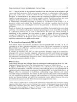
Fig. 9. Transition states and products of the uncatalyzed reaction of MeNCS and allene with
water.
cyclic TS (see Figure 9), whose strained structure explains the high activation barrier of the
reaction. Typical energy values for these structures can be found in Table 2. The resulting
products are also shown in Figure 9. The addition reactions of allene to the products 6 and 7
are both exergonic (see Table 2) and propene-2-ol 6 tautomerizes under standard conditions
to the more stable acetone. In case of isothiocyanates, the intermediates are still not exergonic,
but after surmounting some minor transition states, several co nformers of the exergonic car-
bamic thio acid can be reached (Eger et al., 2009).
To summarize this, the activation barriers of the uncatalyzed reactions of allenes and isoth-
iocyanates are very high, as they are four-membered cyclic transition states and therefore
possess Gibb’s free energies between 200 and 300 kJ/mol. Keeping the estimated activation
barriers of carbon dioxide and carbonyl sulfide in mind, it should be possible to see a signifi-
cant catalytic effect in the reactions of allenes and isothiocyanates.
5.2 The Selectivity Problem
Contrary to the case of carbon dioxide, allenes or isothiocyanates as educts for the nucle-
ophilic attack of hydroxide or water provide a more complex scenario. As a heterocumu-
lene, iso thiocyanate posses s nitrogen and oxygen on the outer p ositions of the cumulenic
system and additionally has an imine group, which reduces the symmetry of the molecule
and introduces more reaction possibilities (see Figure 9). Looking at allene, all known prob-
lems regarding alkenes and alkynes come to mind, thus chemo- (single or double addition),
regio- (Markovnikov- or anti-Markovnikov products) and stereoselectivity (cis- or trans-ad-
dition products on stereotopic sides) play a role. For substi tuted allenes there exists a posi-
tional selectivity (Hashmi, 2000) as the attack can take place at two different positions of the
allene molecule. Therefore, additions at one of the orthogonal double bonds will lead to con-
stitutional isomers in the case of substituted allenes and as a consequence, this inclusion of
regioselectivity d oubles the number of isomers.
6. Isothiocyanates (R-NCS), the Link to Synthesis
As described previously, the reaction of isothiocyanates with water and other H-X com-
pounds, i. e. alcohols and amines, is kinetically hindered. Water and alcohols do not react
educt MeNCS 1 allene 2
EC
a
uN-1
c
uA-1
d
23 22
TS
a
uN-2(ts)
c
uN-3(ts)
c
uN-4(ts)
c
uA-2(ts)
d
uA-3(ts)
d
220 210 206 263 293
1
X
2
CR
b
144
◦
142
◦
145
◦
157
◦
142
◦
1
X
4
H
3
O
b
117
◦
123
◦
114
◦
122
◦
121
◦
2
C
3
O
4
H
b
81
◦
74
◦
73
◦
69
◦
70
◦
1
X
2
C
3
O
4
H
b
7
◦
2
◦
4
◦
0
◦
0
◦
1
X
4
H
3
O
5
H
b
114
◦
105
◦
105
◦
180
◦
179
◦
1
X
2
C
b
1.716 Å 1.703 Å 1.300 Å 1.386 Å 1.392 Å
2
C
3
O
b
1.526 Å 1.683 Å 1.629 Å 1.833 Å 1.884 Å
3
O
4
H
b
1.179 Å 1.269 Å 1.175 Å 1.181 Å 1.190 Å
1
X
4
H
b
1.724 Å 1.605 Å 1.364 Å 1.449 Å 1.432 Å
product
a
3 4 5 6 7
49 71 -1 -92 -44
X
1
C
2
H
4
O
3
H
5
R
R
a
∆G in kJ/mol
b 1
X
2
C denote t he at tacked double bond, with X=C,N, O, S.
Depending on the selectivity of the reaction the residue R
could be H, CH
2
, NMe or S (see formula left).
c
Calculated at the MP2/aug-CC-pVTZ level of theory
d
Calculated at the mPW1k/aug-CC-pVDZ level of theory
Table 2. Energies and geometr ies of the uncatalyzed reaction of methylisothiocyanate and
allene with water.
under standard conditio ns, even when they are heated, it takes very long to see some prod-
uct (Browne & Dyson, 1931; Hagemann, 1983; Rao & Venkataraghavan, 1962; Walter & Bode,
1967). This is only true as long as there is no acid or base present, which would open up
other reaction possibilities . If the catalysis by a CA mode l is efficient, it would be the method
of choice to hydrolyze or alcoholyze iso thiocyanate systems under neutral conditions. This
might be interesting for the synthesis of complex and acid or base sensitive molecules.
In comparison to carbon dioxide and carbonyl sulfide, isothiocyanates bear a residue on one of
the outstanding hetero atoms. As this is an imine function, it increases the degree of freedom
and therefore produces more possible pathways.
X C Y
carbon dioxide X,Y = O -0.56 1.13 -0.56
carbon oxid sulfid X= O, Y = S -0.48 0.50 -0.01
methylisothiocyanate X = S, Y = N -0.10 0.30 -0.48
allene X,Y = C -0.51 0.07 -0.51
Table 3. Natural Charges δ
NC
for CO
2
, COS, MeNCS, and allene.
TheCarbonicAnhydraseasaParagon:
TheoreticalandExperimentalInvestigationofBiomimeticZinc-catalyzedActivationofCumulenes 181
C
CH
2
H
H
OH
H
CH
2
C
CH
3
O
H
N C
SH
O
H
CH
3
N C
SH
O
H
CH
3
N C
S
O H
CH
3
H
CC
C
H
H
H
H
H
O
H
C C
C
H
H
H
H
O
H
H
SC
N
CH
3
H
O
H
SC
N
H
O
H
CH
3
N C
S
CH
3
H
O
H
uN-2(ts) uN-3(ts)
uN-4(ts)
uA-2(ts)
uA-3(ts)
3
4
5
6
7
Fig. 9. Transition states and products of the uncatalyzed reaction of MeNCS and allene with
water.
cyclic TS (see Figure 9), whose strained structure explains the high activation barrier of the
reaction. Typical energy values for these structures can be found in Table 2. The resulting
products are also shown in Figure 9. The addition reactions of allene to the products 6 and 7
are both exergonic (see Table 2) and propene-2-ol 6 tautomerizes under standard conditions
to the more stable acetone. In case of isothiocyanates, the intermediates are still not exergonic,
but after surmounting some minor transition states, several co nformers of the exergonic car-
bamic thio acid can be reached (Eger et al., 2009).
To summarize this, the activation barriers of the uncatalyzed reactions of allenes and isoth-
iocyanates are very high, as they are four-membered cyclic transition states and therefore
possess Gibb’s free energies between 200 and 300 kJ/mol. Keeping the estimated activation
barriers of carbon dioxide and carbonyl sulfide in mind, it should be possible to see a signifi-
cant catalytic effect in the reactions of allenes and isothiocyanates.
5.2 The Selectivity Problem
Contrary to the case of carbon dioxide, allenes or isothiocyanates as educts for the nucle-
ophilic attack of hydroxide or water provide a more complex scenario. As a heterocumu-
lene, iso thiocyanate posses s nitrogen and oxygen on the outer p ositions of the cumulenic
system and additionally has an imine group, which reduces the symmetry of the molecule
and introduces more reaction possibilities (see Figure 9). Looking at allene, all known prob-
lems regarding alkenes and alkynes come to mind, thus chemo- (single or double addition),
regio- (Markovnikov- or anti-Markovnikov products) and stereoselectivity (cis- or trans-ad-
dition products on stereotopic sides) play a role. For substi tuted allenes there exists a posi-
tional selectivity (Hashmi, 2000) as the attack can take place at two different positions of the
allene molecule. Therefore, additions at one of the orthogonal double bonds will lead to con-
stitutional isomers in the case of substituted allenes and as a consequence, this inclusion of
regioselectivity d oubles the number of isomers.
6. Isothiocyanates (R-NCS), the Link to Synthesis
As described previously, the reaction of isothiocyanates with water and other H-X com-
pounds, i. e. alcohols and amines, is kinetically hindered. Water and alcohols do not react
educt MeNCS 1 allene 2
EC
a
uN-1
c
uA-1
d
23 22
TS
a
uN-2(ts)
c
uN-3(ts)
c
uN-4(ts)
c
uA-2(ts)
d
uA-3(ts)
d
220 210 206 263 293
1
X
2
CR
b
144
◦
142
◦
145
◦
157
◦
142
◦
1
X
4
H
3
O
b
117
◦
123
◦
114
◦
122
◦
121
◦
2
C
3
O
4
H
b
81
◦
74
◦
73
◦
69
◦
70
◦
1
X
2
C
3
O
4
H
b
7
◦
2
◦
4
◦
0
◦
0
◦
1
X
4
H
3
O
5
H
b
114
◦
105
◦
105
◦
180
◦
179
◦
1
X
2
C
b
1.716 Å 1.703 Å 1.300 Å 1.386 Å 1.392 Å
2
C
3
O
b
1.526 Å 1.683 Å 1.629 Å 1.833 Å 1.884 Å
3
O
4
H
b
1.179 Å 1.269 Å 1.175 Å 1.181 Å 1.190 Å
1
X
4
H
b
1.724 Å 1.605 Å 1.364 Å 1.449 Å 1.432 Å
product
a
3 4 5 6 7
49 71 -1 -92 -44
X
1
C
2
H
4
O
3
H
5
R
R
a
∆G in kJ/mol
b 1
X
2
C denote t he at tacked double bond, with X=C,N, O, S.
Depending on the selectivity of the reaction the residue R
could be H, CH
2
, NMe or S (see formula left).
c
Calculated at the MP2/aug-CC-pVTZ level of theory
d
Calculated at the mPW1k/aug-CC-pVDZ level of theory
Table 2. Energies and geometr ies of the uncatalyzed reaction of methylisothiocyanate and
allene with water.
under standard conditio ns, even when they are heated, it takes very long to see some prod-
uct (Browne & Dyson, 1931; Hagemann, 1983; Rao & Venkataraghavan, 1962; Walter & Bode,
1967). This is only true as long as there is no acid or base present, which would open up
other reaction possibilities . If the catalysis by a CA mode l is efficient, it would be the method
of choice to hydrolyze or alcoholyze iso thiocyanate systems under neutral conditions. This
might be interesting for the synthesis of complex and acid or base sensitive molecules.
In comparison to carbon dioxide and carbonyl sulfide, isothiocyanates bear a residue on one of
the outstanding hetero atoms. As this is an imine function, it increases the degree of freedom
and therefore produces more possible pathways.
X C Y
carbon dioxide X,Y = O -0.56 1.13 -0.56
carbon oxid sulfid X= O, Y = S -0.48 0.50 -0.01
methylisothiocyanate X = S, Y = N -0.10 0.30 -0.48
allene X,Y = C -0.51 0.07 -0.51
Table 3. Natural Charges δ
NC
for CO
2
, COS, MeNCS, and allene.
Biomimetics,LearningfromNature182
Zn
O
L
L
L
H
S
N
CH
3
Zn
O
L
L
L
H
S
N
CH
3
Zn
O
L
L
L
H
N
S
CH
3
NCS-a(ts); 82 NCS-b(ts); 89 NCS-c(ts); 97
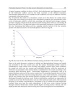
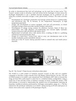
Fig. 10. Rate determining steps in the catalyzed reaction with methyl isothiocyanate. Level of
theory is B3LYP/6-311+G(d,p), given values are Gibb’s free energies ∆G in kJ /mol.
6.1 Calculated Mechanistic Pathway
The calculations show only one encounter complex NCS-1, in which the isothiocyanate coor-
dinates via the sulfur atom to two ammonia ligands using hydrogen bridging bonds. Coming
from this encounter complex, three different transition states could take place. Whereas in
NCS-1(ts) and NCS-2(ts) the C=S do uble bond adds to the Zn-O bond, the C=N double bond
does this in the case of NCS-3(ts) (see Figure 10). These transition states resemble the rate de-
termining steps in the reactions o f carbon dioxid e and carbonyl sulfide and also are the highest
activation barriers in the pathway of i sothiocyanate. Contrary to the situation in case of COS,
which also possesses an unsymmetric cumulenic system, the energies of this transition states
differ not significantly, so a prediction of selectivity dep ends not only on the energies of the
rate determining steps, but also on the further reaction paths and thermodynamic control.
Comparing the free enthalpies of the three transition states and the energies of the following
reaction paths, it becomes obvious, that the attack on the C=S double bond is thermodynam-
ically and kinetically slig htly favored. Contrary to the fact, that the existence of the imine
function makes the situation at the rate determining step more complex, it simplifies it at the
point, where the Lindskog and Lipscomb transition states enter the scenery right after the at-
tack of the C=S double bond. As the disturbed symmetry of isothiocyanate opens up about
eight possible pathways, the kinetically and thermodynamically most favorable will be dis-
cussed shortly here (see Figure 11).
Structure NCS-2(ts) is the rate determining step, as no other transition state builds up a
higher activation barrier. ∆G = 82 kJ/mol relative to the separated educts (ammonia model
and methyl isothiocyanate), is not as good as the corresponding values estimated for carbon
dioxide and carbonyl sulfide, but it is easily surmountable in a normal experimental environ-
ment. The catalytic effect becomes ver y clear, when comparing the activation barriers of the
rate determining steps in the catalyzed and uncatalyzed reaction, as the gap between these
values is about ∆∆G = 76 kJ/mol. This is a significant decrease in energy. The reaction path
proceeds further via a Lindskog reaction mechanism (NCS-4(ts)), which is rather lower than
the corresponding Lipscomb proton shift. Nevertheless, the pathway surmounting NCS-4(ts)
is the thermodynamically and kinetically favored one.
The found selectivity is only true for the reaction with methyl isothiocyanate, as calculation
with several residues showed different results. In general, the inductive effect of the residue
of the isothiocyanate changes the selectivity. The greater the ability of the residue to pull
electrons out o f the cumulenic system, the more an attack of the C=N double bond is preferred.
This is mainly a result of the electronic structure in the cumulenic system. If the residue on the
nitrogen atom pulls electron density out of the double bond system, it is mainly taken from
O
H
H
Zn
N
L
L
L
S
O H
CH
3
Zn
N
L
L
L
S
O
H
CH
3
Zn
N
L
L
L
S
O
H
CH
3
O
H
H
Zn
N
L
L
L
S
O
H
CH
3
O
H
H
Zn
N
L
L
L
S
O
H
CH
3
Zn
O
L
L
L
H
S
N
CH
3
Zn
O
L
L
L
H
S
N
CH
3
Zn
N
L
L
L
S
O
H
CH
3
Zn
N
L
L
L
S
O
H
CH
3
O
H
H
O
H
Zn
L
L
L
H N
S
O
H
CH
3
Zn
O
L
L
L
H
S
N
CH
3
Zn
O
L
L
L
H
S
N
CH
3
NCS-1; 2
NCS-2(ts); 82
NCS-3; 24
NCS-4(ts); 40
NCS-5; 0
NCS-6(ts); 17
NCS-7; -14
NCS-8; -22
NCS-9(ts); -14
NCS-10; -34
NCS-11(ts); -29
NCS-12; -34
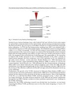
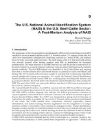
Fig. 11. Pathway of the catalyzed reaction with methyl isothiocyanate. Level of theory is
B3LYP/6-311+G(d,p), given values are Gibb’s free enthalpies in kJ/mol.
the C=S double bond. Thus NBO calculations show, that in such cases the C=N double bond
has a strong triple, and the C=S double bond a strong single bond character (Eger et al., 2009).
Further calculations with complexe s not bearing a hydroxide ion but an hydrosulfide and an
thiolate ion respectively, showed, that the biomimetics of CA are not only limited to hydroxide
bearing complexes and thus the add ition of water to cumulenic system. Furthermore a lot of
different combinations of different nucleophiles and cumulenes are possible.
6.2 Experimental Results
As the reaction with a thiolate complex reduces the number of possible pathways signifi-
cantly and those complexes recently proved their ability simulating CA biomimetic insertion
reactions (e. g. with carbon disulfide) (Notni et al., 2006), this seems to be a goo d model com-
plex to see, if isothiocyanate inserts even similar. Thiolate complexes bearing a four-dentate
[12]aneN
4
ligand are known to work faster than the corresponding three-dentate complexed
compounds (Notni, Günther & Anders, 2007).
TheCarbonicAnhydraseasaParagon:
TheoreticalandExperimentalInvestigationofBiomimeticZinc-catalyzedActivationofCumulenes 183
Zn
O
L
L
L
H
S
N
CH
3
Zn
O
L
L
L
H
S
N
CH
3
Zn
O
L
L
L
H
N
S
CH
3
NCS-a(ts); 82 NCS-b(ts); 89 NCS-c(ts); 97
Fig. 10. Rate determining steps in the catalyzed reaction with methyl isothiocyanate. Level of
theory is B3LYP/6-311+G(d,p), given values are Gibb’s free energies ∆G in kJ /mol.
6.1 Calculated Mechanistic Pathway
The calculations show only one encounter complex NCS-1, in which the isothiocyanate coor-
dinates via the sulfur atom to two ammonia ligands using hydrogen bridging bonds. Coming
from this encounter complex, three different transition states could take place. Whereas in
NCS-1(ts) and NCS-2(ts) the C=S do uble bond adds to the Zn-O bond, the C=N double bond
does this in the case of NCS-3(ts) (see Figure 10). These transition states resemble the rate de-
termining steps in the reactions o f carbon dioxid e and carbonyl sulfide and also are the highest
activation barriers in the pathway of i sothiocyanate. Contrary to the situation in case of COS,
which also possesses an unsymmetric cumulenic system, the energies of this transition states
differ not significantly, so a prediction of selectivity dep ends not only on the energies of the
rate determining steps, but also on the further reaction paths and thermodynamic control.
Comparing the free enthalpies of the three transition states and the energies of the following
reaction paths, it becomes obvious, that the attack on the C=S double bond is thermodynam-
ically and kinetically slig htly favored. Contrary to the fact, that the existence of the imine
function makes the situation at the rate determining step more complex, it simplifies it at the
point, where the Lindskog and Lipscomb transition states enter the scenery right after the at-
tack of the C=S double bond. As the disturbed symmetry of isothiocyanate opens up about
eight possible pathways, the kinetically and thermodynamically most favorable will be dis-
cussed shortly here (see Figure 11).
Structure NCS-2(ts) is the rate determining step, as no other transition state builds up a
higher activation barrier. ∆G = 82 kJ/mol relative to the separated educts (ammonia model
and methyl isothiocyanate), is not as good as the corresponding values estimated for carbon
dioxide and carbonyl sulfide, but it is easily surmountable in a normal experimental environ-
ment. The catalytic effect becomes ver y clear, when comparing the activation barriers of the
rate determining steps in the catalyzed and uncatalyzed reaction, as the gap between these
values is about ∆∆G = 76 kJ/mol. This is a significant decrease in energy. The reaction path
proceeds further via a Lindskog reaction mechanism (NCS-4(ts)), which is rather lower than
the corresponding Lipscomb proton shift. Nevertheless, the pathway surmounting NCS-4(ts)
is the thermodynamically and kinetically favored one.
The found selectivity is only true for the reaction with methyl isothiocyanate, as calculation
with several residues showed different results. In general, the inductive effect of the residue
of the isothiocyanate changes the selectivity. The greater the ability of the residue to pull
electrons out o f the cumulenic system, the more an attack of the C=N double bond is preferred.
This is mainly a result of the electronic structure in the cumulenic system. If the residue on the
nitrogen atom pulls electron density out of the double bond system, it is mainly taken from
O
H
H
Zn
N
L
L
L
S
O H
CH
3
Zn
N
L
L
L
S
O
H
CH
3
Zn
N
L
L
L
S
O
H
CH
3
O
H
H
Zn
N
L
L
L
S
O
H
CH
3
O
H
H
Zn
N
L
L
L
S
O
H
CH
3
Zn
O
L
L
L
H
S
N
CH
3
Zn
O
L
L
L
H
S
N
CH
3
Zn
N
L
L
L
S
O
H
CH
3
Zn
N
L
L
L
S
O
H
CH
3
O
H
H
O
H
Zn
L
L
L
H N
S
O
H
CH
3
Zn
O
L
L
L
H
S
N
CH
3
Zn
O
L
L
L
H
S
N
CH
3
NCS-1; 2
NCS-2(ts); 82
NCS-3; 24
NCS-4(ts); 40
NCS-5; 0
NCS-6(ts); 17
NCS-7; -14
NCS-8; -22
NCS-9(ts); -14
NCS-10; -34
NCS-11(ts); -29
NCS-12; -34
Fig. 11. Pathway of the catalyzed reaction with methyl isothiocyanate. Level of theory is
B3LYP/6-311+G(d,p), given values are Gibb’s free enthalpies in kJ/mol.
the C=S double bond. Thus NBO calculations show, that in such cases the C=N double bond
has a strong triple, and the C=S double bond a strong single bond character (Eger et al., 2009).
Further calculations with complexe s not bearing a hydroxide ion but an hydrosulfide and an
thiolate ion respectively, showed, that the biomimetics of CA are not only limited to hydroxide
bearing complexes and thus the add ition of water to cumulenic system. Furthermore a lot of
different combinations of different nucleophiles and cumulenes are possible.
6.2 Experimental Results
As the reaction with a thiolate complex reduces the number of possible pathways signifi-
cantly and those complexes recently proved their ability simulating CA biomimetic insertion
reactions (e. g. with carbon disulfide) (Notni et al., 2006), this seems to be a goo d model com-
plex to see, if isothiocyanate inserts even similar. Thiolate complexes bearing a four-dentate
[12]aneN
4
ligand are known to work faster than the corresponding three-dentate complexed
compounds (Notni, Günther & Anders, 2007).
Biomimetics,LearningfromNature184
Zn
S
L
L
L
R
L
S
N
R
Zn
L
L
L
L
S
S
R
N
R
Zn
L
L
L
L
N
S
R
S
R
eplacements
[Zn([12]aneN
4
)SR]
+
NCS
C=S addition C=N addi tio n
+
and
+ HX
Fig. 12. Insertion possibilities of isothiocyanate to a zinc thiolate complex.
The reaction shown in Figure 12 was carried out in dimethyl sulf oxide under standard con-
ditions at room temperature. The insertion could be proved using GC/MS and Raman spec-
troscopy. For different isothiocyanates different reaction rates could be determined, as mostly
isothiocyanates with an electron withdrawing residue as phenyl or p-nitro phenyl were able
to insert easily at room temperature. Depending on the purpose of the reaction those activated
cumulenes can react further with an HX compound, e. g. an alcohol or mercaptan.
7. Allene
Allene is the simplest hydrocarbon with cumulated double bonds. Since van’t Hoff has pre-
dicted the correct structures of allene and higher cumulenes, chemists are fascinated by the
extraordinary properties like axial chirality of the elongated tetrahedron, if two different sub-
stituents at every terminal carbon exist. Allene with its isomer methyl acetylene accrues in
large amounts in the C3-cut of the naphtha distill ation. Currently both compounds are only
hydrated to propene and propane respectively or flared off. Therefore the activation of allene
has additionally to the biomimetic a strong economical aspect.
Allene could be estimated as the parent compound for heterocumulenes with two cumulated
double bonds . By the formal exchange of one o r both terminal carbon atoms a vast number of
heterocumulenes are available.
The first investigation of a possible biomimetic activation of allene with zinc catalysts was
undertaken by Breuer et al. (1999). They found catalytical activity of zinc silicates with zinc
acetate in me thanol to give 2-methoxypropene and 2,2-dimethoxypropene in 85 % yield .
7.1 Calculated Mechanistic Pathway
The presentation of the whole calculated reaction mechanism of the addition of water to al-
lene goes beyond the scope of this chapter due to the immense number of reaction steps (see
(Jahn et al., 2008) for further reading). Therefore the description of mechanistical pathways is
confined to the variants of the initial nucleophili c attack, which lead to mechanistical impor-
tant intermediates. The results show that the initial attack is the rate determining step for the
whole catalytic cycle.
The zinc catalyzed addition starts with an encounter complex A-1 between the zinc hydroxide
complex and allene. This structure is the starting point for the different reaction variants, com-
parable to the uncatalyzed reaction described in section 5.1. Corresponding to the regio selec-
tivity problem the attack to alle ne can take place at either the central or the terminal carbon
atom (see Figure 13). The attack of the hydroxide on the terminal carbons leads to a concerted
four-membered cyclic transition state A-2(ts) with an activation barrier of ∆G = 139 kJ/mol.
H
H
H
H
Zn
O
L
L
L
H
Zn
O
L
L
L
H
Zn
O
L
N
L
H
C C
C
H
H
H
H
H
Zn
O
L
N
L
H
C C
C
H
H
H
H
H
Zn
O
L
L
L
H
H
H
H
H
Zn
L
L
L
CH
2
H
H
OH
Zn
O
L
L
L
H
H
H
H
H
Zn
L
L
L
H
H
OH
H
H
H
O
H
Zn
L
L
L
H
H
OH
H
H
L
O
Zn
L
L
CH
2
CH
3
AG
AH
Z
allene
A-1; 15
A-2(ts); 139
A-3; -20
A-5(ts); 124
A-9; -57
H
2
O
H
2
O
H
2
O
A-4(ts); 82
7; -44
A-7; -120
6; -92
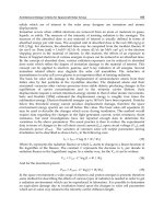
Fig. 13. Calculated mechanism of the initial, rate determining steps of the activation of allene.
∆G in kJ/mol. Level of theory is mPW1k/aug-CC-pVDZ.
This structure relaxes to the C
2v
-symmetric, slightly exergonic intermediate A-3, in which the
carbon backbone, the hydroxyl group, the metal ion and one nitrogen of the ligand span the
symmetry plane. The hydroxyl group is placed between and in front of the ligands. There is
only one possibility to close the catalytic cycle starting from intermediate A-3. This mecha-
nism is an attack of a water molecule, which leads to a cleavage of the Zn-C bond. One water
proton is shifted to the central carbon atom to give allylalcohol 7 and the remaining hydroxide
regenerates the catalytic model.
TheCarbonicAnhydraseasaParagon:
TheoreticalandExperimentalInvestigationofBiomimeticZinc-catalyzedActivationofCumulenes 185
Zn
S
L
L
L
R
L
S
N
R
Zn
L
L
L
L
S
S
R
N
R
Zn
L
L
L
L
N
S
R
S
R
eplacements
[Zn([12]aneN
4
)SR]
+
NCS
C=S addition C=N addi tio n
+
and
+ HX
Fig. 12. Insertion possibilities of isothiocyanate to a zinc thiolate complex.
The reaction shown in Figure 12 was carried out in dimethyl sulf oxide under standard con-
ditions at room temperature. The insertion could be proved using GC/MS and Raman spec-
troscopy. For different isothiocyanates different reaction rates could be determined, as mostly
isothiocyanates with an electron withdrawing residue as phenyl or p-nitro phenyl were able
to insert easily at room temperature. Depending on the purpose of the reaction those activated
cumulenes can react further with an HX compound, e. g. an alcohol or mercaptan.
7. Allene
Allene is the simplest hydrocarbon with cumulated double bonds. Since van’t Hoff has pre-
dicted the correct structures of allene and higher cumulenes, chemists are fascinated by the
extraordinary properties like axial chirality of the elongated tetrahedron, if two different sub-
stituents at every terminal carbon exist. Allene with its isomer methyl acetylene accrues in
large amounts in the C3-cut of the naphtha distill ation. Currently both compounds are only
hydrated to propene and propane respectively or flared off. Therefore the activation of allene
has additionally to the biomimetic a strong economical aspect.
Allene could be estimated as the parent compound for heterocumulenes with two cumulated
double bonds . By the formal exchange of one o r both terminal carbon atoms a vast number of
heterocumulenes are available.
The first investigation of a possible biomimetic activation of allene with zinc catalysts was
undertaken by Breuer et al. (1999). They found catalytical activity of zinc silicates with zinc
acetate in me thanol to give 2-methoxypropene and 2,2-dimethoxypropene in 85 % yield .
7.1 Calculated Mechanistic Pathway
The presentation of the whole calculated reaction mechanism of the addition of water to al-
lene goes beyond the scope of this chapter due to the immense number of reaction steps (see
(Jahn et al., 2008) for further reading). Therefore the description of mechanistical pathways is
confined to the variants of the initial nucleophili c attack, which lead to mechanistical impor-
tant intermediates. The results show that the initial attack is the rate determining step for the
whole catalytic cycle.
The zinc catalyzed addition starts with an encounter complex A-1 between the zinc hydroxide
complex and allene. This structure is the starting point for the different reaction variants, com-
parable to the uncatalyzed reaction described in section 5.1. Corresponding to the regio selec-
tivity problem the attack to alle ne can take place at either the central or the terminal carbon
atom (see Figure 13). The attack of the hydroxide on the terminal carbons leads to a concerted
four-membered cyclic transition state A-2(ts) with an activation barrier of ∆G = 139 kJ/mol.
H
H
H
H
Zn
O
L
L
L
H
Zn
O
L
L
L
H
Zn
O
L
N
L
H
C C
C
H
H
H
H
H
Zn
O
L
N
L
H
C C
C
H
H
H
H
H
Zn
O
L
L
L
H
H
H
H
H
Zn
L
L
L
CH
2
H
H
OH
Zn
O
L
L
L
H
H
H
H
H
Zn
L
L
L
H
H
OH
H
H
H
O
H
Zn
L
L
L
H
H
OH
H
H
L
O
Zn
L
L
CH
2
CH
3
AG
AH
Z
allene
A-1; 15
A-2(ts); 139
A-3; -20
A-5(ts); 124
A-9; -57
H
2
O
H
2
O
H
2
O
A-4(ts); 82
7; -44
A-7; -120
6; -92
Fig. 13. Calculated mechanism of the initial, rate determining steps of the activation of allene.
∆G in kJ/mol. Level of theory is mPW1k/aug-CC-pVDZ.
This structure relaxes to the C
2v
-symmetric, slightly exergonic intermediate A-3, in which the
carbon backbone, the hydroxyl group, the metal ion and one nitrogen of the ligand span the
symmetry plane. The hydroxyl group is placed between and in front of the ligands. There is
only one possibility to close the catalytic cycle starting from intermediate A-3. This mecha-
nism is an attack of a water molecule, which leads to a cleavage of the Zn-C bond. One water
proton is shifted to the central carbon atom to give allylalcohol 7 and the remaining hydroxide
regenerates the catalytic model.
Biomimetics,LearningfromNature186
L
L
L
Zn
OH
CH
2
H
H
L
L
L
Zn
OH
CH
2
H
H
L
L
L
Zn
CH
2
OH
H
H
L
L
L
Zn
CH
2
OH
H
H
L
L
L
Zn
OH
CH
2
H
H
L
L
L
Zn
OH
CH
2
H
H
rotTS-I; -41
(pR-)A-9; -57
(pS-)A-9; -57
rotTS-II; -29
(pR-)A-9
(pS-)A-9
mirror plane
Fig. 14. Mechanism of the racemization of A-9 via rotTS-I and rotTS-II. ∆G in kJ/mol. Level
of theory is mPW1k/aug-CC-pVDZ.
Alternatively, the initial nucleophilic attack on the CA model comp lex can take place at the
central carbon atom. Depending on the kind o f the model complex, two different mechanisms
of the initial reaction step can be found. This reaction path can either proceed via a stepwise
or a concerted reaction mechanism, whereas the stepwise mechanism can only be found using
the azamacrocyclic models. Contrary, the concerted one can be fo und in all cases. This shows
the restrictions of the ammonia model.
Structure A-5(ts) is the first transition state of the stepwise variant. The activation barrier is
∆∆G = 18 kJ/mol) higher than for the concerted TS A-8(ts), which is interesting, as this TS
has no ster ical restrictions. As its carbon backbone stands approximatively perpendicular
to the Zn-O bond, structure A-5(ts) differs fundamentally in its geometry compared to the
cyclic concerted TSs. The reaction coordinate is only defined by the difference of the distance
between oxygen and the central carbon atom of allene. The TS relaxes to the intermediate A-6.
With ∆G = 113 kJ/mol relative to the Gibb’s free energy of the separated reactants allene and
zinc hydroxide complex, this intermediate is only poorly stable. Intermediate A-6 rearranges
by a cascade of proton transfer steps between the substrate and the ligand to the intermediate
A-7, which is one of the most stable structures in the calculated reaction path variants (∆G = -
120 kJ/mol). Subsequently, the direct formation of acetone is facilitated by a proton shift from
an attacking water molecule to the free methylidene group.
The third and most probable transition state between allene and the CA model complex is
the concerted four-membered cyclic T S A-8(ts). Comparably to me thylisothiocyanate, A-8(ts)
resembles the rate determining step in the reactions of carbon dioxide and carbonyl sulfide. A-
8(ts) possesses the lowest activation barrier of all three initial TSs (∆G = 124 kJ/mol). It finally
relaxes into the intermediate A-9.
Contrary to all other intermediates of different heterocumulenes at comparable points of the
reaction coordinate, structure A-9 has an outmost geometry. Whereas in all geometries of in-
termediates connected to the zinc ion by a heteroatom the former cumulated system and the
metal ion are located in a plane, intermed iate A-9 has a carbon atom connected to the zinc
instead, which forces the plane spanned by the carbon backbone of the allene to stand per-
pendicular to the Zn-C bond and parallel to the plane spanned by the lig and respectively. A
reason for that is the partial double bond character of the bonding between the central and
the zinc-bound carbon atoms. As a consequence, A-9 is a chiral structure without an asym-
metric center and therefore an example of planar chirality. Ho wever, the activation barrier of
the racemization TS is not high enough to ensure a separation of the enantiomers (pR-)A-9
and (pS-)A-9 ( see Figure 14). Isomerization around the single bond between zinc and the zinc
bound carbon can occur clockwise or counter-clockwise. As a result, two rotational transi-
tion states exist (rotTS-I and rotTS-II). TS rotTS-I is slightly preferred, as hydroge n bridg-
ing bonds between the hydroxyl group and the ligand lower the energy. Comparing their
geometries, the propos ed analogous transition state for catalytic cycle of the CO
2
hydration
(Mauksch et al., 2001) and the transition state A-8(ts) are quite similar. In contrast to A-9,
the following so-called Lindskog-type intermediate possesses a C
2v
symmetry like rotTS-I.
The geometry of intermediate A-9 is comparable to the Lindskog-type rotational TS, which
leads to the Lipscomb product. T he latter is a geometrical equivalent to rotTS-II. D ue to the
different geometry, an alternative way like the Lipscomb mechanism (proton shift) (Liang &
Lipscomb, 1987; Lipscomb, 1983) appears to be impossible for intermediate A-9.
Intermediate A-9 could be identified as the the key intermediate for the further possible reac-
tion paths. Starting from here, hydrolysis recreates allene and the CA model complex, whereas
another pathway directly leads to acetone. The catalytic product of all remaining possible
pathways is 6. Thus the water attack can take place on the methylidene group with and with-
out a preceding rotation of the hydroxyl group. Further, an i ntramolecular proton shift from
the hydroxyl to the methylidene group under generation of a carbonyl and methyl group is
another possible pathway. The carbonyl group can also be attacked by a water molecule. Al-
ternatively, a coordination change from the oxygen to the zinc bound carbon can occur. This
step generates the stable structure A-7, which is als o accessible from the initial stepwise mech-
anism.
7.2 Experimental Results
The reaction of allene and [Zn([12]aneN
3
)OH]ClO
4
as the CA model complex was investi-
gated under heterogeneous conditions. Due to the gaseous aggregation state of the unsub-
stituted allene, a pessure cell was used. The analysis was done with Raman spectroscopic
methods.
8. Conclusion
In summary, we have shown that the transformation of COS by carbonic anhydrase, which
finally yields H
2
S and CO
2
, requires no further reactant than water in order to regenerate
the most important zinc-bound hydroxide [L
3
ZnOH]
+
from the hydrosulfide complex. We
conclude that CA is perfectly equipped by nature to perform the task of transformation of
COS into H
2
S. F urthermore, we regard this special function of CA to be perfectly linked to
the plant sulfur metabolism. Therefore, this regeneration mechanism can be regarded as the
missing link between CA-catalyzed COS fixation and plant sulfur metabolism; an aspect of
fundamental significance for the understanding of some very important biological processes.
Nature has chosen an elegant and efficient system for the hydration of CO
2
and COS, the
[L
3
ZnOH]
+
/CO
2
or COS/H
2
O group of reactants. The catalyst is able to transform both
cumulenes, though the relative energies of the corresponding reactions steps differ in some
details significantly. Further we have shown that it is possible to apply biomime tic princi-
ples of high optimized, biochemical processes to the laboratory as well as industriall y usable
syntheses. The reaction principle of carbonic anhydrase is applicable to other isoelectronic
molecules than CO
2
, which are normally not processed by the enzyme. These biomimetic in-
vestigations about the enzyme carbonic anhydrase could serve as a paragon for the further
research on biochemical model systems .
TheCarbonicAnhydraseasaParagon:
TheoreticalandExperimentalInvestigationofBiomimeticZinc-catalyzedActivationofCumulenes 187
L
L
L
Zn
OH
CH
2
H
H
L
L
L
Zn
OH
CH
2
H
H
L
L
L
Zn
CH
2
OH
H
H
L
L
L
Zn
CH
2
OH
H
H
L
L
L
Zn
OH
CH
2
H
H
L
L
L
Zn
OH
CH
2
H
H
rotTS-I; -41
(pR-)A-9; -57
(pS-)A-9; -57
rotTS-II; -29
(pR-)A-9
(pS-)A-9
mirror plane
Fig. 14. Mechanism of the racemization of A-9 via rotTS-I and rotTS-II. ∆G in kJ/mol. Level
of theory is mPW1k/aug-CC-pVDZ.
Alternatively, the initial nucleophilic attack on the CA model comp lex can take place at the
central carbon atom. Depending on the kind o f the model complex, two different mechanisms
of the initial reaction step can be found. This reaction path can either proceed via a stepwise
or a concerted reaction mechanism, whereas the stepwise mechanism can only be found using
the azamacrocyclic models. Contrary, the concerted one can be fo und in all cases. This shows
the restrictions of the ammonia model.
Structure A-5(ts) is the first transition state of the stepwise variant. The activation barrier is
∆∆G = 18 kJ/mol) higher than for the concerted TS A-8(ts), which is interesting, as this TS
has no ster ical restrictions. As its carbon backbone stands approximatively perpendicular
to the Zn-O bond, structure A-5(ts) differs fundamentally in its geometry compared to the
cyclic concerted TSs. The reaction coordinate is only defined by the difference of the distance
between oxygen and the central carbon atom of allene. The TS relaxes to the intermediate A-6.
With ∆G = 113 kJ/mol relative to the Gibb’s free energy of the separated reactants allene and
zinc hydroxide complex, this intermediate is only poorly stable. Intermediate A-6 rearranges
by a cascade of proton transfer steps between the substrate and the ligand to the intermediate
A-7, which is one of the most stable structures in the calculated reaction path variants (∆G = -
120 kJ/mol). Subsequently, the direct formation of acetone is facilitated by a proton shift from
an attacking water molecule to the free methylidene group.
The third and most probable transition state between allene and the CA model complex is
the concerted four-membered cyclic TS A-8(ts). Comparably to methylisothiocyanate, A-8(ts)
resembles the rate determining step in the reactions of carbon dioxide and carbonyl sulfide. A-
8(ts) possesses the lowest activation barrier of all three initial TSs (∆G = 124 kJ/mol). It finally
relaxes into the intermediate A-9.
Contrary to all other intermediates of different heterocumulenes at comparable points of the
reaction coordinate, structure A-9 has an outmost geometry. Whereas in all geometries of in-
termediates connected to the zinc ion by a heteroatom the former cumulated system and the
metal ion are located in a plane, intermed iate A-9 has a carbon atom connected to the zinc
instead, which forces the plane spanned by the carbon backbone of the allene to stand per-
pendicular to the Zn-C bond and parallel to the plane spanned by the lig and respectively. A
reason for that is the partial double bond character of the bonding between the central and
the zinc-bound carbon atoms. As a consequence, A-9 is a chiral structure without an asym-
metric center and therefore an example of planar chirality. Ho wever, the activation barrier of
the racemization TS is not high enough to ensure a separation of the enantiomers (pR-)A-9
and (pS-)A-9 ( see Figure 14). Isomerization around the single bond between zinc and the zinc
bound carbon can occur clockwise or counter-clockwise. As a result, two rotational transi-
tion states exist (rotTS-I and rotTS-II). TS rotTS-I is slightly preferred, as hydroge n bridg-
ing bonds between the hydroxyl group and the ligand lower the energy. Comparing their
geometries, the propos ed analogous transition state for catalytic cycle of the CO
2
hydration
(Mauksch et al., 2001) and the transition state A-8(ts) are quite similar. In contrast to A-9,
the following so-called Lindskog-type intermediate possesses a C
2v
symmetry like rotTS-I.
The geometry of intermediate A-9 is comparable to the Lindskog-type rotational TS, which
leads to the Lipscomb product. T he latter is a geometrical equivalent to rotTS-II. D ue to the
different geometry, an alternative way like the Lipscomb mechanism (proton shift) (Liang &
Lipscomb, 1987; Lipscomb, 1983) appears to be impossible for intermediate A-9.
Intermediate A-9 could be identified as the the key intermediate for the further possible reac-
tion paths. Starting from here, hydrolysis recreates allene and the CA model complex, whereas
another pathway directly leads to acetone. The catalytic product of all remaining possible
pathways is 6. Thus the water attack can take place on the methylidene group with and with-
out a preceding rotation of the hydroxyl group. Further, an i ntramolecular proton shift from
the hydroxyl to the methylidene group under generation of a carbonyl and methyl group is
another possible pathway. The carbonyl group can also be attacked by a water molecule. Al-
ternatively, a coordination change from the oxygen to the zinc bound carbon can occur. This
step generates the stable structure A-7, which is als o accessible from the initial stepwise mech-
anism.
7.2 Experimental Results
The reaction of allene and [Zn([12]aneN
3
)OH]ClO
4
as the CA model complex was investi-
gated under heterogeneous conditions. Due to the gaseous aggregation state of the unsub-
stituted allene, a pessure cell was used. The analysis was done with Raman spectroscopic
methods.
8. Conclusion
In summary, we have shown that the transformation of COS by carbonic anhydrase, which
finally yields H
2
S and CO
2
, requires no further reactant than water in order to regenerate
the most important zinc-bound hydroxide [L
3
ZnOH]
+
from the hydrosulfide complex. We
conclude that CA is perfectly equipped by nature to perform the task of transformation of
COS into H
2
S. F urthermore, we regard this special function of CA to be perfectly linked to
the plant sulfur metabolism. Therefore, this regeneration mechanism can be regarded as the
missing link between CA-catalyzed COS fixation and plant sulfur metabolism; an aspect of
fundamental significance for the understanding of some very important biological processes.
Nature has chosen an elegant and efficient system for the hydration of CO
2
and COS, the
[L
3
ZnOH]
+
/CO
2
or COS/H
2
O group of reactants. The catalyst is able to transform both
cumulenes, though the relative energies of the corresponding reactions steps differ in some
details significantly. Further we have shown that it is possible to apply biomime tic princi-
ples of high optimized, biochemical processes to the laboratory as well as industriall y usable
syntheses. The reaction principle of carbonic anhydrase is applicable to other isoelectronic
molecules than CO
2
, which are normally not processed by the enzyme. These biomimetic in-
vestigations about the enzyme carbonic anhydrase could serve as a paragon for the further
research on biochemical model systems .
Biomimetics,LearningfromNature188
Acknowledgement
These investigations are part of the gener al research field of the Collaborative Research Centre
Metal Mediated Reactions Model ed after Nature (CRC 436, University of Jena, Germany, since
1997 though 2006 supported by the Deutsche Forschungsgemeinschaft).
9. References
Barnett, D. H., Sheng, S., Howe Charn, T., Waheed, A., Sly, W. S., Lin, C Y., Liu, E. T. &
Katzenellenbogen, B. S. (2008). Estrogen Receptor Regulation of Carbonic Anhydrase
XII through a D istal Enhancer in Breast Cancer, Cancer Research 68(9): 3505–3515.
Bergquist, C., Fillebeen, T., Morlok, M. M. & Parkin, G. (2003). Protonation and Reactivity
towards Carbon Diox ide of the Mononuclear Tetrahedral Zinc and Cobalt Hydrox-
ide Complexes, [Tp
But,Me
]ZnOH and [Tp
But,Me
]CoOH: Comparison of the Reactivity
of the Metal Hydroxide Function in Synthetic Analogues of Carbonic Anhydrase,
Journal of the American Chemical Society 125(20): 6189–6199.
Bertini, I., Dei, A., Luchinat, C. & Monnanni, R. (1985). Acid-Base Properties of Cobalt(II)-
Substituted Carbonic Anhydrases, Inorganic Chemistry 24(3): 301–303.
Bertran, J., Sola, M., Lledos, A. & Duran, M. (1992). Ab Initio Study of the Hydration of CO
2
by Carbonic Anhydr ase. A Compari son between the Lipscomb and the Lindskog
Mechanisms, Journal of the American Chemical Society 114(3): 869–877.
Blezinger, S., Wilhelm, C. & Kesselmeier, J. (2000). Enzymatic Consumption of Carbonyl Sul-
fide (COS) by Marine Algae., Biogeochemistry 48: 185–197.
Bottoni, A., Lanza, C. Z., Miscione, G. P. & Spinell i, D. (2004). New Mode l for a Theoretical
Density Functional Theory Investigation of the Mechanism of the Carbonic Anhy-
drase, Journal of the American Chemical Society 126: 1542–1550.
Bowen, T., Planalp, R. P. & Brechbiel, M. W. (1996). An Improved Synthesis of cis,cis-1,3,5-
triaminocyclohexane. Synthesis of Novel Hexadentate Ligand D erivatives for the
Preparation of Gallium Radiopharmaceuticals, Bioorganic & Medicinal Chemistry Let-
ters 6(7): 807–810.
Brandsch, T., Schell, F A., Weis, K., Ruf, M., Miller, B. & Vahrenkamp, H. (1997). On the
Ligating Properties of Sulfonate and Perchlorate Anions Towards Zinc, Chemische
Berichte 130(2): 283–289.
Bräuer, M., Pérez-Lustres, J. L., Weston, J. & Anders, E. (2002). Quantitative Reactivity
Model for the Hydration of Carbon Dioxide by Biomimetic Zinc Complexes., Inor-
ganic Chemistry 41(6): 1454–1463.
Brennan, D. J. , Jirstrom, K., Kronblad, A., Millikan, R. C., Landberg, G., Duffy, M . J., Ryden, L.,
Gallagher, W. M. & O’Brien, S. L. (2006). CA IX is an Independent Prognostic Marker
in Premenopausal Breast Cancer Patients with One to Three Positive Lymph Nodes
and a Putative Marker of Radiation Resistance, Clinical Cancer Research 12(21): 6421–
6431.
Breuer, K., Teles, J. H., Demuth, D., Hibst, H., Schäfer, A. , Brode, S. & Domgörgen, H. (1999).
Zinksilicate: hochwirksame heterogene Katalysatoren für die Addition primärer
Alkohole an Alkine und Al lene, Angewandte Chemie 111(10): 1497–1502.
Browne, D. W. & Dyson, G. M. (1931). CCCCLVII. â
˘
A
ˇ
T The Inhibitory Effect of Substituents in
Chemical Reactions. Part II. The Reactivity of the Isothiocyano-Group in Substituted
Arylthiocarbimides, Journal of the Chemical Society p. 3285.
Chengelis, C. P. & Neal, R. A. (1980). Studies of Carbonyl Sulfide Toxicity: Metabolism by
Carbonic Anhydrase, Toxicology and Applied Pharmacology 55(1): 198–202.
Chrastina, A., Závada, J., Parkkila, S., Kaluz, S., Kaluzová, M., Rajccaronà ˛ani, J., Pastorek,
J. & Pastoreková, S. (2003). Biodistribution and Pharmacokinetics of
125
I-Labeled
Monoclonal Antibody M75 Speci fic for Car bonic Anhydrase IX, an Intrinsic Marker
of Hypoxia, in Nude Mice Xenografted with Human Colorectal Carcinoma, Interna-
tional Journal of Cancer 105(6): 873–881.
Cronin, L., Greener, B., Moore, M. H. & Walton, P. H. (1996). Preparations and Structures
of Two cis,cis-1,3,5-triaminocyclohexane-Based Comple xes Containing Hydrogen-
Bonded Solvent Molecules, Dalton Transactions pp. 3337–3339.
Cronin, L. & Walton, P. H. (2003). Synthesis and Structure of [Zn(OMe)(L)] *[ Zn(OH)(L)]-
*2(BPh
4
), L = cis,cis-1,3,5-tris[(e,e)-3-(2-Furyl)acrylideneamino]cyclohexane: Struc-
tural Models of Carbonic Anhydrase and Liver Alcohol Dehydrogenase, Chemical
Communications pp. 1572–1573.
Dodgson, S. J. & Forster, R. E., n. ( 1986). Carbonic Anhydrase: Inhibition Results in Decreased
Urea Production by Hepatocytes, Journal of Applied Physiology 60(2): 646–652.
Echizen, T., Ibrahim, M. M., Nakata, K., Izumi, M., Ichikawa, K. & Shiro, M. (2004). Nucle-
ophilic Reaction by Carbonic Anhydrase Model Zinc Compound: Characterization of
Intermediates for CO
2
Hydration and Phosphoester Hydrolysis, Journ al of Inorganic
Biochemistry 98(8): 1347–1360.
Eger, W. A., J ahn, B. O. & Anders, E. (2009). The Zinc Complex Catalyzed Hydration of Alkyl
Isothiocyanates, Journal of Molecular Modeling 15: 433–446.
Erikss on, A. E., Jones, T. A. & Liljas, A. (1988). Refined Structure of Human Carbonic Anhy-
drase II at 2.0 Ã
ˇ
E Resolution, Proteins: Structure, Function, and Genetics 4(4): 274–282.
Frisch, M. J., Trucks, G. W., Schlegel, H. B., Scuseria, G. E., Robb, M. A., Cheeseman, J. R.,
J. A. Montgomery, Jr., J. A., Vreven, T., Kudin, K. N., Burant, J. C., Millam, J. M.,
Iyengar, S. S., Tomasi, J., Barone, V., Mennucci, B., Cossi, M., Scalmani, G., Rega,
N., Petersson, G. A., Nakatsuji, H., Hada, M., Ehara, M., Toyota, K., Fukuda, R.,
Hasegawa, J., Ishida, M., Nakajima, T., Honda, Y., Kitao, O., Nakai, H., Klene, M., Li,
X., Knox, J. E., Hratchian, H. P., Cross, J. B., Adamo, C., Jaramillo, J ., Gomperts, R.,
Stratmann, R. E., Yazyev, O., Austin, A. J., Cammi, R., Pomelli, C., Ochterski, J . W.,
Ayala, P. Y., Morokuma, K., Voth, G. A., Salvador, P., Dannenberg, J . J., Zakrzewski,
V. G., Dapprich, S., Daniels, A. D., Strain, M. C., Farkas, O., Malick, D. K., Rabuck,
A. D., Raghavachari, K. , Foresman, J. B. , Ortiz, J. V., Cui, Q., Baboul, A. G., Clifford,
S., Cioslowski, J., Stefanov, B. B., Liu, G., Liashenko, A., Piskorz, P., Komaromi, I.,
Martin, R. L., Fox, D. J., Keith, T., Al-Laham, M. A., Peng, C. Y., Nanayakkara, A.,
Challacombe, M., Gill, P. M. W. , Johnson, B., Chen, W. , Wong, M. W., Gonzalez, C. &
Pople, J. A. (2004). Gaussian03.
URL:
Gibbons, B. H. & Edsall, J. T. (1964). Kinetic Studies of Human Car bonic Anhydrases B and C,
Journal of Biological Chemistry 239(8): 2539–2544.
Glendening, E. D., Badenhoop, J. K., Reed, A. E., Carpenter, J. E., Bohmann, J. A., Morales,
C. M. & Weinhold, F. (2001). Nbo 5.0.
URL: nbo5
Hagemann, H. (1983). Methoden der Organischen Chemie (Houben-Weyl): Kohlensäure-Derivate,
Vol. E4, Georg Thieme Verlag, Stuttgart.
TheCarbonicAnhydraseasaParagon:
TheoreticalandExperimentalInvestigationofBiomimeticZinc-catalyzedActivationofCumulenes 189
Acknowledgement
These investigations are part of the gener al research field of the Collaborative Research Centre
Metal Mediated Reactions Model ed after Nature (CRC 436, University of Jena, Germany, since
1997 though 2006 supported by the Deutsche Forschungsgemeinschaft).
9. References
Barnett, D . H., Sheng, S., Howe Charn, T., Waheed, A., Sly, W. S., Lin, C Y., Liu, E. T. &
Katzenellenbogen, B. S. (2008). Estrogen Receptor Regulation of Carbonic Anhydrase
XII through a D istal Enhancer in Breast Cancer, Cancer Research 68(9): 3505–3515.
Bergquist, C., Fillebeen, T., Morlok, M. M. & Parkin, G. (2003). Protonation and Reactivity
towards Carbon Diox ide of the Mononuclear Tetrahedral Zinc and Cobalt Hydrox-
ide Complexes, [Tp
But,Me
]ZnOH and [Tp
But,Me
]CoOH: Comparison of the Reactivity
of the Metal Hydroxide Function in Synthetic Analogues of Carbonic Anhydrase,
Journal of the American Chemical Society 125(20): 6189–6199.
Bertini, I., Dei, A., Luchinat, C. & Monnanni, R. (1985). Acid-Base Properties of Cobalt(II)-
Substituted Carbonic Anhydrases, Inorganic Chemistry 24(3): 301–303.
Bertran, J., Sola, M., Lledos, A. & Duran, M. (1992). Ab Initio Study of the Hydration of CO
2
by Carbonic Anhydr ase. A Compari son between the Lipscomb and the Lindskog
Mechanisms, Journal of the American Chemical Society 114(3): 869–877.
Blezinger, S., Wilhelm, C. & Kesselmeier, J. (2000). Enzymatic Consumption of Carbonyl Sul-
fide (COS) by Marine Algae., Biogeochemistry 48: 185–197.
Bottoni, A., Lanza, C. Z., Miscione, G. P. & Spinell i, D. (2004). New Mode l for a Theoretical
Density Functional Theory Investigation of the Mechanism of the Carbonic Anhy-
drase, Journal of the American Chemical Society 126: 1542–1550.
Bowen, T., Planalp, R. P. & Brechbiel, M. W. (1996). An Improved Synthesis of cis,cis-1,3,5-
triaminocyclohexane. Synthesis of Novel Hexadentate Ligand D erivatives for the
Preparation of Gallium Radiopharmaceuticals, Bioorganic & Medicinal Chemistry Let-
ters 6(7): 807–810.
Brandsch, T., Schell, F A., Weis, K., Ruf, M., Miller, B. & Vahrenkamp, H. (1997). On the
Ligating Properties of Sulfonate and Perchlorate Anions Towards Zinc, Chemische
Berichte 130(2): 283–289.
Bräuer, M., Pérez-Lustres, J. L., Weston, J. & Anders, E. (2002). Quantitative Reactivity
Model for the Hydration of Carbon Dioxide by Biomimetic Zinc Complexes., Inor-
ganic Chemistry 41(6): 1454–1463.
Brennan, D. J. , Jirstrom, K., Kronblad, A., Millikan, R. C., Landberg, G., Duffy, M . J., Ryden, L.,
Gallagher, W. M. & O’Brien, S. L. (2006). CA IX is an Independent Prognostic Marker
in Premenopausal Breast Cancer Patients with One to Three Positive Lymph Nodes
and a Putative Marker of Radiation Resistance, Clinical Cancer Research 12(21): 6421–
6431.
Breuer, K., Teles, J. H., Demuth, D., Hibst, H., Schäfer, A. , Brode, S. & Domgörgen, H. (1999).
Zinksilicate: hochwirksame heterogene Katalysatoren für die Addition primärer
Alkohole an Alkine und Al lene, Angewandte Chemie 111(10): 1497–1502.
Browne, D. W. & Dyson, G. M. (1931). CCCCLVII. â
˘
A
ˇ
T The Inhibitory Effect of Substituents in
Chemical Reactions. Part II. The Reactivity of the Isothiocyano-Group in Substituted
Arylthiocarbimides, Journal of the Chemical Society p. 3285.
Chengelis, C. P. & Neal, R. A. (1980). Studies of Carbonyl Sulfide Toxicity: Metabolism by
Carbonic Anhydrase, Toxicology and Applied Pharmacology 55(1): 198–202.
Chrastina, A., Závada, J., Parkkila, S., Kaluz, S., Kaluzová, M., Rajccaronà ˛ani, J. , Pastorek,
J. & Pastoreková, S. (2003). Biodistribution and Pharmacokinetics of
125
I-Labeled
Monoclonal Antibody M75 Specific for Carbonic Anhydrase IX, an Intrinsic Marker
of Hypoxia, in Nude Mice Xenografted with Human Colorectal Carcinoma, Interna-
tional Journal of Cancer 105(6): 873–881.
Cronin, L., Greener, B., Moore, M. H. & Walton, P. H. (1996). Preparations and Structures
of Two cis,cis-1,3,5-triaminocyclohexane-Based Complexes Containing Hydrogen-
Bonded Solvent Molecules, Dalton Transactions pp. 3337–3339.
Cronin, L. & Walton, P. H. (2003). Synthesis and Structure of [Zn(OMe)(L)] *[ Zn(OH)(L)]-
*2(BPh
4
), L = cis,cis-1,3,5-tris[(e,e)-3-(2-Furyl)acrylideneamino]cyclohexane: Struc-
tural Models of Carbonic Anhydrase and Liver Alcohol Dehydrogenase, Chemical
Communications pp. 1572–1573.
Dodgson, S. J. & Forster, R. E., n. ( 1986). Carbonic Anhydrase: Inhibition Results in Decreased
Urea Production by Hepatocytes, Journal of Applied Physiology 60(2): 646–652.
Echizen, T., Ibrahim, M. M., Nakata, K., Izumi, M., Ichikawa, K. & Shiro, M. (2004). Nucle-
ophilic Reaction by Carbonic Anhydrase Model Zinc Compound: Characterization of
Intermediates for CO
2
Hydration and Phosphoester Hydrolysis, Journ al of Inorganic
Biochemistry 98(8): 1347–1360.
Eger, W. A., J ahn, B. O. & Anders, E. (2009). The Zinc Complex Catalyzed Hydration of Alkyl
Isothiocyanates, Journal of Molecular Modeling 15: 433–446.
Erikss on, A. E., Jones, T. A. & Liljas, A. (1988). Refined Structure of Human Carbonic Anhy-
drase II at 2.0 Ã
ˇ
E Resolution, Proteins: Structure, Function, and Genetics 4(4): 274–282.
Frisch, M. J., Trucks, G. W., Schlegel, H. B., Scuseria, G. E., Robb, M. A., Cheeseman, J. R.,
J. A. Montgomery, Jr., J. A., Vreven, T., Kudin, K. N., Burant, J. C., Millam, J. M.,
Iyengar, S. S., Tomasi, J., Barone, V., Mennucci, B., Cossi, M., Scalmani, G., Rega,
N., Petersson, G. A., Nakatsuji, H., Hada, M., Ehara, M., Toyota, K., Fukuda, R.,
Hasegawa, J., Ishid a, M., Nakajima, T., Honda, Y., Kitao, O., Nakai, H., Klene, M., Li,
X., Knox, J. E., Hratchian, H. P., Cross, J. B., Adamo, C., Jaramillo, J ., Gomperts, R.,
Stratmann, R. E., Yazyev, O., Austin, A. J., Cammi, R., Pomelli, C., Ochterski, J . W.,
Ayala, P. Y., Morokuma, K., Voth, G. A., Salvador, P., Dannenberg, J . J., Zakrzewski,
V. G., Dapprich, S., Daniels, A. D., Strain, M. C., Farkas, O., Malick, D. K., Rabuck,
A. D., Raghavachari, K. , Foresman, J. B. , Ortiz, J. V., Cui, Q., Baboul, A. G., Clifford,
S., Cioslowski, J., Stefanov, B. B., Liu, G., Liashenko, A., Piskorz, P., Komaromi, I.,
Martin, R. L., Fox, D. J., Keith, T., Al-Laham, M. A., Peng, C. Y., Nanayakkara, A.,
Challacombe, M., Gill, P. M. W. , Johnson, B., Chen, W. , Wong, M. W., Gonzalez, C. &
Pople, J. A. (2004). Gaussian03.
URL:
Gibbons, B. H. & Edsall, J. T. (1964). Kinetic Studies of Human Car bonic Anhydrases B and C,
Journal of Biological Chemistry 239(8): 2539–2544.
Glendening, E. D., Badenhoop, J. K., Reed, A. E., Carpenter, J. E., Bohmann, J. A., Morales,
C. M. & Weinhold, F. (2001). Nbo 5.0.
URL: nbo5
Hagemann, H. (1983). Methoden der Organischen Chemie (Houben-Weyl): Kohlensäure-Derivate,
Vol. E4, Georg Thieme Verlag, Stuttgart.
Biomimetics,LearningfromNature190
Hammes, B. S., Luo, X., Car rano, M. W. & Carrano, C. J. (2002). Zi nc Complexes of Hydro-
gen Bond Accepting Ester Substituted Trispyrazolylborates, Inorganica Chimica Acta
341: 33–38.
Hashmi, A. S. K. (2000). Neue und Selektive Übergangsmetall-Katalysierte Reaktionen von
Allenen, Angewandte Chemie 112(20): 3737–3740. Angew. Chem. Int. Ed. Engl. 2000,
39, 3590-3593.
Hewett-Emmett, D. & Tashian, R. E. (1996). Functional Diversi ty, Conser vation, and Con-
vergence in the Evolution of the α-, β-, and γ-Carbonic Anhydrase Gene Families,
Molecular Phylogenetics and Evolution 5(1): 50–77.
Jahn, B. O., Eger, W. A. & Anders, E. (2008). Allene as the Parent Substrate in Zinc-
Mediated Biomimetic Hydration Reactions of Cumul enes, Journal of Organic Chem-
istry 73(21): 8265–8278.
Jarvela, S., Parkkila, S., Bragge, H., Kahkonen, M., Parkki la, A K., Soini, Y., Pastorekova,
S., Pastorek, J. & Haapasalo, H. (2008). Carbonic Anhydrase IX in Oligodendroglial
Brain Tumors, BMC Cancer 8(1): 1.
Keilin, D. & Mann, T. (1939). Carbonic Anhydrase , Nature 144: 442–443.
Kimura, E., Shiota, T., Koike, T., Shire, M. & Kodama, M. (1990). A Zinc(II) Complex of 1,5,9-
Triazacyclododecane ([12]aneN
3
) as a Model for Carbonic Anhydrase, Journal of the
American Chemical Society 112(15): 5805–5811.
Kläui, W., Berghahn, M., Rheinwald, G. & Lang, H. (2000). Tris(pyrazolyl)me thanesulfonates:
A Novel Class of Water-Soluble Ligands, Angewandte Chemie International Edition
39(14): 2464–2466.
Krishnamurthy, V., Kaufman, G., Urbach, A., Gitlin, I., Gudiksen, K., Weibel, D. & Whitesides,
G. (2008). Carbonic Anhydr ase as a Mo del for Biophysical and Physical-Organic
Studies of Proteins and Protein-Ligand Bi ndi ng, Chemical Reviews 108(3): 946–1051.
Kuhn, U. & Kesselmeier, J. (2000). Environmental Variables Controlling the Uptake of Car-
bonyl Sulfide by Lichens., Journal of Geophysical Research – Atmospheres 105: 26783–
26792.
Kunz, P., Reiß, G., Frank, W. & Kläui, W. ( 2003). A Novel Water-Soluble Tripodal Imidazolyl
Ligand as a Model for the Tris(histidine) Motif of Zinc Enzymes: Nickel, Cobalt and
Zinc Complexe s and a Comparison with Metal Binding in Carbonic Anhydrase, Eu-
ropean J ournal of Inorganic Chemistry 2003(21): 3945–3951.
Lane, T. W., Saito, M. A., George, G. N., Pickering, I. J., Prince, R. C. & Morel, F. M. M. (2005).
Biochemistry A Cadmium Enzyme from a Marine Diatom, Nature 435(7038): 42–42.
Liang, J. Y. & Lipscomb, W. N. (1987). Hydration of Carbon Dioxide by Carbonic Anhydrase:
Internal Protein Transfer of Zinc(2+)-Bound Bicarbonate, Biochemistry 26(17): 5293 –
5301.
Lindskog, S. (1984). The Kinetic Mechanisms of Human Carbonic Anhydrases I and II: A
Computer Approach, Journal of Molecular Catalysis 23(2-3): 357–368.
Lindskog, S. (1997). Structure and Mechanism of Carbonic Anhydrase, Pharmacology & Thera-
peutics 74(1): 1–20.
Lipscomb, W. N. (1983). Structure and Catalysis of Enzymes, Annual Review of Biochemistry
52: 17–34.
Lowe, N., Brady, H. J. M., Barlow, J. H., Sowdes, J. C., Edwards, M. & Butterworth, P. H. W.
(1990). Structure and Methylation Patterns of the Gene Encoding Human Carbonic
Anhydrase I, Gene 93(2): 277–283.
Mauksch, M., Br äuer, M., We ston, J. & Anders, E. (2001). New Insights i nto the M echa-
nistic Details of the Carbonic Anhydrase Cycle as Derived from the Model System
[(NH
3
)
3
Zn(OH)]
+
/CO
2
, ChemBioChem 2: 190–198.
Nakata, K., Uddin, M. K., Ogawa, K. & Ichikawa, K. (1997). CO
2
Hydration by Mimic Zinc
Complex for Active Site of Carbonic Anhydrase, Chemistry Letters 26(10): 991–992.
Notni, J., Günther, W. & Anders, E. (2007). Zinc Thiolate Complexes [ZnL
n
(SR)]
+
with Aza-
macrocyclic Ligands, Part III: The Influence of the Ligand L
n
on the Reactivity of
Zinc-Bound Thiolate, European Journal of Inorganic Chemistry 7(7): 985–993.
Notni, J., Schenk, S., Protoschill-Krebs, G., Kesselmeier, J. & Anders , E. (2007). The Missing
Link in COS Metabolism: A Model Study on the Reactivation of Carbonic Anhydrase
from its Hydrosulfide Analogue, ChemBioChem 8(5): 530–536.
Notni, J., Schenk, S., Roth, A., Plass, W., Görls, H., Uhlemann, U., Walter, A., Schmitt, M.,
Popp, J., Chatzipapadopoulos, S., Emmler, T., Breitzke, H., Leppert, J., Buntkowsky,
G., Kempe, K. & Anders, E. (2006). Zinc Thiolate Complexes [ZnL
n
(SR)]
+
with Aza-
macrocyclic Ligands, Part II: Mechanism of the Reaction with CS
2
, European Journal
of Inorganic Chemistry 14(14): 2783–2791.
Parkin, G. (2000). The Bioinorganic Chemistry of Zinc: Synthetic Analogues of Zinc Enzymes
that Feature Tripodal Ligands, Chemical Communications pp. 1971–1985.
Parkin, G. (2004). Synthetic Analogues Relevant to the Structure and Function of Zinc En-
zymes, Chemical Reviews 104(2): 699–768.
Rao, C. N. R. & Venkataraghavan, R. (1962). Mechanism of the Addition of Alcohols to Sub-
stituted Phenylisothiocyanates: Electrical Effects of the Substituents on the R eaction,
Tetrahedron 18(5): 531–537.
Riccardi, D. & Cui, Q. (2007). pK
a
Analysis for the Zinc-Bound Water in Human Carbonic
Anhydrase II: Benchmark for Multiscale QM/MM Simulations and Mechanistic Im-
plications, The Jou rnal of P hysical Chemistry A 111(26): 5703–5711.
Richman, J. E. & Atkins, T. J. (1974). Nitrogen Analogs of Crown Ethers., Journal of the American
Chemical Society 96: 2268–2270.
Ruusuvuori, E., L i, H. , Huttu, K., Palva, J. M., Smirnov, S., Rivera, C., Kaila, K. & Voipio, J.
(2004). Carbonic Anhydrase Isoform VII Acts as a Molecular Switch in the Devel-
opment of Synchronous Gamma-Frequency Firing of Hippocampal CA1 Pyramidal
Cells, Journal of Neuroscience 24(11): 2699–2707.
Saito, K. (2004). Sulfur Assimilatory Metabolism. The Long and Smelling Road, Plant Physiol-
ogy 136(1): 2443–2450.
Sánchez-Méndez, A., Silvestri, G., de¢aJesÞs, E., de¢ala¢aMata, F., Flores, J., Gómez, R. &
Gómez-Sal, P. (2004). Tris(pyrazolyl)methane Ligands: Syntheses and Structures of
Monometallic and Metallodendritic Complexes, European Journal of Inorganic Chem-
istry 2004(16): 3287–3296.
Sandoval-Soto, L., Stanimirov, M., von Hobe, M., Schmitt, V., Valdes, J., Wild, A. &
Kesselmeier, J. (2005). Global Uptake of Carbonyl Sulfide (COS) by Terrestrial Veg-
etation: Estimates Corrected by Deposition Velocities Normalized to the Uptake of
Carbon Dioxide (CO
2
), Biogeosciences Discussion 2: 183–201.
Schenk, S., Kesselmeier, J. & Anders, E. (2004). How Does the Exchange of One Oxygen
Atom with Sulfur Affect the Catalytic Cy cl e of Carbonic Anhydrase?, Chemistry – A
European Journal 10(12): 3091–3105.
TheCarbonicAnhydraseasaParagon:
TheoreticalandExperimentalInvestigationofBiomimeticZinc-catalyzedActivationofCumulenes 191
Hammes, B. S., Luo, X., Car rano, M. W. & Carrano, C. J. (2002). Zi nc Complexes of Hydro-
gen Bond Accepting Ester Substituted Trispyrazolylborates, Inorganica Chimica Acta
341: 33–38.
Hashmi, A. S. K. (2000). Neue und Selektive Übergangsmetall-Katalysierte Reaktionen von
Allenen, Angewandte Chemie 112(20): 3737–3740. Angew. Chem. Int. Ed. Engl. 2000,
39, 3590-3593.
Hewett-Emmett, D. & Tashian, R. E. (1996). Functional Diversity, Conser vation, and Con-
vergence in the Evolution of the α-, β-, and γ-Carbonic Anhydrase Gene Families,
Molecular Phylogenetics and Evolution 5(1): 50–77.
Jahn, B. O., Eger, W. A. & Anders, E. (2008). Allene as the Parent Substrate in Zinc-
Mediated Biomimetic Hydration Reactions of Cumul enes, Journal of Organic Chem-
istry 73(21): 8265–8278.
Jarvela, S., Parkkila, S., Bragge, H., Kahkonen, M., Parkki la, A K., Soini, Y., Pastorekova,
S., Pastorek, J. & Haapasalo, H. (2008). Carbonic Anhydrase IX in Oligodendroglial
Brain Tumors, BMC Cancer 8(1): 1.
Keilin, D. & Mann, T. (1939). Carbonic Anhydrase , Nature 144: 442–443.
Kimura, E., Shiota, T., Koike, T., Shire, M. & Kodama, M. (1990). A Zinc(II) Complex of 1,5,9-
Triazacyclododecane ([12]aneN
3
) as a Model for Carbonic Anhydrase, Journal of the
American Chemical Society 112(15): 5805–5811.
Kläui, W., Berghahn, M., Rheinwald, G. & Lang, H. (2000). Tris(pyrazolyl)me thanesulfonates:
A Novel Class of Water-Soluble Ligands, Angewandte Chemie International Edition
39(14): 2464–2466.
Krishnamurthy, V., Kaufman, G., Urbach, A., Gitlin, I., Gudiksen, K., Weibel, D. & Whitesides,
G. (2008). Carbonic Anhydr ase as a Mo del for Biophysical and Physical-Organic
Studies of Proteins and Protein-Ligand Bi ndi ng, Chemical Reviews 108(3): 946–1051.
Kuhn, U. & Kesselmeier, J. (2000). Environmental Variables Controlling the Uptake of Car-
bonyl Sulfide by Lichens., Journal of Geophysical Research – Atmospheres 105: 26783–
26792.
Kunz, P., Reiß, G., Frank, W. & Kläui, W. ( 2003). A Novel Water-Soluble Tripodal Imidazolyl
Ligand as a Model for the Tris(histidine) Motif of Zinc Enzymes: Nickel, Cobalt and
Zinc Complexe s and a Comparison with Metal Binding in Carbonic Anhydrase, Eu-
ropean J ournal of Inorganic Chemistry 2003(21): 3945–3951.
Lane, T. W., Saito, M. A., George, G. N., Pickering, I. J., Prince, R. C. & Morel, F. M. M. (2005).
Biochemistry A Cadmium Enzyme from a Marine Diatom, Nature 435(7038): 42–42.
Liang, J. Y. & Lipscomb, W. N. (1987). Hydration of Carbon Dioxide by Carbonic Anhydrase:
Internal Protein Transfer of Zinc(2+)-Bound Bicarbonate, Biochemistry 26(17): 5293 –
5301.
Lindskog, S. (1984). The Kinetic Mechanisms of Human Carbonic Anhydrases I and II: A
Computer Approach, Journal of Molecular Catalysis 23(2-3): 357–368.
Lindskog, S. (1997). Structure and Mechanism of Carbonic Anhydrase, Pharmacology & Thera-
peutics 74(1): 1–20.
Lipscomb, W. N. (1983). Structure and Catalysis of Enzymes, Annual Review of Biochemistry
52: 17–34.
Lowe, N., Brady, H. J. M., Barlow, J. H., Sowdes, J. C., Edwards, M. & Butterworth, P. H. W.
(1990). Structure and Methylation Patterns of the Gene Encoding Human Carbonic
Anhydrase I, Gene 93(2): 277–283.
Mauksch, M., Br äuer, M., We ston, J. & Anders, E. (2001). New Insights i nto the M echa-
nistic Details of the Carbonic Anhydrase Cycle as Derived from the Model System
[(NH
3
)
3
Zn(OH)]
+
/CO
2
, ChemBioChem 2: 190–198.
Nakata, K., Uddin, M. K., Ogawa, K. & Ichikawa, K. (1997). CO
2
Hydration by Mimic Zinc
Complex for Active Site of Carbonic Anhydrase, Chemistry Letters 26(10): 991–992.
Notni, J., Günther, W. & Anders, E. (2007). Zinc Thiolate Complexes [ZnL
n
(SR)]
+
with Aza-
macrocyclic Ligands, Part III: The Influence of the Ligand L
n
on the Reactivity of
Zinc-Bound Thiolate, European Journal of Inorganic Chemistry 7(7): 985–993.
Notni, J., Schenk, S., Protoschill-Krebs, G., Kesselmeier, J. & Anders , E. (2007). The Missing
Link in COS Metabolism: A Model Study on the Reactivation of Carbonic Anhydrase
from its Hydrosulfide Analogue, ChemBioChem 8(5): 530–536.
Notni, J., Schenk, S., Roth, A., Plass, W., Görls, H., Uhlemann, U., Walter, A., Schmitt, M.,
Popp, J., Chatzipapadopoulos, S., Emmler, T., Breitzke, H., Leppert, J., Buntkowsky,
G., Kempe, K. & Anders, E. (2006). Zinc Thiolate Complexes [ZnL
n
(SR)]
+
with Aza-
macrocyclic Ligands, Part II: Mechanism of the Reaction with CS
2
, European Journal
of Inorganic Chemistry 14(14): 2783–2791.
Parkin, G. (2000). The Bioinorganic Chemistry of Zinc: Synthetic Analogues of Zinc Enzymes
that Feature Tripodal Ligands, Chemical Communications pp. 1971–1985.
Parkin, G. (2004). Synthetic Analogues Relevant to the Structure and Function of Zinc En-
zymes, Chemical Reviews 104(2): 699–768.
Rao, C. N. R. & Venkataraghavan, R. (1962). Mechanism of the Addition of Alcohols to Sub-
stituted Phenylisothiocyanates: Electrical Effects of the Substituents on the R eaction,
Tetrahedron 18(5): 531–537.
Riccardi, D. & Cui, Q. (2007). pK
a
Analysis for the Zinc-Bound Water in Human Carbonic
Anhydrase II: Benchmark for Multiscale QM/MM Simulations and Mechanistic Im-
plications, The Jou rnal of P hysical Chemistry A 111(26): 5703–5711.
Richman, J. E. & Atkins, T. J. (1974). Nitrogen Analogs of Crown Ethers., Journal of the American
Chemical Society 96: 2268–2270.
Ruusuvuori, E., L i, H. , Huttu, K., Palva, J. M., Smirnov, S., Rivera, C., Kaila, K. & Voipio, J.
(2004). Carbonic Anhydrase Isoform VII Acts as a Molecular Switch in the Devel-
opment of Synchronous Gamma-Frequency Firing of Hippocampal CA1 Pyramidal
Cells, Journal of Neuroscience 24(11): 2699–2707.
Saito, K. (2004). Sulfur Assimilatory Metabolism. The Long and Smelling Road, Plant Physiol-
ogy 136(1): 2443–2450.
Sánchez-Méndez, A., Silvestri, G., de¢aJesÞs, E., de¢ala¢aMata, F., Flores, J., Gómez, R. &
Gómez-Sal, P. (2004). Tris(pyrazolyl)methane Ligands: Syntheses and Structures of
Monometallic and Metallodendritic Complexes, European Journal of Inorganic Chem-
istry 2004(16): 3287–3296.
Sandoval-Soto, L., Stanimirov, M., von Hobe, M., Schmitt, V., Valdes, J., Wild, A. &
Kesselmeier, J. (2005). Global Uptake of Carbonyl Sulfide (COS) by Terrestrial Veg-
etation: Estimates Corrected by Deposition Velocities Normalized to the Uptake of
Carbon Dioxide (CO
2
), Biogeosciences Discussion 2: 183–201.
Schenk, S., Kesselmeier, J. & Anders, E. (2004). How D oes the Exchange of One Oxygen
Atom with Sulfur Affect the Catalytic Cy cl e of Carbonic Anhydrase?, Chemistry – A
European Journal 10(12): 3091–3105.
Biomimetics,LearningfromNature192
Schenk, S., Kesselmeier, J. & Anders, E. (2005). Chemical Model ling of the Uptake Process of
Carbonyl Sulphide (COS) by the Enzyme Carbonic Anhydrase, Geophysical Research
Abstracts 7.
Schröder, D., Schwarz, H., Schenk, S. & Anders, E. (2003). A Gas-Phase Reaction as a Func-
tional Model for the Activation of Carbon Dioxide by Carbonic Anhydrase., Angewe-
wandte Chemie International Edition 42: 5087–5090.
Schrodt, A., Neubrand, A. & van Eldik, R. (1997). Fixation of CO
2
by Zinc(II) Chelates
in Alcoholic Medium. X-ray Structures of {[Zn(cyclen)]
3
(µ
3
-CO
3
)}(ClO
4
)
4
and
[Zn(cyclen)EtOH](ClO
4
)
2
., Inorganic Chemistry 36: 4579–4584.
Sinnecker, S., Bräuer, M., Koch, W. & Anders, E. (2001). CS
2
Fixation by Carbonic Anhy-
drase Model Systems - A New Substrate in the Catalytic Cycl e., Inorganic Chemistry
40: 1006–1013.
Sly, W. S., Hewett-Emmett, D., W hyte, M. P., Yu, Y. S. & Tashian, R. E . ( 1983). Carbonic An-
hydrase II Deficiency Identified as the Primary Defect in the Autosomal Recessive
Syndrome of Osteopetrosis with Renal Tubular Acidosis and Cerebral Calcification,
Proceedings of the National Academy of Sciences of the United States of America 80(9): 2752–
2756.
Sun, X. C., Li, J. , Cui, M. & Bonanno, J. A. (2008). Role of Carbonic Anhydrase IV in Corneal En-
dothelial HCO
−
3
Transport, Investigative Ophthalmology and Visual Science 49(3): 1048–
1055.
Tang, C. C., Davalian, D., Huang, P. & Breslow, R. (1978). Models for Metal Binding
Sites in Zinc Enzymes. Syntheses of tris[4(5)-Imidazolyl]carbinol (4-TIC), tris(2-
Imidazolyl)carbinol (2-TIC), and Related Li gands, and studies on metal complex
binding constants and spectra, Journal of the American Chemical Society 100(12): 3918–
3922.
Tautermann, C. S., Loferer, M. J., Voegele, A. F. & Lied l, K. R. (2003). About the Kinetic Fea-
sibility of the Lipscomb Mechanism in Human Carbonic Anhydrase II, The Journ al of
Physical Chemistry B 107(43): 12013–12020.
Trofimenko, S. (1966). Boron-Pyrazole Chemistry, Journal of the American Chemical Society
88(8): 1842–1844.
Trofimenko, S. (1967). Boron-Pyrazole Chemistry II. Poly(1-pyrazolyl)-bor ates, Journal of the
American Chemical Society 89(13): 3170–3177.
Urbach, F. L., Sarneski, J. E., Turner, L. J. . & Busch, D. H. (1968). Nickel(II) Complexes with
the Two Isomers of 1,3,5-Triaminocyclohexan, Inorganic C hemistry 7(10): 2169–2171.
Walter, W. & Bode, K D. (1967). Neuere Methoden der Präparativen Organischen Chemie VI.
Synthesen von Thiourethanen, Angewandte Chemie 79(7): 285–297.
Yan, J., Jiao, Y., J iao, F., Stuart, J., Donahue, L. R., Beamer, W. G., Li, X., Roe, B. A., LeDoux,
M. S. & Gu, W. (2007). Effects of Carbonic Anhydrase VIII Deficiency on Cerebellar
Gene Expression Profiles in the WDL Mouse, Neu roscience Letters 413(3): 196–201.
Yang, K., Yin, Y. & Jin, D. (1995). An Active Site Model for Carbonic Anhydrase. Synthesis and
Crystal Structure of [[η]
3
-HB(3-Phpz)
3
]ZnBr, Polyhedron 14(8): 1021–1023.
Zhang, X. & van Eldik, R. (1995). A Functional Model for Car bo nic Anhydrase, Inorganic
Chemistry 34: 5606–5614.
Zvargulis, E. S., Buys, I. E. & Hambley, T. W. (1995). Models of the Active Sites of Zinc Con-
taining Enzymes: The Crystal Structures of Two bis(Tripod)zinc(II) Complexes, Poly-
hedron 14(15-16): 2267–2273.
BiomimeticLessonsLearntfromNacre 193
BiomimeticLessonsLearntfromNacre
KalpanaS.Katti,DineshR.KattiandBedabibhasMohanty
X
Biomimetic Lessons Learnt from Nacre
Kalpana S. Katti, Dinesh R. Katti and Bedabibhas Mohanty
Department of Civil Engineering, North Dakota State University
USA
1. Introduction
Nacre, the inner iridescent layer of molluscan shells has been investigated for many decades
due to its exceptional mechanical properties, tremendous structural redundancy and
complex hierarchical structure that spans nanometer to millimeter length scale. This chapter
gives an overview of past and current literature on advancements in evolution of
understanding of the hierarchical microstructure of nacre, the molecular makeup of mineral
and organic components, as well as recent efforts on biomimicking this structure for a
variety of applications. In addition, we will also describe multiscale modeling efforts in
simulating the mechanical response of this material. Modeling efforts in literature include
fracture mechanics based continuum theories to molecular dynamics studies on mineral-
protein interactions in nacre. The goal of this chapter would be to give the reader an in
depth understanding of the existing knowledge on architecture of nacre and the structure
property relationships therein. Lessons from nature to accomplish optimized mechanical
response, structural redundancy and fracture toughness will be illustrated for this important
material system. Also described are efforts in literature on mimicking the structure of nacre.
2. Structure and properties of nacre
2.1 Evolution
Nacre is the inner iridescent layer in many molluscan shells as shown in Figure 1. It is a type
of structure which is commonly found in the molluscan classes of Gastrpoda, Bivalvia and
Cephalopoda (Boggild 1930, Taylor, Kennedy and Hall 1969, Mutvei 1970, Erben 1972,
Taylor, Kennedy and Hall 1973, Currey 1977). The other structural type that is found in all
classes of molluscan shells has the crossed lamellar structure. Nacre is considered to be the
primitive structural type and it is found in those groups that have undergone less
evolutionary diversification and modification. Review of the history of various structures in
bivalves can be found elsewhere (Taylor 1973). It has been reported that the organic matrix
components of fossil mollusk shell is preserved for 80 million years (Weiner et al. 1979). This
indicates that mollusk shells have been around for at least 80 million years. Furthermore,
presence of preserved amino acids in fossil shells as old as 360 million years is reported in
literature (Abelson 1954). Unfortunately, a good knowledge about the history of various
structural types found in molluscan shells is not available. Most of them are made of
aragonite (a crystallographic form of calcium carbonate), which is less stable than calcite.
9
Biomimetics,LearningfromNature194
Aragonite always tends to metamorphose into calcite with disruption of it structure (Currey
1977).
Fig. 1. Picture of a seashell showing shiny nacreous layer.
2.2. Hierarchical Structure of Nacre
Nacre exhibits a work of fracture about significantly higher than that of pure ceramic
(Jackson, Vincent and Turner 1988, Jackson, Vincent and Turner 1990, Currey 1977) which is
its major constituent. This is the reason why it is extensively studied in the literature for
over four decades. Nacre is made up of 95% of aragonitic form of calcium carbonate
(CaCO
3
) which is a ceramic and 5% organic material primarily composed of proteins and
polysaccharides. The studies in literature suggest that the main strengthening and
toughening mechanisms of nacre result from its unique micro-architecture (Jackson et al.
1988, Jackson et al. 1990, Jackson et al. 1986, Wang et al. 1995, Currey 1980). Structural
hierarchy is an important characteristic of all structural materials in nature such as bone,
teeth and other tissues. Nacre has a very complex hierarchical microarchitecture that spans
over multiple length scales from nanoscale to macroscale. At the lowest length scale, it is
considered as a nanocomposite material and the microarchitecture of nacre is often
described as ‘brick-and-mortar’ structure as shown in Figure 2. The bricks are made of the
mineral phase and the organic matrix forms the mortar. The mineral platelets are ~5-8 µm
long and ~200-500 nm thick, depending on the species and age of the shell and they are
separated by layer of the organic matrix which is ~20 nm thick (Currey 1977, Jackson et al.
1988, Jackson, Vincent and Turner 1989, Jackson et al. 1990, Sarikaya et al. 1990, Currey
1980). Katti et al. (Katti et al. 2005) discovered that nacre has an “interlocked” brick-and-
mortar structure with interlocking influencing the mechanical response. These platelets are
mostly pseudo-hexagonal, but they may be triangular, square, or pentagonal depending on
the degree of twinning on {110} planes of orthorhombic lattice (Heuer et al. 1992, Sarikaya et
al. 1990). From the X-ray diffraction results, it has been found that the aragonite platelets are
organized with their [001] axis perpendicular to the layers. Sarikaya (Sarikaya 1994)
reported that the adjacent platelets in nacre belong to the same [001] zone axis with a slight
rotation among the platelets about this axis with respect to each other. Electron diffraction
revealed that there is a crystallographic relationship between the adjacent platelets, i.e. each
platelet is twin-related to the platelet next to it with a twin plane of {110} type of the
orthorhombic unit cell. This indicates that all the platelets on the same layer are twin-related
whether they share a boundary or not. Further analysis indicated the presence of several
domains with in each platelet that are crystallographically coupled. Diffraction patterns
showed that two superimposed patterns can be correlated with a twin relationship with
{110} twin plane parallel to [001] direction of the unit cell. Recorded patterns from all the
domain boundaries showed identical twin reflections indicating that each domain is twin-
related to the one next to it by a {110} twin relation.
Fig. 2. Schematic representation of hierarchical structure of nacre.
Due to the large span of length scales of the hierarchical structure of nacre, various
characterization methods have been utilized. Schaffer et al. (Schaffer et al. 1997) investigated
the organic matrix layers of nacre by using atomic force microscope (AFM) and scanning ion
conductance microscope (SICM). They observed many nanopores in the inter-lamellar
organic matrix sheets. They suggested that there might be a number of mineral bridges in
the organic matrix layers of nacre and proposed that the formation of nacre may be due to
~ 5µm
~ 0.2-0.5 µm
~ 20 nm
Nano
g
rains
aragonite platelets
or
g
anic matrix
BiomimeticLessonsLearntfromNacre 195
Aragonite always tends to metamorphose into calcite with disruption of it structure (Currey
1977).
Fig. 1. Picture of a seashell showing shiny nacreous layer.
2.2. Hierarchical Structure of Nacre
Nacre exhibits a work of fracture about significantly higher than that of pure ceramic
(Jackson, Vincent and Turner 1988, Jackson, Vincent and Turner 1990, Currey 1977) which is
its major constituent. This is the reason why it is extensively studied in the literature for
over four decades. Nacre is made up of 95% of aragonitic form of calcium carbonate
(CaCO
3
) which is a ceramic and 5% organic material primarily composed of proteins and
polysaccharides. The studies in literature suggest that the main strengthening and
toughening mechanisms of nacre result from its unique micro-architecture (Jackson et al.
1988, Jackson et al. 1990, Jackson et al. 1986, Wang et al. 1995, Currey 1980). Structural
hierarchy is an important characteristic of all structural materials in nature such as bone,
teeth and other tissues. Nacre has a very complex hierarchical microarchitecture that spans
over multiple length scales from nanoscale to macroscale. At the lowest length scale, it is
considered as a nanocomposite material and the microarchitecture of nacre is often
described as ‘brick-and-mortar’ structure as shown in Figure 2. The bricks are made of the
mineral phase and the organic matrix forms the mortar. The mineral platelets are ~5-8 µm
long and ~200-500 nm thick, depending on the species and age of the shell and they are
separated by layer of the organic matrix which is ~20 nm thick (Currey 1977, Jackson et al.
1988, Jackson, Vincent and Turner 1989, Jackson et al. 1990, Sarikaya et al. 1990, Currey
1980). Katti et al. (Katti et al. 2005) discovered that nacre has an “interlocked” brick-and-
mortar structure with interlocking influencing the mechanical response. These platelets are
mostly pseudo-hexagonal, but they may be triangular, square, or pentagonal depending on
the degree of twinning on {110} planes of orthorhombic lattice (Heuer et al. 1992, Sarikaya et
al. 1990). From the X-ray diffraction results, it has been found that the aragonite platelets are
organized with their [001] axis perpendicular to the layers. Sarikaya (Sarikaya 1994)
reported that the adjacent platelets in nacre belong to the same [001] zone axis with a slight
rotation among the platelets about this axis with respect to each other. Electron diffraction
revealed that there is a crystallographic relationship between the adjacent platelets, i.e. each
platelet is twin-related to the platelet next to it with a twin plane of {110} type of the
orthorhombic unit cell. This indicates that all the platelets on the same layer are twin-related
whether they share a boundary or not. Further analysis indicated the presence of several
domains with in each platelet that are crystallographically coupled. Diffraction patterns
showed that two superimposed patterns can be correlated with a twin relationship with
{110} twin plane parallel to [001] direction of the unit cell. Recorded patterns from all the
domain boundaries showed identical twin reflections indicating that each domain is twin-
related to the one next to it by a {110} twin relation.
Fig. 2. Schematic representation of hierarchical structure of nacre.
Due to the large span of length scales of the hierarchical structure of nacre, various
characterization methods have been utilized. Schaffer et al. (Schaffer et al. 1997) investigated
the organic matrix layers of nacre by using atomic force microscope (AFM) and scanning ion
conductance microscope (SICM). They observed many nanopores in the inter-lamellar
organic matrix sheets. They suggested that there might be a number of mineral bridges in
the organic matrix layers of nacre and proposed that the formation of nacre may be due to
~ 5µm
~ 0.2-0.5 µm
~ 20 nm
Nano
g
rains
aragonite platelets
or
g
anic matrix
Biomimetics,LearningfromNature196
continuous growth of mineral bridges through these nanopores in organic matrix layers.
Later Song et al. (Song, Zhang and Bai 2002, Song and Bai 2001, Song, Soh and Bai 2003)
observed nacre under transmission electron microscope (TEM) and confirmed the existence
of mineral bridges in the organic matrix layers. They suggested that the microarchitecture of
nacre should be considered as “brick-bridge-mortar” structure rather than the traditional
“brick and mortar” arrangement. Based on their observation, they proposed a distribution
law of mineral bridges in the organic matrix layers using statistical methods. These mineral
bridges were circular in shape with a diameter of ~25-34 nm and the height being equal to
the thickness of organic matrix. Density of the mineral bridges was estimated to be
approximately 91-116 µm
-2
. Nanoscale mineral island-like structures were observed using
atomic force microscopy (AFM) and scanning electron microscopy (SEM) (Wang et al. 2001,
Evans et al. 2001). These were called as nanoasperities and were about 30-100 nm in
diameter, 10 nm in amplitude, and separated 60-120 nm. Later, Kattis and co-workers (Katti
et al. 2005) discovered the presence of interlocking between the platelets of nacre. The
mineral platelets are rotated by a small angle relative to each other and form the interlocks
by interpenetrating into each other. Scanning electron microscopy imaging of fractured
surface of nacre first revealed the presence of extensive interlocking in nacre as shown in
Figure 3. They showed that the bricks are not only stacked upon one another to form a brick
wall but also interpenetrated into one another to form interlocks. Further, Li et al. (Li et al.
2004) observed that the aragonite platelets consist of polygonal nanograins which are the
basic building blocks in nacre. Observation of screw dislocations in nacre has been reported
as early as the 1950s in literature (Watabe 1955). Wise et al. (Wise and Devillie.J 1971)
showed the significance of these screw dislocations in the crystal growth of the aragonite
platelets. Yao et al. (Yao, Epstein and Akey 2006) confirmed the presence of these screw
dislocations by excavating individual aragonite platelets using a nanomanipulator with in-
situ SEM/FIB. They suggested that the crystal growth in nacre occurs via spiral motion
which is responsible for the identical crystal orientation in the successive layers. Nacre
contains 106 screw dislocations per square centimeter which is three orders of magnitude
higher as compared to typical ionic or covalent crystals. Thus the architecture of nacre spans
from molecular, nanograin, defect structures of crystals, mineral bridges, asperities,
polygonal, interlocked bricks over the atomic to 10s of microns of length scale with
strengthening and toughening mechanisms associated with each length scale.
Fig. 3. SEM of nacre showing interlocks (Katti et al. 2005).
2.3. Molecular Structure
Although the molecular structures of components of nacre: the mineral platelets and organic
mortar are known to have very significant role on its mechanics, far lesser studies have been
conducted on evaluation of molecular structures as compared to the architectures. Weiner
and co-workers (Weiner 1979, Weiner and Traub 1980, Weiner and Traub 1981, Weiner,
Talmon and Traub 1983, Weiner 1983, Weiner and Traub 1984, Weiner 1984, Addadi and
Weiner 1985, Addadi et al. 1987, Addadi and Weiner 1997, Nudelman et al. 2006), as well as
Morse and co-workers (Fritz et al. 1994, Zaremba et al. 1995, Zaremba et al. 1996, Schaffer et
al. 1997, Shen et al. 1997, Smith et al. 1999, Belcher et al. 1996, Su et al. 2002) have made
significant efforts in understanding the organic matrix and biomineralization in molluscan
shells. The organic matrix in nacre primarily consists of proteins, with some glycoproteins
and chitin. Different proteins play different roles such as: some provide nucleation sites for
the growth of aragonite crystals, some are responsible for the secretion of calcium carbonate,
and many of them help in stabilizing the aragonite crystal arrangement. Presence of β-sheet
structures in an antiparallel conformation was indicated by the infrared spectroscopic
results and the amino acid compositions of insoluble shell matrices resembled those of silk
fibroins in their content of glycine, alanine and serine, which together constitute more than
50% of the total (Weiner and Traub 1980). The silk-fibroin like protein, in some cases
associated with chitin may contribute to the structural basis for the elaboration of the
organic matrix framework around which the mineral is deposited. X-ray diffraction studies
revealed a well defined spatial relationship between orientations of the protein, chitin and
aragonite. The a-axis of the aragonite orthorhombic cell is lined up with the b-axis of chitin
and the b- and c- axes of the aragonite lie along the b- and c- axes of the protein respectively.
The identification of proteins present in the organic matrix has only been partially done to
this date. Although researchers have succeeded in extracting protein from the shells and
causing precipitation from the supersaturated solutions of Ca
2+
and CO
3
2-
(Addadi and
Weiner 1985). Weiner and co-workers (Addadi and Weiner 1985, Weiner and Traub 1980,
Addadi et al. 1987) have suggested the presence of aspartic acid-rich proteins (Asp-Y)n, and
serine-rich proteins, where Y is an amino acid. The aspartic acid-rich proteins have amino
acid composition: Aspartic acid, 32%; Serine, 10%; Glutamic acid, 17%; Glycine, 7% and are
often associated with small amounts of polysaccharides. The serine-rich proteins have a
amino acid composition: Aspartic acid, 7%; Serine, 25%; Glutamic acid, 8%; Glycine, 19%.
Constituents of both these proteins bind to Ca
2+
and undergo conformational changes. It is
believed that the aspartic acid-rich proteins adopt the β-sheet conformation (Addadi and
Weiner 1985). The phenomenon of stereo-selectivity has been described in considerable
detail and three possible reasons have been suggested: (1) The aspartic acid rich proteins
bind preferentially to the calcium atoms, (2) A favorable electric charge is created on the
(001) face for the protein adsorption due to the relative position of calcium and carbonate
ions, and (3) The coordination around the protein-bound calcium atoms are completed as
the carboxylate groups (CO
3
-
) are oriented perpendicular to the (001) face. It has been
further proposed that the aspartic acid-rich domains are covalently bonded to sulfated
polysaccharides, and these sulfates cooperate with β-sheet structured carboxylates for
oriented calcite crystal nucleation (Addadi et al. 1987). The sulfates help concentrate
calcium, creating the super-saturation required for nucleation on the structured carboxylate
domains. The basis of cooperative mechanism is attributed to the distinct ways in which
carboxylates and the sulfates interact with calcium ions. The sulfates are strongly associated
BiomimeticLessonsLearntfromNacre 197
continuous growth of mineral bridges through these nanopores in organic matrix layers.
Later Song et al. (Song, Zhang and Bai 2002, Song and Bai 2001, Song, Soh and Bai 2003)
observed nacre under transmission electron microscope (TEM) and confirmed the existence
of mineral bridges in the organic matrix layers. They suggested that the microarchitecture of
nacre should be considered as “brick-bridge-mortar” structure rather than the traditional
“brick and mortar” arrangement. Based on their observation, they proposed a distribution
law of mineral bridges in the organic matrix layers using statistical methods. These mineral
bridges were circular in shape with a diameter of ~25-34 nm and the height being equal to
the thickness of organic matrix. Density of the mineral bridges was estimated to be
approximately 91-116 µm
-2
. Nanoscale mineral island-like structures were observed using
atomic force microscopy (AFM) and scanning electron microscopy (SEM) (Wang et al. 2001,
Evans et al. 2001). These were called as nanoasperities and were about 30-100 nm in
diameter, 10 nm in amplitude, and separated 60-120 nm. Later, Kattis and co-workers (Katti
et al. 2005) discovered the presence of interlocking between the platelets of nacre. The
mineral platelets are rotated by a small angle relative to each other and form the interlocks
by interpenetrating into each other. Scanning electron microscopy imaging of fractured
surface of nacre first revealed the presence of extensive interlocking in nacre as shown in
Figure 3. They showed that the bricks are not only stacked upon one another to form a brick
wall but also interpenetrated into one another to form interlocks. Further, Li et al. (Li et al.
2004) observed that the aragonite platelets consist of polygonal nanograins which are the
basic building blocks in nacre. Observation of screw dislocations in nacre has been reported
as early as the 1950s in literature (Watabe 1955). Wise et al. (Wise and Devillie.J 1971)
showed the significance of these screw dislocations in the crystal growth of the aragonite
platelets. Yao et al. (Yao, Epstein and Akey 2006) confirmed the presence of these screw
dislocations by excavating individual aragonite platelets using a nanomanipulator with in-
situ SEM/FIB. They suggested that the crystal growth in nacre occurs via spiral motion
which is responsible for the identical crystal orientation in the successive layers. Nacre
contains 106 screw dislocations per square centimeter which is three orders of magnitude
higher as compared to typical ionic or covalent crystals. Thus the architecture of nacre spans
from molecular, nanograin, defect structures of crystals, mineral bridges, asperities,
polygonal, interlocked bricks over the atomic to 10s of microns of length scale with
strengthening and toughening mechanisms associated with each length scale.
Fig. 3. SEM of nacre showing interlocks (Katti et al. 2005).
2.3. Molecular Structure
Although the molecular structures of components of nacre: the mineral platelets and organic
mortar are known to have very significant role on its mechanics, far lesser studies have been
conducted on evaluation of molecular structures as compared to the architectures. Weiner
and co-workers (Weiner 1979, Weiner and Traub 1980, Weiner and Traub 1981, Weiner,
Talmon and Traub 1983, Weiner 1983, Weiner and Traub 1984, Weiner 1984, Addadi and
Weiner 1985, Addadi et al. 1987, Addadi and Weiner 1997, Nudelman et al. 2006), as well as
Morse and co-workers (Fritz et al. 1994, Zaremba et al. 1995, Zaremba et al. 1996, Schaffer et
al. 1997, Shen et al. 1997, Smith et al. 1999, Belcher et al. 1996, Su et al. 2002) have made
significant efforts in understanding the organic matrix and biomineralization in molluscan
shells. The organic matrix in nacre primarily consists of proteins, with some glycoproteins
and chitin. Different proteins play different roles such as: some provide nucleation sites for
the growth of aragonite crystals, some are responsible for the secretion of calcium carbonate,
and many of them help in stabilizing the aragonite crystal arrangement. Presence of β-sheet
structures in an antiparallel conformation was indicated by the infrared spectroscopic
results and the amino acid compositions of insoluble shell matrices resembled those of silk
fibroins in their content of glycine, alanine and serine, which together constitute more than
50% of the total (Weiner and Traub 1980). The silk-fibroin like protein, in some cases
associated with chitin may contribute to the structural basis for the elaboration of the
organic matrix framework around which the mineral is deposited. X-ray diffraction studies
revealed a well defined spatial relationship between orientations of the protein, chitin and
aragonite. The a-axis of the aragonite orthorhombic cell is lined up with the b-axis of chitin
and the b- and c- axes of the aragonite lie along the b- and c- axes of the protein respectively.
The identification of proteins present in the organic matrix has only been partially done to
this date. Although researchers have succeeded in extracting protein from the shells and
causing precipitation from the supersaturated solutions of Ca
2+
and CO
3
2-
(Addadi and
Weiner 1985). Weiner and co-workers (Addadi and Weiner 1985, Weiner and Traub 1980,
Addadi et al. 1987) have suggested the presence of aspartic acid-rich proteins (Asp-Y)n, and
serine-rich proteins, where Y is an amino acid. The aspartic acid-rich proteins have amino
acid composition: Aspartic acid, 32%; Serine, 10%; Glutamic acid, 17%; Glycine, 7% and are
often associated with small amounts of polysaccharides. The serine-rich proteins have a
amino acid composition: Aspartic acid, 7%; Serine, 25%; Glutamic acid, 8%; Glycine, 19%.
Constituents of both these proteins bind to Ca
2+
and undergo conformational changes. It is
believed that the aspartic acid-rich proteins adopt the β-sheet conformation (Addadi and
Weiner 1985). The phenomenon of stereo-selectivity has been described in considerable
detail and three possible reasons have been suggested: (1) The aspartic acid rich proteins
bind preferentially to the calcium atoms, (2) A favorable electric charge is created on the
(001) face for the protein adsorption due to the relative position of calcium and carbonate
ions, and (3) The coordination around the protein-bound calcium atoms are completed as
the carboxylate groups (CO
3
-
) are oriented perpendicular to the (001) face. It has been
further proposed that the aspartic acid-rich domains are covalently bonded to sulfated
polysaccharides, and these sulfates cooperate with β-sheet structured carboxylates for
oriented calcite crystal nucleation (Addadi et al. 1987). The sulfates help concentrate
calcium, creating the super-saturation required for nucleation on the structured carboxylate
domains. The basis of cooperative mechanism is attributed to the distinct ways in which
carboxylates and the sulfates interact with calcium ions. The sulfates are strongly associated
Biomimetics,LearningfromNature198
with calcium, and carboxylates are relatively weak binders of calcium. Carboxylates being a
part of an ordered protein structure help in binding calcium ions in an ordered array.
Morse and co-workers investigated the biomineralization process in red abalone shells by
inserting different materials including flat pearls as substrates between the mantle and shell
(Fritz et al. 1994, Belcher et al. 1996, Zaremba et al. 1996). Exposure of mantle epithelial cells
to a foreign material causes the secretion of proteins that activate growth of calcite layer
with preferred {104} orientation, which is followed by the growth of nacreous aragonite. The
transition from {104} oriented calcite growth to aragonite growth is abrupt. It is suggested
that the formation of mineral structure and the molluscan shell architecture are controlled
by the interaction at the cell-mineral interface. Belcher et al. performed in vitro
crystallization of calcium carbonate in the presence of water-soluble polyanionic proteins
extracted from abalone shell. It was found that these proteins are sufficient to control the
crystal phase and govern the transition between calcite and aragonite growth. These
proteins are also necessary for the control of nucleation and crystal orientation. Shen et al.
characterized the cDNA coding for Lustrin A, which is a protein they identified and is
present in the organic matrix in the nacreous layer of red abalone (Shen et al. 1997). Analysis
of amino acid sequence of this protein revealed that it exhibits a highly modular structure
with a high proportions of Serine (16%), Proline (14%), Glycine (13%) and Cystine (9%). It
contains ten cystine-rich domains (C1-C10) and eight proline-rich domains (P1-P8). These
praline-rich domains are present between the cystine-rich domains and act as extenders
allowing them to work independently. The gylcine- and serine-rich domains lie between the
cystine-rich domains near the C terminus. It was shown that the mineralization of nacre is
controlled by the cystine-rich domain, and the proline-rich domains act as spacers between
the cystine domains. Further, Lustrin A is multifunctional protein that combines several
structural elements into a single molecule. Su et al. (Su et al. 2002) characterized the growth
lines in red abalone shell using X-ray diffraction, and scanning and transmission electron
microscopy. The growth lines were observed to consist of two types of structures: blocklike
and spherulitic, separated by a green organic matrix interlayer. Both these structures are
composed of aragonite, the same CaCO
3
polymorph as in the nacreous layer. The spherulitic
structure is composed of radially distributed elongated crystals and the block-like structure
is made up of crystalline aggregates with irregular shape. The size of the individual
aggregates is similar to that of a single crystal and the orientation is identical to that of the
adjacent stack of tablets in the nacreous structure. Nudelman et al. (Nudelman et al. 2006)
mapped the distribution of organic matrix components underlying a single aragonite
platelet in nacre. Four different zones were observed under a single aragonite platelet: a
central spot rich in carboxylates which is surrounded by a ring-shaped area rich in sulfates,
third zone is the area between the central nucleation region and the imprint periphery
containing carboxylates, and the fourth zone is the intertabular matrix which is rich in
carboxylates and sulfates. Gilbert et al. (Gilbert et al. 2008) investigated red abalone shells
using X-ray photoelectron emission spectromicroscopy and suggested that orientational
ordering of aragonite tablets in nacre do not occur abruptly but gradually over a distance of
50 µm from the prismatic boundary. They suggested that different crystal orientations in
nacre tablets correspond to different growth rates. All the tablets try to grow and compete
for space. The oriented tablets grow faster than the misoriented ones and create ordering in
nacre. It was also suggested that the ordering of the mineral phase may be independent of
biological or organic-molecule control.
Kattis and co-workers have performed FT-IR (Fourier transform infrared) spectroscopic
experiments (Verma, Katti and Katti 2006, Verma, Katti and Katti 2007) and molecular
dynamics (MD) simulations (Ghosh, Katti and Katti 2007, Ghosh, Katti and Katti 2008) to
understand the organic-inorganic interactions in nacre at molecular level. Verma et al.
(Verma et al. 2006) performed PA-FTIR (photoacoustics FTIR) experiments on undisturbed
nacre as well as powdered nacre and compared the results. The observed differences in PA-
FTIR spectra of nacre powder and undisturbed nacre are believed to arise from two sources:
breaking of bonds between organic and inorganic phases, and relaxation of residual stress
that exists in the structure of nacre. They also investigated the stratification in nacre using
PA-FTIR experiments in step-scan mode. Results did not indicate any significant
compositional changes in the mineral and protein layers. In another study, Kattis and
coworkers (Verma et al. 2007) performed 2D-FTIR spectroscopy and deconvolution analysis
to investigate the nature and location of water present in nacre. They found three different
forms of water present at various locations in nacre as shown in Figure 4. One of the forms
is partially hydrogen bonded possibly hydrogen bonded with the organic matrix. Second
form of water is fully hydrogen bonded with the surrounding water molecules and is
similar to bulk water. This form of water is possibly located in the pores of the organic
matrix and the organic platelets. Third form of water is the chemisorbed water present on
the surface of the aragonite platelets. Polarization experiments indicated that the water
present in nacre exhibits a preferred orientation. The H-O-H plane of water molecule is
oriented parallel to the c-axis of aragonite platelets. Furthermore, molecular models of
organic and mineral phase were built and steered molecular dynamics simulations were
performed to understand the effect of mineral-protein interaction in the work of Kattis and
co-workers (Ghosh et al. 2007). In this work, glycine-serine domain of a nacre protein
Lustrin A was used as a model system. The protein molecule was pulled in absence and
presence of mineral phase as shown in Figure 5. Obtained load-displacement curves
indicated that the mechanical response of the organic phase in nacre is significantly
influenced by the mineral proximity. It was observed that the energy required to pull the
protein molecule in the proximity of mineral is several times higher than when the mineral
is absent. Further, the pulling velocity of the protein molecule influences the factor by which
additional amount of energy is required to unfold a protein domain. In another study, Kattis
and co-workers (Ghosh et al. 2008) quantitatively described the specific mechanisms
responsible for the differences in load-displacement (L-D) responses of protein at mineral
proximity and absence of mineral. It was shown that the peaks in the L-D plot can be
directly correlated to the interaction energies between the atoms involved in the latching
phenomenon of amino acid side chain to aragonite surface during the early stage of pulling.
Further, water plays a significant role in the mineral-protein interaction. Water close to the
mineral phase is highly oriented and does not move while the protein is being pulled. The
layer of water around the protein strands moves with the strand as the protein is pulled.
Attractive interactions between the various constituents, the protein, protein-bound water,
and the mineral are primarily responsible for the high magnitude of load required for a
given displacement. These studies indicate a significant role of organic-inorganic
interactions in the mechanical response of nacre and the important role of water in these
interactions.
BiomimeticLessonsLearntfromNacre 199
with calcium, and carboxylates are relatively weak binders of calcium. Carboxylates being a
part of an ordered protein structure help in binding calcium ions in an ordered array.
Morse and co-workers investigated the biomineralization process in red abalone shells by
inserting different materials including flat pearls as substrates between the mantle and shell
(Fritz et al. 1994, Belcher et al. 1996, Zaremba et al. 1996). Exposure of mantle epithelial cells
to a foreign material causes the secretion of proteins that activate growth of calcite layer
with preferred {104} orientation, which is followed by the growth of nacreous aragonite. The
transition from {104} oriented calcite growth to aragonite growth is abrupt. It is suggested
that the formation of mineral structure and the molluscan shell architecture are controlled
by the interaction at the cell-mineral interface. Belcher et al. performed in vitro
crystallization of calcium carbonate in the presence of water-soluble polyanionic proteins
extracted from abalone shell. It was found that these proteins are sufficient to control the
crystal phase and govern the transition between calcite and aragonite growth. These
proteins are also necessary for the control of nucleation and crystal orientation. Shen et al.
characterized the cDNA coding for Lustrin A, which is a protein they identified and is
present in the organic matrix in the nacreous layer of red abalone (Shen et al. 1997). Analysis
of amino acid sequence of this protein revealed that it exhibits a highly modular structure
with a high proportions of Serine (16%), Proline (14%), Glycine (13%) and Cystine (9%). It
contains ten cystine-rich domains (C1-C10) and eight proline-rich domains (P1-P8). These
praline-rich domains are present between the cystine-rich domains and act as extenders
allowing them to work independently. The gylcine- and serine-rich domains lie between the
cystine-rich domains near the C terminus. It was shown that the mineralization of nacre is
controlled by the cystine-rich domain, and the proline-rich domains act as spacers between
the cystine domains. Further, Lustrin A is multifunctional protein that combines several
structural elements into a single molecule. Su et al. (Su et al. 2002) characterized the growth
lines in red abalone shell using X-ray diffraction, and scanning and transmission electron
microscopy. The growth lines were observed to consist of two types of structures: blocklike
and spherulitic, separated by a green organic matrix interlayer. Both these structures are
composed of aragonite, the same CaCO
3
polymorph as in the nacreous layer. The spherulitic
structure is composed of radially distributed elongated crystals and the block-like structure
is made up of crystalline aggregates with irregular shape. The size of the individual
aggregates is similar to that of a single crystal and the orientation is identical to that of the
adjacent stack of tablets in the nacreous structure. Nudelman et al. (Nudelman et al. 2006)
mapped the distribution of organic matrix components underlying a single aragonite
platelet in nacre. Four different zones were observed under a single aragonite platelet: a
central spot rich in carboxylates which is surrounded by a ring-shaped area rich in sulfates,
third zone is the area between the central nucleation region and the imprint periphery
containing carboxylates, and the fourth zone is the intertabular matrix which is rich in
carboxylates and sulfates. Gilbert et al. (Gilbert et al. 2008) investigated red abalone shells
using X-ray photoelectron emission spectromicroscopy and suggested that orientational
ordering of aragonite tablets in nacre do not occur abruptly but gradually over a distance of
50 µm from the prismatic boundary. They suggested that different crystal orientations in
nacre tablets correspond to different growth rates. All the tablets try to grow and compete
for space. The oriented tablets grow faster than the misoriented ones and create ordering in
nacre. It was also suggested that the ordering of the mineral phase may be independent of
biological or organic-molecule control.
Kattis and co-workers have performed FT-IR (Fourier transform infrared) spectroscopic
experiments (Verma, Katti and Katti 2006, Verma, Katti and Katti 2007) and molecular
dynamics (MD) simulations (Ghosh, Katti and Katti 2007, Ghosh, Katti and Katti 2008) to
understand the organic-inorganic interactions in nacre at molecular level. Verma et al.
(Verma et al. 2006) performed PA-FTIR (photoacoustics FTIR) experiments on undisturbed
nacre as well as powdered nacre and compared the results. The observed differences in PA-
FTIR spectra of nacre powder and undisturbed nacre are believed to arise from two sources:
breaking of bonds between organic and inorganic phases, and relaxation of residual stress
that exists in the structure of nacre. They also investigated the stratification in nacre using
PA-FTIR experiments in step-scan mode. Results did not indicate any significant
compositional changes in the mineral and protein layers. In another study, Kattis and
coworkers (Verma et al. 2007) performed 2D-FTIR spectroscopy and deconvolution analysis
to investigate the nature and location of water present in nacre. They found three different
forms of water present at various locations in nacre as shown in Figure 4. One of the forms
is partially hydrogen bonded possibly hydrogen bonded with the organic matrix. Second
form of water is fully hydrogen bonded with the surrounding water molecules and is
similar to bulk water. This form of water is possibly located in the pores of the organic
matrix and the organic platelets. Third form of water is the chemisorbed water present on
the surface of the aragonite platelets. Polarization experiments indicated that the water
present in nacre exhibits a preferred orientation. The H-O-H plane of water molecule is
oriented parallel to the c-axis of aragonite platelets. Furthermore, molecular models of
organic and mineral phase were built and steered molecular dynamics simulations were
performed to understand the effect of mineral-protein interaction in the work of Kattis and
co-workers (Ghosh et al. 2007). In this work, glycine-serine domain of a nacre protein
Lustrin A was used as a model system. The protein molecule was pulled in absence and
presence of mineral phase as shown in Figure 5. Obtained load-displacement curves
indicated that the mechanical response of the organic phase in nacre is significantly
influenced by the mineral proximity. It was observed that the energy required to pull the
protein molecule in the proximity of mineral is several times higher than when the mineral
is absent. Further, the pulling velocity of the protein molecule influences the factor by which
additional amount of energy is required to unfold a protein domain. In another study, Kattis
and co-workers (Ghosh et al. 2008) quantitatively described the specific mechanisms
responsible for the differences in load-displacement (L-D) responses of protein at mineral
proximity and absence of mineral. It was shown that the peaks in the L-D plot can be
directly correlated to the interaction energies between the atoms involved in the latching
phenomenon of amino acid side chain to aragonite surface during the early stage of pulling.
Further, water plays a significant role in the mineral-protein interaction. Water close to the
mineral phase is highly oriented and does not move while the protein is being pulled. The
layer of water around the protein strands moves with the strand as the protein is pulled.
Attractive interactions between the various constituents, the protein, protein-bound water,
and the mineral are primarily responsible for the high magnitude of load required for a
given displacement. These studies indicate a significant role of organic-inorganic
interactions in the mechanical response of nacre and the important role of water in these
interactions.
Biomimetics,LearningfromNature200
Fig. 4. Schematic showing presence of water clusters in the protein matrix and chemisorbed
water molecules at the interface (Verma et al. 2007)
Fig. 5. Model of Glycine – Serine (GS) domain at mineral proximity as the GS domain is
pulled. The ball form with the cyan, red, and green combination is atoms in aragonite, the
ribbon form in green is the GS domain, and the line form with red and cyan lines represents
water (Ghosh et al. 2007).
2.4. Mechanical properties
Literature shows several comparative studies on the mechanical properties of various
structural types found in molluscan shells (Taylor and Layman 1972, Currey and Taylor
1974, Currey 1976). It is clear that nacre is stronger than other structures irrespective of the
loading type. The extraordinary toughness possessed by nacre was first described by Currey
(Currey 1977). He suggested that the mechanical properties of nacre result from its structure
and specifically, the highly organized micro-architecture with precise arrangement of
mineral platelets. This arrangement of mineral platelets obstructs crack propagation. Later,
Jackson et al. (Jackson et al. 1988) performed an extensive experimental study on the
mechanical properties of nacre and tested nacre in dry and wet conditions. They reported
Young’s moduli values of 70 GPa and 60 GPa for dry and wet samples respectively. The
tensile strength of nacre was found to be 170 MPa for dry and 140 MPa for wet samples.
Depending on the span-to-depth (S/D) ratio and degree of hydration, the work of fracture
varied between 350 to 1240 J/m
2
. In contrast, the observed work of fracture for monolithic
CaCO
3
was about 3000 times less than that of nacre. They found that the water absorbed in
the organic matrix of nacre plays a significant role in the mechanical response of nacre.
Presence of water reduces the Young’s modulus and tensile strength, where as the work of
fracture shows pronounced increment. Nacre exhibits a work of fracture which is almost
three times when wet as compared to dry nacre. Water may cause reduction in shear
modulus and shear strength of the organic matrix which in turn reduces the Young’s
modulus and tensile strength. Significant improvement in the toughness might be due to the
plasticizing of organic matrix by water. This plasticizing of the organic matrix may cause
debonding around the crack tip. They also pointed out that the nacre platelets have small
S/D ratios making them more ductile and there by increasing the ability to absorb more
energy. In another study Jackson et al. (Jackson et al. 1990) compared the properties of nacre
with different synthetic composites that had high volume percent of ceramic phase and a
minor organic phase as the matrix. The closely packed layered structure and soft organic
matrix were the two features that distinguished nacre from other synthetic composites used
in the study. Further, they found that nacre is stiffer, stronger and tougher than the synthetic
composites. After these key studies by Currey and Jackson et al., literature was deluged
with various kinds of studies involving experiments and modeling to understand the
mechanisms responsible for the high toughness in nacre. Various experimental techniques
have been used to understand the mechanical behavior of nacre at different length scales
and their relationship to the microstructure. Modeling techniques such as finite element
analysis (FEA) and molecular dynamics (MD) have been used to get an insight into the
mechanisms enhancing the mechanical behavior of nacre.
Experimental evaluation using fractographic and ultramicrostructural analysis using
scanning electron microscopy (SEM) and transmission electron microscopy respectively
have also been conducted (Sarikaya et al. 1990). conducted mechanical tests on nacre
samples. Similar tests were also carried out on alumina and partially stabilized zirconia
samples for comparison. From the indentation experiments it was observed that cracks
extend radially from the corners of the indentation in case of the pure ceramic samples,
indicating brittle behavior. Similar tests on transverse direction in nacre indicated that
cracks do not extend from corners of the indentation, but from regions close to corners and
propagate in various directions. They suggested that these directions might be the high
strain regions formed due to the complex stress distribution in the nacre structure. The path
of the cracks and microcracks were very tortuous which indicated a large amount of energy
absorption in the form of deformation during the crack propagation. This resulted in higher
fracture toughness as compared to other monolithic ceramics. From the fractographic
analysis of the fracture surfaces and indentation cracks, they suggested several possible
BiomimeticLessonsLearntfromNacre 201
Fig. 4. Schematic showing presence of water clusters in the protein matrix and chemisorbed
water molecules at the interface (Verma et al. 2007)
Fig. 5. Model of Glycine – Serine (GS) domain at mineral proximity as the GS domain is
pulled. The ball form with the cyan, red, and green combination is atoms in aragonite, the
ribbon form in green is the GS domain, and the line form with red and cyan lines represents
water (Ghosh et al. 2007).
2.4. Mechanical properties
Literature shows several comparative studies on the mechanical properties of various
structural types found in molluscan shells (Taylor and Layman 1972, Currey and Taylor
1974, Currey 1976). It is clear that nacre is stronger than other structures irrespective of the
loading type. The extraordinary toughness possessed by nacre was first described by Currey
(Currey 1977). He suggested that the mechanical properties of nacre result from its structure
and specifically, the highly organized micro-architecture with precise arrangement of
mineral platelets. This arrangement of mineral platelets obstructs crack propagation. Later,
Jackson et al. (Jackson et al. 1988) performed an extensive experimental study on the
mechanical properties of nacre and tested nacre in dry and wet conditions. They reported
Young’s moduli values of 70 GPa and 60 GPa for dry and wet samples respectively. The
tensile strength of nacre was found to be 170 MPa for dry and 140 MPa for wet samples.
Depending on the span-to-depth (S/D) ratio and degree of hydration, the work of fracture
varied between 350 to 1240 J/m
2
. In contrast, the observed work of fracture for monolithic
CaCO
3
was about 3000 times less than that of nacre. They found that the water absorbed in
the organic matrix of nacre plays a significant role in the mechanical response of nacre.
Presence of water reduces the Young’s modulus and tensile strength, where as the work of
fracture shows pronounced increment. Nacre exhibits a work of fracture which is almost
three times when wet as compared to dry nacre. Water may cause reduction in shear
modulus and shear strength of the organic matrix which in turn reduces the Young’s
modulus and tensile strength. Significant improvement in the toughness might be due to the
plasticizing of organic matrix by water. This plasticizing of the organic matrix may cause
debonding around the crack tip. They also pointed out that the nacre platelets have small
S/D ratios making them more ductile and there by increasing the ability to absorb more
energy. In another study Jackson et al. (Jackson et al. 1990) compared the properties of nacre
with different synthetic composites that had high volume percent of ceramic phase and a
minor organic phase as the matrix. The closely packed layered structure and soft organic
matrix were the two features that distinguished nacre from other synthetic composites used
in the study. Further, they found that nacre is stiffer, stronger and tougher than the synthetic
composites. After these key studies by Currey and Jackson et al., literature was deluged
with various kinds of studies involving experiments and modeling to understand the
mechanisms responsible for the high toughness in nacre. Various experimental techniques
have been used to understand the mechanical behavior of nacre at different length scales
and their relationship to the microstructure. Modeling techniques such as finite element
analysis (FEA) and molecular dynamics (MD) have been used to get an insight into the
mechanisms enhancing the mechanical behavior of nacre.
Experimental evaluation using fractographic and ultramicrostructural analysis using
scanning electron microscopy (SEM) and transmission electron microscopy respectively
have also been conducted (Sarikaya et al. 1990). conducted mechanical tests on nacre
samples. Similar tests were also carried out on alumina and partially stabilized zirconia
samples for comparison. From the indentation experiments it was observed that cracks
extend radially from the corners of the indentation in case of the pure ceramic samples,
indicating brittle behavior. Similar tests on transverse direction in nacre indicated that
cracks do not extend from corners of the indentation, but from regions close to corners and
propagate in various directions. They suggested that these directions might be the high
strain regions formed due to the complex stress distribution in the nacre structure. The path
of the cracks and microcracks were very tortuous which indicated a large amount of energy
absorption in the form of deformation during the crack propagation. This resulted in higher
fracture toughness as compared to other monolithic ceramics. From the fractographic
analysis of the fracture surfaces and indentation cracks, they suggested several possible
Biomimetics,LearningfromNature202
mechanisms for toughening of nacre. These mechanisms are crack blunting/branching,
microcrack formation, platelet pull-outs, crack bridging (ligament formation), and sliding of
layers. They concluded that all these mechanisms have to be operative in nacre to increase
the fracture toughness and strength. High tortuousity seen in the crack propagation was
mainly due to crack blunting and branching, and the totuousity was not considered the
major toughening mechanism. Wang et al. (Wang et al. 1995) used SEM, TEM and
microindentation tests to study the deformation, fracture and toughening mechanisms in
nacre. Their results revealed anisotropic nature in fracture and microindentation
morphologies, as well as the cracking behavior of nacre. And this reflects the
microstructural character of nacre. It was observed that the fracture surface parallel to the
cross-sectional surface is much more tortuous as compared to the surface that is parallel to
the platelet surface. Step-like crack lines were seen on the cross-sectional surface where as it
is polygonal on the platelet surface. They suggested that the major plastic deformation
mechanism in nacre is the sliding of the aragonite layers combined with plastic deformation
in the organic matrix. They concluded that there are three major toughening mechanisms in
nacre acting simultaneously: crack deflection, fiber pull-out, and organic matrix bridging.
Smith et al. (Smith et al. 1999) pulled the organic molecules from a freshly cleaved nacre
surface using atomic force microscope (AFM) and obtained consecutive force-extension
curves without touching the surface between the pulls. They observed rupture events with a
saw-tooth appearance in each of the obtained force-extension curves. Observed hysteresis
after completion of a pulling cycle demonstrated that work has been done on the shell which
is irreversible and is dissipated in the form of heat. The dissipated heat was found from the
area between the retracting and approaching parts of the curve, and it was found to be of
the order of (0.4-1) x 10
-17
J per cycle. Each peak in saw-tooth curve indicates opening of
intra-chain loops or folded domains within a single molecule. This may also indicate the
successive release of sacrificial inter-chain bonds crosslinking multiple chains together in the
matrix. They concluded that breaking of sacrificial inter-chain bonds and opening of folded
domains absorb energy and contribute towards the high toughness of nacre. Song et al.
(Song et al. 2002, Song and Bai 2001, Song et al. 2003) observed the presence of mineral
bridges using TEM. To investigate the effect of mineral bridges, they performed tension and
three-point bend tests on dry nacre samples and examined the fracture surface morphology.
They found that the organic matrix layer is linear elastic in the direction of mineral bridges
and undergoes very small deformation before crack extension. All the cracks were observed
to propagate through the organic matrix layers. They suggested that the presence of mineral
bridges significantly affects the organic matrix layers by enhancing the stiffness, strength
and toughness. Mineral bridges in organic matrix layers are intimately associated with the
two major toughening mechanisms in nacre: crack deflection and platelet pull-out, because
of the unique microstructure of nacre. They concluded that nacre possesses a high toughness
because of the existence of mineral bridges in the weak layers where as other synthetic
materials with “brick and mortar” structure do not have a toughness comparable to nacre. It
has been discussed in the previous section that nanoasperities were observed on the surface
of platelets in nacre (Wang et al. 2001, Evans et al. 2001). Wang et al. (Wang et al. 2001) and
Evans et al. (Evans et al. 2001) showed that deformation in nacre is inelastic both in shear
and tension. And the nanoasperities on the surface of aragonite platelets govern the stress at
which the inelastic deformation proceeds. The interposing arrangements of nanoasperities
control the sliding resistance that facilitates the observed ductility in nacre. They suggested
that the organic matrix and nanoasperities play a key role in toughening of nacre. Based on
their study, they identified four design principles that impart high toughness to nacre. (i)
The mineral phase possesses tabular morphology and is optimized with plate size, aspect
ratio, and topological arrangements to maximize the inelastic strain. (ii) The amplitudes and
wavelengths of nanoscale asperities on the platelet surface cause strain hardening large
enough to form multiple dilatation bands, but not so large which may lead to internal
fracture of the platelets. (iii) Organic interlayer has sufficient adherence and transverse
stretch that keeps the platelets intact in the regions between dilatation bands, where
transverse tensile strains are generated. (iv) Organic matrix provides high lubrication that
makes the interface slip frictionless. Meyers and co-workers studied the quasi-static as well
as dynamic mechanical response of Haliotis rufescens (abalone) (Menig et al. 2000) and
Strombus gigas (Menig et al. 2001) (conch) shells. From their observations, they suggested
that the shell structure imparts a significant increase in the toughness of the brittle aragonite
mineral. They identified two primary toughening mechanisms in nacre: (i) sliding of mineral
platelets by means of viscoplastic deformation of the organic interfacial layers; (ii) arrest and
deflection of cracks by the viscoplastic organic layers. These two mechanisms and the highly
organized microstructure, consisting of mineral platelets, lead to delocalization of failure.
Due to this delocalization of failure, one single sharp crack is replaced by a large number of
smaller cracks in a broader region. Kamat et al. (Kamat et al. 2000) indicated that the cracks
do not propagate catastrophically through the middle layers of mollusk shells and the crack
propagation retarded by the bridging action of the first-order lamellae.
Kotha et al. (Kotha, Li and Guzelsu 2001) modeled the tensile behavior of nacre using a
modified shear lag theory. They used a two-dimensional model to analyze the stress transfer
between the aragonite platelets. They suggested that composites having high toughness can
be made using platelets with small aspect ratios, but the matrix should be designed to have
high shear strains. Organic matrix can have high shear strains through the presence of loops
or domains that break at different strains.
Katti et al. (Katti et al. 2001, Katti and Katti 2001, Katti, Pradhan and Katti 2004, Katti et al.
2005) studied the mechanical response of nacre using finite element modeling and
experimental techniques to understand underlying mechanisms responsible for the high
toughness of nacre. Their results led to important findings that give a better insight about
the mechanical behavior of nacre. 3D finite element modeling results indicated that a very
high (400 MPa) yield stress of the organic phase is needed to obtain the stress-strain
behavior and yield stress of nacre observed experimentally (Katti et al. 2001). This value of
yield stress is higher than the observed values for real proteins and biological tissues.
Necessity of the high yield stress is due to higher effective local stresses in organic phase
than applied. They suggested that the organic phase might be a composite containing a high
yield stress component. Also, high yield stress of the organic phase may be due to the
presence of possible hard contacts between the mineral platelets through the organic layer
causing higher stress in organic phase than applied. In another study using finite element
simulations, Katti et al. (Katti and Katti 2001) showed that the organic phase possesses an
elastic modulus of ~15 GPa. This value of elastic modulus is higher than the reported values
for the organic phase by three orders of magnitude. Katti et al. (Katti et al. 2004) evaluated
the role of nanoscale asperities present on the aragonite platelet surface. These