Báo cáo hóa học: " Fluorescence Quenching of Alpha-Fetoprotein by Gold Nanoparticles: Effect of Dielectric Shell on Non-Radiative Decay" ppt
Bạn đang xem bản rút gọn của tài liệu. Xem và tải ngay bản đầy đủ của tài liệu tại đây (370.53 KB, 6 trang )
NANO EXPRESS
Fluorescence Quenching of Alpha-Fetoprotein by Gold
Nanoparticles: Effect of Dielectric Shell on Non-Radiative Decay
Jian Zhu
•
Jian-jun Li
•
A-qing Wang
•
Yu Chen
•
Jun-wu Zhao
Received: 13 April 2010 / Accepted: 7 June 2010 / Published online: 15 June 2010
Ó The Author(s) 2010. This article is published with open access at Springerlink.com
Abstract Fluorescence quenching spectrometry was applied
to study the interactions between gold colloidal nanoparticles
and alpha-fetoprotein (AFP). Experimental results show that
the gold nanoparticles can quench the fluorescence emission
of adsorbed AFP effectively. Furthermore, the intensity of
fluorescence emission peak decreases monotonously with
the increasing gold nanoparticles content. A mechanism
based on surface plasmon resonance–induced non-radiative
decay was investigated to illuminate the effect of a dielectric
shell on the fluorescence quenching ability of gold nano-
particles. The calculation results show that the increasing
dielectric shell thickness may improve the monochromatic-
ity of fluorescence quenching. However, high energy trans-
fer efficiency can be obtained within a wide wavelength band
by coating a thinner dielectric shell.
Keywords Fluorescence quenching Á Gold
nanoparticles Á Alpha-fetoprotein (AFP) Á Non-radiative
decay Á Dielectric shell
Introduction
Noble metal colloids, such as gold and silver nanoparticles,
allow effective fluorescence quenching over a broad range
of wavelengths, which is to be used across a vast spectrum
of applications such as energy transfer assays for
the detection of proteins [1–4]. As we know, sensitive
analytical technology for quantification of protein con-
centration in solution is important in biological science [5].
The application of fluorescence quenching is a powerful
technique for protein measurement and analysis [6, 7].
Comparing with other commonly used methods to deter-
mine protein concentration, the method based on fluores-
cence resonance energy transfer has a greatly improved
sensitivity [3]. For example, Pihlasalo et al. [3] reported a
new and highly sensitive method to detect protein con-
centrations relying on protein adsorption on gold colloids
and quenching of fluorescently labeled protein. This assay
allowed the determination of picogram quantities of pro-
teins with an average variation of 4.5% in a 10-min assay.
Mayilo et al. [8] report the homogeneous sandwich
immunoassay with gold nanoparticles (AuNPs) as fluores-
cence quenchers. A limit of detection of 0.7 ng/ml was
obtained for protein cardiac troponin T (cTnT), which is
the lowest value reported for a homogeneous sandwich
assay for cTnT. Guan et al. [9] utilize the ‘‘superquen-
ching’’ property of AuNPs to polythiophene derivatives for
detecting aspartic acid (Asp) and glutamic acid (Glu) in
pure water. A sensitive method for detecting Asp and Glu
is established with 32 nMand 57 nM as limit of detection
for Asp and Glu, respectively.
A resonance energy transfer model based on non-radi-
ative decay provides a theoretical understanding of these
observations of fluorescence quenching. The optical prop-
erties of molecules adsorbed on or enclosed in metallic and
dielectric particles have been investigated both experi-
mentally and theoretically in recent years [10–13]. When a
particle has been excited and is oscillating in the incident
electromagnetic field, the excited system may have a
fluctuating electric dipole moment and causes the radiation
[10]. This light radiation from dipole moment provides the
channel for radiative decay. On the other hand, the Joule
J. Zhu Á J. Li Á A q. Wang Á Y. Chen Á J. Zhao (&)
The Key Laboratory of Biomedical Information Engineering
of Ministry of Education, School of Life Science
and Technology, Xi’an Jiaotong University, Xian Ning West
Road 28#, 710049 Xi’an, People’s Republic of China
e-mail:
123
Nanoscale Res Lett (2010) 5:1496–1501
DOI 10.1007/s11671-010-9668-0
heating and plasmon absorption caused by these fields open
the non-radiative decay channels [14, 15]. The competition
between radiative decay and non-radiative decay energy
transfer affects the fluorescence emission of the molecules
located near the particle. If the non-radiative takes the
dominating effect, fluorescence quenching occurs. The dif-
ferent distance behavior of the radiative and non-radiative
rates explains why the apparent quantum yield always
vanishes at short distance from a metallic nanoparticle [11].
Alpha-fetoprotein (AFP) is an oncofetal protein, which
has been widely used as a tumor marker for diagnosis and
management of hepatocellular carcinoma [16–18]. Many
efforts such as amperometric immunosensor [19], enhanced
chemiluminescent (CL) immunoassay [16] and fluoroim-
munoassay [2] have been developed to improve the sensi-
tivity on detecting AFP level in human serum. Although the
fluorescence spectral properties of AFP have already been
studied [20], the effect of gold nanoparticles on the fluores-
cence emission of AFP has seldom been reported. Espe-
cially, when protein molecules such as AFP are adsorbed on
the gold particle, there will be a dielectric shell. How does the
dielectric shell affect the non-radiative energy transfer and
fluorescence quenching is also an interesting topic. In this
paper, we studied the effect of gold colloids with different
concentration on the fluorescence quenching of AFP. By
calculating the quantum efficiency as a function of shell
thickness, we discuss in detail the quenching mechanism
based on SPR-induced non-radiative decay of the dielectric
shell-coated gold nanospheres.
Experimental
Gold colloid nanoparticles with spherical shape were syn-
thesized by sodium citrate reduction of HAuCl
4
as reported
earlier [9, 21]. The AFP standard samples were obtained
from Biocell Biotechnology Co. Ltd (China). The solutions
of AFP were prepared in ultra-pure water at room tem-
perature by directly dissolved to prepare stock solutions of
3, 6, 9, and 40 ng/ml, respectively. When the comparison
of fluorescence spectra between pure AFP (6 ng/ml) and
solution containing both AFP and gold colloid was studied,
the solution containing both AFP and gold colloid was
obtained by mixing 1 ml gold colloid with 2 ml pure AFP
solution (9 ng/ml). So AFP concentration was kept fixed at
6 ng/ml for all samples. When the fluorescence spectra of
solution containing both AFP and gold colloid with dif-
ferent gold particle content were studied, the high AuNPs
concentration sample was obtained by mixing 2 ml pure
AFP (40 ng/ml) with 1.5 ml gold colloid and 0.5 ml ultra-
pure water; the medium AuNPs concentration sample was
obtained by mixing 2 ml pure AFP (40 ng/ml) with 1.0 ml
gold colloid and 1.0 ml ultra-pure water; the low AuNPs
concentration sample was obtained by mixing 2 ml pure
AFP (40 ng/ml) with 0.5 ml gold colloid and 1.5 ml ultra-
pure water. Fluorescence emission and excitation spectra
were carried out on a Perkin–Elmer LS 55 spectrophoto-
fluorometer. The fluorescence excitation spectra were
registered in the range from 250 to 320 nm. The fluores-
cence emission spectra were registered in the range from
250 to 500 nm.
Results and Discussion
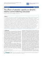
The fluorescence excitation spectrum of pure AFP with a
concentration of 3 ng/ml in Fig. 1 is the scanning excited
wavelength from 200 to 320 nm when the detection
wavelength was located at 345 nm (the fluorescence
emission peak of AFP usually takes place at the wave-
length range from 320 to 350 nm [20]). The experimental
result in Fig. 1 shows that there is a broad exciting band
with two peaks at around 260 and 293 nm, respectively,
which indicates that the fluorescence emission of AFP at
345 nm is sensitive to the excitation from 260 to 293 nm.
The fluorescence emission spectrum of pure AFP with a
concentration of 6 ng/ml in Fig. 2 is the scanning detection
wavelength from 250 to 500 nm when the exciting wave-
length was located at 293 nm. It is obvious that there is a
strong fluorescence emission peak noted at 345 nm. How-
ever, when amount of gold colloids were dropped into the
AFP solution (the concentration of AFP is kept at 6 ng/ml),
the emission peak at 345 nm decreases distinctly, as shown
in Fig. 2. This experimental result indicates that the gold
nanoparticles can quench the fluorescence of AFP. Fluo-
rescence emission spectra of solution containing both AFP
and gold colloid with different gold particle content are
compared in Fig. 3. In this comparison, all the samples have
the same AFP concentration and the exciting wavelength
was located at 260 nm. It is interesting to note that the
Fig. 1 Fluorescence excitation spectrum of pure AFP solution with a
concentration of 3 ng/ml, detection wavelength is 345 nm
Nanoscale Res Lett (2010) 5:1496–1501 1497
123
increasing gold colloid content leads to a decrease in the
fluorescence emission peak, as shown in Fig. 3.
The observed fluorescence quenching is attributed to the
resonance energy transfer from AFP to gold nanoparticles.
This non-radiative decay can be theoretically studied by
using the Fo
¨
rster energy transfer theory [11, 22]. When
some amounts of gold colloidal nanoparticles are dropped
into the solutions of AFP, AFP molecules would tend to
cluster around gold particles due to physical adsorption.
Increasing the AFP concentration leads to more and more
molecules adsorb on the gold particles, so the gold particle
will be coated by a dielectric shell. The thickness and
dielectric constant of the shell are controlled by the con-
centration of AFP and gold colloid content. In order to find
the effect of the dielectric shell on the fluorescence
quenching from gold particle, we calculated the quantum
efficiency of the shell-coated gold nanosphere [11],
In Eq. 1, C
R
denotes the radiative decay rate, C
NR
denotes
the non-radiative decay rate, k = 2p/k denotes the wave
number of the light, z denotes the distance from particle
center to the attached molecule. In our calculation, we
study the attached molecule at the outer surface of the
shell. So the value of z is equal to the radius of the
dielectric shell r
2
, which is changing from 15 to 65 nm.
The polarizability a of this dielectric shell-coated gold
sphere can be obtained from the quasi-static theory [23],
a ¼
4pe
0
r
3
2
½r
3
2
ðe
1
þ 2e
2
Þðe
2
À e
3
Þþr
3
1
ðe
1
À e
2
Þð2e
2
þ e
3
Þ
2r
3
1
ðe
1
À e
2
Þðe
2
À e
3
Þþr
3
2
ðe
1
þ 2e
2
Þðe
2
þ 2e
3
Þ
ð2Þ
In this calculation, the gold core has radius r
1
= 15 nm
and dielectric function e
1
, the dielectric shell has a thickness
r
2
- r
1
and dielectric constant e
2
(when e
2
= 2.0, the gold
particle is coated by a shell; when e
2
= e
3
= 1.0, no
dielectric shell is coated on the gold particle), the
embedding medium has dielectric function e
3
= 1.0. In
Drude model, this frequency-dependent complex dielectric
constant of gold particle can be written as [24]
e
1
ðxÞ¼e
1r
þ ie
1i
¼ e
b
ðxÞÀ
x
2
p
x
2
1 þ
1
x
2
s
2
þ i
x
2
p
x
2
xs 1 þ
1
x
2
s
2
ÀÁ
;
ð3Þ
where e
b
(x) is dielectric function of bulk metal which
is due to inter-band transition and varies with
frequency, these numerical parameters are given in
[25]. x
p
= 9 eV denotes the plasmon frequency of the
bulk metal [26], s is the size limit relaxation time of
gold nanoparticle [27, 28]andx is the frequency of
electromagnetic wave.
Fig. 2 Comparision of fluorescence emission spectra between pure
AFP and solution containing both AFP and gold colloid, exciting
wavelength is 293 nm
Fig. 3 Fluorescence emission spectra of solution containing both
AFP and gold colloid with different gold nanoparticle content,
exciting wavelength is 260 nm
Q ¼
C
R
C
R
þ C
NR
¼
1 þ
k
6
4p
2
a
jj
2
½ðkzÞ
À6
þðkzÞ
À4
þ
k
3
p
Re½aðkzÞ
À3
1 þ
k
6
4p
2
a
jj
2
½ðkzÞ
À6
þðkzÞ
À4
þ
k
3
p
Re½aðkzÞ
À3
þ
3k
3
2p
½Im½aÀ
k
3
6p
a
jj
2
½ðkzÞ
À6
þðkzÞ
À4
ð1Þ
1498 Nanoscale Res Lett (2010) 5:1496–1501
123
As shown in Fig. 4, the quantum efficiency at SPR
frequency is calculated as a function of separation distance
from the particle center to the outer surface of the dielectric
shell. Increasing the separation distance leads to a non-
linear increase in quantum efficiency. The changing speed
is relatively weak at very short and very far distance. These
results are similar to the reports of [29]. In order to find the
effect of the dielectric shell on this distance-dependent
quantum efficiency, the curves corresponding to gold
sphere with a dielectric shell and without a shell are
compared in Fig. 4. When e
2
= e
3
, the gold sphere is
immersed in a dielectric environment and no shell coated
on the gold sphere indeed. In this case, r
2
only denotes the
distance from particle center to the attached molecule.
When e
2
= e
3
, the gold sphere is coated with a dielectric
shell (the dielectric constant is e
2
= 2.0) first and then
immersed in a dielectric environment (the dielectric con-
stant is e
3
= 1.0). The calculated results show that the
existence of dielectric shell reduces the quantum effi-
ciency. This reduction begins to take effect when the
shell thickness exceeds 10 nm and gets intense with the
increasing shell thickness. This reduction of quantum
efficiency also indicates the quenching efficiency of a
shell-coated gold particle starts to decrease at a farer dis-
tance at resonance frequency.
As we know, the fluorescence wavelength is not always
matching the SPR frequency of gold nanoparicle. Espe-
cially, the fluorescence wavelength of the attached mole-
cule is fixed, whereas the SPR frequency of coated gold
nanosphere is tunable by the shell thickness. In order to
find the quantum efficiency at different frequency, we
plotted the quantum efficiency as a function of wavelength
with different shell thickness, as shown in Fig. 5.Itis
interesting to note that increasing the shell thickness leads
to the quantum efficiency peak red shifts, attenuates and
narrows down. The shift and narrow down speed is fast
with thinner shell and slow with thicker shell. However, the
attenuate speed is slow with thinner shell and fast with
thicker shell. These results show that increasing the
dielectric shell thickness may improve the monochroma-
ticity of fluorescence quenching. High energy transfer
efficiency can be obtained within a wide wavelength band
when coated by a thinner shell. This conclusion is in
Fig. 4 Quantum efficiency as a function of separation distance at
SPR frequency
Fig. 5 Quantum efficiency as a function of wavelength with different
dielectric shell thickness
Fig. 6 Absorption cross-section as a function of wavelength and
distance from the gold particle center, a e
2
= e
3
, b e
2
[ e
3
Nanoscale Res Lett (2010) 5:1496–1501 1499
123
agreement with our experimental results. Increasing the
gold particle content leads to a decrease in particle sepa-
ration and reduces the shell thickness. Therefore, the
fluorescence emission decreases with the increasing gold
colloids.
Our next goal is to find the physical origin of the
quantum efficiency of dielectric shell-coated gold nano-
sphere. We believe the SPR absorption is the most
important factor that affects the quantum efficiency of a
single dipole emitter close to a gold nanoparticle. There-
fore, we plotted the absorption cross-section as a function
of wavelength and separation distance, as shown in Fig. 6.
When e
2
= e
3
, the shell has the same dielectric constant of
the embedding medium, thus there is no shell coated on the
gold particle indeed. However, in order to make a com-
parison, we still assumed that there is a shell and calculated
absorption cross-section of this dielectric shell-coated gold
particle on the condition of e
2
= e
3
, as shown in Fig. 6a. In
this case, the absorption intensity decreases rapidly with
the increasing separation distance. However, when e
2
[ e
3
,
the existence of the dielectric shell may slow down the
decreasing speed of the absorption cross-section and then
reduces the quantum efficiency, as shown in Fig. 6b.
Therefore, the existence of dielectric shell may weaken
the quantum efficiency of gold nanosphere, which is in
agreement with the results in Fig. 4. Figure 6b also shows
that the resonance absorption at SPR frequency is intense
with thin dielectric shell and decreases as the shell gets
thicker. However, the off-resonance absorption, which is
far away from SPR frequency, is very weak and is not
sensitive to the shell thickness. Therefore, the changing
range of absorption intensity is larger for thinner shell but
smaller for thicker shell, which results in the narrow down
of the quantum efficiency band with the increasing shell
thickness. This conclusion is in agreement with the result
in Fig. 5.
Conclusion
Fluorescence quenching of AFP has been observed in the
presence of colloidal gold nanoparticles. The quenching
effect can be improved by increasing the gold nanoparticle
content. Based on non-radiative energy transfer theory, we
explained the observed fluorescence quenching characters
by calculating the quantum efficiency as a function of
dielectric shell thickness. The calculated results show that,
because of the SPR-induced non-radiative decay, high
energy transfer efficiency and intense fluorescence
quenching can be obtained within a wide wavelength band
when the gold particles are coated by a thinner dielectric
shell.
Acknowledgments This work was supported by the National High-
tech Research and Development Program (863 Program) of China
under grant No. 2009AA04Z314 and the Fundamental Research
Funds for the Central Universities under grant No. xjj20100049.
Open Access This article is distributed under the terms of the
Creative Commons Attribution Noncommercial License which per-
mits any noncommercial use, distribution, and reproduction in any
medium, provided the original author(s) and source are credited.
References
1. T. Soller, M. Ringler, M. Wunderlich, T.A. Klar, J. Feldmann,
H.P. Josel, Y. Markert, A. Nichtl, K. Kurzinger, Radiative and
nonradiative rates of phosphors attached to gold nanoparticles.
Nano Lett. 7, 1941–1946 (2007)
2. L. Ao, F. Gao, B. Pan, R. He, D. Cui, Fluoroimmunoassay for
antigen based on fluorescence quenching signal of gold nano-
particles. Anal. Chem. 78, 1104 (2006)
3. S. Pihlasalo, J. Kirjavainen, P. Ha
¨
nninen, H. Ha
¨
rma
¨
, Ultrasensi-
tive protein concentration measurement based on particle
adsorption and fluorescence quenching. Anal. Chem. 81, 4995–
5000 (2009)
4. I. Delfino, S. Cannistraro, Optical investigation of the electron
transfer protein azurin-gold nanoparticle system. Biophys. Chem.
139, 1–7 (2009)
5. S. Freddi, L. D’Alfonso, M. Collini, M. Caccia, L. Sironi, G.
Tallarida, S. Caprioli, G. Chirico, Excited-state lifetime assay for
protein detection on gold colloids-fluorophore complexes.
J. Phys. Chem. C 113, 2722–2730 (2009)
6. C.C. Huang, C.K. Chiang, Z.H. Lin, K.H. Lee, H.T. Chang,
Bioconjugated gold nanodots and nanoparticles for protein assays
based on photoluminescence quenching. Anal. Chem. 80, 1497–
1504 (2008)
7. B.N. Giepmans, S.R. Adams, M.H. Ellisman, R.Y. Tsien, The
fluorescent toolbox for assessing protein location and function.
Science 312, 217–224 (2006)
8. S. Mayilo, M.A. Kloster, M. Wunderlich, A. Lutich, T.A. Klar, A.
Nichtl, K. Ku
¨
rzinger, F.D. Stefani, J. Feldmann, Long-range
fluorescence quenching by gold nanoparticles in a sandwich
immunoassay for cardiac Troponin T. Nano Lett. 9, 4558–4563
(2009)
9. H.L. Guan, P. Zhou, X.L. Zhou, Z.K. He, Sensitive and selective
detection of aspartic acid and glutamic acid based on polythio-
phene-gold nanoparticles composite. Talanta 77, 319–324 (2008)
10. J. Gersten, A. Nitzan, Spectroscopic properties of molecules
interacting with small dielectric particles. J. Chem. Phys. 75,
1139–1152 (1981)
11. R. Carminati, J.J. Greffet, C. Henkel, J.M. Vigoureux, Radiative
and non-radiative decay of a single molecule close to a metallic
nanoparticle. Opt. Commun. 261, 368–375 (2006)
12. J. Zhu, Enhanced fluorescence from Dy
3?
owing to surface
plasmon resonance of Au colloid nanoparticles. Mater. Lett. 59,
1413–1416 (2005)
13. T. Pons, I.L. Medintz, K.E. Sapsford, S. Higashiya, A.F. Grimes,
D.S. English, H. Mattoussi, On the quenching of semiconductor
quantum dot photoluminescence by proximal gold nanoparticles.
Nano Lett. 7, 3157–3164 (2007)
14. E. Dulkeith, A.C. Morteani, T. Niedereichholz, T.A. Klar, J.
Feldmann, S.A. Levi, F.C.J.M. van Veggel, D.N. Reinhoudt, M.
Moller, D.I. Gittins, Fluorescence quenching of dye molecules
near gold nanoparticles: radiative and nonradiative effects. Phys.
Rev. Lett. 89, 203002 (2002)
1500 Nanoscale Res Lett (2010) 5:1496–1501
123
15. Y. Chen, K. Munechika, D.S. Ginger, Dependence of fluores-
cence intensity on the spectral overlap between fluorophores and
plasmon resonant single silver nanoparticles. Nano Lett. 7, 690–
696 (2007)
16. X.Y. Yang, Y.S. Guo, S. Bi, S.S. Zhang, Ultrasensitive enhanced
chemiluminescence enzyme immunoassay for the determination
of a-fetoprotein amplified by double-codified gold nanoparticles
labels. Biosens. Bioelectron. 24, 2707–2711 (2009)
17. W.C. Tsai, I.C. Lin, Development of a piezoelectric immuno-
sensor for the detection of alpha-fetoprotein. Sens. Actuator B
Chem. 106, 455–460 (2005)
18. Y.F. Chang, R.C. Chen, Y.J. Lee, S.C. Chao, L.C. Su, Y.C. Li, C.
Chou, Localized surface plasmon coupled fluorescence fiber-
optic biosensor for alpha-fetoprotein detection in human serum.
Biosens. Bioelectron. 24, 1610–1614 (2009)
19. Y.Y. Xu, C. Bian, S. Chen, S. Xia, A microelectronic technology
based amperometric immunosensor for a-fetoprotein using mixed
self-assembled monolayers and gold nanoparticles. Anal. Chim.
Acta 561, 48–54 (2006)
20. S.S.J. Leong, A.P.J. Middelberg, Dilution versus dialysis: a
quantitative study of the oxidative refolding of recombinant
human alpha-fetoprotein. Food Bioprod. Process. 84, 9–17 (2006)
21. K.C. Grabar, R.G. Freeman, M.B. Hommer, M.J. Natan, Prepa-
ration and characterization of Au colloid monolayers. Anal.
Chem. 67, 735–743 (1995)
22. T. Fo
¨
rster, Zwischenmolekulare Energiewanderung und Fluo-
reszenz. Ann. Physik. 2, 55–75 (1948)
23. R.D. Averitt, S.L. Westcott, N.J. Halas, Linear optical properties
of gold nanoshells. J. Opt. Soc. Am. B 16, 1824–1832 (1999)
24. J.A.A.J. Perenboom, P. Wyder, F. Meier, Electronic properties of
small metallic particles. Phys. Rep. 78, 173 (1981)
25. P.B. Johnson, R.W. Christy, Optical constants of the noble met-
als. Phys. Rev. B 6, 4370–4379 (1972)
26. V.I. Belotelova, G. Carotenuto, L. Nicolais, A. Longo, G.P. Pepe,
P. Perlo, A.K. Zvezdin, Online monitoring of alloyed bimetallic
nanoparticle formation by optical spectroscopy. J. Appl. Phys. 99,
044304 (2006)
27. D. Canchal-Arias, P. Dawson, Measurement and interpretation of
the mid-infrared properties of single crystal and polycrystalline
gold. Surf. Sci. 577, 95–111 (2005)
28. J. Zhu, Y.C. Wang, L.Q. Huang, Y.M. Lu, Resonance light
scattering characters of core–shell structure of Au–Ag nanopar-
ticles. Phys. Lett. A 323, 455–459 (2004)
29. Z. Gueroui, A. Libchaber, Single-molecule measurements of
gold-quenched quantum dots. Phys. Rev. Lett. 93, 166108 (2004)
Nanoscale Res Lett (2010) 5:1496–1501 1501
123