Báo cáo hóa học: "Identification of cytochrome P450 monooxygenase genes from the white-rot fungus Phlebia brevispora" pptx
Bạn đang xem bản rút gọn của tài liệu. Xem và tải ngay bản đầy đủ của tài liệu tại đây (487.59 KB, 35 trang )
This Provisional PDF corresponds to the article as it appeared upon acceptance. Fully formatted
PDF and full text (HTML) versions will be made available soon.
Identification of cytochrome P450 monooxygenase genes from the white-rot
fungus Phlebia brevispora
AMB Express 2012, 2:8 doi:10.1186/2191-0855-2-8
Ryoich Nakamura ()
Ryuichiro Kondo ()
Ming-hao Shen ()
Hideharu Ochiai ()
Shin Hisamatsu ()
Shigenori Sonoki ()
ISSN 2191-0855
Article type Original
Submission date 9 December 2011
Acceptance date 25 January 2012
Publication date 25 January 2012
Article URL />This peer-reviewed article was published immediately upon acceptance. It can be downloaded,
printed and distributed freely for any purposes (see copyright notice below).
Articles in AMB Express are listed in PubMed and archived at PubMed Central.
For information about publishing your research in AMB Express go to
/>For information about other SpringerOpen publications go to
AMB Express
© 2012 Nakamura et al. ; licensee Springer.
This is an open access article distributed under the terms of the Creative Commons Attribution License ( />which permits unrestricted use, distribution, and reproduction in any medium, provided the original work is properly cited.
252-5201, Japan
6-10-1 Hakozaki, Higashi-ku, Fukuoka 812-8581, Japan
1
Identification of cytochrome P450 monooxygenase genes from the white-rot fungus
Phlebia brevispora
Ryoich Nakamura
1
, Ryuichiro Kondo
2
, Ming-hao Shen
3
, Hideharu Ochiai
4
, Shin
Hisamatsu
1
, Shigenori Sonoki
1,*
1
Department of Environmental Sciences, School of Life and Environmental Science,
Azabu University, 1-17-71 Fuchinobe, Sagamihara 252-5201, Japan
2
Department of Forest Products Sciences, Faculty of Agriculture, Kyushu University,
3
College of Food Science and Engineering, Jilin Agriculture University, No.2888
Xincheng Street, Changchun, Jilin Province P.R.130118, China
4
Research Institute of Biosciences, Azabu University, 1-17-71 Fuchinobe, Sagamihara
2
RN:
M-HS:
RK:
SH:
*Corresponding author:
Email addresses:
HO:
3
Abstract
Three cytochrome P450 monooxygenase (CYP) genes, designated pb-1, pb-2 and pb-3,
were isolated from the white-rot fungus, Phlebia brevispora, using reverse transcription
PCR with degenerate primers constructed based on the consensus amino acid sequence of
eukaryotic CYPs in the O
2
-binding, meander and heme-binding regions. Individual
full-length CYP cDNAs were cloned and sequenced, and the relative nucleotide sequence
similarity of pb-1 (1788 bp), pb-2 (1881 bp) and pb-3 (1791 bp) was more than 58%.
Alignment of the deduced amino acid (aa) sequences of pb-1–pb-3 showed that these
three CYPs belong to the same family with >40% aa sequence similarity, and pb-1 and
pb-3 are in the same subfamily, with >55% aa sequence similarity. Furthermore,
pb-1–pb-3 appeared to be a subfamily of CYP63A (CYP63A1–CYP63A4), found in
Phanerochaete chrysosporium. The phylogenetic tree constructed by 500 bootstrap
replications using the neighbor-joining method showed that the evolutionary distance
between pb-1 and pb-3 was shorter than that between pb-2 and pb-1 (or pb-3).
Exon-intron analysis of pb-1 and pb-3 showed that both genes have nearly the same
number, size and order of exons and the types of introns, also indicating both genes
appear to be evolutionarily close. It is interesting that the transcription level of pb-3 was
evidently increased above the pb-1 transcription level by exposure to 12 coplanar PCB
congeners and 2,3,7,8-tetrachlorodibenzo-p-dioxin, though the two genes were
evolutionarily close.
4
Keywords:
cytochrome P450 monooxygenase; Phlebia brevispora; gene cloning; real-time RT-PCR;
dioxins; CYP63A
5
Introduction
Cytochrome P450 enzymes (CYPs) constitute a large superfamily of heme-containing
monooxygenases that are widely distributed in all kingdoms of life (Nelson 2009). CYPs
are involved in the metabolism of a wide variety of endogenous and xenobiotic
compounds by catalyzing regio- and stereospecific monooxygenation with an oxygen
atom generated from molecular oxygen. Mammalian CYPs have been studied extensively
because of their leading role in drug and xenobiotic metabolism and detoxification (Allis
et al. 2002; Inouye et al. 2002; McGraw JE and Waller 2006; Shimada 2006; Vrba et al.
2004; Warner et al. 2009; Yamazaki 2000; Zhang et al. 2006). CYPs from bacteria, yeast
and fungi have also been well studied in the biosynthesis of essential compounds like
ergosterol, which is a constituent of fungal cell membranes, and in the detoxification and
biodegradation of a broad spectrum of environmental chemical pollutants (Kelly et al.
1997; Kelly et al. 2003; Lamb et al. 2000; Seth-Smith et al. 2008; van den Brink et al.
1998).
The wood-rotting Basidiomycetes, white-rot fungi, have been extensively used for
biodegradation of various chemical pollutants. The ability to degrade such structurally
diverse chemical pollutants has generally been attributed to a lignin-degrading enzyme
system, including mainly lignin peroxidase, manganese-dependent peroxidase and
laccase produced by these fungi (Cameron et al. 2000; Fujihiro et al. 2009; Han et al.
2004; Mayer and Staples 2002; Takagi et al. 2007; Van Aken et al. 1999). However,
several studies pointed out that white-rot fungi are capable of degrading certain
xenobiotics under culturing conditions that did not induce the production of lignin
peroxidase, manganese-dependent peroxidase or laccase (Bumpus and Brock 1988;
6
Mileski et al. 1988; Yadav and Reddy 1993; Yadav et al. 1995). Therefore, besides such
lignin-degrading enzymes, alternative oxygenases, CYPs, are apparently involved in
catalyzing degradation of several xenobiotics. In particular, several specific CYPs from
Phanerochaete chrysosporium, the model white-rot fungus, have been studied in the
metabolism of xenobiotics (Chigu et al. 2010; Kasai et al. 2010; Matsuzaki and Wariishi
2005; Ning et al. 2010; Subramanian and Yadav 2009; Syed et al. 2010). Since whole
genome sequencing of P. chrysosporium has been completed, the molecular diversity of
CYPs and the presence of at least 150 CYP genes have been elucidated (Nelson 2009).
A previous report described the fungal metabolism of coplanar PCBs (Co-PCBs) by the
white-rot fungus Phlebia brevispora (Kamei et al. 2006). In addition, the
monomethoxylated metabolite was detected in cultures containing each congener by gas
chromatography and mass spectrometry, suggesting the involvement of CYP in the
transformation of Co-PCBs to methoxylated compounds via hydroxylation. This result
led us to search for the CYP system in P. brevispora involved in the metabolism of
xenobiotics. Here, we describe the identification, cloning, and sequence analysis of three
CYP genes from P. brevispora.
Materials and methods
Chemicals
Twelve Co-PCB congeners and 2,3,7,8-tetrachlorodibenzo-p-dioxin (TCDD) were
purchased from Wellington Labs (Ontario, Canada). Each congener was mixed in
dimethylsulfoxide (DMSO) at a concentration of 2 µg/ml for experimental use.
7
Strain and culture conditions
P. brevispora TMIC33929 was obtained from the Tottori Mycological Institute (Tottori,
Japan). The fungus was maintained as a culture on potato dextrose agar medium (Difco
Laboratories, MI, USA). The fungus was grown on a potato dextrose agar plate at 26°C
for 2 weeks. Then, the fungus mycelium was inoculated into Kirk liquid medium and
incubated statically at 26°C for 2 to 3 weeks; an additional incubation was carried out for
2 days in Kirk liquid medium (Tien and Kirk 1988) containing all of 12 Co-PCB
congeners and TCDD at a concentration of 0.25 ng/ml each. Fungal mycelium was
harvested from cultures by vacuum filtration and ground in a mortar and pestle with the
aid of liquid nitrogen. The ground mycelium was immediately used for RNA preparation.
Construction of degenerate primers for cDNA isolation of CYP genes
In a previous study to search for unknown CYP genes in cultures of P. chrysosporium, a
degenerate primer set was constructed based on the relatively conserved consensus aa
sequences across eukaryotic CYPs in the O
2
-binding and heme-binding regions (Kullman
and Matsumura 1997). Hence, for the first round of PCR of CYP genes, we used the same
degenerate forward and a slightly modified reverse primer (see Table 1) from that used in
the study of P. chrysosporium. For the second nested PCR of CYP genes, a degenerate
forward primer was constructed based on the relatively conserved consensus aa sequence
between the CYP O
2
-binding region and the CYP heme-binding region, which is called a
meander region (Hasemann et al. 1995), as shown in Table 1. The degenerate reverse
8
primer used in the second PCR was constructed for a region slightly upstream of the
heme-binding region.
Isolation, cloning and sequencing of partial cDNA fragments of CYP genes
Total RNA as a template for reverse transcription (RT)-PCR was prepared from the
ground mycelium using an RNeasy Plant Mini kit (QIAGEN Sciences, MD, USA). The
RT mixture (13 µl), containing 1 µl total RNA (>50 ng), 1 µl oligo(dT)
12-18
(0.25 µg), 4 µl
dNTP mixture (2.5 mM) and 7 µl sterile water, was heated at 65°C for 5 min and
incubated on ice for 1 min. After addition of 4 µl 5× first-strand buffer, 1 µl dithiothreitol
(0.1 M), 1 µl RNase inhibitor and 1 µl SuperScript III reverse transcriptase (200 units)
(Invitrogen Corp., CA, USA) to a total volume of 20 µl, the reaction mixture was
incubated at 50°C for 60 min, then at 70°C for 15 min. Finally, 20 µl sterile water was
added to the reaction mixture, which was stored at -20°C. The first PCR for CYP cDNA
amplification was performed in a reaction mixture (20 µl) containing 2 µl cDNA, 1 µl
each of the degenerate forward and reverse primers (10 µM), 2 µl 10× Ex Taq buffer, 2 µl
dNTP mixture (2.5 mM), 0.2 µl Ex Taq HS (TaKaRa Bio Inc., Shiga, Japan) and 11.8 µl
sterile water. The cycling conditions used for the first PCR were as follows: 98°C for 3
min, followed by 30 cycles of 98°C for 30 s, 53°C for 30 s and 72°C for 120 s, with a final
step at 72°C for 7 min. The second nested PCR was performed with the first PCR mixture
as a template and degenerate primers for the second PCR according to the following
procedure: 98°C for 3 min, followed by 30 cycles of 98°C for 30 s, 50°C for 30 s and
72°C for 120 s, with a final step at 72°C for 7 min. This two-round PCR led to the
isolation of a single PCR fragment, which had high sequence homology to CYP genes
9
from P. chrysosporium in BLAST homology searches. Cloning of the partial cDNA
fragment for the CYP gene was performed using a Mighty TA-cloning system (TaKaRa
Bio Inc.). The reaction mixture, containing 2 µl of the partial cDNA fragment, 0.5 µl
pMD20-T vector and 2.5 µl ligation Mighty-Mix was incubated at 16°C for 30 min, then
added to competent DH10B E. coli (Invitrogen Corp., CA, USA) for transformation. The
transformed cells were screened in LB medium containing X-gal, IPTG and ampicillin
according to the LacZ blue/white screening method. The cloned partial cDNA fragment
was prepared from a white transformed colony grown in LB medium containing
ampicillin (100 µg/ml) at 37°C overnight using a QIAprep spin miniprep kit (QIAGEN
Sciences). The cloned partial cDNA fragment was sequenced according to the
dye-terminator method (Sanger and Coulson 1975).
Unknown 5'- and 3'-end sequence determination of cDNAs
The 5'- and 3'-end sequences were determined using a SMARTer RACE cDNA
amplification system (Clontech Laboratories Inc., CA, USA). According to the
manufacturer’s instructions, 5'-RACE-ready cDNA and 3'-RACE-ready cDNA were
separately prepared from total RNA (10 ng to 1 µg). The CYP cDNA-specific primers for
5'-RACE and 3'-RACE PCR were respectively designed according to the base sequence
of partial cDNA as follows: 5'-RACE,
5'-TCGAGCGCGATAGTGTCGAAGTGCTGCAGC-3' (first PCR) and
5'-TGTACGCGAACTGCTGGCCGAGGCAGATG-3' (nested PCR); 3'-RACE,
5'-TCGACGAACGTCTGCACAAGCACCTGACAC-3' (first PCR) and
5'-AGCACCTGACACCGAACCCATTCATC-3' (nested PCR). The cycling conditions
10
used for the both rounds of PCR were: 98°C for 3 min, followed by 30 cycles of 98°C for
30 s, 68°C for 30 s and 72°C for 120 s, with a final step at 72°C for 7 min. The cloning and
sequencing methods were the same as described in the Materials and methods subsection:
Isolation, cloning and sequencing of partial cDNA fragments of CYP genes
.
Cloning and sequencing of full-length cDNAs
Full-length CYP cDNAs were cloned using a universal cloning method based on the
site-specific recombination system of bacteriophage lambda (Invitrogen Corp.). Based on
the 5'- and 3'-end sequences, one primer set for cloning of full-length CYP cDNA was
designed to the 5'-UTR region for the forward primer and to the 3'-UTR region for the
reverse primer. According to the manufacturer’s instructions, CYP gene specific forward
and reverse primers, attached by special sequences called attB1
(5'-GGGGACAAGTTTGTACAAAAAAGCAGGCTTC-3') and attB2
(5'-GGGGACCACTTTGTACAAGAAAGCTGGGT-3') were constructed as follows:
forward,
5'-GGGGACAAGTTTGTACAAAAAAGCAGGCTTCTCTCGACGGAGCCAAGTT
GCCTGTATC-3'; reverse,
5'-GGGGACCACTTTGTACAAGAAAGCTGGGTTCGTCCAAATACAAGATGAAT
CGCGCTAC-3'. PCR for full-length CYP cDNA was performed in a reaction mixture
(50 µl) containing 1 µl cDNA, 1 µl each of the attB1-forward and attB2-reverse primers
(10 µM), 25 µl PrimeSTAR Max DNA polymerase (TaKaRa Bio Inc.) and 22 µl sterile
water. The cycling conditions used for PCR were: 98°C for 3 min, followed by 35 cycles
of 98°C for 20 s, 61°C for 10 s and 72°C for 120 s, with a final step at 72°C for 7 min. The
11
cloning of full-length CYP cDNA was performed using a reaction mixture containing
12 µl amplified PCR product (15150 ng), 1.5 µl cloning vector (P-DONR221, 100
ng/µl), 4.55.5 µl TE buffer (pH 8.0) and 2 µl BP Clonase II enzyme mix (Invitrogen
Corp.). The reaction mixture was incubated at 25°C for 60 min, and 1 µl proteinase K was
added to stop the reaction. For transformation of E. coli, 1 µl of the reaction mixture was
added to competent DH10B cells. The transformed cells were screened in LB medium
containing kanamycin (100 µg/ml) at 37°C overnight. Full-length CYP cDNA was
sequenced according to the dye-terminator sequencing method. The aa sequence was
deduced by GENETYX ver.8 software (GENETYX Corp., Tokyo, Japan)
Isolation, cloning and sequencing of full-length CYP genes from genomic DNA
The cloning and sequencing of full-length CYP genes from genomic DNA was
performed using the same procedure as that described in the Materials and methods
subsection: Cloning and sequencing of full-length cDNAs except that the cDNA was
replaced with genomic DNA as the template in the reaction mixture. The genomic DNA
was prepared from the ground mycelium of P. brevispora using a DNeasy Plant Mini kit
(QIAGEN Sciences).
Quantitative analysis of gene transcripts by real-time RT-PCR
Total RNA as a template for real-time quantitative RT-PCR was prepared from P.
brevispora exposed to all 12 Co-PCB congeners and TCDD for 2 days at a final
concentration of 0.5 ng/ml in Kirk liquid medium using an RNeasy Plant Mini kit. As a
12
control experiment, DMSO was added into Kirk liquid medium instead of the 12 Co-PCB
congeners and TCDD. Target gene-specific primers for quantification of transcripts were
constructed based on <300 bp amplicons using online technical support for design of
real-time PCR assays (Roche Applied Science, Bavaria, Germany). The 18S rRNA gene
was used as an internal control gene in RT-PCR. The constructed primers and amplicon
lengths were: pb-1, 5'-CGCGTACAACGAGATGTCA-3' (forward),
5'-GAGCGCGATAGTGTCGAAGT-3' (reverse) and 64 bp (amplicon); pb-2,
5'-TCATCTTCGTGCCCTTCAAT-3' (forward), 5'-ACGACGCTTCGTTGTATGC-3'
(reverse) and 72 bp (amplicon); pb-3, 5'-TTCTATGACGCGCCCTTT-3' (forward),
5'-CATGCCTATCGAACACCTCA-3' (reverse) and 65 bp (amplicon); 18S rRNA,
5'-AACTTAAAGGAATTGACGGAAGG-3' (forward),
5'-TGAGTTTCCCCGTGTTGAG-3' (reverse) and 77 bp (amplicon). The RT reaction
was performed as described in the Materials and methods subsection: Isolation, cloning
and sequencing of partial cDNA fragments of CYP genes except that oligo(dT)
12-18
primers were replaced with random primers in the reaction mixture. Real-time
quantitative RT-PCR was performed by the detection of the nonspecific dye SYBR Green,
which binds to any double-stranded DNA, using a 7500 Fast Real-Time PCR System
(Applied Biosystems). The reaction mixture (25 µl), containing 2 µl cDNA, 2.5 µl each of
the target gene-specific forward and reverse primers (1 µM), 12.5 µl 2× SYBR Premix Ex
Taq II (TaKaRa Bio Inc.), 0.5 µl ROX Reference Dye II and 5 µl sterile water, was put
into a 96-well reaction plate, which was set in the 7500 Fast Real-Time PCR System. The
cycling conditions used were: 95°C for 30 s, followed by 40 cycles of 95°C for 5 s and
60°C for 40 s. The number of gene transcripts was estimated using a polyA
+
RNA
(Takara Bio Inc.) as a standard reference RNA. The amplicon from a polyA
+
RNA was
13
quantified based on the SYBR green fluorescence signal. The standard curve was
constructed by plotting threshold cycle values (Y-axis), which correspond to the number
of PCR cycles needed to reach the threshold fluorescence, against log number of RNA
molecules (X-axis). The number of gene transcripts in each of the DMSO-treated control
and the12 Co-PCB, TCDD-exposed culture was individually estimated using an equation
of the constructed standard curve, Y = -3.1815X + 34.935, R
2
= 0.99765.
Results
Isolation and sequence analysis of cDNAs for CYP genes pb-1, pb-2 and pb-3
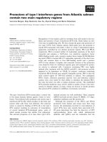
A single cDNA fragment that had an approximate length of 100 bp was obtained by
nested PCR, as shown in Fig. 1. This cDNA fragment showed high nucleotide sequence
homology with the CYP63 family from P. chrysosporium (Yadav et al. 2003). Hence, we
designated this CYP gene from P. brevispora pb-1. Because of high nucleotide sequence
homology between pb-1 and CYP63, degenerate primers were constructed to search for
CYP genes in addition to pb-1 based on the highly conserved consensus sequences in the
O
2
-binding region and heme-binding region of CYP63 (Yadav et al. 2003), as shown in
Table 1. As a result of RT-PCR with these degenerate primers, two more CYP genes
(pb-2, pb-3) were obtained. The nucleotide sequences of the 5'- and 3'-ends of the cDNA
for pb-1, pb-2 and pb-3 were determined by a SMARTer RACE cDNA amplification
system, and finally, full-length cDNAs of pb-1 (1788 bp), pb-2 (1881 bp) and pb-3 (1791
bp) were obtained. The nucleotide sequence similarities of these three genes were 60.9%
between pb-1 and pb-2, 64.6% between pb-1 and pb-3, and 57.9% between pb-2 and pb-3.
14
The nucleotide sequences of the three CYP cDNAs have been registered in the DNA Data
Bank of Japan (DDBJ) and are available under the accession numbers AB634456,
AB634457 and AB634458 for pb-1, pb-2 and pb-3, respectively.
Deduced aa sequence and protein analysis
The aa sequence similarities of pb-1, pb-2 and pb-3 are shown in Table 2. The percentage
of aa sequence similarity was 47.4% between pb-1 and pb-2, 61.4% between pb-1 and
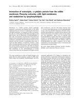
pb-3, and 46.5% between pb-2 and pb-3. The overall aa sequence alignments showed a
lower similarity in the N-terminal region (ca. <140 aa) than in the C-terminal region.
Although the aa sequence similarity was lower between pb-1 and pb-2 and between pb-2
and pb-3, the aa sequences around the meander and heme-binding regions were highly
conserved in the three CYP genes (Fig. 2). Furthermore, pb-1 and pb-3 also showed high
aa sequence similarity to the CYP63 subfamily, CYP63A1–CYP63A3 (Doddapaneni et
al. 2005; Doddapaneni and Yadav 2004), on the other hand, pb-2 showed high aa
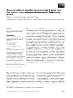
sequence similarity to CYP63A4 (Nelson 2009), as shown in Table 2. Phylogenetic
analysis was performed for pb-1 through pb-3 and CYP63A1 through CYP63A4 using
the neighbor-joining method in MEGA version 5 software (Tamura et al. 2011). A
phylogenetic tree was constructed by 500 bootstrap replications, as shown in Fig. 3. As a
result, three clades appeared with high bootstrap values. CYP pb-1 and CYP63A1 were
siblings in 98% of the bootstrap replications, and CYP pb-2 and CYP63A4 were siblings
in 98% of the bootstrap replications. CYP pb-3 was grouped in a clade that included
CYP63A2 and CYP63A3 in 67% of the bootstrap replications. The deduced CYP
proteins for pb-1, pb-2 and pb-3 had estimated molecular weights of approximately
15
68,400, 71,300 and 68,100 and isoelectric points of 8.46, 6.56 and 6.93, respectively. The
short sequences of hydrophobic aa (ca. 30 bp) at the N-terminal site found in all three
CYP proteins are probably signal peptides for membrane binding.
Cloning and sequence analysis of genomic CYP genes pb-1, pb-2 and pb-3
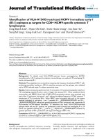
The full-length CYP gene, pb-1, had 16 exons and 15 introns, leading to a predicted
length of 2668 bp, as shown in Fig. 4. Each exon varied in size from 13 bp to 400 bp;
however, the size of the 15 introns was generally around 60 bp (Table 3). The full-length
CYP genes, pb-2 and pb-3, were respectively obtained using attB-sequence attached
primer set prepared as follows: pb-2,
5'-GGGGACAAGTTTGTACAAAAAAGCAGGCTTCACATGGGGACGTCGTCAG
G-3' (forward),
5'-GGGGACCACTTTGTACAAGAAAGCTGGGTTCCCACATAGATACGGCCATC
-3' (reverse); pb-3,
5'-GGGGACAAGTTTGTACAAAAAAGCAGGCTTCTCGAAAGGCGAGCGTCTC
AATTAC-3' (forward),
5'-GGGGACCACTTTGTACAAGAAAGCTGGGTCGGATTCTCCTTTGAATTTGT
TCAC-3' (reverse). CYP pb-2 had 11 exons and 10 introns, with a length of 2871 bp, and
pb-3 had 16 exons and 15 introns, with a length of 2595 bp. As shown in Table 3, the
number, size and order of exons was the same in pb-1 and pb-3, except for three exons of
400, 72 and 45 bp in pb-1. Although each intron that was similar in size in pb-1 was
slightly larger than the corresponding intron in pb-3, each type of intron was in the same
order in pb-1 and pb-3. On the other hand, pb-2 was quite different from the other two
16
CYP genes in all properties of exons and introns. The intron type was defined as follows:
type 0, lies between two codons; type I, lies after the first base in the codon; type II, lies
after the second base in the codon. The relative occurrence of the three intron types was
26.7% (type 0), 46.7% (type I) and 26.7% (type II) for pb-1 and pb-3, and 40% (type 0),
50% (type I) and 10% (type II) for pb-2.
Effect of exposure to dioxins on transcription levels of pb-1, pb-2 and pb-3
The effect of exposure to 12 Co-PCB congeners and TCDD on transcription levels of
pb-1, pb-2 and pb-3 was investigated using real-time quantitative RT-PCR to monitor the
fluorescent intensity of SYBR Green. The ratio of transcription levels following exposure
to 12 Co-PCB congeners and TCDD to that following a control exposure to DMSO, the
solvent for the dioxins, is represented in Fig. 5. Among the three CYP genes, the
transcription of pb-3 was evidently upregulated 2- to 3-fold by exposure to the 12
Co-PCB congeners and TCDD. The transcription rate of pb-2 was slightly increased;
however, pb-1 transcription was unchanged.
Discussion
Kamei et al. (2006) reported the congener-specific metabolism of
3,3',4,4'-tetrachlorobiphenyl, 2,3,3',4,4'-pentachlorobiphenyl,
2,3',4,4',5-pentachlorobiphenyl, 3,3',4,4',5-pentachlorobiphenyl and
2,3',4,4',5,5'-hexachlorobiphenyl in 11 Co-PCBs by P. brevispora and the detection of
methoxylated metabolites in the culture containing each congener, suggesting that these
17
metabolites are probably produced via hydroxylation of Co-PCBs catalyzed by CYPs. To
investigate the involvement of CYPs with the metabolism of dioxins, we first searched
for CYP cDNA in P. brevispora. There is little information concerning CYP genes from
P. brevispora; however, some useful information about nucleotide sequences of CYP
cDNAs from P. chrysosporium is available. Hence, two sets of degenerate primers were
constructed to search for CYP cDNAs from P. brevispora, as shown in Table 1, based on
the nucleotide sequence of CYP cDNAs from P. chrysosporium presented by Kullman
and Matsumura (1997), and the nucleotide sequences registered on the cytochrome P450
homepage organized by Nelson (2009). We describe three unique full-length cDNAs
encoding CYP genes pb-1, pb-2 and pb-3 in P. brevispora. As a result of BLAST
nucleotide sequence homology searching of these three CYP cDNAs, we found they were
closely related to the members of the representative multigene family CYP63,
CYP63A1–CYP63A4, found in P. chrysosporium (Doddapaneni et al. 2005;
Doddapaneni and Yadav 2004; Nelson 2009). CYPs are classified and named based
primarily on the level of aa sequence similarity. A family is generally defined as those
CYPs having >40% aa sequence similarity, and a subfamily is defined as those CYPs
having >55% aa sequence similarity. The deduced aa sequence alignments of pb-1–pb-3
showed that these three CYPs are members of the same family, and pb-1 and pb-3 are in
the same subfamily. Furthermore, pb-1 and pb-3 appeared to belong to the subfamily of
CYP63A1–CYP63A3, and pb-2 to CYP63A4.
Phylogenetic analysis of pb-1–pb-3 and CYP63A1–CYP63A4 with deduced aa
sequence alignment using a neighbor-joining method also indicates that the phylogenetic
tree is constituted of three clades and each pb-1–pb-3 belongs to a different one of the
three clades. Another phylogenetic analysis using a maximum likelihood method showed
18
a different phylogenetic tree from that of the neighbor-joining method, indicating that
pb-3 is grouped in the clade with pb-1 and CYP63A1 in 81% of the bootstrap replications
(data not shown). In both phylogenetic trees, the evolutionary distance between pb-1 and
pb-3 was shorter than that between pb-2 and pb-1 (or pb-3). In addition to having a short
evolutionary distance between pb-1 and pb-3, these two genes were closely located on the
genomic DNA. A PCR fragment that had an approximate length of 700 bp was detected
with the pb-3 forward primer (5'-AGGATTATGGGTCAAGTTCAAGGAAG-3') and
pb-1 reverse primer (5'-CTTATGGACTCTTCCTGCAGCAGAT-3'), indicating that
pb-3 is located upstream of pb-1 with a 613 bp interspace region in the same orientation
(data not shown). Exon-intron analysis of pb-1 and pb-3 indicated that 13 of the 16 exons
of the genes were similar in size and order; the exceptions were three exons: 8 (400 vs.
403 bp), 13 (72 vs. 75 bp) and 16 (45 vs. 42 bp), numbered according to the nucleotide
sequence of pb-1 (Fig. 4, Table 3). Relatively small and similarly sized introns were
found in both pb-1 (49–68 bp) and pb-3 (47–64 bp), and the order of the types of introns
in pb-1 was the same as in pb-3 (Table 3). From these results, the presence of some
interesting variants, which were found in CYP63A1 by Yadav et al. (2003), would also be
expected in the CYP genes of P. brevispora. In a study of intron-exon organization using
the numerous Arabidopsis CYP genes, the intron position and type were well conserved
among both subfamily and family, suggesting that intron position and type can be
correlated with phylogenic relations and CYP functions among the subfamily and family
(Paquette et al. 2000).
Searching for CYP genes involved in the metabolism of dioxins in P. brevispora is an
objective of our studies; one CYP gene (pb-3) found in P. brevispora was especially
upregulated at the level of transcription following exposure to 12 Co-PCB congeners and
19
TCDD. To detect precisely the change in transcription rates of CYP genes by exposure to
12 Co-PCB congeners and TCDD, a control gene that is not influenced by these
chemicals at transcription is essential for correcting the initial level of cDNA in real-time
quantitative RT-PCR. The 18S rRNA gene was not influenced in the transcription step by
12 Co-PCB congeners and TCDD in preliminary experiments; hence, the 18S rRNA gene
was used as an internal control gene. It is interesting that only the transcription level of
pb-3 was evidently increased by exposure to these 12 Co-PCB congeners and TCDD,
though pb-3 and pb-1 were evolutionarily close. In a previous study of xenobiotic
induction of CYP63A1 and CYP63A2, some xenobiotics including PCB (Aroclor 1254),
appeared to be responsible for the induction of only one gene (Doddapaneni and Yadav
2004). It seems that xenobiotic induction is not due to the phylogenetic correlation
between the CYP genes, but rather due to the presence of the transcription regulatory site,
e.g., xenobiotic response elements, located upstream of the CYP genes.
In this study, we have described the presence of three CYP genes in a white-rot fungus,
P. brevispora; one of these genes was upregulated on exposure to dioxins. However, it is
not obvious whether this upregulated CYP gene is involved in the metabolism of dioxins;
so further experiments must be carried out to elucidate the correlation of CYP gene
expression with the metabolism of dioxins.
Competing interests
The authors declare that they have no competing interests.
Acknowledgements
20
This research was partially supported by The Promotion and Mutual Aid Corporation for
Private Schools of Japan, a Grant-in-Aid for Matching Fund Subsidy for Private
Universities.
References
Allis JW, Anderson BP, Zhao G, Ross TM, Pegram RA (2002) Evidence for the
involvement of CYP1A2 in the metabolism of bromodichloromethane in rat liver.
Toxicology 176: 25-37
Bumpus JA, Brock BJ (1988) Biodegradation of crystal violet by the white rot fungus
Phanerochaete chrysosporium. Appl Environ Microbiol 54: 1143-1150
Cameron MD, Timofeevski S, Aust SD (2000) Enzymology of Phanerochaete
chrysosporium with respect to the degradation of recalcitrant compounds and xenobiotics.
Appl Microbiol Biotechnol 54: 751-758
Chigu NL, Hirosue S, Nakamura C, Teramoto H, Ichinose H, Wariishi H (2010)
Cytochrome P450 monooxygenases involved in anthracene metabolism by the white-rot
basidiomycete Phanerochaete chrysosporium. Appl Microbiol Biotechnol 87: 1907-1916
Doddapaneni H, Subramanian V, Yadav JS (2005) Physiological regulation, xenobiotic
induction, and heterologous expression of P450 monooxygenase gene pc-3 (CYP63A3),
a new member of the CYP63 gene cluster in the white-rot fungus Phanerochaete
chrysosporium. Curr Microbiol 50: 292-298
Doddapaneni H, Yadav JS (2004) Differential regulation and xenobiotic induction of
21
tandem P450 monooxygenase genes pc-1 (CYP63A1) and pc-2 (CYP63A2) in the
white-rot fungus Phanerochaete chrysosporium. Appl Microbiol Biotechnol 65: 559-565
Fujihiro S, Higuchi R, Hisamatsu S, Sonoki S (2009) Metabolism of hydroxylated PCB
congeners by cloned laccase isoforms. Appl Microbiol Biotechnol 82: 853-860
Han MJ, Choi HT, Song HG (2004) Degradation of phenanthrene by Trametes versicolor
and its laccase. J Microbiol 42: 94-98
Hasemann CA, Kurumbail RG, Boddupalli SS, Peterson JA, Deisenhofer J (1995)
Structure and function of cytochromes P450: a comparative analysis of three crystal
structures. Structure 3: 41-62
Inouye K, Shinkyo R, Takita T, Ohta M, Sakaki T (2002) Metabolism of polychlorinated
dibenzo-p-dioxins (PCDDs) by human cytochrome P450-dependent monooxygenase
systems. J Agric Food Chem 50: 5496-5502
Kamei I, Sonoki S, Haraguchi K, Kondo R (2006) Fungal bioconversion of toxic
polychlorinated biphenyls by white-rot fungus, Phlebia brevispora. Appl Microbiol
Biotechnol 73: 932-940
Kasai N, Ikushiro S, Shinkyo R, Yasuda K, Hirosue S, Arisawa A, Ichinose H, Wariishi H,
Sakaki T (2010) Metabolism of mono- and dichloro-dibenzo-p-dioxins by Phanerochaete
chrysosporium cytochromes P450. Appl Microbiol Biotechnol 86: 773-780
Kelly SL, Lamb DC, Jackson CJ, Warrilow AG, Kelly DE (2003) The biodiversity of
microbial cytochromes P450. Adv Microb Physiol 47: 131-186
Kelly SL, Lamb DC, Kelly DE (1997) Sterol 22-desaturase, cytochrome P45061,
possesses activity in xenobiotic metabolism. FEBS Lett 412: 233-235
Kullman SW, Matsumura F (1997) Identification of a novel cytochrome P-450 gene from
the white rot fungus Phanerochaete chrysosporium. Appl Environ Microbiol 63:
22
2741-2746
Lamb DC, Kelly DE, Masaphy S, Jones GL, Kelly SL (2000) Engineering of
heterologous cytochrome P450 in Acinetobacter sp.: application for pollutant degradation.
Biochem Biophys Res Commun 276: 797-802
Matsuzaki F, Wariishi H (2005) Molecular characterization of cytochrome P450
catalyzing hydroxylation of benzoates from the white-rot fungus Phanerochaete
chrysosporium. Biochem Biophys Res Commun 334: 1184-1190
Mayer AM, Staples RC (2002) Laccase: new functions for an old enzyme.
Phytochemistry 60: 551-565
McGraw JE S, Waller DP (2006) Specific human CYP 450 isoform metabolism of a
pentachlorobiphenyl (PCB-IUPAC# 101). Biochem Biophys Res Commun 344: 129-133
Mileski GJ, Bumpus JA, Jurek MA, Aust SD (1988) Biodegradation of
pentachlorophenol by the white rot fungus Phanerochaete chrysosporium. Appl Environ
Microbiol 54: 2885-2889
Nelson DR (2009) The cytochrome p450 homepage. Hum Genomics 4: 59-65
Ning D, Wang H, Zhuang Y (2010) Induction of functional cytochrome P450 and its
involvement in degradation of benzoic acid by Phanerochaete chrysosporium.
Biodegradation 21: 297-308
Paquette SM, Bak S, Feyereisen R (2000) Intron-exon organization and phylogeny in a
large superfamily, the paralogous cytochrome P450 genes of Arabidopsis thaliana. DNA
Cell Biol 19: 307-317
Sanger F, Coulson AR (1975) A rapid method for determining sequences in DNA by
primed synthesis with DNA polymerase. J Mol Biol 94: 441-448
Seth-Smith HM, Edwards J, Rosser SJ, Rathbone DA, Bruce NC (2008) The
23
explosive-degrading cytochrome P450 system is highly conserved among strains of
Rhodococcus spp. Appl Environ Microbiol 74: 4550-4552
Shimada T (2006) Xenobiotic-metabolizing enzymes involved in activation and
detoxification of carcinogenic polycyclic aromatic hydrocarbons. Drug Metab
Pharmacokinet 21: 257-276
Subramanian V, Yadav JS (2009) Role of P450 monooxygenases in the degradation of the
endocrine-disrupting chemical nonylphenol by the white rot fungus Phanerochaete
chrysosporium. Appl Environ Microbiol 75: 5570-5580
Syed K, Doddapaneni H, Subramanian V, Lam YW, Yadav JS (2010)
Genome-to-function characterization of novel fungal P450 monooxygenases oxidizing
polycyclic aromatic hydrocarbons (PAHs). Biochem Biophys Res Commun 399: 492-497
Takagi S, Shirota C, Sakaguchi K, Suzuki J, Sue T, Nagasaka H, Hisamatsu S, Sonoki S
(2007) Exoenzymes of Trametes versicolor can metabolize coplanar PCB congeners and
hydroxy PCB. Chemosphere 67: S54-57
Tamura K, Peterson D, Peterson N, Stecher, G, Nei M, Kumar S (2011) MEGA5:
Molecular evolutionary genetics analysis using maximum likelihood, evolutionary
distance, and maximum parsimony methods. Mol Biol Evol 28: 2731-2739
Tien M, Kirk TK (1988) Lignin peroxidase of Phanerochaete chrysosporium. In:
Methods in Enzymology 161: 238-249
Van Aken B, Hofrichter M, Scheibner K, Hatakka AI, Naveau H, Agathos SN (1999)
Transformation and mineralization of 2,4,6-trinitrotoluene (TNT) by manganese
peroxidase from the white-rot basidiomycete Phlebia radiata. Biodegradation 10: 83-91
van den Brink HM, van Gorcom RF, van den Hondel CA, Punt PJ (1998) Cytochrome
P450 enzyme systems in fungi. Fungal Genet Biol 23: 1-17
24
Vrba J, Kosina P, Ulrichova J, Modriansky M (2004) Involvement of cytochrome P450
1A in sanguinarine detoxication. Toxicol Lett 151: 375-387
Warner NA, Martin JW, Wong CS (2009) Chiral polychlorinated biphenyls are
biotransformed enantioselectively by mammalian cytochrome P-450 isozymes to form
hydroxylated metabolites. Environ Sci Technol 43: 114-121
Yadav JS, Quensen JF,3rd, Tiedje JM, Reddy CA (1995) Degradation of polychlorinated
biphenyl mixtures (Aroclors 1242, 1254, and 1260) by the white rot fungus
Phanerochaete chrysosporium as evidenced by congener-specific analysis. Appl Environ
Microbiol 61: 2560-2565
Yadav JS, Reddy CA (1993) Mineralization of 2,4-Dichlorophenoxyacetic Acid (2,4-D)
and Mixtures of 2,4-D and 2,4,5-Trichlorophenoxyacetic Acid by Phanerochaete
chrysosporium. Appl Environ Microbiol 59: 2904-2908
Yadav JS, Soellner MB, Loper JC, Mishra PK (2003) Tandem cytochrome P450
monooxygenase genes and splice variants in the white rot fungus Phanerochaete
chrysosporium: cloning, sequence analysis, and regulation of differential expression.
Fungal Genet Biol 38: 10-21
Yamazaki H (2000) Roles of human cytochrome P450 enzymes involved in drug
metabolism and toxicological studies. Yakugaku Zasshi 120: 1347-1357
Zhang JY, Wang Y, Prakash C (2006) Xenobiotic-metabolizing enzymes in human lung.
Curr Drug Metab 7: 939-948