báo cáo hóa học:" Identification of HLA-A" docx
Bạn đang xem bản rút gọn của tài liệu. Xem và tải ngay bản đầy đủ của tài liệu tại đây (649.96 KB, 11 trang )
BioMed Central
Page 1 of 11
(page number not for citation purposes)
Journal of Translational Medicine
Open Access
Research
Identification of HLA-A*2402-restricted HCMV immediate early-1
(IE-1) epitopes as targets for CD8+ HCMV-specific cytotoxic T
lymphocytes
Jong-Baeck Lim
1
, Hyun Ok Kim
1
, Seok Hoon Jeong
1
, Joo Eun Ha
1
,
Sunphil Jang
1
, Sang-Guk Lee
1
, Kyungwon Lee
1
and David Stroncek*
2
Address:
1
Department of Laboratory Medicine, Yonsei University College of Medicine, Seoul, South Korea and
2
Department of Transfusion
Medicine, Warren G. Magnuson Clinical Center, National Institutes of Health, Bethesda, MD, USA
Email: Jong-Baeck Lim - ; Hyun Ok Kim - ;
Seok Hoon Jeong - ; Joo Eun Ha - ; Sunphil Jang - ; Sang-
Guk Lee - ; Kyungwon Lee - ; David Stroncek* -
* Corresponding author
Abstract
Background: To identify novel HLA-A*2402-restricted human cytomegalovirus (HCMV)
immediate early-1 (IE-1) epitopes for adoptive immunotherapy, we explored 120 overlapping 15-
amino acid spanning IE-1.
Methods: These peptides were screened by measuring the frequency of polyclonal CD8+ T cells
producing intracellular interferon-γ (IFN-γ) using flow cytometry and the epitopes were validated
with a HCMV-infected target Cr release cytotoxicity assay.
Results: Initial screening was performed with 12 mini-pools of 10 consecutive peptides made from
120 overlapping peptides15-amino acids in length that spanned IE-1. When peripheral blood
mononuclear cells (PBMCs) from HLA-A*2402 HCMV-seropositive donors were sensitized with
each of the 12 mini-pools, mini-pools 1 and 2 induced the highest frequency of CD8+ cytotoxic T
lymphocytes (CTLs) producing IFN-γ. When PBMCs were stimulated with each of the twenty
peptides belonging to mini-pools 1 and 2, peptides IE-1
1–15
MESSAKRKMDPDNPD and IE-1
5–
19
AKRKMDPDNPDEGPS induced the greatest quantities of IFN-γ production and cytotoxicity of
HLA-matched HCMV-infected fibroblasts. To determine the exact HLA-A*2402-restricted
epitopes within the two IE-1 proteins, we synthesized a total of twenty-one overlapping 9- or 10
amino acid peptides spanning IE-1
1–15
and IE-1
5–19
. Peptide IE-1
3–12
SSAKRKMDPD induced the
greatest quantities of IFN-γ production and target cell killing by CD8+ CTLs.
Conclusion: HCMV IE-1
3–12
SSAKRKMDPD is a HLA-A*2402-restricted HCMV IE-1 epitope that
can serve as a common target for CD8+ HCMV-specific CTLs.
Background
Human cytomegalovirus (HCMV) infections occurring
after allogeneic hematopoietic stem cell transplantation
(HSCT) are frequently associated with high morbidity and
mortality despite treatment with appropriate antiviral
agents [1-3]. Cytotoxic T lymphocyte (CTL) responses
Published: 23 August 2009
Journal of Translational Medicine 2009, 7:72 doi:10.1186/1479-5876-7-72
Received: 1 June 2009
Accepted: 23 August 2009
This article is available from: />© 2009 Lim et al; licensee BioMed Central Ltd.
This is an Open Access article distributed under the terms of the Creative Commons Attribution License ( />),
which permits unrestricted use, distribution, and reproduction in any medium, provided the original work is properly cited.
Journal of Translational Medicine 2009, 7:72 />Page 2 of 11
(page number not for citation purposes)
have been known to correlate with recovery from HCMV
disease in bone marrow transplant (BMT) recipients [4]
and CD8+ CTLs are believed to play an important role in
suppressing HCMV disease [5-7]. This has led to the devel-
opment of clinical protocols whereby HCMV-specific
CD8+ T cell clones are cultured from the transplant donor
[8] and are administered to the transplant recipient. The
adoptive transfer of these HCMV-specific CD8+ CTLs has
proven to be effective in the prevention of reactivation
and in the treatment of HCMV infections that are unre-
sponsive to antiviral therapy [9-11].
Although HCMV protein pp65 is known to be an impor-
tant target for HCMV-specific CTLs and 70% to 90% of all
HCMV-specific CTLs recognize pp65 epitopes [12-14],
CTLs specific for another HCMV protein, immediate early-
1 (IE-1), occur in infected individuals at frequencies at
least comparable to those of pp65-specific CD8+ T cells
[15,16]. In addition, some recent studies have shown that
the dominance and magnitude of the IE-1 specific CD8+
T cell response more strongly correlates with protection
from HCMV disease than that of CD8+ T cell responses to
pp65 [17-19].
Several alternative approaches have been used to generate
antigen specific cytotoxic T cells. Antigen presenting cells
(APCs) have been genetically modified to which express
HCMV pp65 [20,21]. Epstein-Barr virus (EBV)-trans-
formed B lymphoblastic cell lines (EBV-BLCL) have been
used to generate EBV-specific CTLs. Genetic manipulation
of APCs including dendritic cells (DCs) as well as EBV-
BLCL result in the natural processing and presentation of
HCMV and EBV antigens but their clinical use is compli-
cated by regulatory issues, high cost, and the long dura-
tion of time required to qualify viral supernatants and cell
therapy products [22].
Several reports have proposed the use of donor-derived
HCMV-specific T cells generated by sensitization with
HCMV lysates loaded on either donor peripheral blood
mononuclear cells (PBMCs) or monocyte-derived
cytokine activated dendritic cells [7,8]. However, concerns
have been raised by regulatory agencies regarding the pos-
sibility that lysates of HCMV-infected cells might contain
live viral particles that could be transferred to the host and
HCMV T cells expanded using viral lysate may be predom-
inantly CD4+ cells [7].
The use of immune-dominant HCMV peptides is another
alternative for adoptive immune therapy. Adoptive
immune therapy with peptides is feasible as demonstrated
by the use of several HCMV-specific peptides derived from
pp65 protein to expand large quantities of HCMV-specific
CTLs [9,23].
The immune dominance of pp65 and IE-1 proteins
among HCMV antigens has been reported, but the
number of previously identified CTL epitopes derived
from IE-1 protein is limited. The wide clinical application
of HCMV-peptide, HLA-restricted, adoptive immune ther-
apy requires the identification of at least one immune
dominant HCMV pp65 and IE-1 peptide for each class I
HLA antigen. Especially for epitopes such as HLA-A*2402
which is the most frequent HLA-A allele in many different
races. To this aim, we report a new HLA-A*2402-restricted
pentadecamer peptide from HCMV IE-1, IE-1
3–
12
SSAKRKMDPD, that can be used to stimulate cytotoxic
T cells for adoptive immunotherapy.
Methods
Donor collection and cell preparation
Peripheral blood mononuclear cells (PBMCs) were col-
lected from nineteen HLA-A*2402 donors who were
HCMV seropositive. The presence of IgG and IgM HCMV
antibodies in each donor was analyzed by passive latex
agglutination (CMVSCAN kit, Becton-Dickinson Microbi-
ology System, Cockeysville, MD). MHC Class I genotypes
were determined by sequence-specific primer PCR using
genomic DNA by the HLA laboratory at the Seoul Clinical
Laboratory (Seoul, Korea). PBMCs were isolated from the
donors' peripheral blood by density-gradient centrifuga-
tion using Ficoll-Hypaque 1.077 (Pharmacia Biotech,
Wilkstrom, Sweden). The mononuclear cells were washed
twice with phosphate buffered saline (PBS, Gibco, Grand
Island, NY) and cryopreserved at -160°C in human AB+
serum and basal Iscove's medium (Gibco, Grand Island,
NY) containing 10% DMSO (Sigma, St. Louis, MO). This
research was approved by the institutional review board
of Yonsei University Health System and all participants
gave written informed consent.
Peptide libraries and study design
Peptide library for HCMV IE-1 protein were made up of
15-amino acids in length that overlapped by 11 residues
and covered the complete IE-1 protein (CMV AD169)
[24]. The entire IE-1 library was made up of 120 peptides
and these were commercially synthesized (A & Pep,
Yoengi-gun, Korea). The peptides were diluted in DMSO
to working solution concentrations and pooled into mini-
pools containing 10 consecutive peptides each. For IE-1,
12 mini-pools of 10 peptides were made. We screened
and choose the most immunogenic mini-pools among
the 12 mini-pools by quantifying the IFN-γ production
from the stimulated CD8+ CTLs using flow cytometry as
described below. We then screened and identified the best
15-amino acid peptides among the twenty 15-amino acid
peptides belonging to the selected mini-pools by quanti-
fying the IFN-γ production from the stimulated CD8+
CTLs using flow cytometry and HCMV-infected target cell
killing assay as described below. For further identification
Journal of Translational Medicine 2009, 7:72 />Page 3 of 11
(page number not for citation purposes)
of the exact HLA class I restricted-HLA-A*2402 epitopes,
we tested a total of twenty-one overlapping nona- or
decamer peptides spanning selected 15-amino acid pep-
tides by quantifying the IFN-γ production from the stimu-
lated CD8+ CTLs using flow cytometry and HCMV-
infected target cell killing assay again. Figure 1 briefly
shows the study design.
Generation of autologous dendritic cells (DCs) from
PBMCs
Peptide-loaded autologous DCs were generated as previ-
ously described [3,25]. PBMCs obtained after Ficoll-
Hypaque centrifugation were incubated for 2 hours at
37°C in complete RPMI medium. Adherent monocytes
were resuspended at a concentration of 5 × 10
6
/mL in
serum-free medium, supplemented with GM-CSF (1500
IU/mL, Pepro Tech Inc., Rocky Hill, NJ) and IL-4 (1200
IU/mL, Pepro Tech Inc., Rocky Hill, NJ). On days 2, 4, and
6 of culture, fresh cytokines were added. Fresh medium
was added depending on cell growth. On day 7 of culture,
10 ng/mL tumor necrosis necrosis factor-α (TNF-α, R&D
Systems, Minneapolis, MN) was added for the maturation
of the DCs. After 72-hour maturation, autologous DCs
were pulsed with peptides for at least 3 hours.
Generation of peptide-specific polyclonal CTLs
PBMCs from HCMV-seropositive donors were plated at a
concentration of 2 × 10
6
cells per well in a 24-well culture
plate (Nunc, Roskilde, Denmark) with 2 mL of medium
and directly stimulated with peptides at a concentration
of 10 μg/mL/well (on day 1) and with peptide-pulsed
autologous DCs (4~10 × 10
6
/well, on day 7 for a 1- or 2-
week expansion for flow cytometry analysis or cytotoxicity
assay, respectively). Recombinant human interleukin-2
(rhIL-2, 100 IU/mL, Pepro Tech Inc., Rocky Hill, NJ) was
added to the culture every other day and the cells were cul-
tured for 14 days.
Detection of IFN-
γ
producing CD8+ T cells in response to
peptide stimulation by flow cytometry
Two- week peptide-expanded PBMCs (1 × 10
6
) stimulated
with PHA (Sigma, St. Louis, MO) and PBMCs stimulated
with autologous DCs that were not loaded with any pep-
tide were used as positive and negative controls respec-
tively. HCMV pp65
495–503
(NLVPMVATV, HLA-A*0201)
or pp65
341–350
(QYDPVAALFF, HLA-A*2402), pp65
91–100
(SVNVHNPTGR, HLA-A33) were used as positive or nega-
tive controls according to donor's HLA type [3,24]. One
hour after stimulation, 10 mg of Brefeldin A (Sigma, St.
Louis, MO) was added to each well. After 5 additional
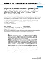
IE-1 peptide library and study designFigure 1
IE-1 peptide library and study design. The library of peptides spanning HCMV IE-1 was made up of 120 peptides 15 amino
acids in length that overlapped by 11 residues which were used to make 12 mini-pools each containing 10 consecutive peptides.
We screened and choose the most immunogenic mini-pools by quantifying IFN-γ production by stimulated CD8+ CTLs using
intracellular flow cytometry analysis. After finding that mini-pools 1 and 2 were the most potent stimulators of IFN-γ, we
screened and choose the best 15-amino acid peptides among twenty 15-amino acid peptides belonging to these two mini-pools
by quantifying IFN-γ production by peptide stimulated CD8+ CTLs using flow cytometry and a HCMV-infected target cell kill-
ing assay. Next, we identified the exact HLA class I restricted-HLA-A*2402 epitope by screening a total of twenty-one overlap-
ping nona- or decamer peptides spanning selected 15-amino acid peptides. These 21 peptides were also tested by intracellular
flow cytometry and cytotoxicity assays.
Journal of Translational Medicine 2009, 7:72 />Page 4 of 11
(page number not for citation purposes)
hours of incubation, PBMCs were washed once with PBS
and were then incubated in PBS containing 1 mM EDTA
for 10 minutes. After two further washes with PBS and 5%
fetal calf serum (FCS, Biosource International, Rockville,
MD) the cells were incubated with fluorescence-labeled
monoclonal antibodies for 15 minutes on ice in the dark.
Staining and analysis was performed as previously
described [3,14,26].
Antibodies and flow cytometry analysis
FITC-conjugated anti-IFN-γ, PerCP-conjugated anti-
CD69, PerCP-conjugated anti-CD3 and PE-conjugated
anti-CD8 were purchased from BD Biosciences. Per sam-
ple, 50,000–100,000 events in the FSC/SSC lymphocyte
gate were acquired on a FACS Calibur flow cytometer
(Becton Dickinson, San Jose, CA). For data analysis (CEL-
LQuest software; Becton Dickinson), CD3+/CD8+ events
were displayed in a CD69+ versus IFN-γ dot plot. CD8+/
IFN-γ cells were expressed as a percent of the respective
reference population. The assessment of responses was
previously described in more detail [3,14,26].
Fibroblast cell lines as target cells
Fibroblasts from allogeneic donor (HLA-A*2402) derived
skin biopsies were used as target cells. The fibroblasts were
propagated in MEM-α supplemented with 1% NEAA
(nonessential amino acid, Sigma, St. Louis, MO), 10%
fetal calf serum, and antibiotics. AD-169 HCMV strain
(VR-538, American Type Culture Collection, Manassas,
VA) was propagated in fibroblasts and the infected cul-
tures were harvested when a cytopathic effect was evident.
The cells were spun at 1500 rpm for 10 minutes and aliq-
uots of supernatant were stored at -80°C until use. HCMV
infectivity of the fibroblasts was confirmed by HCMV-spe-
cific real time RT-PCR testing that targeted the HCMV IE-
1 antigen (Roche, Nutley, NJ)
Cytotoxicity assays
Cytotoxicity assays were performed employing
51
Cr
release as previously described [27,28]. Briefly, HCMV-
infected fibroblasts were labeled overnight with
51
Cr (100
mCi/10
6
cells; PerkinElmer Life and Analytical Science,
Waltharn, MA), washed in PBS, and dispensed in tripli-
cate into 96-well V-bottom plates (Nunc, Roskilde, Den-
mark) at 4 × 10
3
cells/well. CTLs were added to the
infected fibroblasts at an effector to target cell ratio of
10:1, 30:1, 50:1 and 100:1. The cells were pelleted and
after a 5 hour incubation period the supernatant was ana-
lyzed in a gamma counter. Spontaneous and total release
counts for each well were used to calculate percent specific
release with the following formula: % specific release =
(experimental cpm - spontaneous cpm)/(total cpm -
spontaneous cpm).
Results
Screening IE-1 peptide mini-pools by induction of IFN-
γ
production by CD8+T cells
To determine which of the 12 mini-pools contained
potential immune dominant candidate peptides, PBMCs
from five HLA-A*2402 HCMV-seropositive donors
(donor 1–5) were stimulated with each of the 12 mini-
pools. Intracellular IFN-γ production was measured by
flow cytometry. As a positive control, PBMCs from
HCMV-seropositive donors were stimulated with both
phytohemaglutinin (PHA) and pp65
328–335
(QYD-
PVAALF, HLA-A*2402) [24]. In addition, PBMCs from
donors incubated without any peptide or with pp65
91–100
(SVNVHNPTGR, HLA-A33) [3] were used as negative con-
trols. Among the 12 mini-pools, mini-pool 1 induced a
greater frequency of IFN-γ producing CD8+ cytotoxic T
cells than mini-pools 3 through 12 in four of the five
donors. In addition, mini-pool 2 induced a higher fre-
quency of IFN-γ producing CD8+ cytotoxic T cells than
mini-pools 3 through 12 in three of the five donors.
Therefore, both peptide mini-pools 1 and 2 were selected
for further study. A representative experiment is illustrated
in Figure 2.
Identification of specific 15-amino acid candidate
eptitopes by in vitro sensitization and induction of IFN-
γ
production
To determine which 15-amino acid peptides belonging to
mini-pools 1 and 2 had the capacity to specifically re-
induce CTL immune activity, intracellular IFN-γ produc-
tion of CD8+ T cells was measured in HCMV-seropositive
HLA-A*2402 cells from five donors (Donors 2, 3, and 6–
8) that had been in vitro sensitized for a week with each of
the twenty candidate 15-amino acid peptides. After a one
week in vitro sensitization PBMCs were restimulated with
dendritic cells derived from autologous monocytes which
were loaded with each of the twenty 15-amino acid pep-
tides. After a 6-hour resensitization, intracellular IFN-γ
protein production by CD8+ T cells from the HCMV-
seropositve HLA-A*2402 donors was measured by intrac-
ellular flow cytometry. In a representative experiment
illustrated in Figure 3, in all donors peptides IE-1
1–
15
MESSAKRKMDPDNPD and IE-1
5–19
AKRKMDP DNP-
DEGPS consistently induced greater quantities of IFN-γ
production than the other 15-amino acid peptides tested.
As a control, the PBMCs were also sensitized in vitro for a
week with the HLA-A*2402-restricted epitope, pp65
328–
335
QYDPVAALF and the HLA-A*0201-restricted epitope,
pp65
495–503
NLVPMVATV [14] as positive controls and
with the HLA-A*3303-restricted epitope, pp65
91–100
SVN-
VHNPTGR, as a negative control.
These results suggest that IE-1
1–15
MESSAKRKMDPDNPD
and IE-1
5–19
AKRKMDPDNPDEGPS are potential HLA-
Journal of Translational Medicine 2009, 7:72 />Page 5 of 11
(page number not for citation purposes)
A*2402-restricted HCMV IE-1 epitopes and both peptides
were selected for further study.
Analysis of the peptide-specific cytotoxicity of the two 15
amino acid peptides
To confirm that IE-1
1–15
MESSAKRKMDPDNPD and IE-
1
5–19
AKRKMDPDNPDEGPS are immune dominated pep-
tides for HLA-A*2402 subjects, PBMCs from three HLA-
A*2402 HCMV-seropositive donors (Donors 9, 10 and
11) were sensitized in vitro for two weeks with the candi-
date pentadecapeptides. The in vitro sensitized cells were
tested for cytotoxicity against HLA-matched HCMV-
infected targets. The cytotoxicity assay was carried out by
measuring
51
Cr release from HLA-A*2402 HCMV-
infected fibroblasts. For all three donors tested IE-1
1–15
MESSAKRKMDPDNPD- and IE-1
5–19
AKRKMDP DNP-
DEGPS-sensitized CTLs lysed greater quantities of HCMV-
infected fibroblasts than the negative control cells. PBMCs
from donors 9 and 10 that were in vitro sensitized for 2
weeks with IE-1
1–15
MESSAKRKMDPDNPD were highly
cytotoxic to HLA-A*2402 HCMV-infected fibroblasts.
PBMCs in vitro sensitized with IE-1
1–
15
MESSAKRKMDPDNPD lyzed a similar proportion of
HCMV-infected fibroblasts as PBMCs sensitized with
pp65
495–503
which was used as a positive control (Figure
4A, B). However, in donor 11 IE-1
5–19
AKRKMDPDNP
DEGPS showed higher cytotoxicity to HLA-A*2402
HCMV-infected fibroblasts than that of IE-1
1–
15
MESSAKRKMDPDNPD (Figure 4C). These results con-
firmed that both of IE-1
1–15
MESSAKRKMDPDNPD and
IE-1
5–19
AKRKMDPDNPDEGPS were likely to be the best
immunogenic epitopes for HLA-A*2402 among HCMV
IE-1 proteins. Next, we identified the most immunogenic
nona- or decarmer MHC class I-restricted peptides span-
ning IE-1
1–15
and IE-1
5–19
using a HCMV-infected fibrob-
last cytotoxicity assay.
Ex vivo sensitization with 9- and 10 amino acid peptides
spanning IE-
11–15
and IE-
15–19
To determine the exact HLA class I restricted HCMV IE-1
protein epitopes that were immunogenic in HLA-A*2402
subjects, we synthesized and tested a total of twenty-one
overlapping nona- or decamer peptides spanning IE-1
1–15
and IE-1
5–19
. Intracellular IFN-γ protein production was
measured in cells from seven HCMV-seropositive HLA-
A*2402 donors (Donors 12–18) that had been in vitro
sensitized for 2 weeks with each of the twenty-one candi-
date peptides. Among the twenty-one candidate peptides,
IE-1
3–11
SSAKRKMDP, IE-1
3–12
SSAKRKMDPD and IE-1
8–
16
KMDPDNPDE induced greater quantities of IFN-γ pro-
duction than the other peptides tested. Peptide IE-1
3–
12
SSAKRKMDPD was especially potent. It induced greater
quantities of IFN-γ production than the other two pep-
tides in six of seven donors. Therefore, IE-1
3–
12
SSAKRKMDPD was likely the most immunogenic HLA-
A*2402 epitope within HCMV IE-1. A representative
experiment using cells from donor 14 is illustrated in Fig-
ures 5A and 5B. The response of donor 14's CD8+ cells to
IE-1
8–16
KMDPDNPDE was weak (Figure 5A), but IE-1
8–
16
KMDPDNPDE stimulated significant quantities of IFN-
γ in CD8+ cells from five of the seven HLA-A*2402
expressing donors tested.
HCMV IE-
13–12
SSAKRKMDPD specific cytotoxicity
To provide further evidence that IE-1
3–12
SSAKRKMDPD
induced epitope-specific and HLA-A*2402-restricted cyto-
toxicity, PBMCs from a donor expressing HLA-A*2402
(Donor 19) were sensitized in vitro for 2 weeks with IE-1
3–
11
SSAKRKMDP, IE-1
3–12
SSAKRKMDPD and IE-1
8–
16
KMDPDNPDE. The in vitro sensitized cells were tested
Results of screening of the 12 peptide mini-pools by quantify-ing intracellular IFN-γ by CD8+T cellsFigure 2
Results of screening of the 12 peptide mini-pools by
quantifying intracellular IFN-γ by CD8+T cells. To
select the most potential immune-dominant epitopes PBMCs
from five HLA-A*2402 HCMV-seropositive donors (Donors
1–5) were stimulated with each of the 12 mini-pools and
intracellular IFN-γ production was measured by flow cytome-
try. The results of testing cells from Donor 2 who expressed
HLA-A*0201/2402 are shown. Peptide mini-pools 1 and 2
showed a higher frequency of IFN-γ accumulation by CD8+
T cells than the other mini-pools. Therefore, mini-pools 1
and 2 were selected for further study. PHA and HCMV A2
(pp65
495–503
) peptide-stimulated PBMCs were used as posi-
tive controls and HCMV A33 (pp65
91–100
) peptide and IL-2
only stimulated PBMCs (IL-2) were used as negative controls.
Journal of Translational Medicine 2009, 7:72 />Page 6 of 11
(page number not for citation purposes)
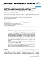
Intracellular IFN-γ protein production by HLA-A*2402 CD8+ CTLs stimulated with the twenty individual 15-amino acid pep-tides included in mini-pools 1 and 2Figure 3
Intracellular IFN-γ protein production by HLA-A*2402 CD8+ CTLs stimulated with the twenty individual 15-
amino acid peptides included in mini-pools 1 and 2. To determine which 15-amino acid peptides belonging to mini-pools
1 and 2 had the capacity to specifically re-induce CTL immune activity, intracellular IFN-γ production by CD8+ T cells was
measured in HCMV-seropositive HLA-A*2402 cells from five donors (Donors 2, 3, and 6–8) that had been in vitro sensitized
for a week with each of the twenty candidate 15-amino acid peptides. The results of testing cells from Donor 2 are shown.
Peptides IE-1
1–15
MESSAKRKMDPDNPD and IE-1
5–19
AKRKMDPDNPDEGPS consistently induced greater quantities of IFN-γ
protein production than the other 15-amino acid peptides tested. PHA and HCMV A2 (pp65
495–503
) peptide-stimulated PBMCs
were used as positive control and HCMV A33 (pp65
91–100
) peptide and IL-2 only stimulated PBMCs (IL-2) were used as nega-
tive controls.
&09$ &09$
,/
3+$
,(
Donor 2 (HLA-A*0201/2402)
CD8
IFN-
,(
,(
,(
,(
,(
,(
,(
,(
,(
,(
,(
,(
,(
,(
,(
,(
,(
,(
,(
Journal of Translational Medicine 2009, 7:72 />Page 7 of 11
(page number not for citation purposes)
for cytotoxicity using a
51
Cr release assay against HLA-
matched HCMV-infected targets. IE-1
3–12
SSAKRKMDPD-
sensitized CTLs lysed greater quantities of HCMV-infected
fibroblasts than the negative control cells. CTLs sensitized
with IE-1
5–16
KMDPDNPDE also lysed greater quantities
of HCMV-infected fibroblasts than the negative control
cells, but they lysed less HCMV-infected fibroblasts than
CTLs sensitized with IE-1
3–12
SSAKRKMDPD (Figure 6).
Discussion
This study focused on the identification of novel HLA-
A*2402 CTL epitopes derived from HCMV IE-1 protein
using pools of overlapping 15-amino acid peptides. These
HCMV-specific HLA-restricted epitopes will be useful for
vaccination, adoptive immunotherapy, and the monitor-
ing of cellular immune response against HCMV disease in
transplant recipients.
Over the last decade vaccination strategies using the
immunogenic peptides derived from several HCMV pro-
teins have been successful in preventing the reactivation
of latent HCMV infection [17-19]. One of the most
important steps in a peptide vaccine approach is the iden-
tification of immunogenic epitopes within HCMV pro-
teins, which bind to HLA Class I molecules that are
expressed by a major proportion of the population
[29,30]. Although HLA-A24 is the most frequent HLA-A
antigen among Asians, HLA-A24-restricted HCMV IE-1
epitopes have not yet been described.
Many current strategies for selecting potentially immuno-
genic epitopes are based on the use of algorithms that pre-
dict the binding affinities of specific peptides to HLA Class
I molecules. Peptides predicted to have a high binding
affinity are tested for their ability to sensitize CTLs. This
strategy can be a very effective way of identifying new
immune dominant peptides, but it has been useful for
only a limited number of peptide sequences and HLA alle-
les [31,32]. Furthermore, as demonstrated by Elkington et
al, even for those HCMV-pp65 peptides that were pre-
dicted to bind to common HLA alleles, only 40% elicited
cytokine-producing T cells detected by enzyme linked
immunospot (ELISPOT) assays, and only a subset of the
T-cell lines generated from HLA-A*0201-seropositive
donors in response to these peptides actually lysed
HCMV-infected cells [33]. We have explored another
method to identify HLA-A24-restricted HCMV IE-1
epitopes. Pools of overlapping 15-amino acid peptides
spanning the sequence of HCMV IE-1 were used for sensi-
tization and generation of HCMV-specific T cells. Such 15-
amino acid peptides previously have been used to identify
immunogenic viral epitopes recognized by T cells in the
Cytotoxic effects of IE-1
1–15
and IE-1
5–19
peptide-specific CTLs against CMV-infected fibroblastFigure 4
Cytotoxic effects of IE-1
1–15
and IE-1
5–19
peptide-specific CTLs against CMV-infected fibroblast. PBMCs from
three HLA-A*2402 HCMV-seropositive donors (Donors 9,10 and11) were sensitized in vitro for two weeks with IE-1
1–
15
MESSAKRKMDPDNPD and IE-1
5–19
AKRKMDPDNPDEGPS and the in vitro sensitized cells were tested for cytotoxicity
against HLA-matched HCMV-infected fibroblast. The cytotoxicity assay was carried out by measuring
51
Cr release from HLA-
A*2402 HCMV-infected fibroblasts. PBMCs from Donor 9 (Figure 4A) and Donor 10 (Figure 4B) that were in vitro sensitized
for 2 weeks with IE-1
1–15
MESSAKRKMDPDNPD were highly cytotoxic to HLA-A*2402 HCMV-infected fibroblasts causing as
much targeted cell lysis as PBMCs sensitized with a positive control. However, in Donor 11, IE-1
5–19
AKRKMDPDNPDEGPS
showed higher cytotoxicity to HCMV-infected fibroblasts than that of IE-1
1–15
MESSAKRKMDPDNPD (Figure 4C). PMBCs
stimulated with the HLA-A24-restricted HCMV pp65 epitope HCMV A24 (pp65
341–350
) was used as positive control and
PBMCs stimulated with the HCMV-A33 restricted epitope CMV A33 (pp65
91–100
) peptide and PBMCs simulated only with IL-2
(IL-2) were used as negative controls.
Donor 9 (HLA-A*1101/2402)
0.0
5.0
10.0
15.0
20.0
25.0
30.0
35.0
% l
y
sis
10:1 CMV-
50:1 CMV-
100:1 CMV-
10:1 CMV+
50:1 CMV+
100:1 CMV+
E:T ratio Target cell
Peptides
&09$ &09$ ,/ ,(
,(
&09$ &09$ ,/ ,(
0.0
5.0
10.0
15.0
20.0
25.0
30.0
35.0
% lysis
10:1 CMV-
50:1 CMV-
100:1 CMV-
10:1 CMV+
50:1 CMV+
100:1 CMV+
E:T ratio, Target cell
Peptides
Donor 10 (HLA-A*0201/2402)
,(
-5.0
0.0
5.0
10.0
15.0
20.0
25.0
10:1, CMV-
50:1, CMV-
100:1, CMV-
10:1, CMV+
50:1, CMV+
100:1, CMV+
% lysis
Peptides
E:T ratio, Target cell
Donor 11 (HLA-A*2402/2402)
&09$ &09$ ,/ ,(
,(
Journal of Translational Medicine 2009, 7:72 />Page 8 of 11
(page number not for citation purposes)
blood of healthy individuals and allograft recipients [25].
By analysis of responses to intersecting mini-pools, spe-
cific 15-amino acid peptides containing immunogenic
epitopes were identified and the epitopes subsequently
defined by testing responses to individual 9 or 10 amino
acid sequences contained in these 15-amino acid peptides
[26].
In our study a total of twelve mini-pools contained 10
consecutive 15-amino acid peptides were prepared using
one hundred-twenty 15-amino acid peptides spanning
HCMV IE-1 protein. The peptide pools were screened by
quantifying the production of IFN-γ by CD8+ T cells from
four HLA-A*2402 donors using flow cytotometry analy-
sis. Mini-pool 1 (Donors 1, 2, 3, and 4) and mini-pool 2
(Donors 1 and 2) induced higher frequencies of CD8+ T
cells producing IFN-γ than the other mini-pools. Mini-
pools 5, 7 and 9 showed a higher frequency IFN-γ produc-
tion in a single donor (Donor 2, Donor 3 and Donor 1,
respectively) (data not shown). Therefore, mini-pools 1
and 2 were selected for further characterization and
all twenty 15-amino acid peptides belonging to these
mini-pools were screened using flow cytometry analysis.
Among twenty 15-amino acid peptides, IE-1
1–
15
MESSAKRKMDPDNPD and IE-1
5–19
AKRKMDPDNP
DEGPS induced the highest frequency of IFN-γ producing
CD8+ T cells and PBMCs sensitized with these two 15-
amino acid peptides showed in vitro cytotoxicity against
HCMV-infected fibroblast.
Virus-infected human cells can be recognized by CD8+ T
cells through antigenic viral protein fragments of 8–12
amino acids in length that are presented on the cell sur-
face in association with HLA class I molecules. Since these
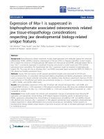
Intracellular IFN-γ analysis of IE-1
1–15
and IE-1
5–19
derived HCMV-specific CTLsFigure 5
Intracellular IFN-γ analysis of IE-1
1–15
and IE-1
5–19
derived HCMV-specific CTLs. To determine the exact HLA class I
restricted- HLA-A*2402 specific IE-1 epitopes, we synthesized a total of twenty-one overlapping nona- or decamer peptides
spanning IE-1
1–15
and IE-1
5–19
. Intracellular IFN-γ protein production was measured in six HCMV-seropositive HLA-A*2402
donors (Donors 12–17). Among the twenty-one candidate peptides, IE-1
3–11
SSAKRKMDP, IE-1
3–12
SSAKRKMDPD and IE-1
8–
16
KMDPDNPDE peptide induced the highest quantities of IFN-γ protein production. Peptide IE-1
3–12
SSAKRKMDPD induced
the greatest quantities of IFN-γ production in five of six donors. The results of testing Donor 14 are shown. The peptide IE-1
3–
12
SSAKRKMDPD induced higher quantities of IFN-γ production by CD8+ CTLs from Donor 14 than any of the other peptides
tested (Panel A). In addition, this peptide also induced the greatest quantities of IFN-γ protein production by CD8+CD69+
CTLs (Panel B). PHA and CMV A24 (pp65
341–350
) peptide-stimulated PBMCs were used as positive control and CMV A2
(pp65
495–503
) peptide and IL-2 only stimulated PBMCs (IL-2) were used as negative controls.
Donor 14 (HLA-A*2402/3303)
CD8
IFN-
&09$ &09$
,/ 3+$
,(
,(
,(
,(
,(
,(
,(
,(
,(
,(
,(
,(
,(
,(
,(
,(
,(
,(
,(
,(
,(
IFN-
CD69
IL-2
PHA CMV A24 CMV A02
IE-1
3-11
Donor 14 (HLA-A*2402/3303)
IE-1
8-16
IE-1
3-12
Journal of Translational Medicine 2009, 7:72 />Page 9 of 11
(page number not for citation purposes)
smaller peptides can be generated by extracellular process-
ing [25], eleven 9-amino acid peptides with 8 overlapping
amino acids and ten 10-amino acid peptides with 9 over-
lapping amino acids spanning IE-1
1–15
MESSAKRKMDP-
DNPD and IE-1
5–19
AKRKMDPDNPDEGPS were
synthesized and tested further for identification of HLA-
A*2402-restricted HCMV IE-1 epitopes. Among the 21
overlapping peptides, IE-1
3–11
SSAKRKMDP, IE-1
3–12
SSAKRKMDPD and IE-1
8–16
KMDPDNPDE induced the
greatest frequencies of IFN-γ producing CD8+ T cells. Pep-
tide IE-1
3–12
SSAKRKMDPD induced the highest frequency
of IFN-γ producing CD8+ T cells. Although when analyzed
by a computer algorithm each of these three peptides
scored a low rank estimated half-time of dissociation from
the HLA-A24 allele, all three peptides induced high fre-
quencies of polycolonal CD8+ T cells producing IFN-γ;
were presented successfully by the HLA-A*2402 allele of
HCMV-infected fibroblast cell lines; and induced strong
cytotoxicity against HCMV-infected fibroblasts. This sug-
gests that these three peptides are processed naturally and
presented successfully in vitro.
In conclusion, we have identified a possible HLA-A*2402
CTL epitope, IE-1
3–12
SSAKRKMDPD, derived from
HCMV IE-1 protein using overlapping peptides 15-amino
acids in length. This peptide was processed naturally in
HCMV-infected human fibroblast and presented success-
fully on the HLA-A*2402 allele and was well recognized
by HCMV-specific polyclonal CD8+ cytotoxic T cells.
Conclusion
HCMV IE-1
3–12
SSAKRKMDPD is a possible HCMV-spe-
cific epitope for vaccination, adoptive immunotherapy,
and the monitoring of cellular immune response against
HCMV disease in transplant recipients.
Conflict of interests
The authors declare that they have no competing interests.
HCMV IE-1
3–12
SSAKRKMDPD specific cytotoxicityFigure 6
HCMV IE-1
3–12
SSAKRKMDPD specific cytotoxicity. PBMCs from donors expressing HLA-A*2402 (Donor 19) were
sensitized in vitro for 2 weeks with IE-1
3–11
SSAKRKMDP, IE-1
3–12
SSAKRKMDPD and IE-1
8–16
KMDPDNPDE and tested for
cytotoxicity using a
51
Cr release assay against HLA-matched HCMV infected fibroblasts. The IE-1
3–12
SSAKRKMDPD sensitized
CTLs lysed greater quantities of HCMV-infected fibroblasts than the negative controls. HCMV A33 (pp65
341–350
) peptide and
IL-2 only stimulated PBMCs (IL-2) were used as negative controls.
% lysis
0
5
10
15
20
25
30
35
10:1, CMV-
30:1, CMV-
50:1, CMV-
10:1, CMV+
30:1, CMV+
50:1, CMV+
Donor 19 (HLA-A*0203/2402)
E:T ratio, Target cell
IL-2
CMV A33
IE-1
3-11
(SSAKRKMDP)
IE-1
3-12
(SSAKRKMDPD)
IE-1
8-16
(KMDPDNPDE)
Journal of Translational Medicine 2009, 7:72 />Page 10 of 11
(page number not for citation purposes)
Authors' contributions
JBL designed the research, preformed research, analyzed
data, and wrote the paper. HOK designed the research,
was responsible for the collection of PBMCs and histo-
compatibility testing, analyzed data, and wrote the paper.
SHJ designed the research, performed research, analyzed
data and wrote the paper. JEH performed research, ana-
lyzed data, and wrote the paper. SJ performed research,
analyzed data and wrote the paper. SGL performed
research, analyzed data and wrote the paper. KL designed
the research and editing the paper. DFS designed the
research and wrote the paper.
Acknowledgements
This work was supported by KOSEF through the National Core Research
Center for Nanomedical Technology (R15-2004024-01001-0).
References
1. Einsele H, Hebart H, Kauffmann-Schneider C, Sinzger C, Jahn G,
Bader P, Klingebiel T, Dietz K, Loffler J, Bokemeyer C, Muller CA,
Kanz L: Risk factors for treatment failures in patients receiv-
ing PCR-based preemptive therapy for CMV infection. Bone
Marrow Transplant 2000, 25:757-763.
2. Szmania S, Galloway A, Bruorton M, Musk P, Aubert G, Arthur A, Pyle
H, Hensel N, Ta N, Lamb L Jr, Dodi T, Madrigal A, Barrett J, Henslee-
Downey J, van Rhee F: Isolation and expansion of cytomegalo-
virus-specific cytotoxic T lymphocytes to clinical scale from
a single blood draw using dendritic cells and HLA-tetramers.
Blood 2001, 98:505-512.
3. Lim JB, Provenzano M, Kwon OH, Bettinotti M, Caruccio L, Nagorsen
D, Stroncek D: Identification of HLA-A33-restricted CMV
pp65 epitopes as common targets for CD8(+) CMV-specific
cytotoxic T lymphocytes. Exp Hematol 2006, 34:296-307.
4. Quinnan GV Jr, Kirmani N, Rook AH, Manischewitz JF, Jackson L,
Moreschi G, Santos GW, Saral R, Burns WH: Cytotoxic t cells in
cytomegalovirus infection: HLA-restricted T-lymphocyte
and non-T-lymphocyte cytotoxic responses correlate with
recovery from cytomegalovirus infection in bone-marrow-
transplant recipients. N Engl J Med 1982, 307:7-13.
5. Riddell SR, Watanabe KS, Goodrich JM, Li CR, Agha ME, Greenberg
PD: Restoration of viral immunity in immunodeficient
humans by the adoptive transfer of T cell clones. Science 1992,
257:238-241.
6. Walter EA, Greenberg PD, Gilbert MJ, Finch RJ, Watanabe KS, Tho-
mas ED, Riddell SR: Reconstitution of cellular immunity against
cytomegalovirus in recipients of allogeneic bone marrow by
transfer of T-cell clones from the donor. N Engl J Med 1995,
333:1038-1044.
7. Einsele H, Roosnek E, Rufer N, Sinzger C, Riegler S, Loffler J, Grigoleit
U, Moris A, Rammensee HG, Kanz L, Kleihauer A, Frank F, Jahn G,
Hebart H: Infusion of cytomegalovirus (CMV)-specific T cells
for the treatment of CMV infection not responding to antivi-
ral chemotherapy. Blood 2002, 99:3916-3922.
8. Peggs K, Verfuerth S, Mackinnon S: Induction of cytomegalovirus
(CMV)-specific T-cell responses using dendritic cells pulsed
with CMV antigen: a novel culture system free of live CMV
virions. Blood 2001, 97:994-1000.
9. Carlsson B, Cheng WS, Totterman TH, Essand M: Ex vivo stimula-
tion of cytomegalovirus (CMV)-specific T cells using CMV
pp65-modified dendritic cells as stimulators. Br J Haematol
2003, 121:428-438.
10. Peggs KS, Mackinnon S: Augmentation of virus-specific immu-
nity after hematopoietic stem cell transplantation by adop-
tive T-cell therapy. Hum Immunol 2004, 65:550-557.
11. Cobbold M, Khan N, Pourgheysari B, Tauro S, McDonald D, Osman
H, Assenmacher M, Billingham L, Steward C, Crawley C, Olavarria E,
Goldman J, Chakraverty R, Mahendra P, Craddock C, Moss PA:
Adoptive transfer of cytomegalovirus-specific CTL to stem
cell transplant patients after selection by HLA-peptide
tetramers. J Exp Med 2005, 202:379-386.
12. Li CR, Greenberg PD, Gilbert MJ, Goodrich JM, Riddell SR: Recovery
of HLA-restricted cytomegalovirus (CMV)-specific T-cell
responses after allogeneic bone marrow transplant: correla-
tion with CMV disease and effect of ganciclovir prophylaxis.
Blood 1994, 83:1971-1979.
13. Wills MR, Carmichael AJ, Mynard K, Jin X, Weekes MP, Plachter B,
Sissons JG: The human cytotoxic T-lymphocyte (CTL)
response to cytomegalovirus is dominated by structural pro-
tein pp65: frequency, specificity, and T-cell receptor usage of
pp65-specific CTL. J Virol 1996, 70:7569-7579.
14. Gratama JW, Kern F: Flow cytometric enumeration of antigen-
specific T lymphocytes. Cytometry A 2004, 58:79-86.
15. Kern F, Surel IP, Faulhaber N, Frommel C, Schneider-Mergener J,
Schonemann C, Reinke P, Volk HD: Target structures of the
CD8(+)-T-cell response to human cytomegalovirus: the 72-
kilodalton major immediate-early protein revisited. J Virol
1999, 73:8179-8184.
16. Slezak SL, Bettinotti M, Selleri S, Adams S, Marincola FM, Stroncek
DF: CMV pp65 and IE-1 T cell epitopes recognized by healthy
subjects. J Transl Med 2007, 5:17.
17. Gibson L, Piccinini G, Lilleri D, Revello MG, Wang Z, Markel S, Dia-
mond DJ, Luzuriaga K: Human cytomegalovirus proteins pp65
and immediate early protein 1 are common targets for
CD8+ T cell responses in children with congenital or postna-
tal human cytomegalovirus infection. J Immunol 2004,
172:2256-2264.
18. Bunde T, Kirchner A, Hoffmeister B, Habedank D, Hetzer R, Cherep-
nev G, Proesch S, Reinke P, Volk HD, Lehmkuhl H, Kern F: Protec-
tion from cytomegalovirus after transplantation is
correlated with immediate early 1-specific CD8 T cells. J Exp
Med 2005, 201:1031-1036.
19. Khan N, Best D, Bruton R, Nayak L, Rickinson AB, Moss PA: T cell
recognition patterns of immunodominant cytomegalovirus
antigens in primary and persistent infection. J Immunol 2007,
178:4455-4465.
20. Sun Q, Burton RL, Dai LJ, Britt WJ, Lucas KG: B lymphoblastoid
cell lines as efficient APC to elicit CD8+ T cell responses
against a cytomegalovirus antigen. J Immunol 2000,
165:4105-4111.
21. Leen AM, Myers GD, Sili U, Huls MH, Weiss H, Leung KS, Carrum G,
Krance RA, Chang CC, Molldrem JJ, Gee AP, Brenner MK, Heslop
HE, Rooney CM, Bollard CM: Monoculture-derived T lym-
phocytes specific for multiple viruses expand and produce
clinically relevant effects in immunocompromised individu-
als. Nat Med 2006, 12:1160-1166.
22. Lucas KG, Sun Q, Burton RL, Tilden A, Vaughan WP, Carabasi M,
Salzman D, Ship A: A phase I-II trial to examine the toxicity of
CMV- and EBV-specific cytotoxic T lymphocytes when used
for prophylaxis against EBV and CMV disease in recipients of
CD34-selected/T cell-depleted stem cell transplants. Hum
Gene Ther 2000, 11:1453-1463.
23. Rauser G, Einsele H, Sinzger C, Wernet D, Kuntz G, Assenmacher M,
Campbell JD, Topp MS: Rapid generation of combined CMV-
specific CD4+ and CD8+ T-cell lines for adoptive transfer
into recipients of allogeneic stem cell transplants. Blood 2004,
103:3565-3572.
24. Rowe WP, Hartley JW, Waterman S, Turner HC, Huebner RJ:
Cytopathogenic agent resembling human salivary gland
virus recovered from tissue cultures of human adenoids. Proc
Soc Exp Biol Med 1956, 92:418-424.
25. Provenzano M, Mocellin S, Bettinotti M, Preuss J, Monsurro V, Marin-
cola FM, Stroncek D: Identification of immune dominant
cytomegalovirus epitopes using quantitative real-time
polymerase chain reactions to measure interferon-gamma
production by peptide-stimulated peripheral blood mononu-
clear cells.
J Immunother 2002, 25:342-351.
26. Kern F, Faulhaber N, Frommel C, Khatamzas E, Prosch S, Schone-
mann C, Kretzschmar I, Volkmer-Engert R, Volk HD, Reinke P: Anal-
ysis of CD8 T cell reactivity to cytomegalovirus using
protein-spanning pools of overlapping pentadecapeptides.
Eur J Immunol 2000, 30:1676-1682.
27. Kuzushima K, Kimura H, Hoshino Y, Yoshimi A, Tsuge I, Horibe K,
Morishima T, Tsurumi T, Kojima S: Longitudinal dynamics of
Epstein-Barr virus-specific cytotoxic T lymphocytes during
posttransplant lymphoproliferative disorder. J Infect Dis 2000,
182:937-940.
Publish with BioMed Central and every
scientist can read your work free of charge
"BioMed Central will be the most significant development for
disseminating the results of biomedical research in our lifetime."
Sir Paul Nurse, Cancer Research UK
Your research papers will be:
available free of charge to the entire biomedical community
peer reviewed and published immediately upon acceptance
cited in PubMed and archived on PubMed Central
yours — you keep the copyright
Submit your manuscript here:
/>BioMedcentral
Journal of Translational Medicine 2009, 7:72 />Page 11 of 11
(page number not for citation purposes)
28. Bao L, Dunham K, Stamer M, Mulieri KM, Lucas KG: Expansion of
cytomegalovirus pp65 and IE-1 specific cytotoxic T lym-
phocytes for cytomegalovirus-specific immunotherapy fol-
lowing allogeneic stem cell transplantation. Biol Blood Marrow
Transplant 2008, 14:1156-1162.
29. Kubo RT, Sette A, Grey HM, Appella E, Sakaguchi K, Zhu NZ, Arnott
D, Sherman N, Shabanowitz J, Michel H, et al.: Definition of specific
peptide motifs for four major HLA-A alleles. J Immunol 1994,
152:3913-3924.
30. Schipper RF, van Els CA, D'Amaro J, Oudshoorn M: Minimal phe-
notype panels. A method for achieving maximum population
coverage with a minimum of HLA antigens. Hum Immunol
1996, 51:95-98.
31. Parker KC, Bednarek MA, Coligan JE: Scheme for ranking poten-
tial HLA-A2 binding peptides based on independent binding
of individual peptide side-chains. J Immunol 1994, 152:163-175.
32. Rammensee H, Bachmann J, Emmerich NP, Bachor OA, Stevanovic S:
SYFPEITHI: database for MHC ligands and peptide motifs.
Immunogenetics 1999, 50:213-219.
33. Elkington R, Walker S, Crough T, Menzies M, Tellam J, Bharadwaj M,
Khanna R: Ex vivo profiling of CD8+-T-cell responses to
human cytomegalovirus reveals broad and multispecific
reactivities in healthy virus carriers. J Virol 2003, 77:5226-5240.