Báo cáo hóa học: " Intense Red Catho- and Photoluminescence from 200 nm Thick Samarium Doped Amorphous AlN Thin Films" ppt
Bạn đang xem bản rút gọn của tài liệu. Xem và tải ngay bản đầy đủ của tài liệu tại đây (362.19 KB, 5 trang )
NANO EXPRESS
Intense Red Catho- and Photoluminescence from 200 nm Thick
Samarium Doped Amorphous AlN Thin Films
Muhammad Maqbool Æ Tariq Ali
Received: 21 January 2009 / Accepted: 2 April 2009 / Published online: 25 April 2009
Ó to the authors 2009
Abstract Samarium (Sm) doped aluminum nitride (AlN)
thin films are deposited on silicon (100) substrates at 77 K
by rf magnetron sputtering method. Thick films of 200 nm
are grown at 100–200 watts RF power and 5–8 m Torr
nitrogen, using a metal target of Al with Sm. X-ray dif-
fraction results show that films are amorphous. Cathodo-
luminescence (CL) studies are performed and four peaks
are observed in Sm at 564, 600, 648, and 707 nm as a
result of
4
G
5/2
?
6
H
5/2
,
4
G
5/2
?
6
H
7/2
,
4
G
5/2
?
6
H
9/2
, and
4
G
5/2
?
6
H
11/2
transitions. Photoluminescence (PL) pro-
vides dominant peaks at 600 and 707 nm while CL gives
the intense peaks at 600 nm and 648 nm, respectively.
Films are thermally activated at 1,200 K for half an hour in
a nitrogen atmosphere. Thermal activation enhances the
intensity of luminescence.
Keywords Cathodoluminescence Á Photoluminescence Á
Thermal activation Á XRD Á Samarium Á AlN
Introduction
Rear-earth doped nitride semiconductors thin films are
attracting increasing attention as phosphor materials, and
are used for optical displays [1–5]. Sputter deposited AlN
has been shown to be a viable host for luminescent rare
earth (RE) ions due to its transparency over a wide range,
including the UV, IR, and entire visible range [6–17].
Recent progress toward nitride-based light-emitting diode
and electroluminescent devices (ELDs) has been made
using crystalline and amorphous AlN doped with a variety
of rare-earth elements [1–9]. The electronic structure of the
RE ions differ from the other elements and are character-
ized by an incompletely filled 4f
n
shell. The 4f electrons lay
inside the ion and are shielded from the surroundings by
the filled 5s
2
and 5p
6
electron orbital [17]. When these
materials are excited by various means, intense sharp-line
emission is observed due to intra-4f
n
-shells transitions of
the rare-earth ion core [18–21]. The amorphous III-nitride
semiconductors have the advantage over their crystalline
counterpart because the amorphous material can be grown
at room temperature with little stress due to lattice mis-
match [22]. They may also be more suitable for wave-
guides and cylindrical and spherical laser cavities because
of the elimination of grain boundaries at low-temperature
growth [5].
High thermal conductivity, stability, and chemical
inertness of AlN also make it very useful for its electrical
and thermal applications.
In the present work, luminescence properties of
Samarium (Sm) are studied when deposited in AlN host.
The spectra obtained provide data in a broad range from
300 to 800 nm. Thus luminescence from the films in UV,
visible, and IR are obtained and studied simultaneously.
The effect of thermal activation is also studied by acti-
vating these materials in a tube furnace up to 1,200 K.
Experimental Details
Thin films of amorphous AlN:Sm were prepared at 77 K by
rf magnetron sputtering of an aluminum target of 99.999%
M. Maqbool (&)
Department of Physics and Astronomy, Ball State University,
Muncie, IN 47306, USA
e-mail:
T. Ali
Department of Physics, State University of New York at Buffalo,
Buffalo, NY 14260, USA
123
Nanoscale Res Lett (2009) 4:748–752
DOI 10.1007/s11671-009-9309-7
purity in a pure nitrogen atmosphere. Doping of thin films
with Sm was accomplished by drilling a small hole (0.5 cm
diameter) in the aluminum target (4.2 cm diameter) and
placing a slug of Sm in the hole. Sm was then co-sputtered
with the aluminum. The rf power was varied between
100 and 200 watts. All films were deposited onto
2cm9 2 cm, or less, p-silicon (100) substrates. The
background pressure in the chamber was \3 9 10
-5
Torr.
Liquid nitrogen was used to keep the temperature of the
film at 77 K. The metallic substrate holder was designed
such that it had a half inch diameter cylindrical hole from
the top. The substrate was pasted on the metal base of the
holder below the liquid nitrogen. Liquid nitrogen was
constantly poured in the holder to provide a constant low-
temperature to the substrate during film growth.
The as-deposited films were characterized for their
characteristic emissions. The thickness of the films was
200 nm, measured with a quartz crystal thickness monitor
in the growth chamber. X-rays diffraction (XRD) was used
to determine the structure of the films. No diffraction peaks
were observed, indicating that the as-deposited films were
amorphous.
Cathodoluminescence (CL) studies of the films were
performed at room temperature in a vacuum chamber at a
pressure of about 3 9 10
-6
Torr, which was maintained
with an Alcatel CFF 450 turbo pump. Films were excited
with electron beam energy of 2.85 kV and beam current of
100 lA. The films were placed an angle of 45° to the
incident electron beam coming out of electron gun. The
detector was placed at an angle of 45° to the film such that
lines joining electron gun, the film and detector were
making and angle of 90°. Luminescence from the films was
focused onto the entrance slit of a SPEX Industries double
monochromator with gratings blazed at 500 nm and
detected at a Thorn EMI fast high gain photomultiplier tube
with a range of 200–900 nm. The resolution of the spectra
was 1 nm.
A 488 nm line of Argon laser was used to obtain the
photoluminescence spectra, analyzed by a spectrometer
equipped with a cooled photomultiplier tube. The power of
the laser beam was 9.3 mW.
Thermal activation was accomplished by placing the flat
films in a tube furnace at 1,200 K in a nitrogen atmosphere
for half an hour.
Results and Discussion
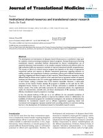
Figure 1 shows the photoluminescence (PL) spectrum of
AlN:Sm when excited with a 488 nm Argon laser. A strong
emission occurred at 598 nm (near 600 nm) which is
indicated by a sharp peak in the figure. This peak corre-
sponds to
4
G
5/2
?
6
H
9/2
transition. The intensity of the
emission is very strong and hence it serves as a potential
candidate for a red laser production at 598 nm. Further the
PL is showing that the material can emit light under photon
excitation and can be optically pumped for a laser con-
struction. This work is still in progress and will be reported
once laser achievement is successful.
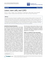
Figure 2 shows the PL spectrum of AlN:Sm when
excited with the same 488 nm Argon laser. A very strong
emission occurred at 707 nm (near 710 nm) which is
indicated by a sharp peak in the figure. This peak corre-
sponds to
4
G
5/2
?
6
H
11/2
transition. The intensity of the
emission is very strong and hence it also serves as a
potential candidate for an orange-red laser production at
707 nm. The intensity of this peak is almost double than
the intensity of the peak at 598 nm with the same power of
excitation sourcing. Thus the
4
G
5/2
?
6
H
11/2
transition has
a strong potential to produce a red-near IR laser under
optimum conditions.
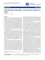
Figure 3 provides CL spectrum of AlN:Sm in 300–
850 nm range at room temperature. It is observed that
Fig. 1 PL spectrum of amorphous AlN:Sm with excitation at 488 nm
and emission at 598 nm
Fig. 2 PL spectrum of amorphous AlN:Sm with excitation at 488 nm
and emission at 707 nm
Nanoscale Res Lett (2009) 4:748–752 749
123
Sm
3?
give four transitions under electron excitation. Three
of these transitions are in the visible range of the spectrum
at 564, 600, and 648 nm as a result from
4
G
5/2
?
6
H
5/2
,
4
G
5/2
?
6
H
7/2
and
4
G
5/2
?
6
H
9/2
transitions, respectively
[7, 20]. The fourth peak falls in the infrared region at
707 nm due to
4
G
5/2
?
6
H
11/2
. The peak at 600 nm is the
strongest while the peak at 707 nm is the weakest amongst
all. The
4
G
5/2
?
6
H
5/2
transition at 564 nm falls in yellow
region of the spectrum. The dominant transition
4
G
5/2
?
6
H
7/2
at 600 nm and the
4
G
5/2
?
6
H
9/2
transitions occur in
red region of the visible spectrum. Because of the combi-
nation of these colors and dominancy of orange-red peak,
the direct observation of AlN:Sm films exposed to electron
beam in CL gives orange-red light to naked eye. All these
transitions and their relative intensities are tabulated in
Table 1.
Figure 4 gives a combined spectra of AlN:Sm before and
after thermal activation. It is clear from the figure that
thermal annealing enhances the luminescence from Sm. It is
observed that thermal annealing doubles the luminescence
intensity from the dominant transition
4
G
5/2
?
6
H
7/2
at
600 nm. The
4
G
5/2
?
6
H
5/2
transition at 564 nm has got
maximum enhancement when annealed thermally at
1,200 K for half an hour. The intensity of luminescence of
this transition increases by a factor of 2.5 after thermal
annealing. The other two transitions are also enhanced sig-
nificantly by thermal annealing.
Figure 5 shows the XRD analysis of the AlN:Sm films
deposited on Si(100) substrate. Only one peak can be
observed in the film at 69.1° that corresponds to Si(100).
No other peak is present in the figure, indicating that the
deposited films are amorphous. Thermal activation of the
films at 1,200 K has not changed the structure of the films.
Table 1 provides detail of all transitions from Sm
3?
.
Column 2 and 3 give all transitions and the corresponding
wavelengths of emission. The relative intensities of non-
annealed and annealed samples are given in column 4 and
5, respectively. These relative intensities are determined by
comparing the intensity of every peak to the intensity
brightest peak (567 nm) in the non-annealed samples.
Column 4 gives the ratio by which the intensity of lumi-
nescence is enhanced by thermal annealing. Careful
0
200
400
600
800
1000
1200
1400
1600
300 323 346 369 393 416 439 462 485 508 532 555 578 601 624 647 671 694 717 740 763 786
Wavelength (nm)
Intensity (a.u)
564 nm
600 nm
648 nm
707 nm
Fig. 3 CL spectrum of
amorphous AlN:Sm films
Table 1 Summary of Sm
3?
ions emissions from AlN:Sm
Material Transition Wavelength
(nm)
Relative intensity
non-annealed films.
Relative intensity
of annealed films
Enhancement
ratio
CL data
AlN:Sm
4
G
5/2
?
6
H
5/2
564 0.425 1.07 2.52
4
G
5/2
?
6
H
7/2
600 1.000 2.3 2.3
4
G
5/2
?
6
H
9/2
648 0.686 1.143 1.66
4
G
5/2
?
6
H
11/2
707 0.312 0.457 1.46
PL data
4
G
5/2
?
6
H
7/2
598 0.61
4
G
5/2
?
6
H
11/2
707 1.00
750 Nanoscale Res Lett (2009) 4:748–752
123
consideration of these ratios tells that enhancement is
higher for lower wavelengths and it goes down when one
moves from ultraviolet to infrared region of the spectrum.
The reason being, with increasing temperature the proba-
bility of populating higher energy levels increases and
hence higher energy levels are thermally more populated as
compared to lower energy levels at high-temperature [21].
These thermally populated higher energy levels give rise to
enhanced emission.
Both PL peaks indicate very strong emission from
AlN:Sm when excited with 488 nm laser. Such a strong
intensity clearly indicates that this material is a potential
candidate for laser production. We are in the process of
providing optimum conditions and laser power to achieve
laser in AlN:Sm. Polarization study is also in progress and
will be published soon once it is complete.
This significant increase in the intensities of lumines-
cence from Sm
3?
ions by thermal annealing has got a
good explanation. Luminescence occurs from Sm
3?
ions
and not from Sm
2?
or Sm
1?
. During the film deposition,
it is most likely that some of Al
3?
of AlN may be
replaced by Sm
3?
but there are also chances for imper-
fections and defects giving rise to Sm
2?
or Sm
1?
during
film growth. These ions do not contribute to lumines-
cence. Smaller the number of these ions, more will be
Sm
3?
ions and hence luminescence will be higher. When
these films are activated thermally at a higher temperature
then most of Sm
2?
or Sm
1?
impurities ionize and con-
verts to Sm
3?
ions giving path to enhanced luminescence
[22–24]. Moreover when the films are transferred to the
furnace and thermally activated after removed from the
deposition chamber, they are exposed to air. Thus oxi-
dation of the surface of the film cannot be ignored.
Oxygen enhances the luminescence of rare-earth ions
giving rise to the enhanced luminescence after thermal
activation of the films [13].
The results show that amorphous AlN:Sm is a promising
candidate for its use in nanoscale optical devices and
communication tools. The strong red emission makes this
material a potential candidate for making quantum dots.
Conclusion
Thin films of amorphous AlN:Sm are deposited by rf
magnetron sputtering. Films were characterized for their
surface morphology and luminescence properties by XRD,
PL, and CL. Samarium ion emits mainly in visible region
with the most intense transition in the orange-red portion of
the spectrum. Thermal activation enhances the lumines-
cence of films. PL provides very sharp emission in red
making it a useful material for nanoscale optical devices
applications.
0
500
1000
1500
2000
2500
3000
3500
300 325 351 376 402 427 453 478 504 529 555 580 606 631 657 682 708 733 759 784
Wavlength (nm)
Intensity (a.u)
Inactivated Film
Thermally Activated Film
564 nm
600 nm
648 nm
707 nm
Fig. 4 CL spectra of thermally
activated and inactivated
amorphous AlN:Sm films
Fig. 5 XRD analysis of the AlN:Sm films deposited on Si(100)
substrates
Nanoscale Res Lett (2009) 4:748–752 751
123
References
1. M. Maqbool, I. Ahmad, H.H. Richardson, M.E. Kordesch, Appl.
Phys. Lett. 91(19), 193511 (2007)
2. M. Maqbool, H.H. Richardson, M.E. Kordesch, J. Mater. Sci.
42(14), 5657–5660 (2007). doi:10.1007/s10853-006-0730-3
3. M. Maqbool, I. Ahmad, Curr. Appl. Phys. 9, 234–237 (2008). doi:
10.1016/j.cap.2008.02.001
4. M. Maqbool, H.H. Richardson, M.E. Kordesch, in Materials
Research Society International Symposium proceedings, vol. 831
Article E8.12.1, @2005 Materials Research Society
5. M. Maqbool, H.H. Richardson, P.G. Van Patten, M.E. Kordesch,
in Materials Research Society International Symposium pro-
ceedings, vol. 798 (Materials Research Society, 2004), pp. 8.5.1–
8.5.5
6. H. Chen, K. Gurumurugan, M.E. Kordesch, W.M. Jadwisienczak,
H.J. Lozykowski, MRS Internet J. Nitride Semicond. Res. 5,
U130 (2000)
7. M. Caldwell, H.H. Richardson, M.E. Kordesch, MRS Internet J.
Nitride Semicond. Res. 5, U142 (2000)
8. W.M. Jadwisienczak, H.J. Lozykowski, F. Perjeru, H. Chen,
M. Kordesch, I. Brown, Appl. Phys. Lett. 76, 3376 (2000). doi:
10.1063/1.126652
9. V. Dimitrova, P.G. Van Patten, H.H. Richardson, M.E. Kordesch,
Appl. Phys. Lett. 77, 478 (2000). doi:10.1063/1.127016
10. V. Dimitrova, P.G. Van Patten, H. Richardson, M.E. Kordesch,
Appl. Surf. Sci. 175–176, 481 (2001). doi:10.1016/S0169-4332
(01)00128-3
11. M.L. Caldwell, A.L. Martin, C.M. Spalding, V.I. Dimitrova, P.G.
Van Patten, M.E. Kordesch, H.H. Richardson, J. Vac. Sci.
Technol. A 19, 1894 (2001)
12. M.L. Caldwell, A.L. Martin, V.I. Dimitrova, P.G. Van Patten,
M.E. Kordesch, H.H. Richardson, Appl. Phys. Lett. 78, 1246
(2001). doi:10.1063/1.1351531
13. M.L. Caldwell, P.G. Van Patten, M.E. Kordesch, H.H. Richard-
son, MRS Internet J. Nitride Semicond. Res. 6, 13 (2001)
14. H.H. Richardson, P.G. van Patten, D.R. Richardson, M.E. Kor-
desch, Appl. Phys. Lett. 80, 2207 (2002). doi:10.1063/1.1464220
15. H. Chen, K. Chen, D.A. Drabold, M.E. Kordesch, Appl. Phys.
Lett. 77, 1117 (2000). doi:10.1063/1.1289496
16. M.E. Little, M.E. Kordesch, Appl. Phys. Lett. 78, 2891 (2001).
doi:10.1063/1.1370548
17. J.F. Suyver, P.G. Kik, T. Kimura, A. Polman, G. Franzo,
S. Coffa, Nucl. Instr. Meth. Phys. Res. B 148, 497–501 (1999)
18. J.B. Gruber, B. Zandi, H.J. Lozykowski, W.M. Jadwisienczak,
J. Appl. Phys. 91(5), 2929–2935 (2002). doi:10.1063/1.1436297
19. H.J. Lozykowski, Phys. Rev. B 48, 17758 (1993). doi:10.1103/
PhysRevB.48.17758
20. A.J. Steckl, R. Birkhahn, Appl. Phys. Lett. 73, 1700 (1998). doi:
10.1063/1.122250
21. M.J.V. Bell, L.A.O. Nunes, A.R. Zanatta, J. Appl. Phys. 86(1),
338–341 (1999). doi:10.1063/1.370734
22. M. Overberg, C.R. Abernathy, J.D. MacKenzie, S.J. Pearton,
R.G. Wilson, J.M. Zavada, Mater. Sci. Eng. B 81, 121–126
(2001). doi:10.1016/S0921-5107(00)00686-3
23. J.D. MacKenzie, C.R. Abernathy, S.J. Pearton, U. Hommerich,
J.T. Seo, R.G. Wilson, J.M. Zavada, Appl. Phys. Lett. 72(21),
2710–2712 (1998). doi:10.1063/1.121107
24. S.Z. Wang, S.F. Yoon, L. He, X.C. Shen, J. Appl. Phys. 90(5),
2314–2320 (2001). doi:10.1063/1.1391213
752 Nanoscale Res Lett (2009) 4:748–752
123