Báo cáo hóa học: " Synthesis of Glass Nanofibers Using Femtosecond Laser Radiation Under Ambient Condition" pptx
Bạn đang xem bản rút gọn của tài liệu. Xem và tải ngay bản đầy đủ của tài liệu tại đây (347.12 KB, 4 trang )
NANO EXPRESS
Synthesis of Glass Nanofibers Using Femtosecond Laser Radiation
Under Ambient Condition
M. Sivakumar Æ K. Venkatakrishnan Æ
B. Tan
Received: 29 May 2009 / Accepted: 3 July 2009 / Published online: 19 July 2009
Ó to the authors 2009
Abstract We report the unique growth of nanofibers in
silica and borosilicate glass using femtosecond laser radi-
ation at 8 MHz repetition rate and a pulse width of 214 fs
in air at atmospheric pressure. The nanofibers are grown
perpendicular to the substrate surface from the molten
material in laser-drilled microvias where they intertwine
and bundle up above the surface. The fibers are few tens of
nanometers in thickness and up to several millimeters in
length. Further, it is found that at some places nanoparticles
are attached to the fiber surface along its length. Nanofiber
growth is explained by the process of nanojets formed in
the molten liquid due to pressure gradient induced from the
laser pulses and subsequently drawn into fibers by the
intense plasma pressure. The attachment of nanoparticles is
due to the condensation of vapor in the plasma.
Keywords Silica nanofibers Á Femtosecond laser
ablation Á Nanostructuring
Introduction
Micro- and nanoscale photonic devices require miniatur-
ized glass-based photonic components. In this context,
silica nanofibers have a great potential as low-loss wave-
guides for nano-optics and microphotonics applications.
The large tensile strength of these fibers allows for the
development of micro- and nanomechanical springs and
levers [1]. Nanofibers can also be used as reinforcement for
the fabrication of dental composites. Various techniques
have been proposed for the fabrication of nanofibers, such
as high-temperature taper-drawing [2] and electrospinning
[3, 4]. Recent investigations suggested that femtosecond
lasers are well suited for nanostructuring of materials [5, 6].
This is due to the fact that femtosecond laser pulses do not
interact with the ejected particles, thus avoiding compli-
cated secondary laser-material interactions. Further, the
material reaches extreme temperature and pressure and
cools down in a very short time. This leads to material
states which cannot be produced using longer pulses of
comparable energy. The fast cooling also results in minimal
heat accumulation and small heat affected zone. Although
previous investigations on femtosecond laser nanostruc-
turing of materials showed the formation of silicon nanotips
[7], nanobumps in thin gold films [8], thin rims in boro-
silicate glass [9, 10] and nanofibers in chalcogenide glass
[11] using femtosecond laser radiation with kHz repetition
rate, the growth of glass nanofibers at MHz repetition rate
under ambient condition has not been reported. In a pre-
vious study, we report the synthesis of weblike nanoparti-
cles aggregate of silicon and metallic materials using MHz
frequency femtosecond laser radiation under ambient con-
dition [5] and is explained by the theory of vapor con-
densation. In the present work, we aim to study the unique
growth of nanofibers of silica and borosilicate glass using
femtosecond laser radiation at MHz repetition rate under
ambient condition, which is defined by rather a different
mechanism. We intend to discuss the growth mechanisms
of the nanofibers.
M. Sivakumar Á B. Tan (&)
Department of Aerospace Engineering, Ryerson University,
350 Victoria Street, Toronto, ON M5B 2K3, Canada
e-mail:
K. Venkatakrishnan
Department of Mechanical and Industrial Engineering, Ryerson
University, 350 Victoria Street, Toronto, ON M5B 2K3, Canada
123
Nanoscale Res Lett (2009) 4:1263–1266
DOI 10.1007/s11671-009-9390-y
Experimental Methods
A direct-diode pumped Yb-doped fiber amplified ultrafast
laser system (k = 1,030 nm) capable of delivering a
maximum output power of 15 W average power at a pulse
repletion rate ranging from 200 kHz to 26 MHz is
employed in this experiment. In the present case, arrays of
microvias were drilled on silica and borosilicate glass
specimens using laser radiation with a repetition of 8 MHz
and pulse width 214 fs. The experimental setup used is
presented in Fig. 1. The laser beam is focused on the
substrate surface and scanned using a computer controlled
galvanometer system. The specimens are processed with
and without nitrogen background gas at atmospheric
pressure.
Results and Discussion
SEM micrographs of nanofibers generated in borosilicate
and silica glass materials using femtosecond laser radiation
at a pulse frequency of 8 MHz and pulse width 214 fs are
presented in Figs. 2 and 3, respectively. It appeared from
SEM observations that nanofibers are grown both parallel
and perpendicular to the substrate surface from the molten
material in laser drilled microvias. Fibers grown perpen-
dicular to the substrate are intertwined and bundle up above
the surface (Figs. 2b, c, 3a–c), while fibers grown parallel
to the surface are attached to the substrate (Fig. 2a). The
nanofibers are of a thickness of few tens of nanometers
(Figs. 2d, 3f) and length up to several millimeters. The
SEM images in Figs. 2d and 3e revealed the attachment of
nanoparticles at some places on fiber surface.
Processing of dielectric materials for example glass using
femtosecond laser radiation involves steps such as nonlinear
absorption, plasma formation, shock wave propagation, melt
propagation, and resolidification. Laser radiation energy is
absorbed by the electrons in materials through multiphoton
and avalanche ionization and then transferred to the lattice
within few picoseconds, after which heat diffusion into
material begins [9]. At the same time, the ionized material is
removed from the surface through ablation in the form of an
expanding high pressure plasma. A smaller portion of the
absorbed energy from laser radiation remains in the material
as thermal energy. Since glass does not have a latent heat of
melting, all of this energy is used for melting.
Fig. 1 Experimental setup. AOM—Acousto-optic modulator
Fig. 2 SEM images of laser-processed borosilicate glass
1264 Nanoscale Res Lett (2009) 4:1263–1266
123
The melt time and the pulse repetition rate have signif-
icant influence on the growth of nanofibers. The lifetime of
the molten material is determined by the time in which the
energy diffuses into the substrate. The estimated melt time
based on one-dimensional heat conduction model for glass
varies between 0.4 and 0.8 ls[9], which is longer than the
pulse separation time of 0.125 ls used in the present
experiment. As a result, resolidification of molten material
throughout the irradiation process is avoided. This helps for
the continuous growth of the nanofibers with successive
laser pulses. Furthermore at 2 MHz, the pulse separation
time is 0.5 ls, which is longer than the melt time, only
resolidified particles are observed on the irradiated surface.
The forces acting on the molten material are interacting
capillary forces coupled with thermal processes (Marangoni
force) [8, 12] and hydrodynamic forces exerted by the
plasma above the surface [13]. The temperature gradient on
the molten surface, which follows the Gaussian intensity
profile of the laser beam induces the thermocapillary flow
[10]. The temperature gradient in turn creates surface ten-
sion gradient that drives material from the hot center to the
cold periphery. This behavior is expected in most materials
where the surface tension decreases as the fluid gets hotter
in the center [10]. However, with glass the surface tension
gradient is positive, and as a result the thermocapillary flow
would actually drive fluid from the cold periphery to the hot
center [14]. This is in contrast to the one observed in our
experiments where the nanofibers are grown along and
perpendicular to the substrate surface. By taking into
account of the pressure gradient created by ablation plume,
the fiber growth can be explained as follows.
The strong temperature gradients produced by the
tightly focused femtosecond laser pulses generate acceler-
ation of the molten liquid at the melt/air interface. This
acceleration as well as the plasma plume induces a pressure
gradient from the center toward the periphery in the molten
liquid of the laser-drilled microvias. The highly energetic
droplets that are formed inside the molten material are
moved to the exterior of the laser-drilled microvias due to
pressure gradient in the molten liquid and provide the
heads of nanojets which eventually draws the fiber from
the melt and because of shorter duration of the laser pulse
the fibers drawn are immediately solidified. Further, sub-
sequent laser pulses are not interacting with the nanofibers
that are grown perpendicular to the surface. This argument
is supported by the calculated timescale for Marangoni
Fig. 3 SEM images of laser-
processed silica glass
Nanoscale Res Lett (2009) 4:1263–1266 1265
123
flow in glass which is three orders of magnitude longer
than the pressure driven flow [10]. Therefore, Marangoni
effect is not playing a significant role in the formation of
these nanofibers for the conditions used in the experiment.
Hence, the large plasma pressure above the molten surface
acts to move the fluid more quickly than do the surface
tension gradients.
Further, it is observed that the use of nitrogen back-
ground gas at atmospheric pressure suppresses the fiber
growth. With nitrogen background gas the plume expan-
sion will be slowed down due to collisions between vapor
species and the gas atoms [15]. Moreover, condensation of
vapor present in the plasma leads to nanoparticles gener-
ation [5, 16]. The temperature of these nanoparticles is high
enough to attach with the fibers. Hence, condensation is not
playing a significant role in nanofiber generation. Since the
repetition rate is in MHz range, laser dwell time (interac-
tion time) also plays a role in nanofiber growth. For a dwell
time of 0.1 ms, the laser radiation is not making any
change in the surface. For 0.5 ms, nanoparticles aggregate
are generated and fiber growth is not observed. Fiber
formation is observed at a dwell time of 1 ms. The melting
threshold reached within few microseconds of irradiation
and thereafter the melt is maintained by the subsequent
laser pulses.
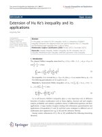
EDX analysis of the nanofibers shows that there is no
significant change in composition when compared to the
untreated glass surfaces. Microraman spectra of the unpro-
cessed substrate and processed nanofibers of silica glass are
presented in Fig. 4. The intensity of the spectrum of nanof-
ibers in laser-irradiated surfaces is much higher than the
untreated surface. Moreover, a slight increase in intensity at
603 cm
-1
peak is observed in the spectrum of nanofibers.
The Raman peaks at 487 and 603 cm
-1
are due to breathing
modes from 4- to 3-membered ring structures in the silica
network [17]. The increase in intensity corresponds to an
increase in relative number of these ring structures in the
glass network. Similar changes are observed for silica sub-
jected to femtosecond laser treatment [18].
Conclusions
In summary, we report a characteristic growth of nanofi-
bers of silica and borosilicate glass using femtosecond laser
radiation at 8 MHz repetition rate and a pulse width of
214 fs under atmospheric pressure. The fibers are grown
perpendicular to the substrate, intertwined and stands
above the surface. The nanofiber growth is explained by the
formation of nanojets in the molten material and drawn into
fibers by high plasma pressure. Further studies are required
to find the suitability of these nanofibers as waveguides for
nanophotonic applications.
Acknowledgments This research is funded by Natural Science and
Engineering Research Council of Canada.
References
1. L. Tong, R.R. Gattass, J.B. Ashcom, S. He, J. Lou, M. Shen,
I. Maxwell, E. Mazur, Nature 426, 816 (2003)
2. L.M. Tong, E. Mazur, J. Non-Cryst. Solids 354, 1240 (2008)
3. Y. Liu, S. Sagi, R. Chandrasekar, L.F. Zhang, N.E. Hedin,
H. Fong, J. Nanosci. Nanotechnol. 8, 1528 (2008)
4. J. Kameoka, S.S. Verbridge, H.Q. Liu, D.A. Czaplewski,
H.G. Craighead, Nano Lett. 4, 2105 (2004)
5. B. Tan, K. Venkatakrishnan, Opt. Express 17, 1064 (2009)
6. B. Tan, A. Dalili, K. Venkatakrishnan, Appl. Phys. A—Mater.
Sci. Process. 95, 537 (2009)
7. D.G. Georgiev, R.J. Baird, I. Avrutsky, G. Auner, G. Newaz,
Appl. Phys. Lett. 84, 4881 (2004)
8. F. Korte, J. Koch, B.N. Chichkov, Appl. Phys. A: Mater. Sci.
Process. 79, 879 (2004)
9. B Y. Adela, L.B. Robert, H. Anthony, A. Jacqueline, A.S. Howard,
S. Mengyan, M. Eric, Appl. Phys. Lett. 83, 3030 (2003)
10. A. Ben-Yakar, A. Harkin, J. Ashmore, R.L. Byer, H.A. Stone, J.
Phys. D Appl. Phys. 40, 1447 (2007)
11. S. Juodkazis, H. Misawa, O.A. Louchev, K. Kitamura, Nano-
technology 17, 4802 (2006)
12. J. Koch, F. Korte, T. Bauer, C. Fallnich, A. Ostendorf,
B.N. Chichkov, Appl. Phys. A: Mater. Sci. Process. 81, 325 (2005)
13. D.S. Ivanov, B. Rethfeld, G.M. O’Connor, T.J. Glynn, A.N.
Volkov, L.V. Zhigilei, Appl. Phys. A—Mater. Sci. Process. 92,
791 (2008)
14. W.D. Kingery, J. Am. Ceram. Soc. 42, 6 (1959)
15. B. Luk’yanchuk, W. Marine, Appl. Surf. Sci. 154, 314 (2000)
16. T. Takiya, I. Umezu, M. Yaga, M. Han, J. Phys: Conf. Ser. 59,
445 (2007)
17. F.L. Galeener, Solid State Commun. 44, 1037 (1982)
18. J.W. Chan, T. Huser, S. Risbud, D.M. Krol, Opt. Lett. 26, 1726
(2001)
Fig. 4 Microraman spectra of laser-processed and unprocessed silica
glass
1266 Nanoscale Res Lett (2009) 4:1263–1266
123