Báo cáo hóa học: " Self-Assembled Polymeric Micellar Nanoparticles as Nanocarriers for Poorly Soluble Anticancer Drug Ethaselen" pdf
Bạn đang xem bản rút gọn của tài liệu. Xem và tải ngay bản đầy đủ của tài liệu tại đây (301.74 KB, 10 trang )
NANO EXPRESS
Self-Assembled Polymeric Micellar Nanoparticles as Nanocarriers
for Poorly Soluble Anticancer Drug Ethaselen
Xinru Li Æ Zhuoli Yang Æ Kewei Yang Æ Yanxia Zhou Æ
Xingwei Chen Æ Yanhui Zhang Æ Fei Wang Æ
Yan Liu Æ Lijun Ren
Received: 10 June 2009 / Accepted: 19 August 2009 / Published online: 16 September 2009
Ó to the authors 2009
Abstract A series of monomethoxy poly(ethylene glycol)-
poly(lactide) (mPEG-PLA) diblock copolymers were syn-
thesized, and mPEG-PLA micelle was fabricated and used as
a nanocarrier for solubilization and delivery of a promising
anticancer drug ethaselen. Ethaselen was efficiently encap-
sulated into the micelles by the dialysis method, and the
solubility of ethaselen in water was remarkably increased up
to 82 lg/mL before freeze-drying. The mean diameter of
ethaselen-loaded micelles ranged from 51 to 98 nm with a
narrow size distribution and depended on the length of PLA
block. In vitro hemolysis study indicated that mPEG-PLA
copolymers and ethaselen-loaded polymeric micelles had no
hemolytic effect on the erythrocyte. The enhanced antitumor
efficacy and reduced toxic effect of ethaselen-loaded poly-
meric micelle when compared with ethaselen-HP-b-CD
inclusion were observed at the same dose in H
22
human liver
cancer cell bearing mouse models. These suggested that
mPEG-PLA polymeric micelle nanoparticles had great
potential as nanocarriers for effective solubilization of
poorly soluble ethaselen and further reducing side effects
and toxicities of the drug.
Keywords Monomethoxy poly(ethylene glycol)-poly
(lactide) Á Polymeric micelles Á Hemolysis Á Ethaselen Á
Antitumor efficacy
Introduction
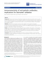
BBSKE (Fig. 1), chemically named (1,2-[bis(1,2-ben-
zisoselenazol-3(2H)-one)]ethane), is a novel organic sele-
nium compound, which is one of the derivatives of ethaselen
(For convenience, ethaselen is referred to BBSKE in this
study). It showed a positive antitumor activity and became a
potential anticancer agent with lower toxicity and side
effects [1, 2]. Unfortunately, ethaselen is poorly soluble in
water (2.57 lg/mL) and in commonly used organic solvents
such as methanol, ethanol, ether and chloroform. Its bio-
availability by oral administration is also considerably low.
Poor solubility creates major obstacles for formulations and
successful chemotherapy with ethaselen. Although several
methods including drug delivery systems were investigated
[3], developing a ethaselen delivery system for higher
selectivity, efficient solubilization and delivery of ethaselen
to the intended site without provoking any adverse reactions
is still a challenge.
Recently, polymeric micelles as a means to solubilize
poorly water-soluble drugs have attracted much attention
[4–6]. Generally, block copolymers with concentration
above the critical association concentration (CAC) self-
assemble into spherical polymeric micelles with a core–shell
structure in water: the hydrophobic segments aggregate to
form an inner core being able to accommodate hydrophobic
drugs with improved solubility by hydrophobic interactions;
X. Li Á Y. Zhou Á X. Chen Á Y. Zhang Á F. Wang Á Y. Liu (&)
Department of Pharmaceutics, School of Pharmaceutical
Sciences, Peking University, Xueyuan Road 38, 100191 Beijing,
Haidian, People’s Republic of China
e-mail:
Z. Yang
TEAM Academy of Pharmaceutical Sciences, Beijing,
People’s Republic of China
K. Yang
Department of Pharmaceutical Technology,
Institute of Pharmacy, University Jena, Jena, Germany
L. Ren
Department of Three, Institute of Chemical Defence,
Beijing, People’s Republic of China
123
Nanoscale Res Lett (2009) 4:1502–1511
DOI 10.1007/s11671-009-9427-2
the hydrophilic shell consists of a brush-like protective
corona that stabilizes the micelles in aqueous solution [7–9].
Polymeric micelles as novel drug vehicles present numerous
advantages, such as reduced side effects of anticancer drugs,
selective targeting, stable storage and prolonged blood cir-
culation time [9, 10]. Furthermore, polymeric micelles
possess a nanoscale size range with a narrow distribution,
and they can achieve higher accumulation at the target site
through an enhanced permeation retention effect (EPR
effect) [11]. They can protect drugs against premature
degradation in vivo owing to their core–shell architecture
[12, 13]. More importantly, polymeric micelles are fabri-
cated according to the physicochemical properties of drugs
and the compatibility between the core of micelles and drug
molecules [9, 14].
Biodegradable polymers, especially aliphatic polyesters
such as polylactide (PLA), poly(DL-lactic-co-glycolic
acid) (PLGA) and poly(e-caprolactone) (PCL) have
attracted much attention as biomaterials due to their bio-
compatibility, degradability and nontoxicity. They have
been applied in a wide range of biological systems ranging
from drug delivery to tissue engineering. PLA is the most
attractive candidate. A number of PLA-based amphiphilic
block copolymer micelles have been commonly used for
solubilization of hydrophobic drugs [15–17]. In our
research, PLA was also chosen as hydrophobic segment of
a block copolymer due to the higher compatibility between
the micelle core-forming PLA and the ethaselen molecules.
In previous work, our group has prepared micellar eth-
aselen formulation employing PLA-based copolymers and
characterized its drug loading contents and stability (data
not shown), which have demonstrated the compatibility
between the core of micelles and the ethaselen molecules.
Polyethylene glycol (PEG) is frequently chosen as a
hydrophilic segment to complement a hydrophobic seg-
ment due to the outstanding physicochemical and biologi-
cal properties including solubility in water and in organic
solvents, nontoxicity and filterability through kidney when
the molar mass is below 30,000 [18]. In addition, PEG is
able to form a palisade avoiding protein adsorption and
subsequent nonspecific uptake by the reticuloendothelial
system (RES) after intravenous injection.
In the present work, amphiphilic diblock copolymers
monomethoxy poly(ethylene glycol)-poly(lactide) (mPEG-
PLA) with molecular weight of 2,500, 5,000, 10,000 and
15,000 for PLA block were strategically designed and
synthesized by ring-opening polymerization of
D,L-dilac-
tide (
D,L-LA) in the presence of mPEG with molecular
weight of 5,000, respectively. The micelles preparation,
ethaselen solubilization and micelles properties were
investigated by the UV–vis assay, size measurement and
TEM in the micellar solution or in the state of lyophilized
powder. The CAC of mPEG-PLA was measured by using
the standard fluorescence substance of pyrene. Ethaselen-
loaded polymeric micelle was evaluated with respect to its
hemolytic toxicity and antitumor efficacy.
Materials and Methods
Materials
D,L-Dilactide was obtained from Fluka. Stannous 2-ethyl-
hexanoate and monomethoxy poly(ethylene glycol) with
molecular weight of 5,000 were purchased from Sigma–
Aldrich. All other chemicals and reagents were of analyt-
ical grade or better and used without further purification.
Animals
Male Kunming mice were obtained from Experimental
Animal Center of Peking University and acclimatized for
7 days after arrival. New Zealand White rabbits were
purchased from the same supplier and acclimatized for
2 days after arrival. All animals were provided with stan-
dard food and water ad libitum and were exposed to
alternating 12-h periods of light and darkness. Temperature
and relative humidity were maintained at 25 °C and 50%,
respectively. All care and handling of animals were per-
formed with the approval of Institutional Authority for
Laboratory Animal Care of Peking University.
Synthesis and Characterization of mPEG-PLA
mPEG-PLA diblock copolymers were synthesized from
D,L-dilactide and mPEG using stannous 2-ethylhexanoateas
as a catalyst as described previously [19] with modifica-
tion. Briefly,
D,L-dilactide was recrystallized in ethyl ace-
tate at room temperature until the racemic mixture melting
point was attained (124–126 °C), and mPEG was used
without further purification. The synthesized diblock
copolymers were referred to as mPEGx-PLAy. x and y
represented the weight-averaged molecular weight of the
mPEG and PLA block in kDa. mPEG5-PLA2.5, for
Fig. 1 Chemical structure of BBSKE
Nanoscale Res Lett (2009) 4:1502–1511 1503
123
example, consisted of a 5 kDa mPEG block connected to a
2.5 kDa PLA block.
mPEGx-PLAy was obtained by modulating the feed
ratio of
D,L-dilactide and mPEG. The molecular weight of
mPEG block was 5,000 (mPEG5). The molecular weights
of PLA block were 2,500 (PLA2.5), 5,000 (PLA5), 10,000
(PLA10) and 15,000 (PLA15), respectively. As a typical
example, the synthesis of mPEG5-PLA15 was carried out
as follows: to remove any trace of water, the starting
materials (5 g of mPEG5 and 15 g of
D,L-dilactide) were
each dissolved in 100 mL toluene in a round-bottomed
flask. About 40 mL of toluene was distilled off using a
water separator. The water-free solutions were united in a
three-neck flask, a precisely weighed amount of 25 mg
stannous 2-ethylhexanoate was added, and the mixture was
refluxed for 24 h at 120 °C under nitrogen atmosphere.
After toluene was distilled off with a rotary evaporator, the
residue was redissolved by the addition of appropriate
amount of chloroform, and vigorously stirred and precipi-
tated in diethyl ether at 0 °C, and then filtered. The pre-
cipitated polymer was dried in a desiccator at room
temperature under high vacuum. After drying for 2 days,
white solid powder of the copolymer was obtained. The
resulting copolymers were dissolved in CDCl
3
, and
1
H-
NMR spectra were taken at 300 MHz with trimethylsilane
(TMS) as internal reference standard using a Bruker
MSL2300 spectrometer (Bruker, Germany). The copoly-
mer molecular weights were determined by gel permeation
chromatography (GPC).
Determination of Critical Association Concentration
(CAC)
The CAC values of mPEG-PLA diblock copolymers were
determined by fluorescence spectroscopy using pyrene
(Fluka, [99%) as a hydrophobic probe [12]. Briefly, a
known amount of pyrene in acetone was added to each of a
series of 50 mL vials, and the acetone was evaporated, then
a known amount of various concentrations of mPEG-PLA
solutions in acetone were added to each vial, and the
acetone was evaporated. The appropriate amount of dis-
tilled water was then added to each vial to obtain polymeric
aqueous solutions with final concentration of 2.24 9 10
-4
–
224 mg/L. The final concentration of pyrene was
6.0 9 10
-7
mol/L. The sample solutions were kept in a
constant temperature shaking water bath at 37 °C for 24 h
to equilibrate the pyrene and the micelles, and cooled
overnight at room temperature. The solutions were filtered
with a 0.22 lm pore-sized filtration membrane (Millex-
GV, Millipore, USA). Fluorescence spectra of pyrene were
recorded with a Shimadzu RF-5301 PC fluorescence
spectrometer. The excitation wavelength used was 333 and
335 nm, and the emission spectra were recorded at
390 nm. The peak height intensity ratio (I
335
/I
333
) of the
peak of 335 nm to the peak of 333 nm was plotted against
the logarithm of polymer concentration. Two tangents were
then drawn, one to the curve at high concentrations and
another through the points at low concentrations. The CAC
value was taken from the intersection between the two
tangents.
Preparation of Ethaselen-Loaded Polymeric Micelles
Ethaselen-loaded polymeric micelle (ethaselen-PM) was
prepared by the dialysis method [20]. Briefly, 100 mg of
mPEG-PLA and 20 mg of ethaselen were dissolved in
45 mL dimethyl sulfoxide (DMSO). The mixture was
introduced into a dialysis bag (Spectrapor, MWCO =
3,500 g/mol), and then dialyzed against 4 L of physiological
saline, which was replaced every 12 h in the course over
48 h. The suspension in the dialysis bag was then filtered
through a 0.22 lm filter to remove aggregates. To determine
drug loading content (LC, w/w %) and entrapment efficiency
(EE, w/w %) of micelles, the ethaselen-loaded micelle
solution was lyophilized using PEG6000 as a lyoprotectant,
and then dissolved in DMSO by ultrasonication for 15 min,
and ethaselen content was measured with ultraviolet–visible
spectrophotometer (Agilent 8453, Agilent Technologies,
UK) at 320 nm. The LC and EE of the micelles were then
calculated based on the following formula:
LCð%Þ
¼
mass of ethaselenextracted fromfreezeÀdried micelles
totalmass of freezeÀdried micelle
Â100%
EE ð%Þ
¼
mass of ethaselen extracted from freezeÀdriedmicelles
total mass of Eb loadedmicelle initially used
Â100%:
Particle Size and Morphology Analysis
The average hydrodynamic radius of the micelles was
determined by dynamic light scattering (DLS) (Zetasizer
ZEN 3500, Malvern, UK). All DLS measurements were
done with an angle detection of 173° at 25 °C after diluting
the dispersion to an appropriate volume with water. The
results were the mean values of three experiments for the
same sample. The morphology of micellar nanoparticles
was also observed by transmission electron microscopy
(TEM) (Hitachi-500, Hitachi, Japan). To improve the
contrast, the samples were treated with a 1 wt% phospho-
tungstic acid solution for 2 h, deposited on copper grids,
and allowed to dry for 48 h before TEM examination.
1504 Nanoscale Res Lett (2009) 4:1502–1511
123
Physical Stability of Ethaselen-Loaded Micelles
The lyophilized powder and the polymeric micelle solution
were stored at room temperature. Their physical stability
was monitored over time by dynamic light scattering and
visually for signs of opalescence and precipitation. The
leakage percent was also measured by using the same
method as the determination of LC and EE described ear-
lier. In addition, the concentrated magnitude of micelle
solution by freeze-drying compared with initially prepared
micelle solution was evaluated by comparing the volume of
the reconstituted micelle solution of lyophilized powder
with that of initially prepared micelle solution. It should be
noted that the volume of the reconstituted micelle solution
of lyophilized powder was the smallest volume of physi-
ological saline in which the obtained lyophilized powder
was redissolved to produce a clear micelle solution.
Hemolysis Assay
The effect of the copolymers on the integrity of erythrocyte
membranes was investigated by in vitro hemolysis assay
[21]. The release of hemoglobin from the erythrocytes
(RBC) was used as a measure of toxicity of these copoly-
mers. Briefly, rabbit RBC was separated from 20 mL fresh
rabbit blood by centrifugation at 1,500 rpm for 15 min and
then washed three times with 20 mL of normal saline. The
purified RBC was resuspended in normal saline to obtain
2% (v/v) of RBC suspension. Then 2 mL of the RBC sus-
pension was incubated with 3 mL of the drug-free mPEG-
PLA micelle solution (copolymer concentration: 0.5 and
1 mg/mL) or ethaselen-loaded mPEG-PLA micelle solution
(ethaselen concentration: 0.1 and 0.2 mg/mL) at 37 °C for
1 h in an incubator shaker and then centrifuged at
5,000 rpm for 10 min. The percentage of hemolysis was
measured by UV–vis analysis of the supernatant at 576 nm
absorbance. Normal saline was used as the negative control
with 0% hemolysis, and distilled water was used as the
positive control with 100% hemolysis. All hemolysis data
points were presented as the percentage of the complete
hemolysis. Hemolysis percent (HP%) was calculated
according to the following equation:
HP% ¼
ABS
sample
À ABS
saline
ABS
distilled water
À ABS
saline
 100:
Assay for Antitumor Efficacy
Sixty of male Kunming mice (body weight = 18–22 g,
Peking University Experimental Animal Center, SPF-level,
Quality certificated Number: SCXK 2007-0008) were ran-
domly divided into six groups. And seven-day-old liver
cancer H
22
ascites (0.2 mL, 2 9 10
6
cells) were trans-
planted subcutaneously into the right axilla of each mouse
of the groups. The mice were treated as follows: negative
control group (normal saline); ethaselen-HP-b-CD group
(1 mg/kg body weight); ethaselen-loaded mPEG5-PLA2.5
micelle group (1 mg/kg body weight, low dose); ethaselen-
loaded mPEG5-PLA2.5 micelle group (2 mg/kg body
weight, middle dose); ethaselen-loaded mPEG5-PLA2.5
micelle group (4 mg/kg body weight, high dose). All the
groups were administered through the tail vein of animals
once daily for 10 days, starting 24 h after tumor implanta-
tion. At day 11, all the mice were killed by cervical dislo-
cation following by separation and measurement of the
tumor block. The antitumor efficacies of each formulation
were evaluated by tumor inhibition rate, which was calcu-
lated by the following formula: inhibition rate = (tumor
weight of test group - tumor weight of negative control
group)/tumor weight of negative control group 9 100%.
Statistical Analysis
All data were expressed as mean ± SD (standard devia-
tion). Comparisons between the group means were evalu-
ated by the unpaired t test. The statistical significance of
differences among more than two groups was determined
by one-way ANOVA. A value of p \ 0.05 was regarded as
significant.
Results and Discussion
Synthesis and Characterization of mPEG-PLA
mPEG-PLA copolymers were synthesized by ring-opening
polymerization of
D,L-dilactide by using mPEG as initiator.
Various chain lengths of PLA in the copolymers were
obtained by modulating the feed ratio of mPEG and
D,L-
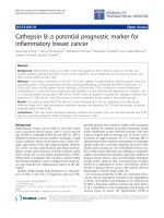
dilactide. Figure 2 showed the
1
H-NMR spectrum of
mPEG5-PLA15, which was representative for all synthe-
sized mPEG-PLAs: the peaks at 3.65 and 3.36 ppm corre-
sponded to methylene units and CH
3
O- in the mPEG
blocks, signals at 1.58 and 5.18 ppm could be attributed to
the hydrogen atoms of CH
3
- and CH-groups for PLA seg-
ments, respectively. From the peak integrity ratio of their
methylene and methyl groups, the mass ratio of repeating
units in mPEG and PLA blocks could be calculated in each
polymer. The results of this analysis were summarized in
Table 1. The values were very close to those of feed com-
positions. Furthermore, the molecular weight data (M
w
, M
n
)
and polydispersity indexes (PI = M
w
/M
n
) resulting from
GPC analysis of the copolymers after synthesis were also
listed in Table 1. The number-averaged molecular weight
(M
n
) of the mPEG and PLA blocks of each polymer was
calculated from
1
H-NMR data. The last column showed the
actual weight ratio of mPEG/PLA of the polymers (as
Nanoscale Res Lett (2009) 4:1502–1511 1505
123
determined from
1
H-NMR data). The PI of the mPEG-PLAs
indicated a narrow molecular weight distribution for all
polymers. Altogether the results confirmed that the
copolymers could be synthesized reliably. The yield of the
reactions ranged from 89 to 94%.
Polymeric micelles can be formed only when the block
copolymer concentration is higher than CAC, which
characterizes the micelle stability [6]. Compared with low
molar mass surfactant micelles, polymeric micelles are
generally more stable, exhibiting a remarkably lower CAC
[22]. They are liable to retain thermodynamic stability even
after intravenous injection, which induces severe dilution
[7, 23]. Table 2 summarized the CAC values of the various
synthesized mPEG-PLA diblock copolymers ranging from
0.96 to 2.31 9 10
-7
mol/L or 1.71 to 2.12 mg/L. These
values appeared much lower than those of low molar mass
surfactants, indicating that micelles formed from mPEG-
PLA copolymers as drug carriers could preserve stability
without dissociation after dilution, which was of major
interest for intravenous injection. Moreover, the hydro-
philicity of mPEG-PLA copolymers mainly depending on
the mass ratio of mPEG/PLA or mPEG content had much
Fig. 2
1
H NMR spectrum of
mPEG5-PLA15 copolymer in
CDCl
3
Table 1 Survey on the composition of mPEG-PLAs
Feed ratio
mPEG/DL-LA
mPEGx-PLAyM
w
a
M
n
b
PI
c
M
n
(mPEG) M
n
(PLA) Found ratio
mPEG/PLA
2:1 mPEG5-PLA2.5 9,300 7,500 1.16 5,000 2,450 67:33
1:1 mPEG5-PLA5 13,500 11,500 1.26 5,000 4,560 52:48
1:2 mPEG5-PLA10 19,600 13,800 1.54 5,000 9,650 34:66
1:3 mPEG5-PLA15 25,300 16,200 1.66 5,000 15,060 25:75
a
Number-average molecular weight of mPEGx-PLAy
b
Weight-average molecular weight of mPEGx-PLAy
c
Polydispersity index
1506 Nanoscale Res Lett (2009) 4:1502–1511
123
influence on the CAC value [24]. As shown in Table 2, the
CAC values of copolymers appeared to decrease with
increasing in PLA block length. mPEG5-PLA15 had longer
hydrophobic PLA blocks and thus, could self-assemble
more easily to form micelles, leading to lower CAC values.
This was consistent with previous reports [25, 26].
Characterization of mPEG-PLA Micelles
Core–shell type polymeric micelles were prepared by
dialysis method. Size and size distribution of micelles were
measured by DLS. The mean diameter ranged from 44 to
85 nm for drug-free micelles and from 51 to 98 nm for
drug-loaded micelles (Table 2). It appeared that the micelle
size gradually increased with increasing in the length of
PLA chains. This result was in agreement with the char-
acteristic of amphiphilic copolymer micelles, i.e., the
shorter the hydrophobic block length, the smaller the
micelles. It might be attributed to the fact that it is difficult
to form compact polymeric micelles for amphiphilic
copolymers with longer hydrophobic chain length. In
addition, similar to drug-loaded micelles reported earlier
[15, 27], the size of drug-loaded micelles was about 10 nm
bigger than that of drug-free micelles, suggesting that
ethaselen molecules were trapped in the hydrophobic inner
cores and that these entrapped ethaselen molecules
increased the average size of ethaselen-loaded polymeric
micelles. Importantly, the PI values of ethaselen-loaded
polymeric micelles were fairly low, indicating a mono-
disperse distribution [28]. Among four polymeric micelles,
the size of mPEG5-PLA2.5 micelles was smallest.
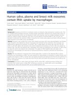
The morphology of the ethaselen-loaded micelles was
examined by using TEM. As shown in Fig. 3, these
micelles were clearly distinguished as bright and discrete
spots with nearly spherical shape and equal granule. The
diameter derived from TEM was lower than that from DLS,
which could be assigned to the dehydration and shrinkage
of the micelles during drying. These results indicated that
the ethaselen-loaded polymeric micelles were well dis-
persed in aqueous media and formed homogeneous nano-
sized micelle structures.
Drug loading content (LC, w/w%) and entrapment
efficiency (EE, w/w%) of polymeric micelles were calcu-
lated by absorbance of ethaselen at 320 nm from UV. The
results were also summarized in Table 2. It could be found
that mPEG5-PLA2.5 micelles exhibited the highest LC and
EE, about 16 and 24%, respectively, while only 1.61% of
LC for hydroxylpropyl beta cyclodextrin (HP-b-CD)
inclusion [3]. The solubility of ethaselen in water was
improved to be about 82 lg/mL before freeze-drying,
which was about 32 times higher than that of ethaselen in
water. Notably, the results presented in Table 2 were not
the general trend that LC and EE of polymeric micelles
increased with increasing hydrophobic chain length. This
finding may be assigned to the factors contributing to LC
and EE. In general, LC and EE depend on the composition
Table 2 Characterization of micelles prepared from a series of mPEG-PLA copolymers
Copolymers Drug-free Drug-loaded CAC
c
(10
-7
mol/L)
LC
d
(%) EE
e
(%)
d
a
(nm) PI
b
d (nm) PI
mPEG5-PLA2.5 44.33 0.137 51.57 0.122 2.31 16.43 24.22
mPEG5-PLA5 61.80 0.164 68.41 0.154 1.87 14.21 20.29
mPEG5-PLA10 79.36 0.127 84.25 0.106 1.45 15.52 19.89
mPEG5-PLA15 84.55 0.091 97.54 0.118 0.96 14.67 20.32
a
Diameter of micelles
b
Polydispersity index
c
Critical association concentration
d
Drug loading content
e
Entrapment efficiency
Fig. 3 TEM photography of ethaselen-loaded mPEG5-PLA2.5
micelles (9100,000)
Nanoscale Res Lett (2009) 4:1502–1511 1507
123
of the copolymers, initial diblock copolymeric concentra-
tion or the feed weight ratio of the drug to the copolymer,
solvent used in formulation process and micelle prepara-
tion method and so on. Therefore, the length of hydro-
phobic chains was not the only one factor influencing LC
and EE. When the factors mentioned earlier except the
composition of the mPEG-PLA copolymers are optimal for
all copolymers, the length of hydrophobic chains is a vital
one. In case of our experiment, all micelles were prepared
in the same conditions, which may not be optimal for all
copolymers, thereby the result was not the general trend.
The other possible reason might be filtration step removed
larger particles. This phenomenon was also seen in previ-
ous report [29]. Nevertheless, the result would not have
influence on the subsequent experiments.
The physical stabilities of both the lyophilized powder
and micelle solution of ethaselen were evaluated by deter-
mination of leakage percent and mean size during storage.
Figure 4 showed the representative change profiles of
leakage percent and mean size. It could be seen that the
lyophilized powder exhibited far improved physical stability
compared to micelle solution. It could be stored at room
temperature for at least 2 months without significant chan-
ges of entrapment efficiency and micelle size (p [0.05). In
addition, the micelle solution could be concentrated by
reconstitution of lyophilized samples in physiological saline
at least one time when compared with the micelle solution
before freeze-drying, hence, the solubility of the drug in
water would be further increased. However, the physical
stability of mPEG-PLA micelle solutions was poor at room
temperature and retained only for 3 days. After storage for
5 days the drug started to leak from micelles (Fig. 4a), the
leakage percent ranged from 1.37 to 2.49%. And the initial
transparent micelle solution became translucent or turbid,
thereby the size could not be determined by DLS. The
leakage percent markedly increased to 9.1–17.4% and sed-
imentation appeared after 15 days. The similar results were
reported previously [30, 31], where mPEG-PLA micelles
maintained their stability after drug loading only for several
hours or days. It was attributed to the lost of a hydrophilic
and hydrophobic balance, which was the critical influence
factor for micelle stability owing to the encapsulation of
hydrophobic drugs, and drug-loaded PLA-PEG polymer
micelles broke up to result in drug precipitation. For this
reason, it was suggested that the obtained polymeric
micelles might be freeze-dried for a longer storage and
reconstituted in aqueous solutions prior to use.
Hemolytic Toxicity of Micelles
The block copolymers in this study were amphiphilic and
could solubilize lipids or insert into phospholipid mem-
branes to destabilize them [32–34]. When the micelles are
injected into the blood for drug delivery or drug detoxifi-
cation, detrimental interaction of these particles with blood
constituents must be avoided. Therefore, the hemolysis
assay would give additional information about the bio-
compatibility in the case of an in vivo application.
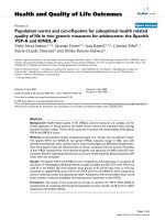
Although the concentrations of the copolymers were very
high, micelles did not show any observational hemolytic
activities in the RBC in the experimental range. Figure 5
showed the hemolytic activities of drug-free micelle solu-
tions and ethaselen-PM with different PLA block lengths.
It was observed that the hemolytic percentage of mPEG-
PLA diblock copolymers seemed not to depend on their
concentrations. The hemolytic percentage was always
lower than 5% in the whole tested concentration range.
According to the Guiding Principles of Hemolysis Test
[H]GPT4-1, the samples were considered as hemolytic if
the hemolytic percentage was above 5%. Consequently, the
mPEG-PLA diblock copolymers had no hemolytic effect
on the RBC. The results also suggested that the mPEG-
PLA micelles were suitable for intravenous administration.
In addition, the hemolytic activity of ethaselen-loaded
Fig. 4 Leakage percent (a) and mean size (b) changes of ethaselen-
loaded mPEG5-PLA2.5 micelles in 2 months at 25°
1508 Nanoscale Res Lett (2009) 4:1502–1511
123
polymeric micelles slightly increased, and it appeared to
ethaselen-loaded polymeric micelles that the hemolysis
was ethaselen concentration-dependent to a certain degree.
The dilution stability and hemolytic potential of micelle
formulation indicated that ethaselen-loaded polymeric
micelles could be administered intravenously at a wide
range of drug concentrations so that a precise dilution is not
required.
In vivo Antitumor Efficacy
According to the results earlier mentioned and favorable
pharmacokinetics characteristic (dada not shown) ethaselen-
loaded mPEG5-PLA2.5 micelle was chosen to evaluate the
in vivo antitumor efficacy with the animal tumor models set
up by inoculation of H
22
human liver cancer cell by mea-
suring tumor weight or relative tumor inhibition rate after
tumor implantation. None of the animals treated with tested
formulations died during the experimental period. As shown
in Table 3, ethaselen-HP-b-CD and ethaselen-loaded
mPEG5-PLA2.5 micelle significantly inhibited the growth
of tumor compared with the control group (p \ 0.05). After
11 days, ethaselen-HP-b-CD at 1 mg/kg suppressed tumor
growth by 36.93%, and ethaselen-loaded mPEG5-PLA2.5
micelle at 1 mg/kg, 2 and 4 mg/kg suppressed tumor growth
by 45.10, 55.60 and 58.47%, respectively, compared with
the control group, indicating that ethaselen-loaded mPEG5-
PLA2.5 micelle inhibited tumor growth in a dose-dependent
manner to some extent. More importantly, ethaselen-loaded
mPEG5-PLA2.5 micelle had significantly stronger inhibi-
tory effect on the tumor growth compared with ethaselen-
HP-b-CD at the same dose of ethaselen (p \ 0.05), rein-
forcing that ethaselen-loaded polymeric micelles had more
effective antitumor activity than ethaselen-HP-b-CD. This
could be attributed to the ‘‘enhanced permeation and reten-
tion’’ (EPR) effect of nano-sized micellar delivery systems
[35–37]. Fast growing tumor tissues need a tremendous
amount of oxygen and nutrients supplied by blood vessels.
They release special growth factors including vascular
endothelial cell growth factor (VEGF) to facilitate neo-
vascularization. As a result, many new vessels are formed,
but their cell junctions are not as tight as those of normal
tissues. Ethaselen-loaded mPEG5-PLA2.5 micelle with a
size of about 51 nm was likely to freely pass through the
endothelial junctions of the capillaries in tumor tissue. In
addition, the prevention of hydrophobic interactions
between vascular endothelial cell in tumor tissues and the
drug by hydrophilic and flexile PEG shell of polymeric
micelle made the drug enter into the tumor tissue success-
fully [38]. On the other hand, it has been reported that CD
Fig. 5 Hemolysis activity of
copolymers and ethaselen-
loaded mPEG-PLA micelles
Table 3 In vivo antitumor effect of ethaselen-loaded mPEG5-PLA2.5 micelle and ethaselen-HP-b-CD in H
22
human liver cancer cell bearing
mice model (
"
x Æs, n = 10)
Formulation Dose (mg/kg) Body weight (g) Tumor weight (g) Inhibition rate (%)
Before administration After administration
Physiological saline 0 20.38 ± 0.94 28.01 ± 2.24 1.29 ± 0.26 –
Ethaselen-HP-b-CD 1 19.94 ± 0.86 26.94 ± 2.11* 0.81 ± 0.13* 36.93
Ethaselen-micelle (L) 1 20.52 ± 0.66 28.10 ± 1.65 0.71 ± 0.12*
,
** 45.10
Ethaselen-micelle (M) 2 20.92 ± 0.58 28.75 ± 1.76 0.57 ± 0.17*
,
**
,
*** 55.60
Ethaselen-micelle (H) 4 20.20 ± 0.42 28.42 ± 1.81 0.53 ± 0.11*
,
**
,
***
,
**** 58.47
* p \ 0.05, versus physiological saline; ** p \0.05, versus ethaselen-HP-b-CD; *** p \0.05, versus ethaselen-micelle (L); **** p [ 0.05,
versus ethaselen-micelle (M)
Nanoscale Res Lett (2009) 4:1502–1511 1509
123
couldn’t influence the pharmacokinetics of drugs [39, 40],
while much more drug would be accumulated in the solid
tumor region due to delay of the circulation time of drugs for
polymeric micelles [22, 27, 39, 40]. A combined effect of
improved pharmacokinetics and enhanced cellular uptake
would be the main reason for the suppression of tumor
growth. It could also be seen that the middle and high dose
groups suppressed the tumor growth more significantly than
the low one (p \ 0.05), but there was no significant differ-
ence of the tumor inhibition rate for the middle and high dose
group (p [ 0.05). Taken together, the finding that ethaselen-
loaded mPEG5-PLA2.5 micelle at 2 mg/kg significantly
improved antitumor efficacy could have important clinical
implications.
The body weight of mice treated with physiological
saline without drug continuously increased due to probably
its nontoxic effect as well as the rapid growth of tumor
(Table 3). In ethaselen-loaded polymeric micelle treated
groups at all doses, the body weight of mice did not sig-
nificantly increase. This might be due to their antitumor
efficacies thereby the slower growth of tumor. On the other
hand, ethaselen-HP-b-CD group appeared to have signifi-
cant weight loss, resulting from the toxic effect of the drug,
indicating that this micelle-based drug delivery system
could reduce unwanted side effects of anticancer drugs
during cancer therapy.
Overall, ethaselen-loaded polymeric micelle possessed
improved antitumor activity and reduced toxic side effects
of anticancer drug than ethaselen-HP-b-CD mainly due to
the enhanced vascular permeability and EPR effect, and
passive targeting function although they do not have active
targeting function [41–43]. Furthermore, tumor tissues are
characterized with leaky blood vessels and the premature
lymphatic drainage [44]. Resultantly, we speculated that
ethaselen-loaded polymeric micelles would also be a
superior formulation for other tumor models.
Nevertheless, ethaselen, as a poorly water-soluble drug,
might be physically incorporated into the inner core of the
polymeric micelles by hydrophobic interactions. Further, it
may avoid RES recognition due to a size smaller than
200 nm. Therefore, it is advantageous for ethaselen to be
efficiently encapsulated in micelles. In case of our exper-
iment, the EE was unfavorable, thereby the enhancing of
hydrophobic interaction between ethaselen and the inner
core of the polymeric micelles would be vital by chemical
modification the hydrophobic block of mPEG-PLA.
Conclusions
We have successfully synthesized a series of mPEG-PLA
copolymers. It was found that monodispersed micelles self-
assembled from mPEG-PLA could effectively solubilize
the anticancer drug ethaselen when compared with HP-b-
CD inclusion. The hemolysis assay indicated that ethase-
len-loaded mPEG-PLA micelles could be administered
intravenously at a wide range of drug concentrations. In
mice, ethaselen-loaded polymeric micelles showed
noticeable antitumor efficacy, and reduced the toxic effect
of the drug, compared with ethaselen-HP-b-CD inclusion.
These results suggested that polymeric micelles might be
an effective drug delivery system for ethaselen for cancer
chemotherapy. Nevertheless, the micelles could still be
improved, especially with respect to enhancing their
entrapment efficiency by modification of inner core of
micelles, which are in progress. Better drug retention in the
micelle core is a key to ensure prolonged circulation time
and eventually maximize drug accumulation at the tumor
site via the enhanced permeation and retention effect.
Acknowledgments The authors wish to thank Prof. Huihui Zeng
from Department of Chemical Biology, Peking University for friendly
providing ethaselen.
References
1. C.J. Shi, L.Z. Yu, F.G. Yang, H.H. Zeng, Biochem. Biophy. Res.
Commun. 309, 578 (2003)
2. J. Yan, S.J. Deng, H.H. Zeng, J. Chin. Pharm. Sci. 13, 199 (2004)
3. H.M. Cui, C.G. Zhang, F.L. Wu, Chin. Pharm. Sci. 42, 765
(2007)
4. V.P. Torchilin, Adv. Drug Deliv. Rev. 54, 235 (2002)
5. A. Ro
¨
sler, G.W. Vandermeulen, H.A. Klok, Adv. Drug Deliv.
Rev. 53, 95 (2001)
6. M. Jones, J. Leroux, Eur. J. Pharm. Biopharm. 48, 101 (1999)
7. X. Shuai, T. Merdan, A.K. Schaper, F. Xi, T. Kissel, Bioconjug.
Chem. 15, 441 (2004)
8. T. Riley, C.R. Heald, S. Stolnik, M.C. Garnett, L. Illum, S.S.
Davis, Langmuir 19, 8428 (2003)
9. L. Liu, C. Li, X. Li, Z. Yuan, Y. An, B. He, J. Appl. Polym. Sci.
80, 1976 (2001)
10. E. Pierri, K. Avgoustakis, J. Biomed. Mater. Res. A 75, 639
(2005)
11. K. Kataoka, A. Harada, Y. Nagasaki, Adv. Drug Deliv. Rev. 47,
113 (2001)
12. M. Wilhelm, C.L. Zhao, Y.C. Wang, R.L. Xu, M.A. Winnik, J.L.
Mura et al., Macromolecules 24, 1033 (1991)
13. R. Barreiro-Iglesias, L. Bromberg, M. Temchenko, T.A. Hatton,
A. Concheiro, C. Alvarez-Lorenzo, J. Control. Release 97, 537
(2004)
14. P.Z. Li, X.R. Li, H.X. Zhou, Y.H. Zhang, F. Wang, Y. Liu, Chin.
J. New Drug 18, 262 (2009)
15. Y. Zhang, T. Jin, R.X. Zhuo, Colloids Surf. B: Biointerfaces 44,
104 (2005)
16. E.S. Lee, K. Na, Y.H. Bae, J. Control. Release 91, 103 (2003)
17. D. Le Garrec, S. Gori, L. Luo, D. Lessard, D.C. Smith, M.A.
Yessine et al., J. Control. Release 99, 83 (2004)
18. Y. Hu, X. Jiang, Y. Ding, L. Zhang, C. Yang, J. Zhang et al.,
Biomaterials 24, 2395 (2003)
19. A. Lucke, J. Tessmar, E. Schnell, G. Schmeer, A. Go
¨
pferich,
Biomaterials 21, 2361 (2000)
20. B.G. Yu, T. Okano, K. Kataoka, G. Kwon, J. Control. Release 53,
131 (1998)
1510 Nanoscale Res Lett (2009) 4:1502–1511
123
21. S. Zhu, F. Qian, Y. Zhang, C. Tang, C. Yin, Eur. Polym. J. 43,
2244 (2007)
22. G.S. Kwon, K. Kataoka, Adv. Drug Deliv. Rev. 16, 295 (1995)
23. S.Y. Kim, I.G. Shin, Y.M. Lee, J. Control. Release 56, 197 (1998)
24. S. Li, M. Vert, Macromolecules 36, 8008 (2003)
25. X. Zhang, J.K. Jackson, H.M. Burt, Inter. J. Pharm. 132, 195
(1996)
26. B. Jeong, Y. Han Bae, S. Wan Kim, Colloids Surf. B: Biointer-
faces 16, 185 (1999)
27. K.W. Yang, X.R. Li, Z.L. Yang, P.Z. Li, F. Wang, Y. Liu, J.
Biomed. Mater. Res. Part A 87A, 140 (2009)
28. A. Harada, K. Kataoka, Macromolecules 28, 5294 (1995)
29. Y.C. Chang, I.M. Chu, Eur. Polym. J. 44, 3922 (2008)
30. K.M. Huh, S.C. Lee, Y.W. Cho, J. Lee, J.H. Jeong, K. Park, J.
Control. Release 101, 59 (2005)
31. H.M. Burt, X. Zhang, P. Toleikis, L. Embree, W.L. Hunter,
Colloids Surf. B: Biointerfaces 16, 161 (1999)
32. J. Zastre, J. Jackson, M. Bajwa, R. Liggins, F. Iqbal, H. Burt, Eur.
J. Pharm. Biopharm. 54, 299 (2002)
33. R. Savic, L. Luo, A. Eisenberg, D. Maysinger, Science 300, 615
(2003)
34. M.K. Pratten, J.B. Lloyd, G. Ho
¨
rpel, H. Ringsdorf, Die Makro-
mol. Chem. 186, 725 (1985)
35. J.G. Shiah, M. Dvora
´
k, P. Kopeckova
´
, Y. Sun, C.M. Peterson, J.
Kopecek, Eur. J. Cancer 37, 131 (2001)
36. J.J. Shiah, Y. Sun, C.M. Peterson, J. Kopecek, J. Control. Release
61, 145 (1999)
37. M. Yokoyama, S. Fukushima, R. Uehara, K. Okamoto, K. Kat-
aoka, Y. Sakurai et al., J. Control. Release 50, 79 (1998)
38. Z. Zuo, Y.K. Tam, J. Diakur, L.I. Wiebe, J. Pharm. Pharm. Sci. 5,
292 (2002)
39. H.W. Frijlink, E.J. Franssen, A.C. Eissens, R. Oosting, C.F. Lerk,
D.K. Meijer, Pharm. Res. 8, 380 (1991)
40. G. Piel, B. Evrard, T. Van Hees, L. Delattre, Int. J. Pharm. 180,
41 (1999)
41. Y. Jeong, H.S. Na, K.O. Cho, H.C. Lee, J.W. Nah, C.S. Cho, Int.
J. Pharm. 365, 150 (2009)
42. K. Greish, A. Nagamitsu, J. Fang, H. Maeda, Bioconjug. Chem.
16, 230 (2005)
43. H. Maeda, J. Wu, T. Sawa, Y. Matsumura, K. Hori, J. Control.
Release 65, 271 (2000)
44. Y. Bae, K. Kataoka, Adv. Drug Deliv. Rev. 61, 768 (2009)
Nanoscale Res Lett (2009) 4:1502–1511 1511
123