Báo cáo hóa học: "Single-component Polymer Nanocapsules for Drug Delivery Application" docx
Bạn đang xem bản rút gọn của tài liệu. Xem và tải ngay bản đầy đủ của tài liệu tại đây (163.6 KB, 3 trang )
NANO SPOTLIGHTS
Single-component Polymer Nanocapsules for Drug Delivery
Application
Published online: 18 July 2008
Ó to the author 2008
The design of delivery vehicles for transporting anticancer
drugs to tumor sites has gained traction during the past few
decades. Utilizing polymer-based materials has played an
important role in the development of such systems, largely
because of the ability to prepare polymers with tailored
properties, including biocompatibility, size, structure, and
functionality. Several polymer-based vehicles have been
reported, including polymer particles, polymer-based
micelles, polymer-drug conjugates, and polymer nanocap-
sules. These systems can facilitate higher payloads,
prolong the circulation time of the drugs, improve drug
targeting and solubility, and provide controlled-release of
the therapeutics into the blood stream or the targeted tumor
tissues. Among these, the polymer capsules are particularly
attractive candidates for drug delivery applications.
‘‘Layer-by-layer (LbL) assembly processes have been
widely used by our group and others to prepare polymer
capsules with well-defined chemical and structural prop-
erties. In LbL assembly, a nonporous sacrificial colloidal
template is generally used to sequentially deposit multiple
polymer layers one after another, followed by removal of
the core, leading to well-defined polymer capsules with
nanometer-thick walls’’, Prof. Frank Caruso, Director of
the Centre for Nanoscience and Nanotechnology at the
University of Melbourne, Australia, explains to Nano-
spotlight. ‘‘Multiple assembly steps required in the LbL
assembly often require the use of more than one polymer
and can make the process relatively intensive and time-
consuming, particularly for the synthesis of thick walled
polymer capsules.’’
To overcome these limitations, Prof. Caruso’s team used
a novel silica particle template with a solid core and
mesoporous shell (SC/MS) for polymer nanocapsules
synthesis. Prof. Caruso further explains to Nanospotlight:
‘‘The use of SC/MS template allows a ‘single polymer’ to
be infiltrated in the mesoporous shells of SC/MS particles
in a ‘single macromolecular assembly step’ by solution
adsorption, followed by cross-linking of the macromole-
cules in the mesoporous silica shells, and subsequent
removal of the SC/MS templates, thus leading to mon-
odispersed, single-component and thick-walled polymer
nanocapsules (see Scheme 1).’’
‘‘This approach offers a number of advantages over the
conventional LbL technique to prepare capsules. Firstly,
uniform nanocapsules of various macromolecules are
obtained by a single macromolecular assembly step of a
single macromolecule type, eliminating the need for mul-
tiple polymers and/or multiple polymer adsorption steps.
Secondly, the nanocapsules derived from the SC/MS tem-
plates have porous walls that are significantly thicker than
those prepared by LbL assembly (e.g., more than an order
of magnitude for a single adsorption step), thus offering a
simple approach to regulate the physical properties (e.g.,
structure, permeability, payloads) of the nanocapsules.’’
The SC/MS particles can be prepared with different
particle size, shell thickness, and solid core composition
(e.g., silica, gold and Fe
3
O
4
nanoparticles). The size and
thickness of the nanocapsules can be controlled by
choosing the appropriate size SC/MS template and type
and molecular weight of the polymers. For instance, the
thickness of the capsule shells increases as the molecular
weight of the PAH decreases because of more efficient
adsorption of smaller species of PAH in the mesoporous
shells of SC/MS templates (*45 nm and *16 nm thick
capsules with a diameter of *400 nm size were obtained
for PAH of 5 and 70 kDa, respectively). Furthermore, the
macromolecules assembled in the capsules can be stabi-
lized via engineered cleavable covalent linker (e.g.,
123
Nanoscale Res Lett (2008) 3:265–267
DOI 10.1007/s11671-008-9145-1
disulfide), which would add tunable stability and degra-
dability characteristics to the capsules, leading to another
level of control over the release properties of encapsulated
substances.
The researchers have recently published their findings in
Nano Letters (Wang et al., 2008, 8, 1741–1745) and
demonstrated the general applicability of this approach by
preparing nanocapsules using various macromolecules,
including synthetic polyelectrolytes [polyallylamine
hydrochloride (PAH)], polypeptides [poly-
L-lysine (PLL),
and poly-
L-glutamic acid (PGA)], and polypeptide-drug
conjugates [PGA-Doxorubicin (Dox)].
The researchers also investigated the applicability of
thick-walled polymer nanocapsules for tumor therapy via
drug delivery. They prepared drug-loaded polymer nano-
capsules according to the outlined approach by
preconjugating a model anticancer drug (Dox) to a model
polymer system (PGA), which is structurally related to
natural proteins and is generally considered to be bio-
compatible, nonimmunogenic and biodegradable. The
potential of Dox-loaded PGA nanocapsules in tumor ther-
apy applications was demonstrated via in vitro capsule
degradation and Dox-release studies at conditions resem-
bling those within the living cells, nanocapsule uptake by
LIM1215 human colorectal tumor cells, and delivery of the
anticancer drug into the tumor cells, leading to tumor cell
death.
Bansal notes that it is highly desirable for antitumor
applications, that the size of the delivery vehicle is in the
range capable of exploiting the ‘leaky’ nature of tumor
blood vessels, which have pore diameters of between 400
and 600 nm, allowing accessibility to target tumor cells. In
this study, sub-500 nm size capsules were used for this
purpose. PGA-Dox nanocapsules were internalized in large
numbers by LIM1215 colorectal tumor cells, with most of
the internalized capsules being taken up by the lysosomes.
The uptake of the PGA-Dox particles and capsules by
subcellular lysosomes suggests that once internalized,
hydrolytic enzymes present in the reducing environment of
the lysosomes would facilitate Dox release from nano-
capsules, thus causing tumor cell death.
Drug-release studies confirmed that the Dox was grad-
ually released from PGA-Dox capsules under lysosomal
conditions (pH 5.8/10 mM carboxypeptidase) with a near-
linear drug release kinetics over the first 24 h. ‘‘Moreover,
for a drug delivery vehicle to be highly effective, it is
desirable that it should not degrade in the blood stream;
however, it should be easily degraded and release its cargo
after reaching the lysosomal compartments of the tumor
cells’’, notes Bansal: ‘‘Our control experiments showed
negligible passive release of Dox from nanocapsules under
physiological conditions in the absence of lysosomal
hydrolases.’’
The tumor cell death studies on LIM1215 human colo-
rectal tumor cells showed that the PGA-Dox capsules were
highly effective in controlling tumor cell growth ([85%
cell death within 16 h). When LIM1215 tumor cells were
treated with equivalent amounts of PGA-Dox polymer
conjugates, insignificant tumor cell death was observed.
The researchers speculate that the high negative charge of
the small PGA-Dox polymer chains restricts their uptake
by the negatively charged cell membranes and hence leads
to reduced cell death. However, PGA-Dox loaded SCMS
particles and PGA-Dox capsules can be internalized into
the tumor cells via endocytosis due to their larger sizes,
thus highlighting the important role that polymer capsules
might play in drug delivery applications.
The researchers highlight that although free Dox was
found to be as efficient as PGA-Dox capsules in causing
tumor cell death, Dox is known to cause high systemic
toxicity when administered into animals in its free form.
The PGA-Dox capsules can provide an added advantage of
controlled release, wherein Dox molecules will be released
only after capsules reach the target tumor site, thus mini-
mizing any systemic toxicity. Moreover, significantly
higher amounts and more than one type of drug can be
principally loaded in PGA capsules in a controlled manner,
due to the presence of a large number of free –COOH
groups. In addition, the remaining free –COOH groups of
PGA-Dox capsules can be easily conjugated to targeting
moieties to target PGA-Dox capsules to various tumors,
which is the subject of further investigation.
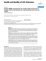
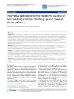
Scheme 1 Schematic representation of the preparation of single-
component macromolecular capsules by using solid core and
mesoporous shell (SC/MS) silica particles as templates. The process
involves the infiltration of polyelectrolyte or polymer-drug conjugates
into mesoporous shells of SC/MS particles (step 1), followed by
crosslinking of the infiltrated polymer chains (step 2) and subsequent
removal of the SC/MS silica template (step 3), leading to thick-walled
polyelectrolyte or drug-conjugated polymer nanocapsules
266 Nanoscale Res Lett (2008) 3:265–267
123
PGA-Dox capsules shown in this study provide a unique
drug delivery system: they remain stable at physiological
pH and are amenable to deconstruction (by disassembly of
PGA-Dox chains due to lysosomal reducing environments)
and degradation (by lysosomal hydrolases) in response to
chemical stimuli within living cells, thereby delivering
Dox to LIM1215 human colorectal tumor cells and causing
tumor cell death. The attachment of targeting ligands to the
drug-conjugated capsules through established coupling
protocols will further provide functional capsules for tar-
geted drug delivery applications.
Overall, the simple, efficient, and general nature of
the approach for the fabrication of a new class of mon-
odispersed, single-component and thick-walled polymer
nanocapsules, coupled with the capability to synthesize a
wide range of materials with tunable properties, and the
additional ability to post-functionalize the thick capsule
shells, provides exciting new opportunities for designing
advanced capsules for use in a range of therapeutic and
diagnostic applications.
Kimberly Sablon
Nanoscale Res Lett (2008) 3:265–267 267
123