Báo cáo hóa học: "Exploring the Immunotoxicity of Carbon Nanotubes" ppt
Bạn đang xem bản rút gọn của tài liệu. Xem và tải ngay bản đầy đủ của tài liệu tại đây (414.85 KB, 7 trang )
NANO REVIEW
Exploring the Immunotoxicity of Carbon Nanotubes
Yanmei Yu Æ Qiu Zhang Æ Qingxin Mu Æ
Bin Zhang Æ Bing Yan
Received: 14 June 2008 / Accepted: 16 July 2008 / Published online: 20 August 2008
Ó to the authors 2008
Abstract Mass production of carbon nanotubes (CNTs)
and their applications in nanomedicine lead to the
increased exposure risk of nanomaterials to human beings.
Although reports on toxicity of nanomaterials are rapidly
growing, there is still a lack of knowledge on the potential
toxicity of such materials to immune systems. This article
reviews some existing studies assessing carbon nanotubes’
toxicity to immune system and provides the potential
mechanistic explanation.
Keywords Nanotube Á Nanoparticle Á Nanomaterial Á
Immunity Á Cytokine Á Macrophage Á Animal study Á
Cell culture Á Nano-combinatorial chemistry Á Nanotoxicity
Introduction
Carbon nanotubes (CNTs) are cylindrical molecules with a
length of up to micrometers and a diameter of 0.4–2 nm for
single-walled carbon nanotubes (SWNTs) and 2–100 nm for
coaxial multi-walled carbon nanotubes (MWNTs). CNTs
have long been speculated and tested as new materials for
biological and biomedical applications. Because of their
abilities to bind cells and across the cell membrane [1, 2],
functionalized CNTs can be used as nanovectors for drug
delivery and cancer phototherapy [3]. On the other hand,
when injected intravenously, CNTs will interact directly
with immune cells and proteins in blood and tissues.
Immunotoxicity is one of the consequences of using nano-
particles. Immunity is the function of the body to recognize
and eliminate pathogens and foreign particles. The immune
system is a tightly regulated network of organs, cells, and
molecules. This system functions through cell-to-cell con-
tacts and communicates via soluble mediators such as
cytokines [4], which play a key role in immune defense,
immunological homeostasis, and immune surveillance.
In Vivo Studies
Potential hazards from carbon nanotube production are
associated with CNT inhalation and epidermal exposure.
Lung Toxicity
SWNT was shown to cause lung inflammation, granuloma
formation [5–20], and mortality by intratracheal instillment
into mice at a dose of 0.1 or 0.5 mg per mice [5]. Mortality in
this study was suggested to be caused by the toxicity of
residual catalyst particles in the sample. However, mortality
found in a rat study [6] was attributed to the blockage of the
upper airways by the instillate and not inherently by SWNTs.
In another report, SWNTs and MWNTs were intrana-
sally instilled into BALB/C mice [7]. Authors detected a
general inflammatory response through air hyper-respon-
siveness and changes in macrophage cell count in
interstitial spaces of the lung. It was also reported that
intratracheally instilled MWNTs into the lung of rats [8]or
pharyngeal aspiration of SWCNT into mice [9, 10] caused
Yanmei Yu and Qiu Zhang contributed equally to this work.
Y. Yu Á Q. Zhang Á Q. Mu Á B. Zhang Á B. Yan
School of Pharmaceutical Sciences, Shandong University,
Jinan, China
Q. Mu Á B. Yan (&)
Department of Chemical Biology and Therapeutics, St. Jude
Children’s Research Hospital, 332 North Lauderdale Street,
Memphis, TN 38105, USA
e-mail:
123
Nanoscale Res Lett (2008) 3:271–277
DOI 10.1007/s11671-008-9153-1
persistent inflammation and fibrosis, and eventually
granulomas.
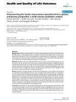
A recent research showed that intratracheal instillation
of 0.5 mg of SWMTs into male ICR mice induced alveolar
macrophage activation [11], various chronic inflammatory
responses, and severe pulmonary granuloma formation
(Fig. 1). The uptake of SWNT into the macrophages is able
to activate various transcription factors such as nuclear
factor-NF-jB and activator protein 1 (AP-1). This led to
oxidative stress, the release of proinflammatory cytokines,
the recruitment of leukocytes, the induction of protective
and antiapoptotic gene expression, and the activation of T
cells. The resulting innate and adaptive immune responses
might explain the chronic pulmonary inflammation and
granuloma formation in vivo caused by SWNTs.
Five different samples of MWNTs were intratracheally
instilled into guinea pigs [12]. Significant pulmonary tox-
icity was observed. Multiple lesions in all CNT-exposed
animals were also observed. The authors concluded that, in
conjunction with their previous report [21], the exposure
time was critical for induction of lung pathological
changes.
The inhalation of MWNTs at particle concentrations
ranging from 0.3 to 5 mg/m
3
did not result in significant
lung inflammation or tissue damage in C57BL/6 adult (10-
to 12-week) male mice, but caused systemic immune
function alterations [13].
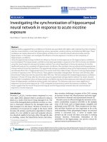
The potential mechanism of pulmonary toxicity of
nanoparticles [14] is tentatively explained in Fig. 2. The
initial acute inflammatory reaction is probably triggered by
damage to pulmonary epithelial type I cells. The response
includes a robust neutrophilic pneumonia followed by
recruitment and activation of macrophages. The unusual
feature of the response is a very early switch from the acute
phase of the response to fibrogenic events resulting in
significant pulmonary deposition of collagen and elastin.
This is accompanied by a characteristic change in the
production and release of proinflammatory (tumor necrosis
factor-a, interleukin-1h) to anti-inflammatory profibrogenic
cytokines (transforming growth factor-b, interleukin-10).
The inflammatory and fibrogenic responses were accom-
panied by a detrimental decline in pulmonary function and
enhanced susceptibility to infection. Other mechanistic
explanations were also provided [10, 15].
Fig. 1 Hematoxylin and eosin staining of mouse lung tissue. (a, e)
Fluronic F-68-treated group acts as the solvent control. (b, f) Early
response (3 days) of the mouse lung tissue to a single dose of 0.5 mg
of SWNT. (c, d, g, h) Two weeks response of the mouse lung tissue to
a single dose of 0.5 mg of SWNT. (f, g) SWNT-loaded foamy-like
macrophages in the alveolae; (h) multifocal macrophage-containing
granuloma around the sites of SWNT aggregates. (a–d) Original
magnification 9100, bar = 100 um; (e–h) 9400. The black arrows
shown in panels b and c indicate the SWNT-loaded foamy-like
macrophages. Reprinted with permission from [23]. Copyright (2004)
American Chemical Society
272 Nanoscale Res Lett (2008) 3:271–277
123
One recent study presented that more dispersed SWNT
structures altered pulmonary distribution and response
[20]. In this test, a dispersed preparation of SWNT with a
mean length of 0.69 micron was given by pharyngeal
aspiration to C57BL/6 mice. Macrophage phagocytosis of
SWNT was rarely observed at any time point. No granu-
lomatous lesions or epithelioid macrophages were detected.
The results demonstrate that dispersed SWNT are rapidly
incorporated into the alveolar interstitium and that they
produce an increase in collagen deposition.
A new research showed that oxidative stress induced by
SWNT in C57BL/6 mice and Vitamin E deficiency
enhances pulmonary inflammatory response [22].
Although there is evidence that it takes energy and
agitation to release fine CNT particles into the air and the
current handling procedures do not produce significant
quantities of airborne CNT [23], an extreme caution is
highly recommended.
Skin Toxicity
If CNTs penetrate the stratum corneum cells and become
lodged into the viable epidermal cell layers of the skin,
they may enter the keratinocytes directly or trigger the
production of proinflammatory cytokines or initiate other
sequela [24].
Studies on skin irritation by CNTs are extremely limited
at this time [16–19]. Skin irritation was evaluated by
conducting two routine dermatological tests among vol-
unteers. Their tests showed no irritation in comparison to a
CNT-free soot control, and it was concluded that no special
precautions have to be taken while handling these carbon
nanostructures [25].
In another experiment, CNTs were subcutaneously
implanted into BALB/c mice and CD4? and CD8? T-cells
in peripheral blood, and the histopathological changes on
skin tissues were measured [13]. SWNTs were shown to
activate major histocompatibility complex (MHC) class I
pathway of antigen–antibody response system resulting in
the appearance of an edematous aspect after one week. After
2 weeks, high values in CD4? and CD4?/CD8? were
detected indicating an activated MHC class II. No death or
body weight changes were observed within 3 months [26].
One recent study illustrated that the length of CNT
modulates inflammation response. When 0.1 mg of CNTs
were implanted in the subcutaneous tissue in the thoracic
region in each rat [27], there were more inflammation
around 825-CNTs (long) than that around 220-CNTs
(short) since macrophages could envelop 220-CNTs more
readily than 825-CNTs. However, no severe inflammatory
response such as necrosis, degeneration, or neutrophil
infiltration in vivo was observed for both CNTs examined
throughout the experimental period.
In Vitro Studies
Potential clinical use of CNTs suggests that a wide range of
biological systems must be evaluated. Some in vitro studies
are summarized below.
Cell Uptake
Mammalian cells have at least five ways to internalize
macromolecules or nanoparticles: phagocytosis (via man-
nose receptor-, complement receptor-, Fcc receptor-, and
scavenger receptor-mediated pathways), macropinocy-
tosis, clathrin-mediated endocytosis, caveolin mediated
pathways, and clathrin/caveolin-independent endocytosis
[28–30].
MWNTs were used to deliver amphotericin B (AmB) to
Human Jurkat lymphoma T cell by linking AmB and
fluorescein to CNTs [31]. Maximum fluorescence was
observed after just 1 h of incubation, indicating fast cell
uptake of FITC-AmB-MWNTs. Most conjugates were
found in the cytoplasm and around the nuclear membrane.
Recently, carbon nanotubes have been shown to traverse
cellular membranes by endocytosis and shuttle biological
molecules, including DNA, siRNA, and proteins, into
Fig. 2 In the lung, the initial target for CNTs is probably type I
epithelial cells whose necrotic death stimulates a proinflammatory
response and recruitment of inflammatory cells. Interactions include
oxidative burst due to activation of NADPH oxidase and possible
interactions of nanoparticles with microbial pathogens. NADPH
oxidase complex is activated in macrophages during inflammation
and acts as the major source for generation of reactive oxygen
species, such as superoxide O
2
–d radicals that disproportionate to
form hydrogen peroxide (H
2
O
2
). Transition metals, through their
interactions with O
2
–d and H
2
O
2
, act as catalysts for the formation of
highly reactive hydroxyl (OH
Á
) radicals. Oxidatively modified lipids
generated by cyclooxygenase (COX-2) and lipooxygenase (LOX)
participate in amplification of the inflammatory response via recruit-
ment of new inflammatory cells
Nanoscale Res Lett (2008) 3:271–277 273
123
immortalized cancer cells [3, 16–19, 21, 32–38]. Single-
walled carbon nanotubes (SWNTs) may serve as nonviral
molecular transporters for the delivery of siRNA into
human T cells and primary cells. Another report presented
that SWNT and SWNT-streptavidin conjugates can be
taken up into human promyelocytic leukemia (HL60) cells
and human T cells (Jurkat) [34]. The uptake was also
suggested to be through endocytosis [34, 39].
However, evidence was also presented that the cell
uptake of CNTs was through nonendocytosis pathway
based on the lack of temperature dependence and lack of
inhibition from endocytosis-specific inhibitor [35, 40].
CNT-induced Oxidative Stress
The oxidative stress is induced by exposing cells to CNTs.
According to the hierarchical oxidative stress hypothesis,
the lowest level of oxidative stress is associated with the
induction of antioxidant and detoxification enzymes
(Table 1)[41]. The genes that encode the phase II enzymes
are under the control of the transcription factor Nrf-2. Nrf-2
activates the promoters of phase II genes via an antioxidant
response element [41]. Defects or aberrancy of this
protective response pathway may determine disease sus-
ceptibility during ambient particle exposure. At higher
levels of oxidative stress, this protective response is
overtaken by inflammation and cytotoxicity (Table 1).
Inflammation is initiated through the activation of proin-
flammatory signaling cascades (e.g., mitogen-activated
protein kinase and NF-jB cascades), whereas programmed
cell death could result from mitochondrial perturbation and
the release of proapoptotic factors.
Cytotoxicity
Using guinea pig alveolar macrophages, cytotoxicity was
detected with SWNTs and MWNTs [16–19, 42]. High
concentration of pristine and oxidized MWNTs have been
shown to generate loss of viability of the human Jurkat T
cells and human peripheral blood lymphocytes [43]. A
comparative study on the toxicity of pristine and oxidized
MWNT in human Jurkat T leukemia cells has shown that
the latter were more toxic [43].
However, in a different report, highly purified SWNTs
was taken up slowly by human macrophage cells with low
toxicity [44]. Similarly, CNTs were found across the cell
membrane of rat macrophages (NR8383) [2], but no
cytotoxicity was observed.
Cherukuri et al. [33] investigated the uptake of pristine
SWNT into the mouse J774.1A macrophage-like cell line
via near infrared fluorescence microscopy. The study
reported that the macrophage-like cells appeared to phag-
ocytose SWNT at a rate of approximately one SWNT per
second, without any apparent cytotoxicity [33]. The SWNT
remained fluorescent, suggesting that the macrophage-like
cells were not capable of breaking them down within the
time period of study. This result is inconsistent with pre-
vious macrophage investigations [8, 42].
Complement Activation
The biochemical cascade that removes pathogens, known as
the complement system, consists of two pathways. The
classical complement pathway is activated by antigen–
antibody complexes, whereas the alternative pathway is
antibody independent. Nanoliposomes and engineered car-
bon nanotubes can activate the complement system.
Intriguingly, both SWNTs and double-walled carbon
nanotubes (DWNTs) stimulated the classical pathway [45],
but only DWNTs triggered the alternative pathway. The
mechanism of this selective complement activation
remains unknown.
Inflammatory Response
Interactions between chemically modified SWNTs and B
and T lymphocytes as well as macrophages were studied at
a concentration of 10–50 lg/mL [1]. These functionalized
SWNTs were taken up by cells without inducing toxicity.
Authors found that only the less soluble ones preserved
lymphocytes’ functionality while provoking secretion of
proinflammatory cytokines by macrophages.
Table 1 The hierarchical oxidative stress model
Level of oxidative stress
Low Medium High
Response pathways: Anti-oxidant defense Inflammation Cytotoxicity
Signaling pathway: Nrf-2 MAP kinase NF-jB cascade Mitochondrial PT pore
Genetic response: Anti-oxidant response element AP-1 NF-jB N/A
Outcome: Phase II enzymes Cytokines chemokines Apoptosis
274 Nanoscale Res Lett (2008) 3:271–277
123
Nitric oxide, TNF-a, and IL-8 are key inflammatory
mediators when macrophages are activated. Interestingly,
after CNTs were taken up by murine and rat macrophage
cells, no inflammatory mediators such as NO, TNF-a, and
IL-8 were observed [2, 44]. However, a dose- and time-
dependent increase of intracellular reactive oxygen species
and a decrease of the mitochondrial membrane potential
occurred. Incubation with the purified CNTs had no effect.
Inflammatory responses were also observed when human
epidermal keratinocytes or human skin fibroblast was
exposed to CNTs [24, 46–50]. The mechanism is likely due
to the production of reactive oxygen species, leading to the
activation of the NF-jB. Another research demonstrated that
30 nm CNTs penetrated skin tissue within 2–3 min during
microimaging MRI experiments [51]. Cell adhesion function
was reported to be altered by nanotubes [52].
Antigenicity
Biotechnology-derived pharmaceuticals can cause specific
antibody response (antigenicity). Antibodies are special-
ized proteins produced by plasma B cells in response to an
antigen or foreign materials. The immune response to a
composite nanoparticle-based drug potentially involves
antibodies for both the particles and the surface groups.
To date, there are very limited studies on the antigenicity
of functionalized nanoparticles and none of them report
CNT-specific antibody generation [16–19]. CNTs func-
tionalized with a peptide antigen (B cell epitope from the
foot-and-mouth disease virus, FMDV) was studied [53, 54].
The CNT-FMDV was recognized by antibodies equally well
as the free peptide and the immunization of mice with the
CNT-FMDV clearly enhanced anti-FMDV peptide antibody
responses. Moreover, no immune response to CNTs was
detected, which is an important issue in view of epitopic
suppression when peptide antigen carriers are used.
A variety of factors, such as particle surface properties
and functional groups, may ultimately affect the systemic
antigenicity of CNTs when it was used as drug carrier.
Causes of CNTs Immunotoxicity and its Control
Size, shape, structure, and surface all play a role in defining
nanotoxicity. The aggregation status and p–p electronic
effects may also be important in case of CNTs. CNTs have
an unusually large surface area/mass ratio. The large sur-
face area gives the particles a greater area to contact with
the cellular membrane and proteins, as well as a greater
capacity for absorption and transport of bioactive sub-
stances. The large surface area also suggests that chemistry
modification may impact significantly the biological
activities of CNTs [16–19].
Impurity Effect
Contamination is one of the reasons for CNTs’ potential
harm. The presence of such impurities interferes with our
study on the inherent toxicity of CNTs. Transition metals
are particularly effective as catalysts of oxidative stress in
cells, tissues, and biofluids. A report [55] compared the
interactions of two types of SWNT (1) iron-rich (non-
purified) SWCNT (26% of iron) and (2) iron-stripped
(purified) SWNT (0.23 wt% of iron) with RAW264.7
macrophages. Each type of SWNT was able to generate
intracellular production of superoxide radicals or nitric
oxide in the cells. Less pure iron-rich SWNT were more
effective in generating hydroxyl radicals, and superoxide
radicals, accumulating lipid hydroperoxides, and causing
significant loss of intracellular low molecular weight thiols
(GSH). Therefore, the inflammatory responses caused by
nanotubes with metals can be particularly damaging. Oxi-
dative species generated during inflammatory response can
interact with transition metals to trigger redox-cycling
cascades with a remarkable oxidizing potential to deplete
endogenous reserves of antioxidants and induce oxidative
damage to macromolecules.
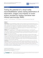
Surface Modifications
Chemical modifications of nanoparticles surface holds
promise to confer them improved biocompatibility. Nano-
combinatorial chemistry approach was used to generate a
MWNT library containing 80 different surface modifica-
tions [56]. In addition to the successful regulation of
protein binding and cytotoxicity, they also showed differ-
ent activity in activating immune systems as measured by
nitric oxide generation (Fig. 3). Compared with the pre-
cursor, MWNT-COOH, many modified MWNTs exhibited
lower immune responses [33]. More biocompatible and
immune-friendly nanomedicine carriers can be developed
through iterative screening and optimization studies.
Conclusions
As nanotechnology-based products and nanomedicine
research are relatively new, there are currently no stan-
dardized guidelines for assessing immunotoxicity
generated by CNTs. Many important issues need to be
addressed in order to develop a new generation of nan-
omedicines. Available data (Table 2) strongly suggest that
CNTs enter cells, cause ROS, and interact with the immune
systems. A better understanding of the mechanisms of
CNTs’ interaction with immune systems is still needed for
developing and optimizing biocompatible nanomedicine
carriers.
Nanoscale Res Lett (2008) 3:271–277 275
123
Acknowledgments This work was supported by Shandong Uni-
versity, the American Lebanese Syrian Associated Charities
(ALSAC), and St. Jude Children’s Research Hospital.
References
1. H. Dumortier, S. Lacotte, G. Pastorin, R. Marega, W. Wu,
D. Bonifazi et al., Nano Lett. 6, 1522 (2006). doi:10.1021/nl061
160x
2. K. Pulskamp, S. Diabate, H.F. Krug, Toxicol. Lett. 168,58
(2007). doi:10.1016/j.toxlet.2006.11.001
3. N.W.S. Kam, M. O’Connell, J.A. Wisdom, H.J. Dai, Proc. Natl
Acad. Sci. USA 102, 11600 (2005). doi:10.1073/pnas.0502680102
4. D.R. Roth, D. Roman, P. Ulrich, A. Mahl, U. Junker, E. Perentes,
Exp. Toxicol. Pathol. 57, 367 (2006). doi:10.1016/j.etp.2006.03.012
5. C.W. Lam, J.T. James, R. McCluskey, R.L. Hunter, Toxicol. Sci.
77, 126 (2004). doi:10.1093/toxsci/kfg243
6. D.B. Warheit, B.R. Laurence, K.L. Reed, D.H. Roach, G.A.M.
Reynolds, T.R. Webb, Toxicol. Sci. 77, 117 (2004). doi:10.1093/
toxsci/kfg228
Fig. 3 Immune responses induced by the functionalized-MWNT
library. Library members were assayed for MWNT-induced NO
release (a and b) in macrophages at either 50 lg/mL or 200 lg/mL.
The basal level of NO release by LPS (100 ng/mL) is marked on
panels a and b. The f-MWNT library-induced NO release in the
presence of LPS (100 ng/mL) is shown as vertical bars. The
precursor, Carboxylated MWNT, generated high response as shown
at the lower right corner. The plots are presented in library format
showing all amine and acylator building blocks used in synthesizing
this MWNT library
Table 2 Pathophysiology and toxicity effects of CNTs
a
Experimental NM effects Possible pathophysiological outcomes
ROS generation
a
Protein, DNA and membrane injury,
a
oxidative stress
b
Oxidative stress
a
Phase II enzyme induction, inflammation,
b
mitochondrial perturbation
a
Mitochondrial perturbation
a
Inner membrane damage,
a
permeability transition (PT) pore opening,
a
energy failure,
a
apoptosis,
a
apo-necrosis, cytotoxicity
Inflammation
a
Tissue infiltration with inflammatory cells,
b
fibrosis,
b
granulomas,
b
atherogenesis,
b
acute phase protein expression (e.g., C-reactive protein)
Uptake by reticulo-endothelial system
a
Asymptomatic sequestration and storage in liver,
a
spleen, lymph nodes,
b
possible organ enlargement and dysfunction
Protein denaturation, degradation
a
Loss of enzyme activity,
a
auto-antigenicity
Nuclear uptake
a
DNA damage, nucleoprotein clumping,
a
autoantigens
Uptake in neuronal tissue
a
Brain and peripheral nervous system injury
Perturbation of phagocytic function,
a
‘‘particle overload,’’
mediator release
a
Chronic inflammation,
b
fibrosis,
b
granulomas,
b
interference in clearance of
infectious agents
b
Endothelial dysfunction, effects on blood clotting
a
Atherogenesis,
a
thrombosis,
a
stroke, myocardial infarction
Generation of neoantigens, breakdown in immune tolerance Autoimmunity, adjuvant effects
Altered cell cycle regulation
DNA damage
Proliferation, cell cycle arrest, senescence
Mutagenesis, metaplasia, carcinogenesis
a
Effects supported by limited experimental evidence;
b
Effects supported by limited clinical evidence. From Andre Nel, Tian Xia, Lutz Madler,
Ning Li, Toxic Potential of Materials at the Nanolevel. Science 2006, 311:622–627. Reprinted with permission from AAAS
276 Nanoscale Res Lett (2008) 3:271–277
123
7. F. Raymond, J.R. Hamilton, M.C. Buford, M.B. Wood, B. Arnone,
M. Morandi et al., Nanotoxicology 1, 104 (2007). doi:10.1080/
17435390600926939
8. J. Muller, F. Huaux, N. Moreau, P. Misson, J.F. Heilier, M. Delos
et al., Toxicol. Appl. Pharmacol. 207, 221 (2005)
9. A.A. Shvedova et al., AJP-Lung Cell Mol. Physiol. 298, L698
(2005)
10. Shvedova AA, Kisin ER, Murray AR, Kommineni C, Castranova
V, Fadeel B, et al., Toxicol. Appl. Pharmacol. (2008) (Epub
ahead of print)
11. C.C. Chou, H.Y. Hsiao, Q.S. Hong, C.H. Chen, Y.W. Peng, H.W.
Chen et al., Nano Lett. 8, 437 (2008). doi:10.1021/nl0723634
12. A. Huczko, H. Lange, M. Bystrzejewski, P. Baranowski, Carbon
Nanostruct. 13, 141 (2005). doi:10.1081/FST-200050691
13. L.A. Mitchell, J. Gao, R.V. Wal, A. Gigliotti, S.W. Burchiel, J.D.
McDonald, Toxicol. Sci. 101, 179 (2008)
14. V.E. Kagan, H. Bayir, A.A. Shvedova, Nanomed. Nanotechnol.
Biol. Med. 1, 313 (2005). doi:10.1016/j.nano.2005.10.003
15. J.B. Mangum, E.A. Turpin, A. Antao-Menezes, M.F. Cesta, E.
Bermudez, J.C. Bonner, Part Fibre Toxicol. 3, 15 (2006). doi:
10.1186/1743-8977-3-15
16. D. Cui, J. Nanosci. Nanotechnol. 7, 1298 (2007)
17. W. Wei, A. Sethuraman, C. Jin, N.A. Monteiro-Riviere, R.J. Nara-
yan, J. Nanosci. Nanotechnol. 7, 1284 (2007). doi:10.1166/jnn.
2007.655
18. S.K. Smart, A.I. Cassady, G.Q. Lu, D.J. Martin, Carbon 44, 1034
(2006)
19. L. Lacerda, A. Bianco, M. Prato, K. Kostarelos, Adv. Drug Deliv.
Rev. 58, 1460 (2006). doi:10.1016/j.addr.2006.09.015
20. R.R. Mercer, J. Scabilloni, L. Wang, E. Kisin, A.R. Murray, D.
Schwegler-Berry et al., Am. J. Physiol. Lung Cell Mol. Physiol.
294, L87 (2008). doi:10.1152/ajplung.00186.2007
21. N.W.S. Kam, Z. Liu, H.J. Dai, J. Am. Chem. Soc. 127, 12492
(2005). doi:10.1021/ja053962k
22. A.A. Shvedova, E.R. Kisin, A.R. Murray, O. Gorelik, S. Arepalli,
V. Castranova et al., Toxicol. Appl. Pharmacol. 221, 339 (2007).
doi:10.1016/j.taap.2007.03.018
23. A.D. Maynard, P.A. Baron, M. Foley, A.A. Shvedova, E.R. Kissin,
V. Castranova, J. Toxicol. Environ. Health A 67, 87 (2004). doi:
10.1080/15287390490253688
24. N.A. Monteiro-Riviere, A.O. Inman, Carbon 44, 1070 (2006).
doi:10.1016/j.carbon.2005.11.004
25. A. Huczko, H. Lange, Fuller Sci. Technol. 9, 247 (2001)
26. S. Koyama, M. Endo, Y.A. Kim, T. Hayashi, T. Yanagisawa, K.
Osaka et al., Carbon 44, 1079 (2006). doi:10.1016/j.carbon.2005.
08.006
27. Y. Sato, A. Yokoyama, K-i Shibata, Y. Akimoto, S-i Ogino, Y.
Nodasaka et al., Mol. Biosyst. 1, 176 (2005). doi:10.1039/b502429c
28. A. Aderem, D.M. Underhill, Annu. Rev. Immunol. 17
, 593
(1999). doi:10.1146/annurev.immunol.17.1.593
29. S.D. Conner, S.L. Schmid, Nature 422, 37 (2003). doi:10.1038/
nature01451
30. J.M. Blander, R. Medzhitov, Nat. Immunol. 7, 1029 (2006). doi:
10.1038/ni1006-1029
31. K. Kostarelos, L. Lacerda, G. Pastorin, W. Wu, S. Wieckowski, J.
Luangsivilay et al., Nat. Nanotechnol. 2, 108 (2007). doi:10.1038/
nnano.2006.209
32. N.W.S. Kam, Z.A. Liu, H.J. Dai, Angew. Chem. Int. Ed. 45, 577
(2006). doi:10.1002/anie.200503389
33. P. Cherukuri, S.M. Bachilo, S.H. Litovsky, R.B. Weisman, J. Am.
Chem. Soc. 126, 15638 (2004). doi:10.1021/ja0466311
34. N.W.S. Kam, T.C. Jessop, P.A. Wender, H.J. Dai, J. Am. Chem.
Soc. 126, 6850 (2004). doi:10.1021/ja0486059
35. D. Pantarotto, J. Briand, M. Prato, A. Bianco, Chem. Commun.
(Camb) 10, 16 (2004). doi:10.1039/b311254c
36. A. Bianco, K. Kostarelos, C.D. Partidos, M. Prato, Chem.
Commun. (Camb) 10, 571 (2005). doi:10.1039/b410943k
37. N.W.S. Kam, Z. Liu, H.J. Dai, Angew. Chem. 118, 591 (2006).
doi:10.1002/ange.200503389
38. M. Prato, K. Kostarelos, A. Bianco, Acc. Chem. Res. 41,60
(2008). doi:10.1021/ar700089b
39. Z. Liu, M. Winters, M. Holodniy, H.J. Dai, Angew. Chem. Int.
Ed. 46, 2023 (2007). doi:10.1002/anie.200604295
40. Bianco A, Kostarelos K, Partidos CD, Prato M, Chem. Commun.
(Camb) 571 (2005)
41. G.G. Xiao, M.Y. Wang, N. Li, J.A. Loo, A.E. Nel, J. Biol. Chem.
278, 50781 (2003). doi:10.1074/jbc.M306423200
42. G. Jia, H.F. Wang, L. Yan, X. Wang, R.J. Pei, T. Yan et al.,
Environ. Sci. Technol. 39, 1378 (2005). doi:10.1021/es048729l
43. M. Bottini,S. Bruckner,K. Nika,N. Bottini, S. Bellucci, A. Magrini
et al., Toxicol. Lett. 160, 121 (2006). doi:10.1016/j.toxlet.2005.
06.020
44. S. Fiorito, A. Serafino, F. Andreola, P. Bernier, Carbon 44, 1100
(2006). doi:10.1016/j.carbon.2005.11.009
45. C. Salvador-Morales, E. Flahaut, E. Sim, J. Sloan, M.L.H. Green,
R.B. Sim, Mol. Immunol. 43, 193 (2006). doi:10.1016/j.molimm.
2005.02.006
46. S.K. Manna, S. Sarkar, J. Barr, K. Wise, E.V. Barrera, O. Jeje-
lowo et al., Nano Lett. 5, 1676 (2005). doi:10.1021/nl0507966
47. A.A. Shvedova, V. Castranova, E.R. Kisin, D. Schwegler-Berry,
A.R. Murray, V.Z. Gandelsman et al., Toxicol. Environ. Health
66, 1909 (2003). doi:10.1080/713853956
48. N.A. Monteiro-Riviere, R.J. Nemanich, A.O. Inman, Y.Y. Wang,
J.E. Riviere, Toxicol. Lett. 155, 377 (2005). doi:10.1016/j.toxlet.
2004.11.004
49. A.A. Shvedova, V. Castranova, E.R. Kisin, D. Schwegler-Berry,
A.R. Murray, V.Z. Gandelsman et al., J. Toxicol. Environ. Health
A 66, 1909 (2003)
50. L. Ding, J. Stilwell, T. Zhang, O. Elboudwarej, H. Jiang, J.P.
Selegue et al., Nano Lett. 5, 2448 (2005). doi:10.1021/nl051748o
51. Sharma R, Shetty K, Liang R, Chen CJ, US Nanotech 2007
Conference, Wednesday May 23.
52. D.X. Cui, F.R. Tian, C.S. Ozkan, M. Wang, H.J. Gao, Toxicol.
Lett. 155, 73 (2005). doi:10.1016/j.toxlet.2004.08.015
53. D. Pantarotto, C.D. Partidos, J. Hoebeke, F. Brown, E. Kramer,
J.P. Briand et al., Chem. Biol. 10, 961 (2003). doi:10.1016/
j.chembiol.2003.09.011
54. D. Pantarotto, C.D. Partidos, R. Graff, J. Hoebeke, J.P. Briand,
M. Prato et al., J. Am. Chem. Soc. 125, 6160 (2003). doi:10.1021/
ja034342r
55. V.E. Kagan, Y.Y. Tyurina, V.A. Tyurin, N.V. Konduru, A.I.
Potapovich, A.N. Osipov et al., Toxicol. Lett. 165, 88 (2006). doi:
10.1016/j.toxlet.2006.02.001
56. H. Zhou, Q. Mu, N. Gao, A. Liu, Y. Xing, S. Gao et al., Nano
Lett. 8, 859 (2008). doi:10.1021/nl0730155
Nanoscale Res Lett (2008) 3:271–277 277
123