Báo cáo hóa học: " Fatty Acid Binding Domain Mediated Conjugation of Ultrafine Magnetic Nanoparticles with Albumin Protein" docx
Bạn đang xem bản rút gọn của tài liệu. Xem và tải ngay bản đầy đủ của tài liệu tại đây (357.54 KB, 6 trang )
NANO EXPRESS
Fatty Acid Binding Domain Mediated Conjugation of Ultrafine
Magnetic Nanoparticles with Albumin Protein
D. K. Bora Æ P. Deb
Received: 4 September 2008 / Accepted: 11 November 2008 / Published online: 22 November 2008
Ó to the authors 2008
Abstract A novel bioconjugate of stearic acid capped
maghemite nanoparticle (c-Fe
2
O
3
) with bovine serum
albumin (BSA) was developed by taking recourse to the
fatty acid binding property of the protein. From FT-IR
study, it was found that conjugation took place covalently
between the amine group of protein molecule and carboxyl
group of stearic acid capped maghemite nanoparticle. TEM
study further signified the morphology of the proposed
nanobioconjuagte. The binding constant of nanoparticle
with protein molecule was evaluated from the optical
property studies. Also, magnetic measurement (M–H)
showed retaining of magnetic property by significant val-
ues of saturation magnetization and other hysteretic
parameters.
Keywords Bioconjugate Á Maghemite nanoparticle Á
Bovine serum albumin Á Covalent interaction Á
Fatty acid binding domain
Introduction
Bioconjugation of magnetic nanoparticles is basically done
to make it compatible for numerous biomedical applica-
tions such as MRI contrast enhancement, drug delivery,
detoxification of biological fluids, immunoassay, tissue
repair, hyperthermia etc. [1–6]. Besides these, bioconjugate
systems are also being applied in various large-scale bio-
processes such as nucleic acid detachment, protein
separation, magnetic biosensor etc. [7–9]. All these biore-
lated applications require the use of magnetic nanoparticles
that should have size smaller than 10 nm with overall
narrow particle size distribution, so that the particles have
uniform and unique properties [10]. This is mainly because
of the fact that particles at this size range have the
advantage of showing well-established magnetic properties
which reduces the possibility of particle aggregation upon
magnetic attraction in a magnetic dispersion [11]. For
fabricating a bioconjugate, it generally involves lots of
surface chemistry work [12]. Normally, these are synthe-
sized by modifying the nanoparticle surface with some
chemical linker molecule, so that it can further interact
with next incoming bio molecular entity with the help of
free functional group of linker molecule. This procedure
was already well established but supposed to be having
some difficulty in the sense of stability of linker molecule
due to various biochemical events. This might occur mostly
under in vivo condition when applying the bioconjugate
system in the targeted delivery of neoplastic compounds to
tumor cell.
To overcome this difficulty, we proposed some biolog-
ically evolved linker moiety (fatty acid binding domain) to
fabricate a novel bioconjugate in covalent fashion by uti-
lizing the molecular recognition property of bio molecular
system, such as bovine serum albumin (BSA) protein. It is
supposed to be an important substitute over the synthetic
linker in designing the bioconjugate covalently so that it
can be applied gently under preceding condition. This can
also be called as natural anchor molecule that is functional
in several of its biological activities. Here, BSA is chosen
as the material of interest for the bioconjugation purpose
D. K. Bora Á P. Deb (&)
Department of Physics, Tezpur University (Central University),
Napaam, Tezpur 784028, India
e-mail:
Present Address:
D. K. Bora
Department of Biotechnology, Indian Institute of Technology
Guwahati, North Guwahati 781039, India
123
Nanoscale Res Lett (2009) 4:138–143
DOI 10.1007/s11671-008-9213-6
because of the fatty acid binding domain of BSA, which
helps in the conjugation of stearic acid capped nanoparticle
with the protein moiety [13]. Also, serum albumin is the
major vehicle for transport of nonesterified fatty acids in
the circulation [14]. Magnetic bioconjugate of stearic acid
capped ultrafine maghemite nanoparticle with BSA mole-
cule is quite advantageous in case of stability because of
the molecular recognition ability of the BSA molecule
towards fatty acid itself and its well stability under physi-
ological condition (pH = 7.4).
Experimental
High purity ([99%) iron (III) nitrate [Fe(NO
3
)
3
Á 9H
2
O],
stearic acid [C
18
H
36
O
2
], and tetrahydrofuran (THF) were
used for the synthesis of maghemite nanoparticle. The
albumin protein required for the bioconjugation purpose
was also of high purity from Spectrochem India Pvt. Ltd.
Ultrafine maghemite nanoparticles (c-Fe
2
O
3
) were syn-
thesized through a gentle chemistry route [15]. High purity
iron (III) nitrate [Fe(NO
3
)
3
Á 9H
2
O] and stearic acid
[C
18
H
36
O
2
] in the ratio of 1:2 were used as initial ingre-
dients. The homogeneous solution of molten mixture was
then heated at 125 °C for 1
1
/2 h to form a reddish brown
viscous mass which then subsequently treated with THF.
The powdery precipitates were collected through centrifu-
gation and dried completely in an air oven at 70 °C. The
dried precipitates were further subjected to heat treatment
at 250 °C for holding time of 30 min inside an electrically
heating furnace to get the nanoparticles. The synthesis of
the conjugate of bovine serum albumin with maghemite
nanoparticles (c-Fe
2
O
3
) was carried out by transferring
3 mmol of BSA into PBS buffer. The mixture was then
kept at 4 °C and allowed to stand for 12 h, so that protein
sample gets completely soluble in the PBS buffer. After
this, 1 mmol of maghemite nanoparticles (c-Fe
2
O
3
) were
mixed with the BSA containing buffer and the mixture was
vortexed for 1 h at room temperature. The vortexed mix-
ture is again stored at 4 °C for 2 h for the stabilization
purpose. This was basically done to control the covalent
interaction taking place between stearic acid coated
maghemite nanoparticles with that of protein molecule.
Further, the mixture was centrifuged at high speed for
15 min to get the magnetic nanobioconjugate consisting of
maghemite nanoparticles (c-Fe
2
O
3
) and BSA. The pellet
portion is collected and allowed to vacuum dried and the
supernatant being kept for further characterization.
The formation of maghemite–BSA nanobioconjugate
had been studied with the help of FT-IR spectrometer. The
spectrum was recorded in the transmission mode on a
Nicollet Impact 410-spectrometer. The dried samples of
maghemite, BSA as well as the magnetic nanobioconjugate
were grounded with KBr and the mixture was compressed
into a pellet for characterization. Transmission electron
microscopy (TEM) study for the conjugate of bovine serum
albumin with maghemite nanoparticle (MNP/BSA) as well
as MNP itself was carried out using a JEM-100CX model
operated at 100 KV. The photoluminescence spectrum for
all samples was taken with Perkin Elmer LS-55 fluores-
cence spectrometer. The magnetic properties of the
resulting bioconjugate as well as maghemite nanoparticles
had been studied with vibrating sample magnetometer
(VSM, Lakeshore, 7410) for confirming the retaining of
magnetic properties by the nanoparticles after the forma-
tion of nanobioconjugate.
Results and Discussion
In this work, we used a versatile technique [15] for the
synthesis of maghemite nanoparticle, which was quite
advantageous in the sense of not forming large aggregates,
occurrence of uncontrolled oxidation, and presence of
matrix etc. The procedure was characterized by a complete
and homogeneous mixing of initial ingredients at molecu-
lar or atomic level. This technique ensured the presence of
uniform particle size distribution as evidenced from the
SAXS study [15]. We adopted the synthesis procedure for
preparing magnetic nanobioconjugate system by taking the
principle of covalent interaction of fatty acid binding
domain of BSA molecule with stearic acid. As the syn-
thesized magnetic nanoparticle was capped with stearic
acid molecule, it can effectively help in the formation of
bioconjugate with protein molecule. The reaction was
carried out at 4 °C by keeping the pH of the reaction
medium constant at 7.4. The precaution was taken with a
motivation to keep the structure of the albumin molecule in
intact from. This was primarily because of the fact that, at
neutral pH(=7.4), a net charge of -10, -8 & 0 for domain
I, II & III for BSA had been obtained. So, to keep the
various domains of the molecule stable, primarily the
domain III; the reaction had been carried out at neutral pH
in PBS buffer. Serum albumin also undergoes reversible
conformational changes with the changes in pH. In this
regard, the % of helix transformation tends to be normal
(55%) at the pH range 4.3–8. The schematic diagram of the
synthesis of magnetic nanobioconjugate is shown in Fig. 1.
Though bioconjugation of BSA on nanosized magnetic
nanoparticles (Fe
3
O
4
) as well as semiconductor nanocrys-
tals (CdTe) had already been done by different approaches
[16, 17], even using oleic acid coated magnetite nanopar-
ticles [18].In these published reports, some of the surface
functionalization reactions to conjugate BSA molecules
onto the support particle surface were very complicated in
comparison to the process described in this paper.
Nanoscale Res Lett (2009) 4:138–143 139
123
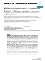
We performed the FTIR study to primarily confirm the
feasibility of the conjugation process. The FTIR spectra of
maghemite/BSA nanobioconjuagte along with maghemite
nanoparticle and BSA were shown in Fig. 2. The FTIR
spectra exhibited strong bands in the low frequency region
due to the iron oxide skeleton. In other regions, the spectra
of iron oxide had weak bands. The presence of free car-
boxyl group on the nanoparticle surface was further
confirmed from the C=O stretching band (1,704 cm
-1
)as
well as OH stretching band (2845.40 cm
-1
) of the carboxyl
group on the stearic acid capped magnetic nanoparticle.
Due to very strong hydrophobic character of the hydro-
carbon chain of stearic acid molecule, it is very difficult for
it to be exposed to the aqueous solution. So, the interaction
of stearic acid molecule with the iron oxide skeleton took
place through the hydrocarbon segment leaving the car-
boxyl group towards the aqueous solution. Comparing the
spectra of maghemite nanoparticles before and after con-
jugation with BSA, the strong absorption bands at 536.84
and 552.88 cm
-1
confirmed the presence of maghemite as
the main phase in both samples. The characteristics band of
the BSA protein at 1,650 and 1,530 cm
-1
are due to C=O
stretching band of carboxyl functional group on the
tryptophan moiety as well as carboxylate group. The con-
jugation of BSA to nanoparticle surface was confirmed by
the appearance of the new N–H stretching band
(3398.4 * 3,400 cm
-1
) as well as vanishing of the C=O
stretching band (1,704 cm
-1
) of carboxyl functional group
on stearic acid molecule. This clearly signified the forma-
tion of the covalent bond between the amino & carboxyl
functional groups of the protein molecule as well as stearic
acid capped maghemite nanoparticle. The new band
(1655.02 cm
-1
) appeared after the formation of the bio-
conjugate was due to C=O stretching pattern of the
secondary amide linkage. On the other hand, new bands
appearing at around 1,100–900 cm
-1
were due to the C–N
stretching (1080.49 cm
-1
) from secondary amide linkage;
C–H deformation out of plane (983.83 cm
-1
) from stearic
acid hydrocarbon chain as well as C–H deformation of
aromatic hydrocarbon (864.58 cm
-1
) from tryptophan
amino acid present over the protein molecule. The
appearance of the C–N stretching band at (1080.49 cm
-1
)
was a strong significance that the conjugation took place in
covalent manner through amide linkage.
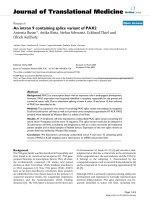
Further, to get the simplified view of the bioconjugate so
prepared, we also carried out the TEM studies of the ma-
ghemite nanoparticle as well as the resulting magnetic
nanobioconjugate. From the TEM micrographs, the con-
jugation of the nanoparticles with the protein molecule can
easily be visualized as shown in Fig. 3b. It clearly illus-
trates the ellipsoidal pattern of BSA molecule over which
the nanoparticles were conjugated and formed an assembly
pattern. It is also evident that the nanoparticles are well
separated from each other, i.e. aggregation has not taken
place. This has occurred as a result of the perturbation of
electron cloud of the bimolecular environment during the
conjugation process.
To understand the nature of interaction between mag-
netic nanoparticles with biomolecule, we had also studied
the optical property of the resulting bioconjugate by car-
rying out the photoluminescence experiment. The
photoluminescence pattern obtained for different samples
Fig. 1 The conjugation scheme
of maghemite nanoparticle with
bovine serum albumin
4000 3500 3000 2500 2000 1500 1000 500
0
10
20
30
40
50
60
70
80
90
100
%Transmittance
Wavelength( cm-
1
)
γ−Fe
2
O
3
Bovine serum albumin
γ−Fe
2
O
3
+BSA
3424.27
2925.63
1654.97
1542.27
1387.77
699.17
535.84
3395.46
1655.02
1080.49
983.93
864.58
536.84
3423.06
2845.46
1517.42
1399.70
552.88
1704.55
Fig. 2 FT-IR spectra of maghemite nanoparticle before and after the
conjugation with BSA
140 Nanoscale Res Lett (2009) 4:138–143
123
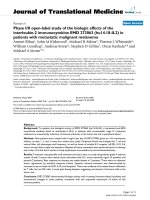
were shown in Fig. 4. From the experimental data of the
photoluminescence pattern, we calculated the binding
constant of the magnetic nanoparticles with the bovine
serum albumin by using the model put forward by Lehrer
and Fashman [19]. According to this method, the trypto-
phan residue fluorescence intensity (F) scaled with the
maghemite/BSA concentration through the following
equation:
F
0
À F
F À F
1
¼
½MNP ÀBSA
K
diss
n
where F
0
and F
?
are the relative luminescence intensities
of the protein alone and the protein conjugated with
maghemite nanoparticles, respectively, n is the stoichi-
ometry of the complex. The reciprocal of the K
diss
is the
binding constant K
b
. The intensity value (F) for the tryp-
tophan residue was obtained from the area under the
fluorescence spectra in the range of our investigation 300–
500 nm on calculation, the binding constant of the nano-
bioconjugate has been found out to be K
b
= 58.13. From
the figure, it became evident that the emission wavelength
of the bare maghemite nanoparticles was 617 nm. While
for the standard BSA sample it was found out to be
365.5 nm. For the maghemite/BSA nanobioconjugate col-
loidal solution, it had been observed that there was
significant shift in the emission wavelength (420.5 nm). On
the other hand, in case of standard maghemite/BSA mix-
ture the emission wavelength was found out to be 343 nm
which was strictly different from that of the optical effect
shown by the bioconjugate. The excitation wavelength
applied in all the samples were: k
Ex
= 320 nm for bare
maghemite nanoparticle, maghemite/BSA nanobioconju-
age, & maghemite/BSA mixture.On the other hand, the
excitation wavelength applied in case of BSA molecule
was k
Ex
= 280 nm. The pattern obtained for bare maghe-
mite nanoparticle was due to the intrinsic property of the
electronic transition in iron oxides such as the ligand to
metal charge transfer transition. This intensity was found to
be low, due to the fast (picosec) overall decay as well as
very efficient nonradiative relaxation raised from the dense
band structure and a high density of trap states in the iron
oxide skeleton as previously reported [20]. Also we had
assumed that there will be substantial improvement in PL
yield as no quenching phenomenon occurring in PL pattern
of the bare nanoparticle [21]. On the other hand, the
emission of BSA at 365.5 nm was due to luminescence
originated from two aromatic tryptophan moieties present
in BSA amino acid sequence: Trp 134 and Trp 214.This
was further shifted to 420.5 nm after the formation of
bioconjugate. This shifting might be due to the increase in
the nanoparticle size during the synthesis of bioconjuagte.
Also the produced BSA–NP conjugate causes effective
interaction between the excited states of the biological and
inorganic parts. This effect should be attributed to the
spatial closeness of BSA and NP in the tightly bound
covalent system [17].
For finally validating the magnetic properties of ultrafine
maghemite nanoparticles before and after conjugation with
bovine serum albumin molecule, we studied the same with
vibrating sample magnetometer (VSM). We had mainly
done it to ensure the existence of magnetic behavior of
maghemite nanoparticle after the conjugation process. The
magnetic behavior of the biomolecule–nanoparticles
assembly depends sensitively on the morphology and the
size of the nanoparticles, where the dipole coupling
between the nanoparticles governs the overall magnetic
behavior [22]. In conjugate of maghemite nanoparticles
with BSA, we observed a formation of unusual self-
alignment of nanoparticles over BSA molecules. Figure 5a,
b showed the room temperature magnetization of both
maghemite and maghemite/BSA nanobioconjugate. The
bare maghemite nanoparticle had saturation magnetization
1.044 emu/g, whereas that of maghemite/BSA nano-
bioconjuagte was found to be 1.196 emu/g. Also, the
retentivity (M
R
) and coercivity (H
C
) values (0.4016 emu/g
Fig. 3 TEM micrograph of the conjugate of maghemite nanoparticle
with elongated bovine serum albumin molecule
Nanoscale Res Lett (2009) 4:138–143 141
123
as well as 25.860 Oe, respectively) of the bioconjugates
were different in comparison to maghemite nanoparticles
(M
R
= 0.2851 emu/g; H
C
= 23.083 Oe). The difference
between these values could be attributed to the change in
the microstructure of the particles due to covalent binding
of BSA to the carboxyl group of stearic acid capped ma-
ghemite nanoparticle and the increase in the interparticle
interactions during the bioconjugation process. This
microstructural variation occurred as a result of the drastic
change in particle surface effect. The particle surface effect
hereby referred to the disordered alignment of surface
atomic spins induced by reduced coordination and broken
exchange bonds between surface spin [23]. The observed
induction in saturation magnetization value could also be
realized from the increase in the interparticle interactions
during the bioconjugation process. It had also been found
out that the obtained saturation magnetization value
(1.196 emu/g) of the prepared bioconjuagte was close to
the value (1.34 emu/g) obtained by Salgueirino-Maceira
et al. [24] for the luminescent magnetic nanoparticle. The
other hysteretic parameter, the coercivity value, also
increased due to the transition of domain boundary to
multidomain regime.
Conclusions
In summary, we developed a simple technique for the
synthesis of bioconjugate of maghemite nanoparticles with
BSA molecule by using the covalent interaction between
the fatty acid binding domains of BSA molecule with
stearic acid capped nanoparticles. This will lead to the
development of non-toxic iron oxide nanoparticles using
BSA as a biocompatible passivating agent. We confirmed
the formation of the same from the FT-IR spectra as well as
TEM micrograph. We also established the well retaining of
magnetic property of nanoparticles after the formation of
bioconjugate from M–H study. It is worth mentioning here
that, this is the first report on conjugation of nanoparticles
with biomolecules by utilizing biologically evolved linker
moiety in covalent fashion. The designed magnetic bio-
conjugate seems to be applicable for targeted delivery
purpose to a neoplastic cell due to the receptor action of the
BSA molecule by binding to a wide variety of lipophilic
compounds such as steroid present over cancer cell.
Acknowledgments One of the author, PD, gratefully acknowledge
the financial support by DAE-BRNS, Govt. of India (vide project no.
Fig. 4 The photoluminescence spectra of a maghemite nanoparticle b BSA c maghemite/BSA nanobioconjugate d Maghemite/BSA mixture
Fig. 5 Room temperature
(300 K) M–H data of a
Maghemite and b Maghemite/
BSA nanobiconjugate
142 Nanoscale Res Lett (2009) 4:138–143
123
2007/20/34/04-BRNS/1865) under DAE Young Scientist Research
Award. The authors would like to extend sincere thanks to CIF, IIT
Guwhati, India and RSIC–NEHU for VSM and TEM facility.
References
1. M. Kumagai, Y. Imai, T. Nakamura, Y. Yamasaki, M. Sekino, S.
Ueno, K. Hanaoka, K. Kikuchi, T. Nagano, E. Kaneko, K.
Shimokado, K. Kataoka, Colloids Surf. B: Biointerfaces 56, 174
(2007). doi:10.1016/j.colsurfb.2006.12.019
2. A.K. Gupta, S. Wells, IEEE. T. Nanobiosci. 3, 1536 (2004). doi:
10.1109/TNB.2003.820277
3. M.D. Kaminski, A.J. Rosengart, J. Magn. Magn. Mater. 293, 398
(2005). doi:10.1016/j.jmmm.2005.02.055
4. J.M. Perez, Nat. Nanotechnol. 2, 535 (2007). doi:10.1038/
nnano.2007.282
5. A. Ito, M. Shinkai, H. Honda, T. Kobayashi, J. Biosci. Bioeng.
100, 1 (2005). doi:10.1263/jbb.100.1
6. S. Yan, D. Zhang, N. Gu, J. Zheng, A. Ding, Z. Wang, B. Xing,
M. Ma, Y. Zhang, J. Nanosci. Nanotechnol. 5, 1185 (2005). doi:
10.1166/jnn.2005.219
7. N. Zhu, A. Zhang, P. He, Y. Fang, Electroanalysis 16, 1925
(2004). doi:10.1002/elan.200303028
8. H. Gu, K. Xu, C. Xu, B. Xu, Chem. Commun. 941 (2006). doi:
10.1039/b514130c
9. L.M. Rossi, A.D. Quach, Z. Rosenzweig, Anal. Bioanal. Chem.
380, 606 (2004). doi:10.1007/s00216-004-2770-3
10. A.K. Gupta, M. Gupta, Biomaterials 26, 3995 (2005). doi:
10.1016/j.biomaterials.2004.10.012
11. S. Yu, G.M. Chow, J. Mater. Chem. 14, 2781 (2004). doi:
10.1039/b404964k
12. Z.G. Peng, K. Hidajat, M.S. Uddin, J. Colloid Interface Sci. 271,
277 (2004). doi:10.1016/j.jcis.2003.12.022
13. R.G. Reed, J. Biochem. 261, 15619 (1986)
14. T. Peters Jr., Adv. Protein Chem. 37, 161 (1985). doi:10.1016/
S0065-3233(08)60065-0
15. P. Deb, A. Basumallick, D. Sen, S. Mazumder, B.K. Nath, D.
Das, Philos. Mag. Lett. 86, 491 (2006). doi:10.1080/09500830
600876573
16. B. Samanta, H. Yan, N.O. Fischer, J. Shi, D.J. Jerry, V.M.
Rotello, J. Mater. Chem. 18, 1204 (2008). doi:10.1039/b718745a
17. N.N. Mamedova, N.A. Kotov, A.L. Rogach, J. Studer, Nano.
Lett. 1, 281 (2001). doi:10.1021/nl015519n
18. C.J. Tan, H.G. Chua, K.H. Ker, Y.W. Tong, Anal. Chem. 80, 683
(2008). doi:10.1021/ac701824u
19. S.S. Lehrer, G.D. Fashman, Biochem. Biophys. Res. Commun. 2,
133 (1966). doi:10.1016/0006-291X(66)90517-1
20. N.J. Cherepy, D.B. Liston, J.A. Lovejoy, H. Deng, J.Z. Zhang,
J. Phys. Chem. B 102, 770 (1998). doi:10.1021/jp973149e
21. Y. Li, H. Yang, Z. He, L. Liu, W. Wang, F. Li, L. Xu, J. Mater.
Res. 20, 2940 (2005). doi:10.1557/JMR.2005.0362
22. T. Kim, L. Reis, K. Rajan, M. Shima, J. Magn. Magn. Mater. 295,
132 (2005). doi:10.1016/j.jmmm.2005.01.004
23. M.P. Morales, S. Veintemillas-Verdaguer, C.J. Serna, J. Mater.
Res. 14, 3066 (1999). doi:10.1557/JMR.1999.0411
24. V. Salgueirino-Maceira, M.A. Correa-Duarte, M. Spasova, L.M.
Liz-Marzan, M. Farle, Adv. Funct. Mater. 16, 509 (2006). doi:
10.1002/adfm.200500565
Nanoscale Res Lett (2009) 4:138–143 143
123