Báo cáo hóa học: " SiC Nanowires Synthesized by Rapidly Heating a Mixture of SiO and Arc-Discharge Plasma Pretreated Carbon Black" ppt
Bạn đang xem bản rút gọn của tài liệu. Xem và tải ngay bản đầy đủ của tài liệu tại đây (302.78 KB, 4 trang )
NANO EXPRESS
SiC Nanowires Synthesized by Rapidly Heating a Mixture
of SiO and Arc-Discharge Plasma Pretreated Carbon Black
Feng-Lei Wang Æ Li-Ying Zhang Æ Ya-Fei Zhang
Received: 17 September 2008 / Accepted: 11 November 2008 / Published online: 22 November 2008
Ó to the authors 2008
Abstract SiC nanowires have been synthesized at
1,600 °C by using a simple and low-cost method in a high-
frequency induction furnace. The commercial SiO powder
and the arc-discharge plasma pretreated carbon black were
mixed and used as the source materials. The heating-up and
reaction time is less than half an hour. It was found that
most of the nanowires have core-shell SiC/SiO
2
nano-
structures. The nucleation, precipitation, and growth
processes were discussed in terms of the oxide-assisted
cluster-solid mechanism.
Keywords Silicon carbide Á Nanowires Á
Induction heating
Introduction
Silicon carbide (SiC) has been widely used in the fields of
electronic and optic devices due to its unique properties,
such as a wide band gap of 2.3–3.3 eV, high strength, and
Young’s modulus, good resistance to oxidation and cor-
rosion, excellent thermal conductivity, and electron
mobility [1–4]. One-dimensional (1D) SiC materials, i.e.,
nanowires, nanofibers, nanorods, and nanocables have
recently attracted much attention because they have been
thought suitable for the fabrication of high temperature,
high frequency, and high power nanoscaled electronic
devices [5–9].
The first successfully synthesis of 1D SiC nanowires
was in 1995 by using carbon nanotube as a template [10].
Up to now, lots of approaches have been developed, for
example, arc-discharge [11], laser ablation [12], sol–gel
method [13], carbon thermal reduction [14], and chemical
vapor deposition [15]. Recently, metal catalyst assisted
synthesis of 1D SiC nanostructures had also been reported
[16, 17]. In most of these methods, expensive raw mate-
rials, catalysts, and sophisticated techniques were used.
These drawbacks may limit the massive fabrication and
application of SiC nanowires. It is still a challenge for
scientists and industrials to synthesize large-scale SiC
nanowires by using a simple and rapid method.
In this paper, we report a novel method to fabricate
b-SiC nanowires by using a high-frequency induction
furnace with a graphite tube. A mixture of commercial SiO
and the carbon black powder with loose structures pre-
treated by an arc-discharge plasma method was used as the
starting materials. After heating the source materials in
graphite tube in argon atmosphere, bright blue powders can
be observed in the tube, which were characterized as b-SiC
nanowires with core-shell structures. The total heating-up
and reaction time is less than 1 h, and more than 200 g
products can obtain per day. The modified oxide-assisted
cluster-solid growth mechanism was used to explain the
formation of core-shell SiC/SiO
2
nanowires.
Experimental
The fabrication of b-SiC nanowires was carried out in a
high-frequency introduction furnace. First, commercial
carbon black was pretreated in order to form porous and
F L. Wang Á L Y. Zhang Á Y F. Zhang (&)
National Key Laboratory of Nano/Micro Fabrication
Technology, Key Laboratory for Thin Film and Microfabrication
of the Ministry of Education, Research Institute of Micro/Nano
Science and Technology, Shanghai Jiao Tong University,
Shanghai 200240, People’s Republic of China
e-mail:
123
Nanoscale Res Lett (2009) 4:153–156
DOI 10.1007/s11671-008-9216-3
loose structures, which can make the reaction much easier.
The carbon black was pressed to a carbon rod and put into
an arc-discharge plasma instrument. After treating for
about 1 h, a black powder with loose structures was
obtained.
The as-prepared carbon black was mixed with the
commercial SiO powder (mass ratio of 1:1) and ball-milled
for several hours. Then, the precursor was loaded in a
graphite boat and located in a high-purity graphite tube. As
a heating crucible, the graphite tube was placed in a hori-
zontal quartz tube and heated in a high-frequency induction
furnace. The furnace was first evacuated to 50 Pa, and then
the argon gas was introduced until the furnace pressure
reached about 4 9 10
4
Pa, which was maintained
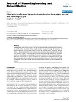
throughout the whole experimental process. The powder
was rapidly heated to 1,600 °C within 3 min and kept for
40 min. A bright blue-colored powder was found in the
graphite boat. The schematic diagram of the apparatus is
shown in Fig. 1.
An energy-dispersive X-ray (EDX, INCA OXFORD)
spectroscopy and an X-ray diffraction (XRD, D/MAX-RA)
were used to characterize the composition and crystal
structure of samples. A field-emission scanning electron
microscopy (SEM, FEI SIRION 200) and a transmission
electron microscopy (TEM, JEM-2010) were employed to
observe the morphology and the detail structure of the
nanowires.
Results and Discussion
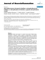
Figure 2 shows the typical SEM image of the carbon black,
which was treated in an arc-discharge plasma instrument.
The loose and porous nanostructures were formed, which
have more surface areas compared with original materials.
This provides more chance for the reaction with SiO vapor.
The inset in Fig. 2 displays the corresponding EDX spec-
trum, indicating only two elements (carbon and oxide)
existed in the pretreated carbon black.
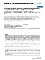
The characteristic XRD pattern of the products is
showed in Fig. 3. The major diffraction peaks can be
indexed as the (1 1 1), (2 0 0), (2 2 0), (3 1 1), and (2 2 2)
reflections of cubic b-SiC (unit cell parameter
a = 0.4370 nm). These values are almost identical to the
known values for standard b-SiC (JCPDS Card No. 29–
1129). Moreover, there is amorphous background in the
XRD pattern, which is similar to amorphous SiO
2
.Fur-
thermore, the diffraction peaks are broadened, which may
be related to the inner thinner b-SiC nanowire and the outer
amorphous silicon oxide wrapping layer.
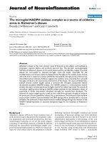
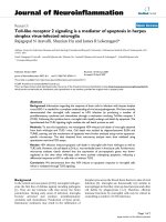
Figure 4 shows the SEM and TEM images of the as-
synthesized nanowires without any other treatments. In
Fig. 4a and b, it can be seen that the nanowires have almost
uniform diameters and smooth surfaces. The diameter of
nanowires can be roughly estimated in the range of 60–
100 nm and the length are several microns. The observed
impurities in SEM images were the intermediate product of
SiO
2
and the residual carbon. To validate the existing of
impurities, high-temperature oxidation and hydrofluoric
acid (5%) treatment were used to get rid of the residual
carbon and SiO
2
, respectively. In the high-temperature
Fig. 1 Schematic diagram of the apparatus for synthesis of SiC
nanowires
Fig. 2 SEM image and EDX pattern of carbon black after arc-
discharge plasma treatment
Fig. 3 XRD pattern of the SiC nanowires
154 Nanoscale Res Lett (2009) 4:153–156
123
oxidation processing, about 72% of the as-synthesized
sample remained as well as 28% of carbon was oxidized.
After dipping in hydrofluoric acid (5%) for 2 h, about 74%
of the residual sample remained when SiO
2
was corroded.
Therefore, it can be concluded that the yield of SiC
nanowires was about 53%. The inset in Fig. 4a displays the
corresponding EDX spectrum, indicating three elements
(silicon, carbon, and oxide) exist in the nanowires. The
TEM image in Fig. 4c shows detailed structure of the
nanowire. One can find that the nanowire has a core-shell
nanocabled structure. According to the component ratio
obtained by EDX results, the core ought to be crystallized
SiC and the shell is amorphous SiO
2
. In fact, the unique
core-shell SiC/SiO
2
structure has also been observed by
other researchers [18–20].
Vapor–liquid–solid (VLS) mechanism has usually been
used to explain the growth process of 1D nanomaterials
[21]. However, it seems unsuitable to interpret our exper-
iments and results because there is no catalyst liquid
droplet available during the high-frequency induction
heating procedure. The oxide-assisted cluster-solid mech-
anism proposed by Zhang et al. [22], which was
established to interpret the growth process of Si/SiO
2
nanowires, may be used to understand the growth process
of core-shell SiC/SiO
2
nanowires. In terms of this mecha-
nism, there exist three processes, that is, nucleation,
precipitation, and growth. Figure 5 illustrates the sche-
matic diagram of growing process. As the temperature is up
to 1,600 °C, SiO powder will vaporize and react with the
carbon source as follows:
3SiO vðÞþ3C sðÞ¼2SiC sðÞþSiO
2
sðÞþCO vðÞ ð1Þ
where v and s refer to vapor and solid states of the material,
respectively. It will generate SiC and SiO
2
nanoparticles in
this process, which provide crystalline nucleus for growth
of nanowires. Actually, three different atoms (silicon,
carbon, and oxygen) contained in the nanoparticles. The
superfluous of any element will lead to the occurrence of
precipitation (separate out) process. Reaction 2 can occur
under a supersaturated condition of CO [23]:
SiO vðÞþ3CO vðÞ¼SiC sðÞþ2CO
2
ðvÞ: ð2Þ
When SiO vapor is prevail, the following reaction will
occur:
3SiO vðÞþCO vðÞ¼SiC sðÞþ2SiO
2
ðsÞ: ð3Þ
No matter what reaction is in the ascendant, SiC can
generate and provide to the nanoparticles. Since there exist
sufficient silica and carbon atoms in the reaction
atmosphere, the precipitation (separate out) of SiC is
possible. When the reaction 3 is dominant, SiO
2
is then the
Fig. 4 a The SEM image of SiC nanowires; b the magnified SEM
image of SiC nanowires; and c the TEM image of SiC nanwires with a
core-shell SiC/SiO
2
structure. The inset in a shows the EDX pattern
of SiC nanowires
Fig. 5 Schematic diagram of growing process of SiC nanowire
Nanoscale Res Lett (2009) 4:153–156 155
123
main resultant and can separate out accompanying with the
growth of SiC nanocrystals. This is why SiC nanowires are
wrapped by SiO
2
layers.
At the same time, the CO
2
gas generated from reaction 2
may react with the carbon source as follows:
CO
2
vðÞþCsðÞ¼2CO vðÞ: ð4Þ
The partial supersaturation of CO gas can lead to a
diameter distribution of the as-synthesized SiC nanowires
[24, 25]. The CO gas is hard to be got rid of from graphite
crucible in our experiment, and therefore, leads to the
distribution of the diameter in as-synthesized SiC/SiO
2
nanowires.
Conclusion
We present a simple, rapid, and low-cost method to syn-
thesize massive b-SiC nanowires by a high-frequency
induction heating procedure. A ball-milled mixture of SiO
and carbon black was used as source materials. The carbon
black were pretreated in an arc-discharge plasma instru-
ment in order to form loose and porous structures. The
heating-up and the reaction time is less than 1 h. The
nanowires have core-shell SiC/SiO
2
structures in which the
core of SiC crystallizes very well, whereas the SiO
2
has
amorphous structure. The diameter of nanowires is ranged
from 60 to 100 nm and the length is up to several microns.
This method provides a promising candidate for industrial
fabrication of b-SiC nanowires.
Acknowledgments This work is supported by the National Basic
Research Program of China (No. 2006CB300406) and the Shanghai
Science and Technology Grant (No: 0752nm015) as well as the
National Natural Science Foundation of China (No. 50730008). The
authors also thank the Instrumental Analysis Center of Shanghai Jiao
Tong University for the Materials Characterization.
References
1. G.L. Harris, Properties of Silicon Carbide (INSPEC, London,
1995). ISBN:0852968701
2. H.L. Heinisch, L.R. Greenwood, W.J. Weber, R.E. Williford,
J. Nucl. Mater. 307, 895 (2002). doi:10.1016/S0022-3115(02)
00962-5
3. H. Morko, S. Strite, G.B. Gao, M.E. Lin, B. Sverdlov, M. Burns,
J. Appl. Phys. 76, 1363 (1994). doi:10.1063/1.358463
4. C. Persson, U. Lindefelt, Phys. Rev. B 54, 10257 (1996). doi:
10.1103/PhysRevB.54.10257
5. N.G. Wright, A.B. Horsfall, J. Phys. D: Appl. Phys. (Berl) 40,
6345 (2007). doi:10.1088/0022-3727/40/20/S17
6. E.W. Wong, P.E. Sheehan, C.M. Lieber, Science 277, 1971
(1997). doi:10.1126/science.277.5334.1971
7. S.M. Pimenov, V.D. Frolov, A.V. Kudryashov, Diam. Relat.
Mater. 17, 758 (2008). doi:10.1016/j.diamond.2007.08.016
8. L.G. Zhang, W.Y. Yang, H. Jin, Z.H. Zheng, Appl. Phys. Lett. 89,
143101 (2006). doi:10.1063/1.2358313
9. W.M. Zhou, L.J. Yan, Y. Wang, Y.F. Zhang, Appl. Phys. Lett.
89, 013105 (2006). doi:10.1063/1.2219139
10. H. Dai, E.W. Wang, Y.Z. Liu, S.S. Fan, C.M. Lieber, Nature 357,
769 (1995). doi:10.1038/375769a0
11. T. Seeger, P. Kohler-Redlich, M. Ruhle, Adv. Mater. 12, 279
(2000). doi:10.1002/(SICI)1521-4095(200002)12:4\279::AID-
ADMA279[3.0.CO;2-1
12. W.S. Shi, Y.F. Zheng, H.Y. Peng, N. Wang, C.S. Lee, S.T. Lee, J.
Am. Ceram. Soc. 83, 3228 (2000). doi:10.1111/j.1151-2916.2000.
tb01714.x
13. X.K. Li, L. Liu, Y.X. Zhang, S.D. Shen, S. Ge, L.C. Ling, Carbon
39, 159 (2001). doi:10.1016/S0008-6223(00)00020-8
14. G.Z. Shen, Y. Bando, C.H. Ye, B.D. Liu, D. Golberg, Nano-
technology 17, 3468 (2006). doi:10.1088/0957-4484/17/14/019
15. R.B. Wu, Y. Pan, G.Y. Yang, M.X. Gao, J.J. Chen, L.L. Wu, R.
Zhai, J. Lin, J. Phys. Chem. C 111, 6233 (2007). doi:10.1021/
jp070115q
16. G.C. Xi, Y.Y. Peng, S.M. Wan, T.W. Li, W.C. Yu, Y.T. Qian, J.
Phys. Chem. B 108, 20102 (2004). doi:10.1021/jp0462153
17. G.C. Xi, Y.K. Liu, X.Y. Liu, X.Q. Wang, Y.T. Qian, J. Phys.
Chem. B 110, 14172 (2006). doi:10.1021/jp0617468
18. G.Z. Shen, D. Chen, K.B. Tang, Y.T. Qian, S.Y. Zhang, Chem.
Phys. Lett. 375, 177 (2003). doi:10.1016/S0009-2614(03)00877-7
19. B.S. Li, R.B. Wu, Y. Pan, L.L. Wu, G.Y. Yang, J.J. Chen, Q. Zhu,
J. Alloy Compd. 462, 446 (2008). doi:10.1016/j.jallcom.2007.
08.076
20. A. Meng, Z.J. Li, J.L. Zhang, L. Gao, H.J. Li, J. Cryst. Growth
308
, 263 (2007). doi:10.1016/j.jcrysgro.2007.08.022
21. R.S. Wagner, W.C. Ellis, Appl. Phys. Lett. 4, 89 (2006). doi:
10.1063/1.1753975
22. Y.F. Zhang, Y.H. Tang, N. Wang, C.S. Lee, I. Bello, S.T. Lee,
J. Cryst. Growth 197, 136 (1999). doi:10.1016/S0022-0248(98)
00953-1
23. W.Q. Han, S.S. Fan, Q.Q. Li, W.J. Liang, B.L. Gu, D.P. Yu,
Chem. Phys. Lett. 265, 374 (1997). doi:10.1016/S0009-2614(96)
01441-8
24. C.C. Tang, S.S. Fan, H.Y. Dang, J.H. Zhao, C. Zhang, P. Li et al.,
J. Cryst. Growth 210, 595 (2000). doi:10.1016/S0022-0248(99)
00737-X
25. Y.H. Gao, Y. Bando, K. Kurashima, T. Sato, J. Mater. Sci. 37,
2023 (2002). doi:10.1023/A:1015207416903
156 Nanoscale Res Lett (2009) 4:153–156
123