Báo cáo hóa học: " Quinoline Group Modified Carbon Nanotubes for the Detection of Zinc Ions" ppt
Bạn đang xem bản rút gọn của tài liệu. Xem và tải ngay bản đầy đủ của tài liệu tại đây (251.92 KB, 6 trang )
NANO EXPRESS
Quinoline Group Modified Carbon Nanotubes for the Detection
of Zinc Ions
Zhengping Dong Æ Bin Yang Æ Jun Jin Æ Jing Li Æ
Hongwei Kang Æ Xing Zhong Æ Rong Li Æ Jiantai Ma
Received: 18 November 2008 / Accepted: 30 December 2008 / Published online: 21 January 2009
Ó to the authors 2009
Abstract Carbon nanotubes (CNTs) were covalently
modified by fluorescence ligand (glycine-N-8-quinolyla-
mide) and formed a hybrid material which could be used as
a selective probe for metal ions detection. The anchoring to
the surface of the CNTs was carried out by the reaction
between the precursor and the carboxyl groups available on
the surface of the support. Fourier transform infrared
spectroscopy (FTIR) and Thermogravimetric analysis
(TGA) unambiguously proved the existence of covalent
bonds between CNTs and functional ligands. Fluorescence
characterization shows that the obtained organic–inorganic
hybrid composite is highly selective and sensitive (0.2 lM)
to Zn(II) detection.
Keywords Carbon nanotubes Á
Glycine-N-8-quinolylamide Á Zn(II) ÁFluorescent material Á
Detection
Introduction
There has been growing interest during the last decade in
the development of fluorescent molecular sensors for cat-
ions and anions in solution [1–8]. Especially, fabricating
fluorescent materials for the detection of Zinc cation has
drawn much more attention [9–13], as Zinc not only plays
important roles in human bodies [14, 15], but also closely
relates to severe pathological diseases such as Alzheimer’s
and Parkinson’s diseases [16]. So far, much study has been
done for the detection and real-time localization of Zn(II).
Yasuhiro Shiraishi’s group has synthesized a quinoline–
polyamine conjugate as a fluorescent chemosensor for
quantitative detection of Zn(II) in water [17]. Maarten
Merkx et al. used chelating fluorescent protein chimeras
for ratiometric detection of Zn(II) in living cells and the
detection range was from 10 nM to 1 mM [18]. Jinshi Ma
and coworkers have synthesized several bis(pyrrol-2-
yl-methyleneamine) ligands as fluorescent sensor for
Zn(II) [11], and their results revealed that the ligands
exhibit excellent fluorescent properties. However, much
of the work was just based on organic molecules as
fluorescent chemosensors. For a few practical applications
the attachment of the fluorescent units to a solid support
has advantages like the possibility of recovering the
materials for their repetitive use. For this point, scientists
chose silica nanoparticles [12], nanosized boehmite par-
ticles [13] and silicon nanowires [19] to support
fluoresence ligands as fluorescence sensors. And these
materials exhibit excellent selectivity and sensitivity to
sense metal ions.
In this study, we chose multi-walled carbon nanotubes
(MWNTs) as fluorescent support. Since its discovery,
surface modification of MWNTs has received considerable
attention [20–24]. The fluorophore in this study is glycine-
N-8-quinolylamide (GNQ) molecule, in light of the fact
that the 8-aminoquinoline derivatives could effectively
coordinate with specific metal ions [25]. The quinoline
group has been covalently grafted to the surface of the
MWNTs that can behave as recognition center for metal
ions depending on its actual protonation state. We find that
this new material (MWNTs-GNQ) has high selectivity and
sensitivity to detect Zn(II), and the sensitivity is down to
Z. Dong Á B. Yang Á J. Jin Á J. Li Á H. Kang Á X. Zhong Á
R. Li Á J. Ma (&)
College of Chemistry and Chemical Engineering, Lanzhou
University, Lanzhou 730000, People’s Republic of China
e-mail:
R. Li
e-mail:
123
Nanoscale Res Lett (2009) 4:335–340
DOI 10.1007/s11671-008-9248-8
0.2 lM., which is about the same as the silica nanoparti-
cles-supported fluorescence sensors [12]. For other sensing
materials [26, 27], the fluorescence enhancement selectiv-
ity is not only for Zn(II) but also for Cd(II), which may
reduce selectivity when they are used just for Zn(II)
detection. On the other hand, the fluorescence enhancement
of MWNTs-GNQ is only for Zn(II).
As the use of organic–inorganic hybrid materials for
bio-application has become a hot subject in the research
field currently [28–31], MWNTs-GNQ may be used to
build nanosensor devices to sense directly in intracellular
environment, because carbon nanotubes (CNTs) can pen-
etrate into cells and almost have no toxicity to organism
[32, 33].
Experimental
Materials
The multi-walled carbon nanotubes (MWNTs, diameters:
20–40 nm, purity: 95–98%) prepared by the catalytic
decomposition of CH
4
were provided by Shengzhen
Nanotech Port Ltd. Co (China). Methanol and Tetrahy-
drofuran were used after distillation. Other reagents were
analytical and used without purification. Glycine-N-8-
quinolylamide (GNQ) was synthesized according to the
known method [34].
Purification of MWNTs
In a typical experiment, 300 mg pristine-MWNTs were
added to a 180 mL 3:1 mixture of concentrated H
2
SO
4
and
HNO
3
. The mixture was treated in an ultrasonic bath
(40 kHz) for 20 min and stirred at 60 °C for 4 h under
reflux. Then, the mixture was vacuum-filtered through a
0.22 lm Millipore polytetrafluoroethylene membrane and
washed with distilled water until pH of the filtrate was 7.
The filter cake was dried under vacuum at 40 °C for 24 h to
obtain MWNTs-COOH.
Functionalization of MWNTs (Scheme 1)
The acid-treated MWNTs-COOH of 200 mg was reacted
with 20 mL SOCl
2
for 24 h under reflux, and then the
residual SOCl
2
was removed by the reduced pressure dis-
tillation, the solid was washed with anhydrous THF several
times until the brown-colored supernatant became to col-
orless. The remaining solid acyl chloride-functionalized
MWNTs (MWNTs-COCl) was dried under vacuum at
20 °C for 24 h.
MWNTs-COCl of 100 mg was dispersed in 10 mL
anhydrous chloroform and the mixture was sonicated for
20 min to create a homogeneous suspension. The mixture
was added with 50 mg GNQ under a nitrogen atmosphere,
and then immersed in an oil bath at 70 °C accompained by
mechanical stirring for 24 h. The resulting reaction med-
ium was vacuum-filtered through a 0.22 lm polycarbonate
membrane three times to yield MWNTs-GNQ.
All the reactions in the experimental procedure were
carried out under a nitrogen atmosphere.
Characterization and Test of the Materials
Fourier transform infrared (FTIR) spectrometer (Bruker
IFS66/S), Dupont-1090 Thermal gravimetric analysis
(TGA) instrument and Gmbh Varioel Elementar Analy-
sensyteme were used to characterize the materials. Perkin
Elmer LS 55 spectrofluorimeter was used to obtain the
fluorescence spectra of the fluorescence material.
Scheme1 Functionalization of
MWNTs
336 Nanoscale Res Lett (2009) 4:335–340
123
Results and Discussion
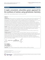
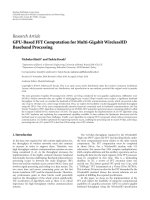
Figure 1 shows the infrared spectra of a GNQ and b
MWNTs-GNQ. In the FTIR spectrum of GNQ, the
absorption peaks at 3383.64 cm
-1
, 3290.48 cm
-1
(–NH
2
),
2890.5 cm
-1
(C–H), 1658.27 cm
-1
(C=O), 1593.29 cm
-1
(N–H), 1523.3 cm
-1
(C–C), and 1059.01 cm
-1
(C–N) are
found. Comparing with the FTIR spectrum of GNQ, the
characteristic peaks of amino groups in the spectrum of
MWNTs-GNQ disappeared, demonstrating that the amino
groups on GNQ have reacted with acyl chloride groups on
the surface of MWNTs. A new peak that appeared around
1686 cm
-1
is attributed to the amide carbonyl (C=O)
stretch. Another new peak at 1629.87 cm
-1
is attributed to
secondary amide band which accompanies the absorption
at 1686 cm
-1
. Another peak at 1082.47 cm
-1
attributed to
(C–N) has also been found, and it has obviously been
enhanced. The results indicate that GNQ has been grafted
to the surface of MWNTs.
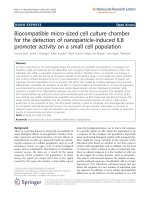
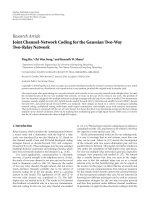
The TGA curves of MWNTs-COOH and MWNTs-GNQ
were recorded on a Dupont-1090 thermogravimeter in Ar
atmosphere at the heating rate 10 ° C/min from 20 °Cto
500 ° C (Fig. 2). According to Fig. 2a, there is a continuous
weight lose of the MWNTs-COOH, and the amount of the
weight loss is about 10% typical for acid-functionalized
MWNTs. The weight loss curves in Fig. 2b, the major
weight loss happened in the temperature range from
200 ° C to 450 °C due to the degradation of the GNQ
grafted to the MWNTs. The content of the GNQ grafted to
the MWNTs is about 12 wt%, which is similar to the cal-
culation result of the elemental microanalysis (Table 1).
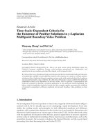
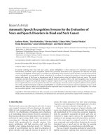
The fluorescence spectra of GNQ and MWNTs-GNQ
are shown in Fig. 3. It can be seen that the fluorescence
peak is red-shifted after modification. Compared with
GNQ, the fluorescence intensity of MWNTs-GNQ is
decreased. It may be affected by the black background of
CNTs.
GNQ is expected to be a stronger coordinating agent
because it is a tridentate ligand. When GNQ is selectively
coordinated with metal ions, the fluorescence from GNQ is
modified appropriately by the metal ions. This phenome-
non can be utilized to construct a material for the detection
of metal ions based on MWNTs (Scheme 2). Accordingly,
Fig. 1 FTIR spectra of a GNQ and b MWNTs-GNQ
Fig. 2 TGA of a MWNTs-COOH and b MWNTs-GNQ
Table 1 Elemental microanalysis for MWNTs-GNQ
Element Content (%)
C 74.27
N 2.629
H 1.238
Fig. 3 Fluorescence spectrum of GNQ (1 9 10
-5
M) and MWNTs-
GNQ (1 9 10
-5
M). Methanol solution. k
ex
= 324 nm
Nanoscale Res Lett (2009) 4:335–340 337
123
titration of various metal ions in the presence of MWNTs-
GNQ in methanol solution was performed, and the results
are summarized in Fig. 4. After titration of various metal
ions, it is observed that the intensity of fluorescence from
MWNTs-GNQ containing Zn(II) is much higher than that
of other metal ions. This is very nice because under many
conditions (e.g., physiological conditions) various metal
ions may exist at certain concentrations compared to
Zn(II).
It was reported that most of the previous Zn(II) sensors
do not exhibit good selectivity to these metal cations [26,
27] (for instance, the selectivity in many cases is close to
1:1 for Zn(II):Cd(II)). This may bring trouble to certain
applications where Co(II), Ni(II), Cu(II), or Cd(II) may
interfere (e.g., in environmental science). Herein, MWNTs-
GNQ shows 2.8-fold fluorescence enhancement for Zn(II)
versus just minimal fluorescence enhancement for Cd(II)
and Ni(II). From these results, it is evident that MWNTs-
GNQ have a high selectivity to Zn(II).
As for selectivity of MWNTs-GNQ to metal ions, when
Zn(II) forms a complex with MWNTs-GNQ with a suitable
radius and an electronic structure 3d
10
4s
0
, the electron-
transfer process of MWNTs-GNQ is forbidden [35], and an
extended p–electron conjugation system is formed syn-
chronously. This conjugation system is involved in an
internal charge transfer process from the ligand donor to
the Zn(II) acceptor, and simultaneously inhibits the exci-
ted-state proton transfer and photo-induced electron
transfer that strongly suppress the fluorescence of
MWNTs-GNQ. Thus Zn(II) considerably enhances the
fluorescence of MWNTs-GNQ.
To further characterize the performance of the sensing
material for Zn(II), a series of comparison experiments
were carried out with MWNTs-GNQ. Because Zn(II)
always coexists with Cu(II) or Cd(II), titration addition of
1 9 10
-5
M Cu(II) and Cd(II) to MWNTs-GNQ solution
containing Zn(II) led to contrary results (Fig. 5). When
Cu(II) was added to the solution, it led to about 97.5%
quenching of the total fluorescence intensity, probably
because Cu(II) formed some complex with GNQ group,
resulting in quenching of fluorescence as reported [36]. But
Cd(II) almost had no influence on the fluorescence inten-
sity of the MWNTs-GNQ solution containing Zn(II),
because the electronic structure of Cd(II) is fairly similar to
that of Zn(II). To resolve the problem of fluorescence
quenching by Cu(II), a masking agent of Na
2
S
2
O
3
was
chosen. As is well known that S
2
O
3
2-
can form a very
stable complex with Cu(II), the coordination number is
three, and Na(I) almost has no influence to fluorescence
intensity of MWNTs-GNQ. The results are shown in
Fig. 5, from which we can see that the masking agent
almost does not affect the experimental results except in
the case when the system contains Zn(II) and Cu(II). When
three stoichiometry S
2
O
3
2-
were added to the Zn(II) and
Scheme 2 Fluorescent
chemosensor (MWNTs-GNQ)
for detection of Zn(II)
Fig. 4 Relative fluorescence intensity of MWNTs-GNQ
(1 9 10
-5
M) in the presence of variouse metal ions alone
(1 9 10
-5
M). Methanol solution. k
ex
= 324 nm
338 Nanoscale Res Lett (2009) 4:335–340
123
Cu(II) solution, the fluorescence intensity was greatly
enhanced. Although, compared to the solution only con-
taining Zn(II), the intensity was a little weaker, Na
2
S
2
O
3
is
still a good masking agent to mask Cu(II). We thus con-
clude that the presence of Cd(II) does not affect the
sensitivity of MWNTs-GNQ for Zn(II) detection, and
Na
2
S
2
O
3
could be used as a masking agent when Cu(II)
coexists in the system.
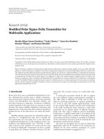
The sensitivity of fluorescence enhancing from
MWNTs-GNQ by Zn(II) was further investigated, and the
results are shown in Fig. 6a. The fluorescence intensity of
MWNTs-GNQ gradually increased with increasing Zn(II)
concentration. When more than 1 eq. Zn(II) was added,
only a marginal increase was observed (Fig. 6b), which
suggested the 1:1 stoichiometry of the ligand to the zinc
ions. Because Zn(II) desires a square planar geometry
when coordinated, while the three nitrogen on MWNTs-
GNQ can only provide a tridentate ligand, the fourth
coordination can come from the solvent methanol oxygen.
It can be seen from Fig. 6b that when the concentration of
Zn(II) is lower than 0.2 lM, the relative fluorescence
intensity is about 1. But when the concentration of Zn(II) is
higher than 0.2 lM, the relative fluorescence intensity is
also increased. It can be expressed by the following
formula:
A ¼À0:012C
2
þ 0:3055C þ 0:9658; R
2
¼ 0:9993: ð1Þ
Wherein, A is the value of the fluorescence intensity, C is
the concentration of Zn(II), and the range of C is from
0.2 lMto10lM in this study. So, it is evident that the
detection limit for Zn(II) is established at 0.2 lM under the
experimental conditions in our study.
Summary
We have prepared a new organic–inorganic hybrid sensing
material based on CNTs as support and glycine-N-8-
quinolylamide as fluorescent center. The results of the
fluorescence characterization show that the composite has a
highly selective and sensitive (0.2 lM) detection for
Zn(II), and reveal that ratiometric Zn(II) sensing is possible
with fluorophore chemically modified carbon nanotubes.
This novel fluorescent material may be used as a fluores-
cent device in intracellular environment for the detection of
Zn(II).
References
1. B. Valeur, I. Leray, Coord. Chem. Rev. 205, 3 (2000)
2. A. Moghimi, B. Maddah, A. Yari, M. Shamsipur, M. Boostani,
M.F. Rastegar, A.R. Ghaderi, J. Mol. Struct. 752, 68 (2005)
3. Z. Liang, Z.L. Liu, L. Jiang, Y.H. Gao, Tetrahedron Lett. 48,
1629 (2007)
4. F.T. Lu, L.N. Gao, H.H. Li, L.P. Ding, Y. Fang, Appl. Surf. Sci
253, 4123 (2007)
Fig. 5 Relative fluorescence intensity of MWNTs-GNQ
(1 9 10
-5
M) or MWNTs-GNQ (1 9 10
-5
M) containing S
2
O
3
2-
(3 9 10
-5
M) in the presence of Zn(II) (1 9 10
-5
M) and interfering
ions with Cu(II) (1 9 10
-5
M) or Cd(II) (1 9 10
-5
M), respectively.
Methanol solution. k
ex
= 324 nm
Fig. 6 a Fluoresence spectra of MWNTs-GNQ (1 9 10
-5
M) with
Zn(II), b Relative fluoresence intensity of MWNTs-GNQ at different
concentration of Zn(II). MWNTs-GNQ (1 9 10
-5
M). Methanol
solution. k
ex
= 324 nm
Nanoscale Res Lett (2009) 4:335–340 339
123
5. R. Martinez, A. Espinosa, A. Tarraga, P. Molina, Tetrahedron 64,
2184 (2008)
6. C.F. Chow, M.H.W. Lam, M.K.P. Leung, Anal. Chim. Acta 466,
17 (2002)
7. Y. Dai, X. Hu, C. Wang, D.P. Chen, X.G. Jiang, C.S. Zhu, B.K.
Yu, J.R. Qiu, Chem. Phys. Lett. 439, 81 (2007)
8. Q.Y. Chen, C.F. Chen, Tetrahedron Lett. 46, 165 (2005)
9. V. Bereau, Inorg. Chem. Comm. 7, 829 (2004)
10. Z.L. Chen, X.L. Li, F.P. Liang, J. Solid State Chem. 181, 2078
(2008)
11. Z.K. Wu, Q.Q. Chen, G.Q. Yang, C.B. Xiao, J.G. Liu, S.Y. Yang,
J.S. Ma, Sensor. Actuat. B 99, 511 (2004)
12. P. Teolato, E. Rampazzo, M. Arduini, F. Mancin, P. Tecilla,
U. Tonellato, Chem. Eur. J. 13, 2238 (2007)
13. R. Aucejo, J. Alarcon, C. Soriano, M.C. Guillem, E.G. Espana,
F. Torres, J. Mater. Chem. 15, 2920 (2005)
14. E.H. Cox, G.L. McLendon, Curr. Opin. Chem. Biol. 4, 162
(2000)
15. P. Jiang, Z. Guo, Coord. Chem. Rev. 248, 205 (2004)
16. S.W. Suh, K.B. Jensen, M.S. Jensen, D.S. Silva, P.J. Kesslak,
G. Danscher, C. Frederickson, J. Brain Res. 852, 274 (2000)
17. Y. Shiraishi, C. Ichimura, T. Hirai, Tetrahedron Lett. 48, 7769
(2007)
18. T.H. Evers, M.A.M. Appelhof, P.T.H.M. de Graaf-Heuvelmans,
E.W. Meijer, M. Merkx, J. Mol. Biol. 374, 411 (2007)
19. L.X. Mu, W.S. Shi, J.C. Chang, S.T. Lee, Nano Lett. 8, 104
(2008)
20. J. Chen, M.A. Hamon, H. Hu, Y.S. Chen, A.M. Rao, P.C. Eklund,
R.C. Haddon, Science 282, 95 (1998)
21. B.P. Singh, D. Singh, R.B. Mathur, T.L. Dham, Nanoscale Res.
Lett. 3, 444 (2008)
22. S.H. LIM, J.Y. LIN, Functional Mater. Lett. 1, 1 (2008)
23. E. Lioudakis, A. Othonos, I. Alexandrou, Nanoscale Res. Lett. 3,
278 (2008)
24. Z.X. Xu, P.A. Hu, S.M. Wang, X.H. Wang, Appl. Surf. Sci. 254,
1915 (2008)
25. Q.E. Cao, K.T. Wang, Z.D. Hu, Q.H. Xu, Talanta 47, 921 (1998)
26. Y. Mikata, M. Wakamatsu, A. Kawamura, N. Yamanaka,
S. Yano, A. Odani, K. Morihiro, S. Tamotsu, Inorg. Chem. 45,
9262 (2006)
27. Y. Mikata, M. Wakamatsu, S. Yano, Dalton Trans. 545 (2005)
28. X. Shi, B. Sitharamana, Q.P. Pham, F. Liang, K. Wu, W.E.
Billups, L.J. Wilson, A.G. Mikos, Biomaterials 28, 4078 (2007)
29. B. Saha, J. Bhattacharya, A. Mukherjee, A.K. Ghosh, C.R.
Santra, A.K. Dasgupta, P. Karmakar, Nanoscale Res. Lett. 2, 614
(2007)
30. B.S. Harrison, A. Atala, Biomaterials 28, 344 (2007)
31. S.A. Corr, Y.P. Rakovich, Y.K. Gun’ko, Nanoscale Res. Lett. 3,
87 (2008)
32. S.T. Yang, X. Wang, G. Jia, Y.Q. Gu, T.C. Wang, H.Y. Nie,
C.C. Ge, H.F. Wang, Y.F. Liu, Toxicol. Lett. 181, 182 (2008)
33. K. Pulskamp, S. Diabate, H.F. Krug, Toxicol. Lett. 168,58
(2007)
34. J.Y. Zhang, X.Y. Wang, C. Tu, J. Lin, J. Ding, L.P. Lin,
Z.M. Wang, C. He, C.H. Yan, X.Z. You, Z.J. Guo, J. Med. Chem.
46, 3502 (2003)
35. L.V. Meervelt, M. Goethals, N. Leroux, T. Zeegers-Huyskens,
J. Phys. Org. Chem. 10, 680 (1997)
36. T. Hirano, K. Kikuchi, Y. Urano, T. Higuch, T. Nagano, J. Am.
Chem. Soc. 122, 12399 (2000)
340 Nanoscale Res Lett (2009) 4:335–340
123