Cell Metabolism Cell Homeostasis and Stress Response Part 2 doc
Bạn đang xem bản rút gọn của tài liệu. Xem và tải ngay bản đầy đủ của tài liệu tại đây (257.63 KB, 15 trang )
Cell Metabolism – Cell Homeostasis and Stress Response
6
fungi or plant cell wall fragments, and then a biological response could be the main factor
determining the survival or decline of plants. Many fungal pathogens have β-glucans as major
components of their cell walls, which are recognized by different plant species (Yoshikawa et
al., 1993). The Albersheim working group, at the middle of 70's, was the first to extract glucans
elicitors of phytoalexins (a natural antimicrobial compound) in soybean from the mycelial
walls of Phytophthora megasperma by heat treatment. These fungal wall structures were
analyzed by Sharp et al., (1984) detailing the primary structure of an active glucan from
Phytophthora megasperma f. sp. glycinea (Pmg) obtained by partial acid hydrolysis, finding that
the hepta-β-glucoside elicitor was the active subunit.
Partial characterization of the fraction with elicitor activity from Pmg walls showed β-
glucans with terminal residues 1-3 (42%), 1-6 (2%) and 1-3, 1-6 (27 %) glycosidic bonds
(Sharp et al., 1984; Waldmüller et al., 1992). They observed that the obtention method of the
cell wall fragments influenced the type of links present in the fungal elicitor. If the elicitor is
released naturally or by heat treatment, then elicitors differ greatly from those glucans
obtained by partial acid hydrolysis. While naturally released glucans have β-(1-3, 1-6)
ramifications, β-(1-6) links are in greater proportion when glucans are released from acid
hydrolysis (Waldmüller et al., 1992).
5.3 Oligoglucan receptors in plants
The recognition of elicitors by plants could be possible if the oligoglucan-receptor
interaction occurs (Yoshikawa et al., 1993). In plants, receptors of fungal elicitors are found
on the cell surface, while bacterial receptors are found within the cell (Ebel & Scheel, 1997).
Other binding sites for oligosaccharides, glycopeptides, peptides and proteins are located on
the cell surface and in the membranes (Cosio et al., 1990). Hence, many defense responses
could be activated against pathogens, if the correct single or complex mixtures of elicitors
are applied in healthy or unhealthy plants.
Binding proteins have been reported in soybean membranes for the hepta-β-glucosides (1-3,
1-6) and their branching fractions (Cosio et al., 1992). Other binding sites for yeast
glycopeptides have been reported in tomato cells (Basse et al., 1993), for chitin-
oligosaccharides these binding proteins have been found in tomato, rice (Baureithel et al.,
1994) and parsley cells (Nürnberger et al., 1994). On the other hand, induction of
phytoalexins by fungal β-glucans showed good correlation with the presence or absence of
high affinity binding sites in several Fabaceae family plants (Cosio et al., 1996). A key
method for assessing the presence of receptors on the membranes is through homogeneous
ligand binding assays in isolated membranes (Yoshikawa et al., 1993). The radiolabeled
ligand competition experiments using non-derivatized hepta-β-glucan as a competitive
agent showed the existence of specific binding in at least four (alfalfa, bean, lupin and pea)
of six species of Fabaceae family plants analyzed (Cosio et al., 1996).
The active oligoglucans can be isolated from the cell wall of algae and phytopathogenic fungi
(Shinya et al., 2006). The oligoglucan laminarin is a β-(1-3)-glucan branching β-(1-6) glucose,
which significantly stimulates defense responses in various crops including tobacco. The best
known fungal elicitor is the heptaglucan (penta-β-(1-6) glucose with two branches
β-(1-3)
glucose) that was isolated from the cell walls of Phytophthora megasperma. This oligoglucan
elicits defense responses in soybean cell cultures but not in cell cultures of tobacco or rice
(Cheong & Hahn, 1991; Klarzinsky et al., 2000, Yamaguchi et al., 2000). A branched
Oligoglucan Elicitor Effects During Plant Oxidative Stress
7
oligoglucan isolated from Pyricularia oryzae induces phytoalexins in rice but not in soybean
(Yamaguchi et al., 2000). Linear oligoglucans were active in tobacco (Klarzinsky et al., 2000),
but not in rice (Yamaguchi et al., 2000) or soybean plants (Cheong & Hahn, 1991). Another
oligoglucans obtained from the cell walls of Colletotrichum lindemuthianum produce oxidative
damage, common plant response to the invasion of pathogens, has been extensively studied in
cell cultures of Phaseolus vulgaris (Sudha & Ravishankar, 2002). This clearly explains the great
diversity of oligoglucans and the various biological effects that can be generated in the plant or
crop to be evaluated. Clearly these facts show that the successful recognition for this kind of
elicitor depends on specific plant receptors among plant species, even within families.
5.4 Oligoglucans action mechanism in plants
At the present time, only few reports about the action mechanisms of oligoglucans have
been described. These reports focused in the final steps of the defense response, mainly
during fungal attack, while other abiotic factors such as stress by uncontrollable
temperatures (heat or cooling) have been less addressed. In order to address these issues,
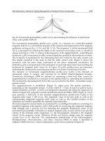
Doke et al., (1996) proposed a mechanism of oxidative damage in plant cells in response to
elicitors derived from fungal cell wall. The invasive fungal elicitor molecule (oligoglucan or,
if the elicitation is mediated by pectic oligogalacturonic from plants) is recognized by the
plasma membrane receptor (peripherial or transmembrane proteins), this recognition
stimulates Ca
2+
influx through Ca
2+
channels. The increase in free Ca
2+
in the cell acts as a
second messenger, together with the activation of calmodulin (CaM) to activate protein
kinases and protein factors by phosphorylation. Then the activated NADPH oxidase
provides electrons through the oxidation of NADPH, and the electron transport system
reduces O
2
molecules generating the radical O
2
•
-
(Figure 3).
Fig. 3. Oligoglucans action mechanism in plants (modified Doke et al., 1996).
Cell Metabolism – Cell Homeostasis and Stress Response
8
6. Fungal glucans and their relationship with the enzymatic antioxidant
system in cold stressed plants
Every day, the non-desirable climate change effects are present in our agriculture and the
worldwide food production suffers the adverse consequences. Therefore, crop yields fell
around fifty percent for several crops (Wahid et al., 2007). Several environmentally agencies
report increments or reductions in temperature along the year. It is crucial to find an
environmental friendly solution to challenge against low crop yields.
Under thermal stress (heat or chilling temperatures), important metabolic and physiologic
plant processes are interrupted. As a consequence, protein aggregation and denaturalization
in chloroplasts and mitochondria, destruction of membrane lipids, production of toxic
compounds and the ROS overproduction (Howarth, 2005) are the most common responses
of plant cells. Those are some reasons of the destructive effects of this kind of abiotic stress.
There are several pre- and postharvest treatments to deal with thermal stress like genetic
modifications, thermal conditioning treatments of seeds and fruits or triggering early
defense systems in plants by exogenous elicitation (Falcón-Rodríguez et al., 2009; Islas-
Osuna et al., 2010). Our work team, evaluated the triggering of some important antioxidant
enzymes in squash (Cucurbita pepo L.) seedlings at low temperature by the spraying of a
novel mixture of fungal glucans isolated from Trichoderma harzianum by chemical and/or
enzymatic fungal cell wall hydrolysis (Cerón-García et al., 2011). Two of the most active
antioxidant enzymes, catalase and ascorbate peroxidase, were triggered by the exogenous
elicitation with fungal oligoglucans in cold-stressed squash seedlings. Both antioxidant
enzymes are the main active H
2
O
2
detoxificant elements in the plant cell. Antioxidant
enzymatic system in plants became unstable under thermal stresses, mainly by the
inhibition of the catalytic activities during extreme temperatures. However, the elicitation
with fungal glucans restored the deficiency of the antioxidant enzymatic system.
7. Conclusion
Biotic and abiotic factors may have a negative effect on plants, favoring the accumulation of
ROS to generate further oxidative stress. Multiple biochemical responses are clearly
generated by the use of oligoglucans as elicitors of defense responses against oxidative
stress. The recognition of elicitors may vary depending on their characteristics, on the plant
species or even for a particularly tissue, where specific receptors enables the generation of
secondary signals that promote the most active plant defense against various biotic and/or
abiotic factors by strengthening the antioxidant system, the accumulation of antimicrobial
compounds such as phytoalexins and the activation of plant defense-related genes. Since
there is little research on plant-oligoglucan interactions, so many questions remain
unanswered.
8. Acknowledgment
Abel Ceron-García thanks the fellowship from Consejo Nacional de Ciencia y Tecnología
(CONACyT). The authors would like to thank Olivia Briceño-Torres, Francisco Soto-
Cordova and Socorro Vallejo-Cohen for technical assistance. We also thank Emmanuel
Aispuro-Hernández for critical reading of the manuscript.
Oligoglucan Elicitor Effects During Plant Oxidative Stress
9
9. References
Alscher, R.G.; Erturk, N. & Heath, L.S. (2002). Role of superoxide dismutases (SODs) in
controlling oxidative stress in plants. Journal of Experimental Botany, Vol.53, No. 372.
pp. 1331-1341.
Apel, K. & Hirt, H. (2004). Reactive oxygen species: Metabolism, oxidative stress, and signal
transduction. Annual Review in Plant Biology, Vol.55, pp. 373-399. ISBN/ISSN 1543-
5008.
Asada, K. (1999). The water–water cycle in chloroplasts: scavenging of active oxygen and
dissipation of excess photons. Annual Review in Plant Physiology and Plant Molecular
Biology, Vol.50, pp. 601-639. DOI: 10.1146/annurev.arplant.50.1.601.
Basse, C.W.; Fath, A. & Boller, T. (1993). High affinity binding of a glycopeptide elicitor to
tomato cells and microsomal membranes and displacement by specific glycan
suppressors. The Journal of Biological Chemistry, Vol.268, pp.14724-14731. ISSN 0021-
9258.
Baureithel, K.; Félix, G. & Boller, T. (1994). Specific high affinity binding of chitin fragments
to tomato cells and membranes. The Journal of Biological Chemistry, Vol.269, pp.
17931-17938. ISSN 0021-9258.
Bolwell, G.P.; Page, A.; Pislewska, M. & Wojtaszek, P. (2001). Pathogenic infection and the
oxidative defences in plant apoplast. Protoplasma, Vol.217. pp. 20-32. ISBN/ISSN
0033-183X.
Cerón-García, A.; Gonzalez-Aguilar, G.A.; Vargas-Arispuro, I.; Islas-Osuna, M.A. & Martinez-
Tellez, M.A. (2011). Oligoglucans as Elicitors of an Enzymatic Antioxidant System in
Zucchini Squash (Cucurbita pepo L.) Seedlings at Low Temperature. American Journal
of Agricultural and Biological Sciences, Vol.6, No. 1. pp. 52-61. ISSN 1557-4989.
Cheong, J.J. & Hahn, M.G. (1991). A specific, high affinity binding site for the hepta-β-
glucoside elicitor exists in soybean membranes. The Plant Cell, Vol.3, pp. 137-147.
ISSN 1040-4651.
Cosio, E.G.; Feger, M.; Miller, C.J.; Antelo, L. & Ebel, J. (1996). High-affinity binding of
fungal β-glucan elicitors to cell membranes of species of the plant family Fabaceae.
Planta, Vol.200, pp. 92-99. DOI: 10.1007/BF00196654.
Cosio, E.G.; Frey, T. & Ebel, J. (1992). Identification of a high-affinity binding protein for a
hepta-β-glucoside phytoalexin elicitor in soybean. European Journal of Biochemistry,
Vol.204, pp. 1115-1123. DOI: 10.1111/j.1432-1033.1992.tb16736.x.
Cosio, E.G.; Frey, T.; Verduyn, R.; Van Boom, J. & Ebel, J. (1990). High-affinity binding of a
synthetic heptaglucoside and fungal glucan phytoalexin elicitors to soybean
membranes. FEBS Letters, Vol.271, pp. 223-226. DOI: 10.1016/0014-5793(90)80411-B.
Coté, F. & Hahn, M.G. (1994). Oligosaccharins: Structure and signal transduction. Plant
Molecular Biology, Vol.26, pp. 1379-1411. DOI: 10.1007/BF00016481.
De Leonardis, S.; Dipierro, N. & Dipierro, S. (2000). Purification and characterization of an
ascorbate peroxidase from potato tuber mitochondria. Plant Physiology and
Biochemistry, Vol.38, pp. 773-779. DOI: 10.1016/S0981-9428(00)01188-8.
Delattre, C.; Michaud, P.; Lion, J. & Courtois, J. (2005). Production of glucuronan
oligosaccharides using a new glucuronan lyase activity from a Trichoderma sp.
strain. Journal of Biotechnology, Vol.118, pp. 448-457. ISBN/ISSN 0168-1656.
Delledonne, M.; Marocco, A. & Lamb, C. (2001). Signal interactions between NO and
reactive oxygen intermediates in the plant hypersensitive disease resistance
Cell Metabolism – Cell Homeostasis and Stress Response
10
response. Proceedings of the National Academy of Sciences of the United States of
America, Vol.98, pp. 13454-13459. DOI: 10.1073/pnas.231178298.
Doke, N.; Miura, Y.; Sanchez, L.M.; Park, H.J.; Noritake, T.; Yoshioka, H. & Kawakita, K.
(1996). The oxidative burst protects plants against pathogen attack: mechanism and
role as an emergency signal for plant bio-defense – a review. Gene, Vol.179, pp. 45-
51. ISBN/ISSN 0378-1119.
Ebel, J. & Scheel, D. (1997). Signals in host-parasite interactions. In: The Mycota V Part A.
Plant Relationships. G C Carroll, T Tudzynski (Eds). pp. 85-105. Springer-Verlag.
Berlin. Heidelberg.
Falcón-Rodríguez, A.B.; Cabrera, J.C.; Ortega, E. & Martinez-Tellez, M.A. (2009).
Concentration and physicochemical properties of chitosan derivatives determine
the induction of defense responses in roots and leaves of tobacco (Nicotiana
tabacum) plants. American Journal of Agricultural and Biological Sciences, Vol.4, pp.
192-200. ISSN 1557-4989.
Gadea, J.; Conejero, V. & Vera, P. (1999), Developmental regulation of a cytosolic ascorbate
peroxidase gene from tomato plants. Molecular Genomics and Genetics, Vol.262, pp.
212-219. DOI: 10.1007/s004380051077.
Girotti, A.W. (2001). Photosensitized oxidation of membrane lipids: reaction pathways,
cytotoxic effects and cytoprotective mechanisms. Journal of Photochemistry &
Photobiology, Vol.63, pp. 103-113. DOI: 10.1016/S1011-1344(01)00207-X.
Halliwell, B. (2006). Reactive species and antioxidants. Redox biology in fundamental theme
of aerobic life. Plant Physiology, Vol.141, pp. 312-322. www.plantphysiol.org/cgi/
doi/10.1104/pp.106.077073.
Hammond-Kosack, K. & Jones, J.D.G. (2000). Responses to plant pathogens. In: Biochemistry
and Molecular Biology of Plants. B.B. Buchanan, W. Gruissem, R.L. Jones (Eds). pp.
1102-1156. American Society of Plant Physiologist. ISBN 0-943088-37-2. Rockville,
MD.
Howarth, C.J. (2005). Genetic Improvements of Tolerance to High Temperature, In: Abiotic
stresses: Plant resistance through breeding and molecular approaches, Ashraf, M. &
Harris, P.J.C. pp. 725. Howarth Press Inc., ISBN: 1-56022-965-9. New York, USA.
Ishikawa, T., Sakai, K, Takeda, T. & Shigeoka, S. (1995). Cloning and expression of cDNA
encoding a new type of ascorbate peroxidase from spinach. FEBS Letters, Vol.367,
pp. 28-32. DOI: 10.1016/0014-5793(95)00539-L.
Ishikawa, T.; Sakai, K.; Yoshimura, K.; Takeda, T. & Shigeoka, S. (1996). cDNAs encoding
spinach stromal and thylakoid-bound ascorbate peroxidase, differing in the
presence or absence of their 3’-coding regions. FEBS Letters, Vol.384, pp. 289-293.
DOI: 10.1016/0014-5793(96)00332-8
Ishikawa, T.; Yoshimura, K.; Sakai, K.; Tamoi, M.; Takeda, T. & Shigeoka, S. (1998).
Molecular characterization and physiological role of a glyoxisome-bound ascorbate
peroxidase from spinach. Plant Cell Physiology, Vol. 30, pp. 23-34. ISSN 0032-0781.
Islas-Osuna, M.A., N.A. Stephens-Camacho, C.A. Contreras-Vergara, M. Rivera Dominguez,
E. Sanchez Sanchez, M.A. Villegas-Ochoa and G.A. Gonzalez Aguilar, 2010. Novel
postharvest treatment reduces ascorbic acid losses in mango (Mangifera indica L.)
Var. Kent. Am. J. Agric. Biol. Sci., 5: 342-349. ISSN: 15574989.
Jespersen, H.; Kjaersgard, I.; Ostergaard, L. & Welinder, K. (1997). From sequence analysis of
three novel ascorbate peroxidases from Arabidopsis thaliana to structure, function
Oligoglucan Elicitor Effects During Plant Oxidative Stress
11
and evolution of seven types of ascorbate peroxidase. Biochemical Journal, Vol.326,
pp. 305-310. PMCID: PMC1218670.
Kawakami, S.; Matsumoto, Y.; Matsunaga, A.; Mayama, S. & Mizuno, M. (2002). Molecular
cloning of ascorbate peroxidase in potato tubers and its response during storage at
low temperature. Plant Science, Vol.163, pp. 829-836.
Klarzinsky, O.; Plesse, B.; Joubert, J.M.; Yvin, J.C.; Kopp, M.; Kloareg, B. & Fritig, B. (2000).
Linear β-1,3 glucans are elicitors of defense responses in tobacco. Plant Physiology,
Vol.124, pp. 1027-1037.
Leon, J.; Lawton, M. & Raskin, I. (1995). Hydrogen peroxide stimulates salicylic acid
biosynthesis in tobacco. Plant Physiology, Vol.108, pp. 1673-1678.
López, F.; Vansuyt, G.; Case-Delbart, F. & Fourcroy, P. (1996). Ascorbate peroxidase activity,
not the mRNA level, is enhanced in salt stressed Raphanus sativas plants.
Physiological Plantarum, Vol.97, pp. 13-20.
Mittler, R. & Zilinskas, B.A. (1992). Molecular cloning and characterization of a gene
encoding pea cytosolic ascorbate peroxidase. The Journal of Biological Chemistry,
Vol.267, pp. 21802-21807. ISSN 0021-9258.
Mittler, R. & Zilinskas, B.A. (1994). Regulation of pea cytosolic ascorbate peroxidase and
other antioxidant enzymes during the progression of drought stress and following
recovery from drought. The Plant Journa,l Vol.5, pp. 397-405. DOI: 10.1111/j.1365-
313X.1994.0
Morita, S.; Kaminaka, H.; Masumura, T. & Tanaka, K. (1999). Induction of rice cytosolic
ascorbate peroxidase mRNA by oxidative stress; the involvement of hydrogen
peroxide in oxidative signal. Plant and Cell Physiology, Vol.1999, No.40, pp. 417-422.
ISSN 0032-0781.
Nürnberger, T.; Nennstiel, D.; Jabs, T.; Sacks, W.R.; Hahlbrock, K. & Scheel, D. (1994). High
affinity binding of a fungal oligopeptide elicitor to parsley plasma membranes
triggers multiple defense responses. Cell, Vol.78, pp. 449-460. DOI: 10.1016/0092-
8674(94)90423-5.
Orozco-Cardenas, M.L.; Narvaez-Vasquez, J. & Ryan, C.A. (2001). Hydrogen peroxide acts
as a second messenger for the induction of defense genes in tomato plants in
response to wounding, systemin, and methyl jasmonate. The Plant Cell, Vol.13, pp.
179-191. DOI: 10.1105/tpc.13.1.179
Orozco-Cardenas, M.L. & Ryan, C.A. (1999). Hydrogen peroxide is generated systemically
in plant leaves by wounding and systemin via the octadecanoid pathway.
Proceedings of the National Academy of Sciences of the United States of America, Vol.96,
pp. 6553-6557. DOI: 10.1073/pnas.96.11.6553
Orvar, B. & Ellis, B. (1995). Isolation of a cDNA encoding cytosolic ascorbate peroxidase in
Tobacco. Plant Physiology, Vol.108, pp. 839-840. PMCID: PMC157414.
Park, S.Y.; Ryu, S.H.; Jang, I.C.; Kwon, S.Y.; Kim, J.G. & Kwak, S.S. (2004). Molecular cloning
of a cytosolic ascorbate peroxidase cDNA from cell cultures of sweetpotato and its
expression in response to stress. Molecular Genetics and Genomics, Vol.271, No. 3. pp.
339-346. DOI 10.1007/s00438-004-0986-8
Qadir, S.; Qureshi, M.I.; Javed, S. & Abdin, M.Z. (2004). Genotypic variation in
phytoremediation potential of Brassica juncea cultivars exposed to Cd-stress. Plant
Science, Vol.167, pp. 1171-1181. DOI: 10.1016/j.plantsci.2004.06.018
Radman, R.; Saez, T.; Bucke, C. & Keshavarz, T. (2003). Elicitacion of plant and microbial cell
systems. Biotechnology Applied Biochemistry, Vol.37, pp. 91-102. ISBN/ISSN 0885-4513.
Cell Metabolism – Cell Homeostasis and Stress Response
12
Reddy, A.M.; Kumar, S.G.; Jyothsnakumari, G.; Thimmanaik, S. & Sudhakar, C. (2005). Lead
induced changes in antioxidant metabolism of horsegram (Macrotyloma uniflorum
(Lam.) Verdc.) and bangalgram (Cicer arietinum L.). Chemosphere, Vol.60, pp. 97-104.
Reilly, K.; Gomez-Vasquez, R.; Buschmann, H. & Beeching, J.R. (2004). Oxidative stress
responses during cassava post-harvest physiological deterioration. Plant Molecular
Biology, Vol.56, pp. 625-641.
Sharp, J.K.; Valent, B. & Albersheim, P. (1984). Purification and partial characterization of a
β-Glucan fragment that elicits phytoalexin accumulation in soybean. The Journal of
Biological Chemistry, Vol.259, No. 18. pp. 11312-11320.
Shigeoka, S.; Ishikawa, T.; Tamoi, M.; Miyagawa, Y.; Takeda, T. & Yoshimura, K. (2002).
Regulation and function of ascorbate peroxidase isoenzymes. Journal of Experimental
Botany, Vol.53, No. 372. pp. 1305-1319.
abstract/53/372/1305
Shinya, T.; Ménard, R.; Kozone, I.; Matsuoka, H.; Shibuya, N.; Kauffmann, S.; Matsuoka, K.
& Saito, M. (2006). Novel β-1,3-, 1,6-oligoglucan elicitor from Alternaria alternata 102
for defense responses in tobacco. FEBS Journal, Vol.273, No. 11. pp. 2421-2431.
ISBN/ISSN 1742-4658.
Sudha, G. & Ravishankar, G.A. (2002). Involvement and interaction of various signaling
compounds on the plant metabolic events during defense response, resistance to
stress factors, formation of secondary metabolites and their molecular aspects. Plant
Cell, Tissue and Organ Culture, Vol.71, pp. 181-212.
Tang, L.; Kwon, S.Y.; Kim, S.H.; Kim, J.S.; Choi, J.S.; Cho, K.Y.; Sung, C.K.; Kwak, S.S. & Lee,
H.S. (2006). Enhanced tolerance of transgenic potato plants expressing both
superoxide dismutase and ascorbate peroxidase in chloroplasts against oxidative
stress and high temperature. Plant Cell Report, Vol.25, No. 12. pp. 1380-1386. DOI
10.1007/s00299-006-0199-1.
The Arabidopsis Genome Initiative. (2000). Analysis of the genome sequences of the
flowering plant Arabidopsis thaliana. Nature, Vol.408, pp. 796-815.
Tsugane, K.; Kobayashi, K.; Niwa, Y.; Ohba, Y.; Wada, K. & Kobayashi, H. (1999). A
recessive Arabidopsis mutant that grows enhanced active oxygen detoxification.
Plant Cell, Vol.11, pp. 1195-206. PMC: 144266.
Wahid, A.; Gelani, S.; Ashraf, M. & Foolad, M.R. (2007). Heat tolerance in plants: An overview.
Environmental & Experimental Botany, Vol.61, pp. 199-223. ISBN/ISSN 0098-8472.
Waldmüller, T.; Cosio, E.G.; Grisebach, H. & Ebel, J. (1992). Release of highly elicitor-active
glucans by germinating zoospores of Phytophthora megasperma glycinea. Planta,
Vol.188, pp. 498-505. DOI: 10.1007/BF00197041.
Webb, R. & Allen, R. (1995). Isolation and characterization of a cDNA for spinach cytosolic
ascorbate peroxidase. Plant Physiology, Vol.108, pp. 1325. PMC: 157502.
Wojtaszek, P. (1997). Oxidative burst: an early plant response to pathogen infection.
Biochemical Journal, Vol.322, pp. 681–692. PMC 1218243.
Yamaguchi, T.; Yamada, A.; Hong, N.; Ogawa, T.; Ishii, T. & Shibuya, N. (2000). Differences
in the recognition of glucan elicitor signals between rice and soybean: beta-glucan
fragments from the rice blast disease fungus Pyricularia oryzae that elicit
phytoalexin biosynthesis in suspension-cultured rice cells. The Plant Cell, Vol.12,
No. 5. pp. 817-826.
Yoshikawa, M.; Yamaoka, N. & Takeuchi, Y. (1993). Elicitors: Their significance and primary
modes of action in the induction of plant defense reactions. Plant Cell Physiology,
Vol.34, No. 8. pp. 1163-1173. ISSN 0032-0781.
2
Regulation of Gene Expression in
Response to Abiotic Stress in Plants
Bruna Carmo Rehem
1
, Fabiana Zanelato Bertolde
1
and
Alex-Alan Furtado de Almeida
2
1
Instituto Federal de Educação, Ciência e Tecnologia da Bahia (IFBA)
2
Universidade Estadual de Santa Cruz
Brazil
1. Introduction
The multiple adverse conditions but not necessarily lethal, that occur sporadically as either
permanently in a location that plants grow are known as "stress." Stress is usually defined as
an external factor that carries a disadvantageous influence on the plant, limiting their
development and their chances of survival. The concept of stress is intimately related to
stress tolerance, which is the plant's ability to confront an unfavorable environment. Stress
is, in most definitions, considered as a significant deviation from the optimal conditions for
life, and induces to changes and responses in all functional levels of the organism, which are
reversible in principle, but may become permanent.
The dynamics of stress include loss of stability, a destructive component, as well as the
promotion of resistance and recovery. According to the dynamic concept of stress, the
organism under stress through a series of characteristic phases. Alarm phase: the start of the
disturbance, which is followed by loss of stability of structures and functions that maintain
the vital activities. A very rapid intensification of the stressor results in an acute collapse of
cellular integrity, before defensive measures become effective. The alarm phase begins with
a stress reaction in which the catabolism predominates over anabolism. If the intensity of the
stressor does not change the restitution in the form of repair processes such as protein
synthesis or synthesis of protective substances, will be quickly initiated. This situation leads
to a resistance phase, in which, under continuous stress, the resistance increases (hardening).
Due to the improved stability, normalization occurs even under continuous stress
(adaptation). The resistance may remain high for some time after the disturbance occurred.
If the state of stress is too lengthy or if the intensity of the stress factor increases, a state of
exhaustion can occur at the final stage, leaving the plant susceptible to infections that occur
as a consequence of reduced host defenses and leading to premature collapse or still a
chronic damage may occur, leading to plant death. However, if the action of the stressor is
only temporary, functional status is restored to its original level. If necessary, any injury
caused can be repaired during the restitution (Larcher, 1995).
The characteristics of the state of stress are manifestations nonspecific, which represent
firstly an expression of the severity of a disturbance. A process can be considered
nonspecific if it can not be characterized as a pattern, whatever the nature of the stressor.
Cell Metabolism – Cell Homeostasis and Stress Response
14
Examples of non-specific indications of the state of stress are: increased respiration,
inhibition of photosynthesis, reduction in dry matter production, growth disorders, low
fertility, premature senescence, leaf chlorosis, anatomical alterations and decreased
intracellular energy availability or increased energy consumption due to repair synthesis.
The cell responses to stress include changes in cell cycle and division, changes in the system
of vacuolization, and changes in cell wall architecture. All this contributes to accentuate
tolerance of cells to stress. Biochemically, plants alter metabolism in several manners, to
accommodate environmental stress (Hirt & Shinozaki, 2004).
Currently, all plant life is being threatened by rapid environmental changes. The gases
associates to global warming as CO
2
and methane have a enormous impact on global
environmental conditions, resulting in extreme changes in temperatures and weather
patterns in many regions of the world (Hirt & Shinozaki, 2004). In contrast to animals, plants
are sessile organisms and can not escape from environmental changes. The greenhouse
effect also affects the ozone layer causing the levels of ultraviolet (UV) are much larger to
reach the ground (Hirt & Shinozaki, 2004). Besides resulting in an increase in the registers of
the occurrence of diseases in humans such as skin cancer. The greenhouse effect also affects
the ozone layer causing the levels of ultraviolet (UV) are much larger to reach the ground.
Another concern is the intense use of chemical fertilizers and artificial irrigation in
agriculture. In many areas of the world, these practices have increased soil salinity. Under
these conditions, resistance to abiotic stress corresponds to a more required to be found in
several plant species (Hirt & Shinozaki, 2004). In short, the factors discussed above, together
with the increasing use of agricultural land cultivated is one of the biggest challenges for the
future humanity with regard to agriculture and conservation of genetic diversity in plant
species.
2. Water stress
Water has a key role in all physiological processes of plants, comprising between 80 and
95% of the biomass of herbaceous plants. If water becomes insufficient to meet the needs of
a particular plant, this will present a water deficit. The water deficit or drought is not caused
only by lack of water but also the environment in low temperature or salinity. These
different tensions negatively affect plant productivity (Hirt & Shinozaki, 2004).
Plants developed different mechanisms to adapt their growth in conditions where water is
limited. These adjustments depend on the severity and duration of drought, as well as the
development phase and morphology and anatomy of plants. The cellular response includes
the action of solute transporters such as aquaporin, activators of transcription, some
enzymes, reactive oxygen species and protective proteins. Two main strategies can be taken
to defend the damage caused by dehydration: synthesis of molecules of protection to
prevent damage and a repair mechanism based on rehydration in order to neutralize the
damage. In the classic signaling pathways, environmental stimuli are captured by receptor
molecules (Hirt & Shinozaki, 2004).
The main response that distinguishes tolerant plants of sensitive plants to drought stress is
the marked intracellular accumulation of osmotically active solutes in tolerant plants. This
mechanism, known as osmotic adjustment, is the ability of many species adjusts their cells
by decreasing the osmotic potential and water potential in response to drought or salinity
without a decrease in cell turgor.
Regulation of Gene Expression in Response to Abiotic Stress in Plants
15
In plants, dehydration activates a protective response to prevent or repair cell damage. The
plant hormone, abscisic acid (ABA) has a central role in this process. The ABA is considered
a "stress hormone" because plants respond to environmental challenges such as water and
salt stress with changes in the availability of ABA, as well as being an endogenous signal
required for adequate development. Dehydration in plants leads to increased levels of ABA,
which in turn induces the expression of several genes involved in defense against the effects
of water deficit. High levels of ABA cause complete closure of stomata and alteration of
gene expression. Stomatal closure reduces water loss through transpiration (Hirt &
Shinozaki, 2004). The ABA signaling is composed of multiple cellular events, including the
regulation of turgor and differential gene expression.
Plants have developed several mechanisms to adapt their growth to the availability of
water. The movement of water molecules is determined by water potential gradient across
the plasma membrane, which in turn is influenced by the concentration of solute molecules
inside and outside the plant cell. Fluctuations in water availability and flows of
transmembrane extracellular solute disrupt cellular structures, altering the composition of
the cytoplasm and modulate cell function (Hirt & Shinozaki, 2004).
One effect of the signal transduction cascade of dehydration is the activation of transcription
factors, which each activates a set of target genes, including those necessary for the synthesis
of protective molecules. Transcription factors that are activated by dehydration are
differentially expressed in tissues. Dehydration causes high level of expression of many
genes, among which the most prominent are the so called late embryogenesis abundant
genes (LEA) (Hirt & Shinozaki, 2004). The last step in the signaling cascade in response to
dehydration is the activation of genes responsible for synthesis of compounds that serve to
protect cellular structures against the deleterious effects of dehydration. Plants that are able
to survive in drought conditions have taken a variety of different strategies. There are three
important mechanisms to allow the plants to resist dehydration: the accumulation of solutes,
elimination of reactive oxygen species and synthesis of proteins with protective functions
(Hirt & Shinozaki, 2004).
In many species, dehydration leads to the accumulation of a variety of compatible solutes.
Compatible solutes are soluble molecules of low molecular weight that are not toxic and do
not interfere with cellular metabolism. The chemical nature of solutes differ among plant
species. They include betaines, including glycine betaine, amino acids (especially proline)
and sugars such as mannitol, sorbitol, sucrose or trehalose. These compounds help to
maintain turgor during dehydration, increasing the number of particles in solution.
Furthermore, can modulate membrane fluidity and protein by keeping it hydrated, allowing
the stabilization of its structure (Hoekstra et al., 2001).
One consequence of dehydration is an increase in the concentration of reactive oxygen
intermediates (ROI) (Mittler, 2002). ROI cause irreversible damage to membranes, proteins,
DNA and RNA. However, a low concentration of ROI is vital to the plant cells, they are
essential components in defense signaling to stress. When the ROI concentration increases
because of dehydration, prevention of damage to competitors is essential for survival. The
accumulation of ROI is largely controlled by intrinsic antioxidant systems that include the
enzymatic action of superoxide dismutase, peroxidases and catalases.
Cell Metabolism – Cell Homeostasis and Stress Response
16
The analysis of differential gene expression and analysis of global patterns of gene
expression using macro and microarray approaches have identified a broad spectrum of
transcripts whose expression is modified in response to dehydration (Fowler & Thomashow,
2002; Kreps et al., 2002; Seki et al., 2002). These studies have provided a fairly
comprehensive overview of the types of transcripts modulated by dehydration plant. They
showed that at least hundreds of genes are affected by dehydration.
3. Oxidative stress
For plants, as for all aerobic organisms, oxygen is required for normal growth and
development, but continuous exposure to oxygen can result in cellular damage and
ultimately death. This is because molecular oxygen is continually reduced within cells by
various forms of reactive oxygen species (ROS), especially the free radical superoxide anion
(O
2
-
) and hydrogen peroxide (H
2
O
2
), which react with many cellular components resulting
in acute or chronic damage resulting in cell death (Scandalios, 2002). Oxidative stress results
from disequilibrium in the generation and removal of ROS within cells. In plant cells, ROS
are generated in large quantities by both constitutive and inducible pathways, but in normal
situations, the cellular redox balance is maintained through the action of a great variety of
antioxidant mechanisms that evolved to remove ROS.
The calcium ions may also be related to oxidative stress and the antioxidant system in
plants. Oxidative stress, many enzymes are involved in the mechanisms of protecting the
protoplasm and cell integrity. This defense includes antioxidant enzymes able to remove or
neutralize free radicals and intermediate compounds that enable their production. Among
these enzymes, highlight the peroxidase (POX), catalase (CAT) and superoxide dismutase
(SOD). The mechanisms of elimination of reactive oxygen involving SOD, whose synthesis
is induced probably by increased production of O
2
-
. In the process, SOD converts O
2
-
to
hydrogen peroxide (H
2
O
2
) and then peroxidase and catalase removes hydrogen peroxide
formed. Hydrogen peroxide and superoxide radicals can exert deleterious effects in cells,
acting on lipid peroxidation of membranes, as well as damaging their DNA.
Several environmental stresses and endogenous stimuli can disrupt the redox balance by
increasing ROS production or reduced antioxidant activity, with continued oxidative stress.
In response toto increased ROS is induced the expression of genes encoding antioxidant
proteins and the genes that encode proteins involved in a variety of cellular processes of
rescue. ROS are produced during photosynthesis and respiration, as a byproduct of
metabolism, or by specific enzymes. Cells are equipped with a variety of effective
antioxidant mechanisms to eliminate ROS. Transcriptome analyses indicate that the
expression of many genes is regulated by ROS. These antioxidants include genes that
encode the rescue of cell defense proteins and signaling proteins. ROS can lead to
programmed cell death, stomatal closure, and gravitropism (Hirt & Shinozaki, 2004).
Oxygen is normally reduced by four electrons to produce water, a reaction catalyzed by
cytochrome oxidase complex and the electron transport chain of mitochondria. It is
relatively unstable and can be converted back to molecular form of oxygen or H
2
O
2
, either
spontaneously or through a reaction catalyzed by the enzyme superoxide dismutase (SOD).
H
2
O
2
in particular, acts as a signaling molecule with regulated synthesis, specific effects and
presents a series of removal mechanisms (Hirt & Shinozaki, 2004).
Regulation of Gene Expression in Response to Abiotic Stress in Plants
17
The evolution of photosynthesis and aerobic metabolism led to the development of
processes of generation of ROS in chloroplasts, mitochondria and peroxisomes. It seems
likely that the antioxidant mechanisms have evolved to combat the negative effects of these
ROS (Scandalios, 2002). As environmental pressures increase the generation of ROS, would
have been the evolutionary pressure for selection of ROS signaling mechanisms inducing
genes encoding antioxidant proteins and cellular defense. This role of "defense" of ROS and
these proteins may be one reason that leads to induction of cellular defense, where many
genes show a common response to various environmental stresses and oxidative stress,
allowing for acclimatization and tolerance (Bowler & Fluhr, 2000). Functions of protection
against ROS may also have been responsible for the evolution of enzymes such as NADPH
oxidase, where the reaction seems to be the key ROS generation, in which the enzyme
activity can be regulated by environmental stresses. Thus, abiotic stress not only increases
the generation of ROS through non-specific mechanisms, but also trigger the signaling of
defense mechanisms that start with the induction of ROS production, continue with the
induction of defense responses and end with removal of ROS to restore the redox status and
cell survival.
Oxidative stress causes the intracellular environment becomes more electropositive, which
may induce a change in the redox environment and thus interfere with signaling pathways.
ROS are generated both electron transport and enzymatic sources. The generation of ROS
occurs through the process of electron transport in chloroplasts and mitochondria. H
2
O
2
is
generated by various enzymatic reactions, from specific enzymes such as NADPH oxidase
(Hirt & Shinozaki, 2004). Plant cells are rich in antioxidants, where the activity and location
of these can affect the concentration of H
2
O
2
.
Gene expression in response to oxidative stress may be coordinated through the interaction
of transcription factors (TF) with cis-elements common to the entity regulatory regions of
these genes. Certainly, the increase of ROS in cellular compartments such as mitochondria
or chloroplasts results in new profiles of transcription (Hirt & Shinozaki, 2004).
4. Flooding stress
The temporary or continuous flooding of the soil resulting from high rainfall, intensive
practice of large-scale irrigation farms or soils with inadequate drainage (Kozlowski, 1997).
In normal drainage, the soil contains air-filled pores that contain content similar to oxygen
from the atmosphere (20%) (Pezeshki, 1994). Excess water replaces the air in these pores,
extremely restricting the flow of oxygen in the soil, creating a condition of hypoxia (low O
2
availability) or, in more severe cases, anoxia (lack of O
2
) (Peng et al., 2005). The gas diffusion
becomes extremely slow in soils saturated with water, about 10,000 times slower than in air
(Armstrong, 1979).
Under natural conditions, the flooding changes numerous physical and chemical properties
of soil through processes of biological reduction, resulting from depletion of available
oxygen (O
2
), increasing the availability of P, Mn and Fe, and decreased availability of Zn
and Cu and the formation of hydrogen sulfide and organic acids (Camargo et al., 1999). This
soil is also characterized by accumulating a larger amount of CO
2
(Jackson, 2004) and
stimulate organic matter decomposition (Kozlowski, 1997; Pezeshki, 2001; Probert &
Keating, 2000). The phytotoxic compounds that accumulate in flooded soils can be produced
Cell Metabolism – Cell Homeostasis and Stress Response
18
both by plant roots (ethanol and acetaldehyde) and the metabolism of anaerobic
microorganisms (methane, ethane, unsaturated acids, aldehydes, ketones), and ethylene
may be produced by by plants and microorganisms (Kozlowski, 1997).
Flooding can devastate vegetation species poorly adapted to this kind of stress (Jackson,
2004). The stress tolerance of hypoxia or anoxia can vary in hours, days or weeks depending
on the species, the organs directly affected the stage of development and external conditions
(Vartapetian & Jackson, 1997). The duration and severity of flooding may be influenced not
only by the rate of influx of water, but also by the rate of water flow around the root zone
and the absorption capacity of soil water (Jackson, 2004).
One major effect of flooding is the privation of O
2
in the root zone, attributed to the slow gas
diffusion in soil saturated with water and O
2
consumption by microorganisms (Folzer et al.,
2005). Higher plants are aerobic and O
2
supplies depend on the environment to support
respiration and several other reactions of oxidation and oxygenation of vital (Vartapetian &
Jackson, 1997). The O
2
as participate of aerobic respiration final electron acceptor in
oxidative phosphorylation, generation of ATP and regeneration of NAD
+
, and in several
crucial biosynthetic pathways as the synthesis of chlorophyll, fatty acids and sterols (Dennis
et al., 2000). Under hypoxia, glycolysis and fermentation can exceed the aerobic metabolism
and become the only way to produce energy (Sousa & Sodek, 2002). The main products of
fermentation in plant tissues are lactate, ethanol and alanine derived from pyruvate, the end
product of glycolysis (Drew, 1997). Evidence suggests that cytosolic acidosis causes lactic
fermentation and therefore the maintenance of cytosolic pH is important for the survival of
plants under waterlogged conditions (Dennis et al., 2000).
The initial responses of many plants to flooding correspond to wilt and to stomatal closure,
starting from one or two days of exposure of roots to this stress, accompanied by decreases
in photosynthetic rate (Chen et al., 2002). These changes promote a decline in growth of
stems and roots and can damage the roots and death of many species of plants (Kozlowski,
1997). The saturation of the soil causes significant decreases in total plant biomass and
biomass allocation to roots and changes in biomass allocation pattern in many woody
species and herbaceous (Pezeshki, 2001; Rubio et al., 1995).
The plant damage induced by hypoxia, have been attributed to physiological dysfunction,
which include the change in the relationship between carbohydrates, minerals, water and
hormones, as well as the reduction or alteration of several metabolic pathways (Kennedy et
al., 1992). Initially, the plant under stress by hypoxia shows decreases in the rate of CO
2
uptake by leaves (Kozlowski, 1997). Some authors suggest that stomatal closure may be
associated with a decrease in hydraulic conductivity of roots (Davies & Flore, 1986;
Kozlowski, 1997), as well as the transmission of hormonal signals from roots to shoots.
Among the hormones involved in signal transmission are the ABA and cytokinin (Else et al.,
1996). During stages of prolonged flooding, the progressive decrease in photosynthetic rate
is attributed to changes in enzyme carboxyl groups and loss of chlorophyll (Drew, 1997;
Kozlowski, 1997). The decrease in activity of ribulose -1,5-bisphosphate carboxylase-
oxygenase (RUBISCO), the enzyme responsible for assimilation of CO
2
in the biochemical
phase of photosynthesis, contributing to losses in photosynthetic capacity (Pezeshki, 1994).
It is known that water temperature, light, O
2
and nutrient availability are the main abiotic
factors that control the growth of woody plants (Kozlowski, 1997). The excess or shortage of
Regulation of Gene Expression in Response to Abiotic Stress in Plants
19
any of these factors, such as water, causes significant deviations in the optimum conditions
for growth for a given species, generating a stress condition. Such a condition, depending on
the level of specialization of the organism, the amplitude and duration of stress, may be
reversible or become permanent (Lichtenthaler, 1996). Excess water in the root zone of
terrestrial plants may be injurious or even lethal because it blocks the transfer of O
2
and
other gases between soil and atmosphere (Drew, 1997). The responses of plants to flooding
vary according to several factors, among which may include the species, genotype and age
of the plant, the properties of water and duration of flooding (Kozlowski, 1997).
Plant growth and primary productivity of ecosystems are ultimately dependent on
photosynthesis (Pereira et al., 2001). Either environmental stressor that, somehow, can
interfere with the photosynthetic rate, will affect the net gain of dry matter and, therefore,
growth (Pereira, 1995). Growth and development of most vascular plant species are
restricted by flooding, particularly when they are completely submerged and may result in
death (Jackson & Colmer, 2005). Generally, waterlogged soils affect the growth of aerial part
of many woody species, suppressing the formation of new leaves, retarding the expansion
of leaves and internodes that formed before flooding and reduce growth of stem diameter of
species not tolerant to flooding, causing senescence and premature leaf abscission
(Kozlowski, 1997). The plant response to flooding during the growing phase, including
injury, inhibiting seed germination, of vegetative and reproductive growth and changes in
plant anatomy (Kozlowski, 1997).
Morphological changes, such as the formation of hypertrophied lenticels, aerenchyma and
adventitious roots, observed during O
2
deficiency in the soil are key to increasing the
availability of O
2
in the tissues of plants. The lenticels participate in the uptake and diffusion
of O
2
to the root system and the liberation of potentially toxic volatile products such as
ethanol, acetaldehyde and ethylene (Medri, 1998). The best strategy of flooding tolerance is
the supply of internal aeration, increased with the formation of hypertrophied lenticels,
which are the main points of entry of O
2
in the plants, associated with the appearance of
intercellular air spaces (White & Ganf, 2002). According Topa & McLeod (1986), the increase
of these air spaces allows for an efficient entry of O
2
causing the lenticels assume the role of
gas exchange in hypoxic conditions. The formation of hypertrophic lenticels occurs in
submerged portions of stems and roots of various woody angiosperms and gymnosperms
(Kozlowski, 1997), and involves both increased activity felogênica as the elongation of
cortical cells (Klok et al., 2002). In addition, participating in the uptake and diffusion of O
2
to
the root system and release potentially toxic volatile products such as ethanol, acetaldehyde
and ethylene (Medri, 1998).
It can be observed the formation of aerenchyma in stems and roots of aquatic species
tolerant to flooding, which usually occurs by cell separation during development
(esquizogeny) or by lysis of cortical cells and cell death (lysogeny) (He et al., 1994; Drew,
1997). The point of view adaptive aerenchyma provides a low resistance to diffusion of air
inside the submerged tissue, promoting survival of plants to flooding (Drew, 1997). The
formation of aerenchyma, recorded during the stress by O
2
deficiency in soil is associated
with the accumulation of ethylene. Roots under flooded soil containing high concentrations
of ethylene, compared with roots in normal, and its precursor (acid-1-aminocyclopropane-1-
carboxylic acid - ACC), and high activity of ACC synthase and ACC oxidase (He et al., 1994;
He et al., 1996). Ethylene induces the activity of different enzymes such as cellulases, and
Cell Metabolism – Cell Homeostasis and Stress Response
20
hydrolases xyloglucanases, enzymes, cell wall loosening related to the formation of
aerenchyma (He et al., 1994; Drew, 1997).
In the submerged portion of the plant there is a dead roots and the production of
adventitious roots on the root system portions of the original stem. These roots induced by
flooding, are usually thickened and exhibit more intercellular spaces of the roots growing in
well drained soils (Hook et al., 1971). The induction of adventitious roots has been reported
in a wide variety of plant species tolerant and non-tolerant, but usually occurs in species
tolerant to flooding (Kozlowski & Pallardy, 1997). According Kozlowski (1997), the
adventitious roots are produced from the original roots and submerged portions of stems.
According to the author in terms of flooding the induction of adventitious root formation
can be reported in both angiosperms and gymnosperms in tolerant and non tolerant to this
type of stress (Kozlowski, 1997). For Chen et al. (2002), the adventitious roots are important
in plants with high root hypoxia, since they are responsible for obtaining O
2
needed for their
development. The increased number of adventitious roots can be accompanied by an
increment of damage and death of the original roots (Chen et al., 2002). Among the root
adaptations to flooding can also cite the development of aerenchyma, induced by increasing
endogenous levels of ethylene (Mckersie, 2001). This tissue serves as an air transport system
in aquatic plants and can develop into plants that grow in hydromorphic soils. The
intercellular spaces are developed primarily by disintegration of cells due to an increase in
cellulase activity, or by increasing the intercellular spaces when there is lack of O
2
and hence
increase in ethylene production (Fahn, 1982). In some plants, such stress induces abnormal
formation of the wood and increase the proportion of parenchymatous tissues of xylem and
phloem (Kozlowski, 1997).
Flooding can also cause a decline in the growth of petioles and leaf stomatal conductance
(Domingo et al., 2002). Moreover, the saturation of the soil (i) interfere in the allocation of
photoassimilates in woody and herbaceous plants, root can decrease metabolism and
oxygen demand (Chen et al., 2002); (ii) inhibits the initiation of flower buds and the increase
in fruit species not tolerant to flooding; (iii) induces abscission of flowers and fruits; (iv)
reduces the quality of fruits due to the reduction of size, changing its appearance and
interfering in its chemical composition (Kozlowski, 1997).
Other responses of plants to flooding include: (i) decreased permeability of the root and the
absorption of water and mineral nutrients, the death and suppression of root metabolism;
(ii) the epinasty, leaf chlorosis and necrosis, and (iii) decrease in fruit production. On the
other hand, several morpho-physiological responses are driven by differential expression of
a large number of genes induced by conditions of hypoxia or anoxia (Vartapetian & Jackson,
1997; Kozlowski, 1997; Holmberg & Büllow, 1998; Vantoai et al., 1994; Klok et al., 2002).
The decreased availability of O
2
also affects different processes of plant genetics (Blom &
Voesenek, 1996; Kozlowski, 2002; Drew, 1997). Saab & Sachs (1995) observed in maize under
conditions of flooding, the 1005 induction of the gene that encodes a homologue of the
xyloglucan endotransglicosilase (x and t), an enzyme potentially involved in cell wall
loosening (Peschke & Sachs, 1994). This gene, which is among the first to be induced by
flooding, does not encode enzymes of glucose metabolism. Believed to be associated with
the onset of the structural changes induced by flooding (Saab & Sachs 1995), because the
substrate of XET and t are the xyloglucans that are part of the cell wall structure. Saab &