Genetic Engineering Basics New Applications and Responsibilities Part 5 potx
Bạn đang xem bản rút gọn của tài liệu. Xem và tải ngay bản đầy đủ của tài liệu tại đây (2.33 MB, 20 trang )
Thermostabilization of Firefly Luciferases Using Genetic Engineering
69
and all of them demonstrated enhanced thermostability (Kajiyama & Nakano, 1994). The L.
lateralis luciferase mutant Ala217Leu retained over 70% of the initial activity after 60 min
incubation at 50°C. Its half-life was about 20 times longer than that of the wild type
L. lateralis luciferase. Its thermostability was superior to that of the L. cruciata luciferase
mutant Thr217Leu.
Random mutagenesis was also used to obtain thermostable mutant of P.pyralis luciferase.
The substitution Glu354Lys increased thermostability of the enzyme 5-fold (White et al.,
1996). The substitution of Glu354 with all possible amino acid residues by site-directed
mutagenesis showed that the most stable mutants contained Lys or Arg residues. Thus, the
substitution of negatively charged residue to positive one in this part of enzyme molecule
increased the thermostability of P.pyralis luciferase. Thermostable P.pyralis luciferase was
also obtained by a combination of random and site-directed mutagenesis. The double
mutant was constructed that contained the substitutions Glu354Lys and Ala215Leu (similar
to Ala217Leu in L. lateralis luciferase). In this case the effect of thermostabilization was not as
high as for . lateralis luciferase. At 37°C the single mutants retained 10-15% of activity after 5
hours, whereas the wild type luciferase was completely inactivated. The double mutant
combined the thermostability gains of the single mutants and retained greater than 50%
activity for over 5 h. At 42°C the half life of the double mutant was reduced to 20 minutes.
At 50°C it was only 4 min (Price et al., 1996). Other point mutations have been identified
(largely by random mutagenesis) that significantly increase the thermostability of the
P.pyralis luciferase: T214A, I232A and F295L. Combining these point mutations with the
E354K mutation into the P.pyralis gene resulted in mutant luciferase (rLucx4ts) that had an
increase in thermostability of about 7°C relative to the wild-type enzyme. Hence, in this case
the multiple point mutations led to a cumulative increase in thermostability (Tisi et al., 2002).
After the spatial structure of luciferase was published, it became possible to rationally select
specific positions for mutagenesis. For example, in molecule of P.pyralis luciferase five bulky
hydrophobic solvent-exposed residues, which are all non-conserved and do not participate
in secondary-structure formation, were substituted by hydrophilic ones, in particular by
charged groups. These substitutions (F16R, L37Q, V183K, I234K and F465R) led to the
enzyme with greatly improved pH-tolerance and stability up to 45°C. The mutant showed
neither a decrease in specific activity relative to the wild-type luciferase (Law et al., 2006).
Introduction of almost all known point mutations (12 residues) enhancing the
thermostability of P. pyralis luciferase resulted in a highly stable mutant with half-time of
inactivation of 15 min at 55°C, whereas wild-type luciferase inactivates within seconds at
this conditions (Tisi et al., 2007).
5. Rational protein design approach to produce the stable and active enzyme
Mutations that are efficient in one particular luciferase do not always lead to successful
results when applied to other homologous luciferases. For example, the mutation E354R
increased the thermal stability of P. pyralis luciferase, whereas the corresponding E356R
substitution did not affect H. parvula luciferase. The substitution A217L in L. lateralis,
L. cruciata and in P. pyralis (A215L) firefly luciferases produced fully active and thermostable
mutants, but in the case of H. parvula luciferase this mutation decreased activity to about 0.1%
of the wild type in spite of some increase in thermal stability (Kitayama, et al. 2003). These
results are of particular interest for the L. mingrelica luciferase because it shares 98%
Genetic Engineering – Basics, New Applications and Responsibilities
70
homology with H. parvula. Hence, both enzymes are considered to be almost identical, and
the similar effect of this mutation could be expected for L. mingrelica luciferase. A rational
protein design approach was used to increase thermal stability of L. mingrelica luciferase and
prevent the detrimental effect of the of the A217L mutation on its activity by combining the
mutation A217L with additional substitutions in its vicinity. The three-dimensional
structure of the firefly luciferase and the multiple sequence alignment of beetle luciferases
were analyzed to identify these additional substitutions (Koksharov & Ugarova, 2011a).
Comparison of the A217 environment in L. mingrelica luciferase with that of L. cruciata and L.
lateralis luciferases showed only 3 significant differences: G216N, I212L, S398M. Another
difference was the change I212L, but it is unlikely to be important because the properties of
Leu and Ile are very close. On the other hand, the neighboring residue G216 and the more
remote S398 are characteristic for the small subgroup of luciferases very close in homology
to L. mingrelica luciferase (including H. parvula luciferase). We surmised that the elimination
of these differences between two groups of luciferases would lead to the A217 environment
similar to that of L. cruciata and L. lateralis luciferases, which could possibly prevent the loss
of activity accompanying the substitution A217L. First, we assumed that that changing the
neighboring residue G216 would be sufficient to retain the enzyme activity/ Therefore, the
double mutant G216N/A217L was constructed. Since this double mutant still showed low
activity, we introduced the additional substitution S398M of the less close residue. This led
to a stable and active mutant of L. mingrelica luciferase (Table 1).
Enzyme Mutant
Relative
specific
activity%
Temperature
of
inactivation
Half-life, min Reference
wild-type 100 ~ 4
Luciola
cruciata
luciferase
T217I 130
50 °C
~ 28
Kajiyama&
Nakano, 1993
wild-type ~ 6
Luciola
lateralis
luciferase
A217L
50 °C
~ 125
Kajiyama &
Nakano, 1994
wild-type 100 ~ 18
Hotaria
parvula
luciferase
A217L 0.074
45°C
~ 60
Kitayama et al.,
2003
wild-type 100 13 ± 1
G216N/A217L 10 280 ± 28
S398M 106 16.1 ± 1.6
Luciola
mingrelica
luciferase
G216N/A217L/S398M 60
45°C
276 ± 28
Koksharov &
Ugarova, 2011a
Table 1. Thermal stability of luciferases with substitution of the residue 217 in a 0.05 M Na-
phosphate buffer, containing 0.4 M (NH
4
)
2
SO
4
, 2 mM EDTA, 0.2 mg/ml BSA, pH 7.8
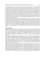
The residues 216, 217, 398 are located near one of the walls of the luciferin-binding channel
(Fig. 4). In the majority of beetle luciferases position 216 is normally occupied with a residue
having a side group but in L. mingrelica and H. parvula luciferases it is occupied with Gly.
Glycine is known to be a very destabilizing residue when in internal position of α-helices
because of the absence of side group and excessive conformational freedom (Fersht &
Serrano, 1993).
Since the G216 is located in the α-helix (Fig. 4) it can be suggested that it makes the
surrounding structure less stable and more sensitive to the substitutions of the neighboring
Thermostabilization of Firefly Luciferases Using Genetic Engineering
71
residues. This can explain the unusual decrease in activity in case of the A217L mutation in
Hotaria parvula luciferase (Kitayama, et al. 2003). The double mutation G216N/A217L
resulted in the significant increase of the thermal stability of L. mingrelica luciferase, but this
mutant retained only 10% of the wild-type activity. The comparison of the environment of
residue 217 in the crystal structure of L. cruciata luciferase (Nakatsu, et al., 2006) with the
homology model of L. mingrelica luciferase (Koksharov & Ugarova, 2008) (Fig. 4) shows that
internal cavities probably exist in L. mingrelica luciferase near the 216 and 398 positions
because of the smaller size side groups of the residues in this positions compared to L.
cruciata luciferase. Additional cavity in the vicinity of S398 could potentially decrease the
local conformational stability, make it more flexible and sensitive to the mutations and the
changes in the environment. This hypothesis is supported by the higher resistance of the
bioluminescence spectrum of the S398M mutant to pH and temperature, which indicates
more rigid and stable microenvironment (Ugarova & Brovko, 2002).
Fig. 4. Structure of L. mingrelica luciferase in complex with oxyluciferin (LO) and AMP. The
residues G216, A217, R220 and S398 are indicated by arrows. 7 Å microenvironment of A217
is indicated by ellipse (Koksharov & Ugarova, 2011a). The large N-terminal and the smaller
C-terminal domains are depicted in grey and orange, respectively
The lowered local conformational stability in the vicinity of G216 and S398 residues can
explain why the A217L mutation in H. parvula and L. mingrelica luciferaess leads to the
decline in activity and red shift of λ
max
that were not observed in the cases of L. cruciata,
L. lateralis, P. pyralis luciferases containing Asn or Thr at the position 216 and Met at the
position 398. In the former case the enzymes are much more likely to loose the conformation
optimal for the activity as a result of residue substitutions. As can be seen the G216, A217,
Genetic Engineering – Basics, New Applications and Responsibilities
72
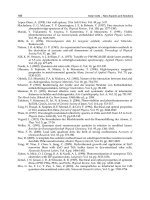
S398 residues are located in one plane with the neighboring residue R220 (Fig. 5). The
residue R220 (the residue R218 in P.pyralis luciferase) is highly conservative and necessary
for the green emission of firefly luciferases. Its substitutions led to the red bioluminescence,
3-15-fold decrease in activity, extended luminescence decay times and dramatic increase in
K
m
values (Branchini et al., 2001). The G216N/A217L double substitution in L. mingrelica
luciferase caused the similar type of effects but of less extent. Thus, in L. mingrelica and
H. parvula luciferases the proper alignment of the R220 residue can be affected by the
substitution of A217L and lead to the observed detrimental effects. Placing Asn and Met at
positions 216 and 398 respectively (as in the triple mutant G216N/A217L/S398M of
L. mingrelica luciferase and in native L. cruciata, L. lateralis luciferases) makes local
microenvironment of A217 sufficiently rigid to retain active conformation in the case of the
A217L mutation.
Fig. 5. Residues 216, 217, 220 and 398 in the structures of L. mingrelica (A) and L. cruciata (B)
luciferases (Koksharov & Ugarova, 2011a). Reproduced by permission of The Royal Society
of Chemistry (RSC)
In conclusion it can be stated that rational protein design of the residue microenvironment
can be an effective strategy when a single mutation in one firefly luciferase does not lead to
the desirable effect reported for the mutation of the homologous residue in the another
firefly luciferase. The constructed triple mutant G216N/A217L/S398M showed significantly
improved thermal stability, high activity and bioluminescence spectrum close to that of the
wild-type enzyme. The improved characteristics of this mutant make it a promising tool for
in vitro and in vivo applications.
6. Site-directed mutagenesis of cysteine residues of Luciola mingrelica firefly
luciferase
The number of Cys residues of luciferases is highly varied (from 4 to 13 residues) depending
on the firefly species. Luciferases contain three absolutely conservative SH groups that do
not belong to the active site. However their mutagenesis was shown to affect activity and
stability of luciferases (Dement’eva et al., 1996; Kumita et al., 2000). For example, the mutant
Photinus pyralis luciferase in which all the four Cys residues were substituted with Ser,
retained only 6.5 % of activity, whereas mutants with single substitutions lost 20-60% of
activity (Kumita et al., 2000; Ohmiya & Tsuji, 1997).
Thermostabilization of Firefly Luciferases Using Genetic Engineering
73
The Luciola mingrelica firefly luciferase contains eight cysteine residues, three of which
correspond to the conservative cysteine residues of P. pyralis firefly luciferase - 82, 260, and
393. Mutant forms of L. mingrelica luciferase containing single substitutions of these cysteine
residues to alanine were obtained previously (Dement’eva et al., 1996). These substitutions
had no effect on bioluminescent and fluorescent spectra of the enzyme and on enzyme
activity. The stability of the C393A mutant was 2-fold higher at 5-35˚C than that of the wild-
type enzyme. The substitutions C82A, C260A did not affect the thermal stability of
luciferase. The pLR plasmid, encoding firefly luciferase with the structure identical to that of
the native enzyme, was previously used for the preparation of the mutant forms of the
enzyme with single substitutions of the non-conserved cysteine residues C62S, C146S
(Lomakina et al., 2008) and C164S (Modestova et al., 2010). These substitutions also had no
significant effect on the catalytic and spectral properties of the luciferase, but they resulted
in an increase of the enzyme thermal stability and in a decrease of the dependence of
inactivation rate constant on the enzyme concentration (unlike the wild-type enzyme).
Moreover, the DTT influence on luciferase stability was diminished. These effects were most
pronounced for the enzyme with the substitution C146S.
The purification of recombinant luciferase obtained using the plasmid pLR is a complicated
multistage process. Therefore, the recombinant L. mingrelica luciferase with C-terminal His
6
-
tag was used for mutagenesis of cysteine residues (Modestova et al., 2011). The wild-type
enzyme and its mutant forms were expressed in E. coli BL21(DE3) cells transformed with the
pETL7 plasmid (Koksharov & Ugarova, 2011a). This approach led to the simpler scheme of
the luciferase purification and to the increase of the enzyme yield due to the use of the
highly efficient pET expression system. The influence of polyhistidine tag on luciferase
properties was not previously analyzed in detail according to the literature. A number of
publications indicate that while his-tags often don’t affect enzyme function, in many cases
the biological or physicochemical properties of the histidine tagged proteins are altered
compared to their native counterparts (Amor-Mahjoub et al., 2006; Carson et al., 2007;
Efremenko et al., 2008; Freydank et al., 2008; Klose et al., 2004; Kuo & Chase, 2011). The goal
of this study was to elucidate the role of non-conserved cysteine residues in the L. mingrelica
firefly luciferase, to study the mutual influence of these residues and the effect of His
6
-tag on
the activity and thermal stability of luciferase (Modestova et al., 2011).
6.1 Analysis of the fragments of luciferase amino acid sequences containing cysteine
residues
Among the firefly luciferases those amino acid sequences are known, firefly luciferases from
Luciola and Hotaria genera, and the Lampyroidea maculata firefly luciferase form a separate
group with more than 80% amino acid identity (Fig. 6). The second group includes
luciferases from firelies of various genera: Nyctophila, Lampyris, Photinus, Pyrocoelia, etc. The
sequence identity of luciferases from the first and the second group does not exceed 70%.
Amino acid sequences of the firefly luciferases belonging to these groups vary significantly.
One of the most evident distinctions is the amount and location of cysteine residues. The
residue С82 is absolutely conserved in all beetle luciferases, and the residue С260 is
absolutely conserved in all firefly luciferases. The residue С393 is conserved in all beetle
luciferases except the Cratomorphus distinctus (Genbank AAV32457) and one (Genbank
U31240) of the P. pennsylvanica luciferases. The C62, 86, and 284 residues are also absolutely
Genetic Engineering – Basics, New Applications and Responsibilities
74
Origin C62 C82, C86 C146 C164 C260 C284 C393
First group of luciferases
Luciola mingrelica
FDIT
CRLAEAM IALCSENCEEFF VQKTVTCIKKIVI NFGGHDCMETFI LGYFACGYRVVML TLQDYKCTSVILV RRGEICVKGPS
Luciola cruciata
LEKS
CCLGKAL IALCSENCEEFF VQKTVTTIKTIVI DYRGYQCLDTFI LGYLICGFRVVML TLQDYKCTSVILV RRGEVCVKGPM
Hotaria parvula
FDIT
CRLAEAM IALCSENCEEFF VQKTVTCIKTIVI NFGGHDCMETFI LGYFACGYRVVML TLQDYKCTSVILV RRGEICVKGPS
Hotaria unmunsana
FDIT
CRLAEAM IALCSENCEEFF VQKTVTCIKTIVI NFGGYDCMETFI LGYFACGYRVVML TMQDYKCTSVILV RRGEICVKGPS
Hotaria tsushimana
FDIT
CHLAEAM IALCSENCEEFF VQKTVTCIKTIVI NFGGYDCMETFI LGYFACGYRVVML TMQDYKCTSVILV RRGEICVKGPS
Luciola italica
FDIT
CRLAEAM IALCSENCEEFF VQKTVTCIKTIVI NFGGYDCVETFI LGYFACGYRIVML TLQDYKCTSVILV RRGEICVKGPS
Lampyroidea
maculata
FDIS
CRLAEAM IALCSENCEEFF VQKTVTCIKTIVI NFGGYDCVETFI LGYFACGYRIVML TMQDYKCTSVILV RRGEICVKGPS
Luciola lateralis
LEKS
CCLGEAL IALCSENCEEFF VQKTVTAIKTIVI DYRGYQSMDNFI LGYLTCGFRIVML TLQDYKCSSVILV RRGEVCVKGPM
Luciola terminalis
LDVS
CRLAQAM IALCSENCEEFF VQKTVTCIKTIVI DYQGYDCLETFI LGYLICGFRIVML TLADYKCNSAILV RRGEICVKGPM
Second group of luciferases (illustrated by Photinus pyralis luciferase)
Photinus pyralis
FEMS
VRLAEAM IVVCSENSLQFF VQKKLPIIQKIII DYQGFQSMYTFV LGYLICGFRVVLM SLQDYKIQSALLV
QRGEL
CVRGPM
Fig. 6. Fragments of amino acid sequence alignment of various firefly luciferases (the
regions containing Cys residues). The numbering corresponds to that of Luciola mingrelica
luciferase
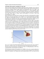
Fig. 7. Fragment of the 3D structure of Luciola mingrelica firefly luciferase containing the
residues C62 and C164
conserved in all luciferases from the first group. The residue C146 is conserved in all
luciferases of the first group, except for the L. lateralis and L. cruciata luciferases, in which
alanine and tyrosine are located at the position 146. The residue C164 is conserved in
luciferases of the first group except for the L. lateralis luciferase, which contains S146. The
C86 residue is located in a highly conserved region of luciferases of the first group, near the
C82 residue, which in its turn is located not far from the active site of the enzyme. Besides,
the C86 residue is located near the surface of the protein, and the surface area of its side
chain, that is accessible to the solvent, is about 11 Å
2
. The residue C146 is of particular
interest because of its surface location. Its side chain is exposed to the solvent with the
accessible surface area as high as 48 Å
2
. As a whole the Luciola luciferases possess high
Thermostabilization of Firefly Luciferases Using Genetic Engineering
75
amino acid sequence identity. However, there are several small areas in their amino acid
sequences the composition of which varies significantly. It is in these areas that the residues
C62 and C164 are located. These residues are positioned in two α-helixes and are in close
proximity with each other (Fig. 7).
The cysteine residues 62, 86, 146, and 164 of L. mingrelica luciferase were chosen for the site-
specific mutagenesis. In terms of the molecule topology the most suitable substitutions of
the Cys are Ser (hydrophilic amino acid) and Val (hydrophobic amino acid). The side chain
sizes of these residues are similar to that of Cys. We considered Ser as the most suitable
substitution for C86 and C146 residues because the side chains of these residues are in
contact with aqueous solution. The residue C164 was also substituted by Ser because its
microenvironment is weakly hydrophilic. Moreover, our previously results (Modestova et
al., 2010) suggest that in certain conditions this residue becomes available to the solvent. In
case of the residue Cys62 two mutants were obtained: C62S and C62V.
6.2 Preparation and physicochemical properties of mutant luciferases
The recombinant L. mingrelica firefly luciferase encoded by the plasmid pETL7 (GenBank
No. HQ007050) (Koksharov & Ugarova, 2011a) served as the parent enzyme (wild-type).
This form contains 4 additional amino acid residues (MASK) on N-terminus as compared to
the native sequence of L. mingrelica firefly luciferase (GeneBank No. S61961). The sequence
AKM at its C-terminus is replaced by the sequence SGPVEHHHHHH. A number of mutants
were obtained by site-directed mutagenesis of the plasmid pETL7: the mutant luciferases
with the single substitutions C62S, C62V, C86S, C146S, C164S, double substitutions
C62/146S, C62/164S, C86/146S, and C146/164S; the triple substitution С62/146/164S. The
wild-type luciferase and its mutant forms were purified using metal chelate
chromatography. The expression level and the specific activity of wild-type and its mutants
C62S, C62V, C164S, C62/146S, and C146S/C164S were the same within an experimental
error. Specific activity of the mutant C146S was ~15% higher than that of the wild-type,
while its expression level was unaltered. Meanwhile, the substitution C86S resulted in the
decrease of the enzyme expression level (62% compared to wild-type) and its specific
activity (30% compared to wild-type). The properties of the firefly luciferase with the double
substitution C86S/146S were similar to those of the mutant C86S. Drastic decrease of the
expression level and of the enzyme specific activity was observed at the introduction of the
double mutation C62S/C164S and the triple mutation С62S/C146S/C164S. Bioluminescence
and intrinsic fluorescence spectra of the wild-type luciferase and its mutant forms were
identical. Single mutations had almost no effect on the K
m
values for both substrates (K
m
ATP
and K
m
LH
2
) with the exception of the mutant C86S, for which, as well as for the mutant
C86S/C146S, 1.5-fold increase of both parameters was observed. The simultaneous
substitution of the residues C62S and C164S in both double and triple mutants led to 30%
increase of K
m
ATP
, but didn’t affect K
m
LH2
.
The irreversible inactivation of the wild-type luciferase and its mutant forms was
measured in 0.05 М Тris-acetate buffer (2 мМ EDTA, 10 мМ MgSO
4
, pH 7.8) at 37° and
42°C at concentration range of 0.01-1.0
µM. The inactivation of the wild-type luciferase
and its mutant forms followed the monoexponential first-order kinetics at all enzyme
concentrations assayed. The k
in
values of the wild-type luciferase and its mutant forms did
not depend on the initial luciferase concentration. The enzyme stabilization was only
Genetic Engineering – Basics, New Applications and Responsibilities
76
observed for the mutant C146S: the k
in
value decreased 2-fold at 37˚C and by 30% - at 42°C
(Table 2). At 37°C the k
in
values of the mutants С62V, C164S and C146S/C164S were
similar to the k
in
of the wild-type luciferase, but at 42°C the k
in
values of these mutants
were higher than that of the wild-type enzyme. All other mutants were less stable than
the wild-type enzyme. The substitution C86S caused a significant destabilizing effect on
the enzyme: the k
in
value increased twofold both at 37° and 42°C. The double mutant
C62S/C164S and the triple mutant С62S/C146S/C164S were the least stable among the
mutants obtained.
k
in
, min
-1
Enzyme
37° 42°
wild-type 0,022 ± 0,004 0,074 ± 0,006
C62V 0,024 ± 0,004 0,135 ± 0,004
C62S 0,036 ± 0,004 0,127 ± 0,004
C86S 0,040 ± 0,002 0,160± 0,006
C146S 0,011 ± 0,002 0,058 ± 0,003
C164S 0,018 ± 0,003 0,108 ± 0,005
C62S/C146S 0,042 ± 0,005 0,108 ± 0,005
C62S/C164S 0,052 ± 0,003 0,153 ± 0,005
C86S/C146S 0,047 ± 0,004 0,120 ± 0,006
C146S/C164S 0,023 ± 0,006 0,086 ± 0,005
C62S/C146S/C164S 0,055 ± 0,005 0,142 ± 0,006
Table 2. Rate constants of irreversible inactivation of wild-type luciferase and its mutant
forms with single and multiple substitutions of the 62, 86, 146, 164 cysteine residues at 37
and 42°C
6.3 The effect of polyhistidine tag on the properties of firefly luciferase
Comparison of the physicochemical properties of luciferases with single substitutions of the
residues C62S, C146S and C164S that were obtained for L. mingrelica luciferase without His
6
-
tag (Lomakina et al., 2008) with that of the mutant enzymes containing C-terminal His
6
-tag
(Modestova et al., 2011) led to a conclusion that the His
6
-tag shows significant influence on
the luciferase properties. Introduction of the His
6
-tag into the luciferase structure leads to
the increase of the K
m
ATP
and K
m
LH2
values. The interaction of the enzyme with the substrates
is known to involve the rotation of a big N-domain and a small C-domain of the luciferase
against each other at almost 90° (Sandalova & Ugarova, 1999). This movement is necessary
for the participation of the residue K531 from C-domain in the formation of enzyme-ATP-
luciferin active complex. The presence of the flexible His
6
-tag on the C-terminus of the
protein molecule might somewhat impede the process of domains rotation, that may result
in a slight increase of Km values for the both substrates.
Thermal inactivation of the firefly luciferase without His
6
-tag is a two-step process, which
includes a fast and a slow inactivation stages. The k
in
values of both stages are dependent
on the enzyme concentration, which is known to be a characteristic feature of oligomeric
Thermostabilization of Firefly Luciferases Using Genetic Engineering
77
enzymes. The single mutations С62S, С146S, С164S result in stabilization of the enzyme at
the slow stage of inactivation and in a decrease of k
in
dependence on the enzyme
concentration (Lomakina et al., 2008). The thermal inactivation of the His
6
-tag containing
wild-type luciferase and its mutants is a one-step process. The k
in
values of these enzymes
do not depend on luciferase concentration and coincide with the k
in
values of the respective
mutants without His
6
-tag that were measured at the increased enzyme concentration (1 µM).
This influence of the His
6
-tag on the inactivation kinetics of the wild-type luciferase and its
mutants may be due to the fact that the presence of the His
6
-tag considerably alters the
process of luciferase oligomerization.
6.4 Effect of the cysteine substitutions on luciferase structure and thermal stability
The substitution C146S results in a 2-fold stabilization of the enzyme at 37°C and in a 30%
increase of the enzyme stability at 42°C. This effect is associated with the surface location of
the side chain of this residue, its large solvent accessible area and the lack of interactions
with other amino acid residues of the enzyme. The C164S substitution doesn’t alter the
enzyme stability at 37°C, but leads to some destabilization at 42°C, though this
destabilization is less than that caused by the substitutions C62V, C62S and C86S. This effect
is, on the one hand, due to the fact, that the C164 residue is located in an area, which is
distant from the enzyme active site. On the other hand, the raise of temperature causes the
increase of solvent accessibility and the replacement of cysteine residue by the hydrophilic
serine improves interactions with the solvent.
Analysis of the luciferase 3D-model shows that it is hard to unambiguously estimate the
properties of the C62 residue microenvironment. This residue contacts with both
hydrophilic and hydrophobic amino acids. Therefore, two enzymes were obtained that carry
a hydrophilic and a hydrophobic side chain in the position 62. The specific activity, the
expression level and the kinetic parameters of the mutants C62S and C62V were similar to
those of the wild-type enzyme. The k
in
values at 42°C were also similar, but the mutant
C62V turned out to be 2-fold more stable than the mutant C62S at 37°C. Therefore, the
hydrophobic valine residue is more advantageous at 37°C in terms of the enzyme stability.
However, at temperature of 42°C the role of the amino acid residue microenvironment in
the enzyme stabilization becomes less pronounced and both modifications – serine or valine
– result in destabilization of the protein globule.
The substitution C86S shows the most significant influence on the luciferase properties. It
results in a decrease of the luciferase expression level and the specific activity, a
deterioration of the K
m
values for both substrates, and a decrease of the enzyme thermal
stability. The C86 residue is located within an unstructured area of the amino acid chain of
the enzyme (Fig. 8). The amino acid sequence forms a loop in this area due to the formation
of a hydrogen bond between the SH-group of the residue C86 and the oxygen atom OE1
belonging to the residue E88. The SH-group of cysteine residue is known to have a tendency
to form non-linear hydrogen bonds due to fact that the deformation of the valence angle has
a relatively small energy cost (Raso et al., 2001). The OH-group of serine residues has no
such tendency. Thereby it may be possible that the hydrogen bond between S86 and E88
residues can’t be formed in the mutant C86S. This may lead to an increase in mobility of the
chain fragment containing the abovementioned residues.
Genetic Engineering – Basics, New Applications and Responsibilities
78
Fig. 8. Fragment of the 3D structure of Luciola mingrelica firefly luciferase containing C82 and
C86 residues (Modestova et al., 2011)
It is important to underline that the C86 residue is located in an absolutely conserved area of
luciferases Luciola genus, not far from the enzyme active site and at the distance of ~15 Å
from T253, F249, F252 residues. These residues participate in the process of luciferase
substrates binding, and it is known that their mutations lead to a drastic alteration of the
enzyme catalytic properties and, in certain cases, to the disturbance of the enzyme
expression process (Freydank et al., 2008). On the basis of the experimental data one can
conclude that disturbance stripping-down of the protein structure (the “untwisting” of the
helix) in the area of the localization of the residue C86 disrupts the native structure of the
firefly luciferase active site area and leads to the deterioration of the luciferase activity and
stability.
Analysis of the properties of the mutants with multiple amino acid substitutions indicates
that in most of the cases the effect of such substitutions is additive. For instance, the
C86S/C146S mutant possesses the properties of the luciferase with single C86S substitution,
because it is the C86S substitution that affects the enzyme properties most significantly. The
mutants C62S/C146S and C146S/C164S also possess the characteristic properties of the
respective mutants with single replacements. However, the combination C62S/C164S leads
to the drastic decrease of the enzyme expression level, to the lowering of its specific activity
and stability and to the increase of the K
m
ATP
in comparison with the enzymes with the
single substitutions C62S and C164S. These facts indicate that the effect of these
substitutions is nonadditive. The analysis of luciferase 3D structure shows that C62 and
C164 residues belong to two closely located α-helixes (Fig. 8). The single mutations of these
residues have no significant effect on the enzyme properties, which is probably due to the
enzyme ability to compensate the effects of these substitutions. Meanwhile, the double
substitutions affect the mutual disposition of two α-helixes, in which these residues are
located.
Thus, the role of each cysteine residue in luciferase molecule is different and is determined
by its location relative to the active site, its microenvironment and even the oligomerization
state of luciferase. For example, in some cases the introduction of Cys residues into internal
protein core can increase the luciferase stability after replacement of hydrophilic residue by
more hydrophobic Cys. Such examples will be shown below.
Thermostabilization of Firefly Luciferases Using Genetic Engineering
79
7. Increase of P. pyralis luciferase thermostability by introduction of disulfide
bridges
It was mentioned above that luciferases are peroxisomal enzymes. They do not form
structural disulfide bonds despite of containing SH-groups (Ohmiya & Tsuji, 1997). When
expressed in E. coli, firefly luciferases cannot form any disulfide bonds due to the reducing
environment of the cytoplasm. On the other hand, introduction of disulfide bridges was
found to be one of the most efficient strategies for increasing protein stability (Eijsink et al.,
2004). Recently, disulfide bridges were introduced into P. pyralis firefly luciferase (Imani et
al., 2010) by site-directed mutagenesis. Two different mutant proteins were made with a
single bridge. P.pyralis firefly luciferase contains four cysteine residues at the positions 81,
216, 258 and 391. To find the residues capable to form disulfide bridges after their mutation
to cysteine, the crystal structure of P. pyralis luciferase was uploaded to the NCBS integrated
Web Server. The results from server showed that there are 150 pairs that could potentially
be selected for disulfide bridge formation. But only two pairs of residues were chosen due to
their similar size to the Cys residues: A103 and S121, located distant from active site region
of the enzyme, and A296 and A326, situated in the vicinity of the active site region. The
ability of mutated sites to form disulfide bridges was analyzed in Swiss-PDB Viewer.
Two mutant luciferases, each containing one S-S bridge, were obtained: A103C/S121C and
A296C/A326C. Relative specific activity showed a 7.25-fold increase for the mutant
A296C/A326C whereas the mutant A103C/S121C showed only 80% of wild-type specific
activity. Both mutants were more stable then the wild-type enzyme. For example, after
incubation at 40
°
C for 5 min the mutants A296C/A326C and A103C/S121C retained ~88%
and 22% of activity respectively, whereas the wild-type enzyme lost nearly all of its activity.
Using circular dichroism spectropolarimetric and fluorescence spectroscopic analysis, the
conformational changes of the enzyme structure were revealed, showing the more fixed
structure of aromatic residues, more compactness of tertiary structure, and a remarkable
increase in α-helix content.
It can be concluded that disulfide bridge formation in mutant A296C/A326C did not have a
destabilizing effect on the enzyme and caused a remarkable change in both secondary and
tertiary structure that is reflected in active site structure. These changes endow the enzyme
with properties that show an increased resistance to pH and temperature without any
stabilizer. On the other hand, the thermal stability of the mutant A103C/S121C arises from
the change of tertiary structure. Finally, these results showed that the engineered disulfide
bridge not only did not destabilize the enzyme but also in one mutant it improved the
specific activity and led to pH-insensitivity of the enzyme (Imani et al., 2010).
8. Thermostabilization of the Luciola mingrelica firefly luciferase by in vivo
directed evolution
Firefly luciferase can be simply screened for its in vivo bioluminescence activity (Wood &
DeLuca, 1987). This makes a directed evolution approach the most promising for
optimization of different luciferase properties including thermostability. This strategy was
shown to successful improve of a wide range of properties for different enzymes, for
example, thermal stability, enantioselectivity, substrate specificity, and activity in non-
natural environments (Jäckel et al., 2008; Turner, 2009). The critical part of a directed
Genetic Engineering – Basics, New Applications and Responsibilities
80
evolution experiment is the availability of a sensitive and efficient screening procedure.
Otherwise identifying the desired mutants within large libraries can become very laborious
and costly. However, there is only one example known when directed evolution was used
for enhancing the thermostability of firefly luciferase. Wood & Hall obtained the
exceptionally stable mutant of Photuris pennsylvanica luciferase by this approach. This
mutant still remains the most stable firefly luciferase to date. In this case a sophisticated
automatic robotic system was implemented to screen mutant libraries. It limits the
possibility of wide application of this technique. However, that system was able to screen
more than 10000 mutants per cycle with a precise measurement of in vitro properties of the
mutants generated such as activity and K
m
. The developed ultra-stable mutant contained 28
substitutions and demonstrated a half-life of about 27 h at 65°C (Wood & Hall, 1999). The
more simple, but efficient screening strategy was successfully used here to evolve a
thermostable form of L. mingrelica luciferase (Koksharov & Ugarova, 2011b).
8.1 Directed evolution of luciferase
Wild-type L. mingrelica luciferase displays rather low thermostability with a half-life of 50
minutes at 37°C. So, the consecutive rounds of random mutagenesis and screening were
used to considerably improve thermostability of L. mingrelica luciferase without compromising
its activity. The fact that E. coli cells withstand temperatures up to about 55°C (Jiang et al.,
2003) and the availability of in vivo bioluminescence assay, allowed to identify thermostable
mutants by a simple non-lethal in vivo screening of E. coli colonies that contained mutant
luciferases. The incubation of E. coli colonies at elevated temperatures resulted in the
inactivation of less stable luciferase mutants. Therefore, thermostable mutants displayed
higher residual bioluminescence activity and could be efficiently detected by a simple
photographic registration of in vivo bioluminescence of colonies. E. coli cells remained viable
after the subjection to elevated temperatures and the subsequent detection of in vivo
bioluminescence. Therefore, there was no need in using replica plates, which simplified the
procedure. Each round of screening could be carried out in a simple and rapid manner
(Koksharov & Ugarova, 2010, 2011b).
The plasmid pLR3 (GenBank No. HQ007051) (Koksharov & Ugarova, 2008), which contains
L. mingrelica luciferase gene, was used in random mutagenesis performed by error-prone
PCR. A mutation rate of about 1 amino acid change (2-3 base changes) per the region
mutated is reported to be most desirable for an efficient selection of improved mutant
(Cirino et al., 2003). It generally gives 30-40% of active clones in the library (Cirino et al.,
2003), so this frequency was targeted in our work. Mutagenesis was applied to a 785 bp
region of the luciferase gene, which corresponds to amino acid residues 130-390 out of 548
residues of L. mingrelica luciferase. This region was chosen because of the convenient
restriction sites available (XhoI and BglII) and because most reported mutants, that increase
the thermostability of firefly luciferases, are located in this region. The results indicate that
the screening of 1000 colonies typically gives a couple of different thermostable mutants. Up
to 2000-3000 mutant colonies could be conveniently screened on a single 90 mm Petri dish.
The mutant S118C was used as a parent enzyme for directed evolution because it
demonstrated slightly higher thermostability compared with the wild-type enzyme
(Koksharov & Ugarova, 2008). The most thermostable mutant identified in each cycle of
mutagenesis was used as a starting point in the following cycle (Table 3).
Thermostabilization of Firefly Luciferases Using Genetic Engineering
81
Cycle Parent
enzyme
Number
of clones
screened
Active
clones
ratio,
%
Incubation
temperature
before
screening
Mutant
enzyme*
)
Substitutions
compared
with the
parent
enzyme
1T1
T213S
S364C
1 S118C 800 53% 37°C
1T2
1T3
S364A
2T1
K156R
A217V
2 1T1 900 53% 50°C
2T2 E356V
3T1
3T2
C146S
E356K
3 2T1 600 65% 50°C
3T3 E356V
4 3T1 1400 65% 55°C 4TS R211L
*
)
For each cycle, the mutant showing the highest stability is shown in bold and underlined. It was used
as a parent for the following cycle.
Table 3. Mutants of Luciola mingrelica firefly luciferase obtained during four cycles of
directed evolution
At the first cycle of mutagenesis the screening of the mutant colonies was performed
directly after their growth at 37°C. The wild-type L. mingrelica luciferase is insufficiently
stable at these conditions, so the in vivo bioluminescence of its colonies is rather dim. Three
clones were identified during screening that produced distinctly brighter colonies because
of the increased thermostability (Table 3). During the second and third cycles of
mutagenesis an additional incubation at 50°C for 40 min was required to detect mutants
showing higher stability. Three mutants obtained at the third cycle displayed similar
brightness after incubation at 50°C but increasing the incubation temperature to 55°C
showed that the mutants 3T1, 3T2 are more stable than 3T3. After the fourth round of
directed evolution the mutant 4TS was identified, which showed the highest in vivo
thermostability among the mutants described in this study. It retained noticeable brightness
of bioluminescence after incubation of its colonies at 55°C for 40 min while all the other
mutants were completely inactivated. Moreover, the mutant 4TS displayed decreased but
noticeable in vivo bioluminescence when its colonies were heated for 20 min at 60°C. E. coli
cells completely lost their viability after 2 min at 60°C. Therefore, further selection of
mutants with even higher stability will require the of replica plates.
8.2 Expression and purification of mutant and wild-type luciferases
The wild-type L. mingrelica luciferase and the mutant 4TS were expressed using the plasmid
pETL7, which was described earlier. Average yields of the purified proteins (mg per 1 L of
culture) were 160 mg for wild-type and 300 mg for te mutant 4TS. As a result of purification
the enzymes were obtained in 20 mM Na-phosphate buffer containing 0.5 M NaCl, pH 7.5
containing 300 mM imidazole, 2 mM EDTA, 1 mM DTT. Generally the luciferases proteins
remained fully active for at least 1 month in this buffer. For the long-term storage the
Genetic Engineering – Basics, New Applications and Responsibilities
82
proteins were transferred to 50 mM Tris-acetate buffer (pH 7.3) containing 100 mM Na
2
SO
4
,
2 mM EDTA and frozen at −80°C. This way they retained full activity for at least 2 years and
tolerated several freeze-thaw cycles without inactivation. Despite the fact that the catalytic
efficiency of the intermediate mutants was not monitored, the resultant mutant 4TS
demonstrated the significant improvement of specific activity as well as K
m
for ATP.
8.3 Thermostability
Comparison of 4TS and wild-type L. mingrelica luciferase thermal stability at 42°C in Tris-
acetate buffer TsB1 (50 mM Tris-acetate buffer containing 20 mM MgSO
4
, 2 mM EDTA, 0.2
mg/ml BSA, pH 7.8) showed a 65-fold the increase in the half-life of L. mingrelica luciferase
at 42°C (from 9.1 to 592 min). Thermal inactivation of the wild-type enzyme and 4TS was
also studied in Na-phosphate buffer TsB2 (50 mM Na-phosphate buffer containing 410 mM
(NH
4
)
2
SO
4
, 2 mM EDTA, 0.2 mg/ml BSA, pH 7.8) to compare these results with other
literature data (Kajiyama & Nakano, 1994; Kitayama, et al., 2003; White, et al., 1996). At all
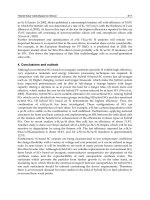
the temperatures studied the mutant 4TS was significantly more stable than the wild-type.
As can be seen from the Arrhenius plot, TsB2 buffer causes significant stabilization of both
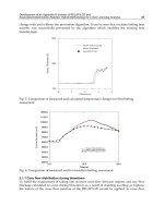
the wild-type enzyme and 4TS compared with TsB1 buffer (Fig. 9)
Fig. 9. Arrhenius plot showing the dependence of rates of inactivation on temperature for
the wild-type luciferase (diamonds) and the mutant 4TS (circles) in buffer TsB1 (closed
symbols) and TsB2 (open symbols) (Koksharov & Ugarova, 2011b). C(enzyme)=13 μg/ml
8.4 Structural analysis
The mutant 4TS contains 7 new substitutions compared with its parent form S118C: T213S,
K156R, R211L, A217V, C146S, E356K, and S364C. All the substitutions are non-conservative
among firefly luciferases. Judging from the order of appearance of these substitutions in the
course of directed evolution (Table 3), literature data and their location in the 3D structure
of the enzyme (Fig. 10), four of these substitutions were suggested to be the key mutations
that cause the high stability of the mutant 4TS: R211L, A217V, E356K, and S364C. The
mutations of the residues A217 (Kajiyama & Nakano, 1993) and E356 (White, et al., 1996) are
known to significantly increase the thermostability of firefly luciferases according to the
Thermostabilization of Firefly Luciferases Using Genetic Engineering
83
previous studies. The effect of the residues R211 and S364 on thermostability is identified for
the first time. The increase in stability by the substitutions R211L, A217V, S364C, and S364A,
can be attributed to the improvement of the internal hydrophobic packing (Fersht & Serrano,
1993). In the case of R211L, S364C, and S364A, the increase of hydrophobicity of the protein
core is achieved by the substitution of the non-conservative buried polar residues by the
hydrophobic ones. As a result of the substitution A217V the larger side group of Val fills the
internal cavity, which is otherwise occupied by a water molecule (Conti et al., 1996). The
surface mutation C146S is known to increase the resistance to oxidative inactivation
(Lomakina et al., 2008). This mutation can explain the increased storage stability of 4TS in
the absence of DTT compared with wild-type. The WT luciferase loses 70% of its activity
within two weeks, whereas the mutant 4TS was remained fully active within one month at
the same conditions (Koksharov & Ugarova, 2011b). The mutants T213S/S364C and S364A
displayed similar in vivo properties. There, it the substitution T213S is unlikely to affect
thermostability. The substitution of the surface residue 156 from positively charged Lys to
similar in properties Arg is also unlikely cause a significant effect on luciferase. The starting
mutant S118C showed only small 1.5-fold increase in stability at 42°C. The mutant 4TS and
its variant without the mutation S118C showed indistinguishable in vivo thermostability at
60°C. Thus, the contribution of S118C seems insignificant. Interestingly, Ser118 is highly
Fig. 10. Homology model of L. mingrelica luciferase showing the location of substitutions in
the mutant 4TS. Four key thermostabilizing mutations are underlined. LO и AMP – luciferyl
and adenylate groups of DLSA (5’-O-[N-(dehydroluciferyl)-sulfamoyl] adenosine).
Subdomains A, B and C are depicted in blue, magenta and orange, respectively
Genetic Engineering – Basics, New Applications and Responsibilities
84
conservative in firefly luciferases. The only exceptions are the similar substitution S118C in
the recently cloned juvenile luciferase from L. cruciata (Oba et al, 2010a) and the substitution
S118T in the luciferase from Lampyroidea maculata (Emamzadeh et al., 2006). However, in
luciferases from non-firefly beetles this position is usually occupied by His or Val.
All four key thermostabilizing substitutions (R211L, A217V, E356K, and S364C) are located
in the second subdomain of firefly luciferase. According to the results of Frydman and
coworkers (Frydman et al., 1999), the fragments of firefly luciferase comprising residues 1-
190 and 422-544 possess high intrinsic stability. These fragments mainly correspond to the
subdomains A and C of firefly luciferase (Fig. 10). That study demonstrated that the middle
subdomain B (192-435) was significantly less stable and that it was the first to unfold under
denaturating conditions. Hence, it likely that the stability of the second subdomain is the
less stable “bottleneck” that determines the stability of the firefly luciferase protein.
Therefore, most of the thermostabilizing mutations would tend to be located in the second
subdomain or at the interface of this subdomain and the remaining parts of the protein. It is
noteworthy that almost all thermostable mutants reported in the literature are located in this
part of the luciferase structure, which is consistent with this hypothesis.
8.5 Conclusion
We have demonstrated that the in vivo directed evolution strategy is a simple and efficient
method to increase thermal stability of firefly luciferase, which allows to obtain highly
thermostable mutants without sacrificing catalytic efficiency. The final mutant obtained here
even displayed superior catalytic properties such as higher specific activity, lower K
m
for
ATP and increased temperature optimum. In typical applications, like ATP-related assays or
reporter genes, beetle luciferases are used at room temperature or 37°C. The mutant 4TS
retains 70% activity after two days of incubation at 37°C. Therefore, its stability is sufficient
for most common in vivo and in vitro applications. The high specific activity, catalytic
efficiency, and improved protein yield make the mutant 4TS an efficient tool for ATP
determination (Ugarova et al., 2010). The increased temperature optimum this mutant can be
an advantage when used for in vivo imaging and in high temperature applications. The new
positions identified in this study can be successfully used for the stabilization of other firefly
luciferases, especially from the Luciola and Hotaria genus’s. The non-lethal in vivo screening
approach described here can be potentially implemented to other beetle or non-beetle
luciferases when the development of thermostable forms of the enzyme is desirable.
9. Acknowledgements
This work was supported by the Russian Foundation for Basic Research (grants 08-04-00624
and 11-04-00698a).
10. References
Amor-Mahjoub, M.; Suppini, J.; Gomez-Vrielyunck, N. & Ladjimi, M. (2006). The effect of
the hexahistidine-tag in the oligomerization of HSC70 constructs. Journal of
Chromatography. B. Analyt. Technol. Biomed. Life Sci., Vol.844, No.2, (December 2006),
pp. 328–334, ISSN 1570-0232
Thermostabilization of Firefly Luciferases Using Genetic Engineering
85
Arnoldi, F.; Neto, A. & Viviani, V. (2010). Molecular insights on the evolution of the lateral
and head lantern luciferases and bioluminescence colors in Mastinocerini railroad-
worms (Coleoptera: Phengodidae). Photochemical and Photobiological Sciences, Vol.9,
No.1, (December 2009), pp. 87-92, ISSN 1474-905X
Binkowski, B.; Fan, F. & Wood, K. (2009). Engineered luciferases for molecular sensing in
living cells. Curr. Opin. Biotechnol., Vol.20, No.1, (February 2009), pp. 14-18, ISSN
0958-1669
Branchini, B.; Magyar, R.; Murtiashow, M.; Anderson, S. & Zimmer, M. (1998). Site-directed
mutagenesis of histidine 245 in firefly luciferase: a proposed model of the active
site. Biochemistry, Vol.37, No.44, (November 1998), pp. 15311-15319, ISSN 0006-2960
Branchini, B.; Magyar, R.; Murtiashaw, M.; Anderson, S.; Helgerson, L. & Zimmer, M.
(1999). Site-directed mutagenesis of firefly luciferase active site amino acids: a
proposed model for bioluminescence color. Biochemistry, Vol.38, No.40, (October
1999), pp. 13223-13230, ISSN 0006-2960
Branchini, B.; Murtiashaw, M.; Magyar, R. & Anderson, S. (2000). The role of lysine 529, a
conserved residue of the acyl-adenylate-forming enzyme superfamily, in firefly
luciferase. Biochemistry, Vol.39, No.18, (May 2000), pp. 5433-5440, ISSN 0006-2960
Branchini, B.; Magyar,R.; Murtiashaw, M. & Portier N. (2001). The role of active site residue
arginine 218 in firefly luciferase bioluminescence. Biochemistry, Vol.40, No.8,
(February 2001), pp. 2410-2418, ISSN 0006-2960
Branchini, B.; Southworth. T.; Murtiashaw, M.; Boije, H. & Fleet S. (2003). A mutagenesis
study of the putative luciferin binding site residues of firefly luciferase.
Biochemistry, Vol.42, No.35, (September 2003), pp. 10429-10436, ISSN 0006-2960
Branchini, B.; Southworth, T.; Murtiashaw, M.; Wilkinson, S.; Khattak, N.; Rosenberg, J. &
Zimmer, M. (2005). Mutagenesis evidence that the partial reactions of firefly
bioluminescence are catalyzed by different conformations of the luciferase C-
terminal domain. Biochemistry, Vol.44, No.5, (January 2005), pp. 1385-1393, ISSN
0006-2960
Branchini, B.; Rosenberg, J.; Fontaine, D.; Southworth, T.; Behney, C. & Uzasci, L. (2011).
Bioluminescence Is Produced from a Trapped Firefly Luciferase Conformation
Predicted by the Domain Alternation Mechanism. Journal of the American Chemical
Society, Vol.133, No.29, (June 2011), pp. 11088-11091, ISSN 0002-7863
Brovko, L.; Belyaeva, E. & Ugarova, N. (1982). Subunit interactions in luciferase from the
firefly Luicola mingrelica. Their role in the manifestation of enzyme activity and the
process of thermal inactivation. Biochemistry (translation from Biokhimiya, USSR),
Vol.47, No.5, (May 1982), pp. 760-766, ISSN 0320-9725
Carson, M.; Johnson, D.; McDonald, H.; Brouillette, C. & Delucas, L. (2007). His-tag impact
on structure. Acta Cryst. D, Vol.63, No.3 (March 2007), pp. 295–230, ISSN 0907-4449
Cirino, P.; Mayer, K. & Umeno, D. (2003). Generating mutant libraries using error-prone
PCR. Methods in Molecular Biology, Vol.231, (April 2003), pp. 3-9, ISSN 1064- 3745
Conti, E.; Franks, N. & Brick P. (1996). Crystal structure of firefly luciferase throws light on a
superfamily of adenylate-forming enzymes. Structure, Vol.4, No.3, (March 1996),
pp. 287-298, ISSN 0969-2126
Conti, E.; Stachelhaus, T.; Marahiel, M. & Brick P. (1997). Structural basis for the activation
of phenylalanine in the non-ribosomal biosynthesis of gramicidin S. EMBO J.,
Vol.16, (July 1997), pp. 4174-4183, ISSN 0261-4189
Genetic Engineering – Basics, New Applications and Responsibilities
86
Dement’eva, E.; Kutuzova, G. & Ugarova, N. (1989). Biochemical properties and stability
of homogeneous luciferase of Luciola mingrelica fireflies. Moscow Universitet
Chemistry Bulletin (translation), Vol.44, No.6, (November 1989), pp. 601-606. ISSN
0027-1314
Dement’eva, E.; Zheleznova, E.; Kutuzova, G.; Lundovskikh, I. & Ugarova, N. (1996).
Physicochemical properties of recombinant Luciola mingrelica luciferase and its
mutant forms, Biochemistry (Moscow), Vol.61, No.1, (January 1996), pp. 115-119,
ISSN 0320-9725
Devine J.; Kutuzova, G.; Green V.; Ugarova N. & Baldwin, T. (1993). Luciferase from the
East European firefly Luciola mingrelica: cloning and nucleotide sequence of the
cDNA, overexpression in Escherichia coli and purification of the enzyme. Biochimica
Biophysica Acta, Gene Structure and Expression, Vol.1173, No.2, (May 1993), pp. 121-
132, ISSN 0006-3002
De Wet, J.; Wood, K.; DeLuca, M.; Helinskii, D. & Subramani, S. (1987). Firefly luciferase
gene: structure and expression in mammalian cells. Mol. Cell. Biol Vol.7, No.2,
(February 1987), pp. 725-737, ISSN 0270-7306
De Wet, J.; Wood, K.; Helinskii, D. & De Luca, M. (1985). Cloning of firefly luciferase cDNA
and the expression of active luciferase in Escherichia coli. Proc. Natl. Acad. Sci. USA,
Vol.82, No.23, (December 1985), pp. 7870-7873. ISSN 0027-8424
Efremenko, E.; Lyagin, I.; Votchitseva, Y.; Gudkov, D.; Peregudov, A.; Aliev, T. &
Varfolomeev, S. (2008). The influence of length and localization of polyhistidine tag
in the molecule of organophosphorus hydrolase on the biosythesis and behavior of
fusion protein. In: Biotechnology: state of the art and prospects for development, G.E.
Zaikov, (Ed.), 87-101, Nova Science Publishers Inc., ISBN 978-1-60456-015-2, New-
York, USA
Eijsink, V.; Bjork, A.; Gaseidnes, S.; Sirevag, R.; Synstad, B.; van den Burg, B. & Vriend, G.
(2004). Rational engineering of enzyme stability. Journal of Biotechnology, Vol.113,
No.1-3, (September 2004), pp. 105-120, ISSN 0168-1656
Emamzadeh, A.; Hosseinkhani, S.; Sadeghizadeh, M.; Nikkhah, M.; Chaichi, M. &
Mortazavi, M. (2006). cDNA cloning, expression and homology modeling of a
luciferase from the firefly Lampyroidea maculata. Journal of Biochemistry and Molecular
Biology. Vol.39, No.5, (September 2006), pp. 578-585, ISSN 1225-8687
Eriksson, J.; Nordstrom, T. & Nyren,P. (2003). Method enabling firefly luciferase-based
bioluminometric assays at elevated temperatures. Anal. Biochem. Vol.314, No.1,
(March 2003), pp. 158-161, ISSN 0003-2697
Fersht, A. & Serrano, L. (1993). Principles of protein stability derived from protein
engineering experiments. Curr. Op. Struct. Biol., Vol.3, No.1, (February 1993), pp.
75-83, ISSN 0959-440X
Forreiter, C.; Kirschner, M. & Nover, L. (1997). Stable transformation of an arabidopsis cell
suspension culture with firefly luciferase providing a cellular system for analysis of
chaperone activity in vivo. The Plant Cell, Vol.9, No.12, (December 1997), pp. 2171-
2181, ISSN 1040-4651
Fraga, H. (2008). Firefly luminescence: A historical perspective and recent developments.
Photochemical and Photobiological Sciences, Vol.7, No.2, (February 2008), pp. 146-158,
ISSN 1474-905X
Thermostabilization of Firefly Luciferases Using Genetic Engineering
87
Freydank, A.; Brandt, W. & Dräger, B. (2008). Protein structure modeling indicates
hexahitidine-tag interference with enzyme activity, Proteins, Vol.72, No.1, (July
2008), pp. 173–183, ISSN 0887-3585
Frydman, J.; Erdjument-Bromage, H.; Tempst P. & Hartl, F. (1999). Co-translational domain
folding as the structural basis for the rapid de novo folding of firefly luciferase. Nat.
Struct. Mol. Biol., Vol.6, No.7, (July 1999), pp. 697-705, ISSN 1072-836
Gould, S.; Keller, G. & Subramani, S. (1987). Identification of a peroxisomal targeting signal
at the carboxy terminus of firefly luciferase. J. Cell Biol., Vol.105, No.6, (December
1987), pp. 2923-2931, ISSN 0021-9525
Gulick, A. (2009). Conformational Dynamics in the Acyl-CoA Synthetases, Adenylation
Domains of Non-ribosomal Peptide Synthetases, and Firefly Luciferase. ACS
Chemical Biology, Vol.4, No.10, (July 19, 2009), pp. 811-827, ISSN 1554-8929
Jäckel, C.; Kast, P. & Hilvert, D. (2008). Protein Design by Directed Evolution. Annual Review
of Biophysics, Vol.37, No.1, (June 2008), pp. 153-173, ISSN 1936-122X
Jiang, X.; Morgan, J. & Doyle, M. (2003). Thermal inactivation of Escherichia coli O157:H7 in
cow manure compost. J. Food. Prot., Vol.66, No.10, (October 2003), pp. 1771-1777,
ISSN 0362-028X
Hall, M. & Leach, F. (1988). Stability of firefly luciferase in Tricine buffer and in a
commercial enzyme stabilizer. J. Biolum. Chemilum., Vol.2, No.1, (January 1988), pp.
41-44, ISSN 0884-3996
Herbst, R.; Gast, K. & Seckler, R. (1998). Folding of Firefly Photinus pyralis Luciferase:
Aggregation and Reactivation of Unfolding Intermediates. Biochemistry, Vol.37,
No.18, (May 1998), pp. 6586-6597, ISSN 0006-2960
Imani, M.; Hosseinkhani, S.; Ahmadian, S. & Nazari, M. (2010). Design and introduction of a
disulfide bridge in firefly luciferase: increase of thermostability and decrease of pH
sensitivity. Photochemical and Photobiological Sciences, Vol.9, No.8, (August 2010), pp.
167-1177, ISSN 1474-905X
Kajiyama, N.; Masuda, T.; Tatsumi, H. & Nakano, E. (1992). Purification and
characterization of luciferases from fireflies, Luciola cruciata and Luciola lateralis.
Biochim.Biophys. Acta, V. 1120, No.2, (April 1992), pp. 228-232, ISSN 0006-3002
Kajiyama, N. & Nakano, E. (1993). Thermostabilization of firefly luciferase by a single amino
acid substitution at position 217. Biochemistry, Vol.32, No.50, (December 1993), pp.
13795-13799, ISSN 0006-2960
Kajiyama, N. & Nakano, E. (1994). Enhancement of thermostability of firefly luciferase from
Luciola lateralis by a single amino acid substitution. Biosci. Biotechnol. Biochem.
Vol.58, No,6, (June 1994), pp. 1170-1171, ISSN 0916-8451
Keller, G.; Gould S.; Deluca M. & Subramani, S. (1987). Firefly Luciferase is Targeted to
Peroxisomes in Mammalian Cell, PNAS, Vol.84, No.10, (May 1987), pp. 3264-3268,
ISSN 0027-8424
Kitayama, A.; Yoshizaki, H.; Ohmiya, Y.; Ueda, H. & Nagamune, T. (2003). Creation of a
thermostable firefly luciferase with pH-insensitive luminescent color.
Photochemistry and Photobiology, Vol.77, No.3, (March 2003), pp. 333-338, ISSN 0031-
8655
Klose, J.; Wendt, N.; Kubald, S.; Krause, E.; Fechner, K.; Beyermann, M.; Bienert, M.;
Rudolph, R. &
Rothemund, S. (2004). Hexahistidin tag position influences disulfide
structure but not binding behavior of in vitro folded N-terminal domain of rat
Genetic Engineering – Basics, New Applications and Responsibilities
88
corticotropin-releasing factor receptor type 2a. Protein Sciences, Vol.13, No.9,
(August 2004), pp. 2470-2475, ISSN 0961-8368
Koksharov, M. & Ugarova, N. (2008). Random mutagenesis of Luciola mingrelica firefly
luciferase. Mutant enzymes whose bioluminescence spectra show low pH-
sensitivity. Biochemistry (Moscow), Vol.73, No.8, (August 2008), pp. 862-869, ISSN
0006-2979
Koksharov, M. & Ugarova, N. (2010). Thermostability enhancement of Luciola mingrelica
firefly by in vivo directed evolution. Luminescense, Vol.25, No.1, (January 2010), pp.
135-136, ISSN 1522-7243
Koksharov, M. & Ugarova, N. (2011a). Triple substitution G216N/A217L/S398M leads to
the active and thermostable Luciola mingrelica firefly luciferase. Photochemical and
Photobiologocal Sciences, Vol.10, No.6, (July 2011), pp. 931-938, ISSN 1474-905X
Koksharov, M. & Ugarova, N. (2011b). Thermostabilization of firefly luciferase by in vivo
directed evolution. Protein Eng. Des. Sel., Vol.24, No.11, (November 2011), pp. 835-
844, ISSN 1741-0126
Kumita, J.; Jain, L.; Safroneeva, E. & Woolley, G. (2000). A cysteine-free firefly luciferase
retains luminescence activity. Biochem. Biophys. Res. Commun., Vol.267, No.4,
(January 2000), pp. 394-397, ISSN 0006-291X
Kuo, W & Chase, H. (2011). Exploiting the interactions between poly-histidine fusion tags
and immobilized metal ions. Biotechnology Letters, Vol.33, No.6, pp. 1075-1084, ISSN
0141-5492
Kutuzova, G.; Skripkin, E.; Tarasova, N.; Ugarova, N. & Bogdanov, A. (1989). Synthesis
and pathway of Luciola mingrelica firefly luciferase in Xenopus laevis frog oocytes
and in cell-free systems. Biochimie, Vol.71, No.4, (April 1989), pp. 579-583, ISSN
0300-9084
Law, G.; Gandelman, O.; Tisi, L.; Lowe, C. & Murray, J. (2006). Mutagenesis of solvent-
exposed amino acids in Photinus pyralis luciferase improves thermostability and pH
tolerance. Biochem. J., Vol.397, No.2, (July 2006), pp. 305-312, ISSN 0264-6021
Leclerc, G.; Boockfor, F.; Faught, W. & Frawley, L. (2000). Development of a destabilized
firefly luciferase enzyme for measurement of gene expression. Biotechniques, Vol.29,
No.3, (September 2000), pp. 590-601, ISSN 0736-6205
Li, X.; Yang, S. & Liang, X. (2006). Phylogenetic relationship of the firefly, Diaphanes
pectinealis (Insecta, Coleoptera, Lampyridae), based on DNA sequence and gene
structure of luciferase. Zoological Research, Vol.27, No.4, (August 2006), pp. 367-374,
ISSN 0254-5853
Lomakina, G.; Modestova, Y. & Ugarova N. (2008). Enhancement of thermostability of the
east european firefly (Luciola mingrelica) luciferase by site-directed mutagenesis of
nonconservative cysteine residues Cys62 and Cys146. Moscow University Chemistry
Bulletin, Vol.63, No.2, (May 2008), pp. 63-66, ISSN 0027-1314
Lundin, A. (2000). Use of firefly luciferase in ATP-related assays of biomass, enzymes, and
metabolites. Methods Enzymol.
, Vol.305, (December 2000), pp. 346-370, ISSN 0076-
6879
Lundovskikh, I.;
Leontieva, O.; Dementieva, E. & Ugarova, N. (1998). Recombinant Luciola
mingrelica firefly luciferase. Folding in vivo, purification and properties. Proceedings
of the 10th International Symposium on Bioluminescence and Chemiluminescence, pp.
420-424, ISBN 0-471-98733-6, Bologna, Italy, September 4-8,1998