Proteomics Human Diseases and Protein Functions Part 3 pdf
Bạn đang xem bản rút gọn của tài liệu. Xem và tải ngay bản đầy đủ của tài liệu tại đây (1.32 MB, 25 trang )
From Biomarker Discovery to
Clinical Evaluation for Early Diagnosis of Lung Surgery-Induced Injury
39
Likewise, the relative expression of α1-antitrypsin at bands 5, 7, and 8 from bronchial
washing was positively correlated with protein concentration, leukocyte number, and the
level of vascular endothelial growth factor (data not shown). These data supported our
hypothesis that the increase of vascular endothelial growth factor after surgery facilitates
leukocyte infiltration and the exudation of acute-phase proteins (such as α1-antitrypsin and
α2-macroglobulin) into alveoli.
3.3 Characterization of α2-macroglobulin and α1-antitrypsin in lobectomized patients
with acute respiratory distress syndrome
Based on the report of the joint American–European Consensus Conference, the acute
respiratory distress syndrome is well defined as follows: bilateral infiltrates on frontal chest
radiography, the absence of left atrial hypertension (pulmonary capillary wedge pressure
<18 mmHg or no clinical signs of left ventricular failure), and severe hypoxemia with a
PaO
2
/FiO
2
ratio <200 mmHg (Bernard et al., 1994). Five patients who received lung surgery
and met these criteria were studied.
3.3.1 Characterization of patients with acute respiratory distress syndrome
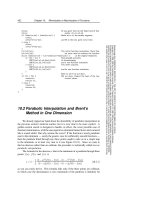
The group with lobectomy free of complications had levels of total protein and total
leukocyte numbers in their bronchial washings similar to those who developed acute
respiratory distress syndrome (P >0.05, Fig. 4). These data indicate that lung surgery induces
inflammation (leukocyte infiltration and protein exudation) in the groups with and without
the complication of acute respiratory distress syndrome. So, factors other than inflammation
contribute to the development of this syndrome.
0
10
20
30
40
50
pre-op post-op ARDS
Total cell number (x10,000)
0
4
8
pre-op post-op ARDS
Total proteins (g/L)
*Significant difference from pre-op.
Fig. 4. Total leukocyte number and protein concentration in patients before (pre-op) and
after lobectomy (post-op) with no complication and those with acute respiratory distress
syndrome (ARDS).
In lung cancer patients, an increase of vascular endothelial growth factor is positively
associated with poor prognosis (P = 0.018; Han et al., 2001) but not with a worse
postoperative year-survival rate (P = 0.0643; Liao et al., 2001). These reports are also
consistent with our finding that the increase of vascular endothelial growth factor after lung
surgery does not contribute to surgery-induced acute respiratory distress syndrome.
*
*
*
*
Total protein (g/L)
Proteomics – Human Diseases and Protein Functions
40
3.3.2 Protein profiling of bronchial washings from lobectomized patients with acute
respiratory distress syndrome
Unlike patients with no complications, those with acute respiratory distress syndrome
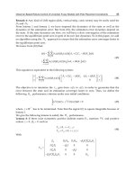
showed white or gray patches on the chest X-ray. In one-dimensional gel electrophoresis,
the protein profiling of bronchial washings from patients without complications showed a
much clearer banding pattern than those from patients with acute respiratory distress
syndrome (Fig. 5). Eight bands from each gel were cut and subjected to LC/MS/MS for
protein identification. No protein was identified in Lane 1. The most significant difference
was that albumin appeared in almost every band of the samples from patients without
complications but not in those with acute respiratory distress syndrome. In contrast, α1-
antitrypsin was identified only in bands 6 and 7 from the group without complications but
was found in bands 2, 3, 4, 5, 6, and 7 in the group with the complication (Fig. 5).
Fig. 5. Comparison of chest X-rays and protein profiling of bronchial washings in
lobectomized patients with no complications (lobectomy, Lob) and those with acute
respiratory distress syndrome (ARDS).
3.3.3 α2-macroglobulin and α1-antitrypsin in bronchial washings from lobectomized
patients with acute respiratory distress syndrome
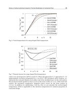
As shown in Fig. 6, both α2-macroglobulin and α1-antitrypsin were detected in bronchial
washings after surgery.
After quantification, the total amounts of α2-macroglobulin at bands 2, 4, and 5 and α1-
antitrypsin at bands 5, 7, and 8 did not show any statistical difference between the groups
with and without complications. The most important finding was lower levels of α1-
antytrypsin at bands 7 and 8 in the group without complications than the acute respiratory
distress syndrome group (Fig. 6). It is likely that α1-antitrypsin variants at bands 5, 7, and 8
can be used as biomarkers for the early detection of acute respiratory distress syndrome.
In bronchial washings collected from the patients with acute respiratory distress syndrome,
leukocyte number was not correlated with the total amounts of α2-macroglobulin or α1-
antitrypsin. Our analyses again supported the notion that surgery-induced inflammation is
not an important indicator in the early phase of acute respiratory distress syndrome.
It has been reported that α1-antitrypsin can be produced by lung epithelial cells (Venember
et al., 1994) but α2-macroglobulin cannot. Our preliminary data confirmed the expression of
Marker Lob ARDS
(kDa)
250
160
105
75
50
35
1
2 α1-antitrypsin
3 α1-antitrypsin
4 α1-antitrypsin
5 α1-antitrypsin
6 α1-antitrypsin
7 α1-antitrypsin
8 β-actin
Lobectomy
Acute respiratory
distress syndrome
From Biomarker Discovery to
Clinical Evaluation for Early Diagnosis of Lung Surgery-Induced Injury
41
Fig. 6. Relative expression of α1-antitrypsin and α2-macroglobulin (macroglobulin) in the
lobectomized group without complications (lobectomy) and in the group with acute
respiratory distress syndrome (ARDS).
α1-antitrypsin in A549, a lung epithelial cell line. The changes in α1-antitrypsin variants
could be due to functional changes in lung epithelial cells.
3.4 Specificity and sensitivity of α1-antitrypsin variants as potential biomarkers for
acute respiratory distress syndrome
It is of importance to turn the relative expression of α1-antitrypsin in bronchial washings
into a measurable outcome because only the measurable outcome is used to determine the
cutoff value. Based on the cutoff value, sensitivity (the proportion of subjects who test
positive among those with the condition) and specificity (the proportion of subjects who test
negative among those without the condition) can be calculated.
As shown in Fig. 6, α1-antitrypsin variants at bands 7 (47 kDa) and 8 (40 kDa) had a lower
abundance in the group without complications than the group with acute respiratory
syndrome. To avoid variations in sample loading and the intensity in each calculation, the
ratio of the expression of α1-antitrypsin at band 5 (70 kDa) to that at bands 7 and 8 was used
as the measurable outcome. Based on this calculation, the cutoff value was 0.5. A ratio <0.5
was considered an indication of acute respiratory distress syndrome.
Table 3 shows the ratio for each patient from the complication-free group. Four out of 7
patients had a ratio <0.5. The specificity of α1-antitrypsin for true negative patients was 0.43
(3/7).
Table 4 shows the ratio for each patient from the complication group. Three out of 5 patients
had a ratio <0.5. The sensitivity of α1-antitrypsin for true positive patients was 0.6 (3/5).
Proteomics – Human Diseases and Protein Functions
42
Patient
No
Ratio of expression of α1-antitrypsin at band
5 to that at bands 7 and 8
Cutoff value = 0.5
1 0.000: 0.027 <0.5
2 0.043: 0.099 <0.5
3 0.019: 0.024 >0.5
4 0.017: 0.023 >0.5
5 0.018: 0.087 <0.5
6 0.000: 0.006 <0.5
7 0.042: 0.053 >0.5
Table 3. Ratio of the expression of α1-antitrypsin at band 5 to that at bands 7 and 8 in the
lobectomized patients without acute respiratory distress syndrome.
Patient No
Ratio of expression of α1-antitrypsin at band
5 to that at bands 7 and 8
Cutoff value = 0.5
A 0.081: 0.177 <0.5
B 0.043: 0.199 <0.5
C 0.081: 0.086 >0.5
D 0.015: 0.040 <0.5
E 0.025: 0.048 >0.5
Table 4. Ratio of the expression of α1-antitrypsin at band 5 to that at bands 7 and 8 in
lobectomized patients with acute respiratory distress syndrome.
3.5 Further improvement of specificity and sensitivity for detecting acute respiratory
distress syndrome using dual biomarkers
As shown in Tables 3 and 4, the sensitivity of α1-antitrypsin variants for detecting acute
respiratory distress syndrome (0.6) was better than the specificity (0.43). The major concern
is how to optimize the cutoff value and improve the specificity. In table 3, patients 1 and 6
with ratios <0.5 showed the lowest values in cell counts and protein concentration.
Meanwhile, the expression of α2-macroglobulin was almost undetectable, which indicates
minor inflammation in the patients. The lower ratio of relative expression of α1-antitrypsin
at band 5 to that at bands 7 and 8 was false-positive.
α1-antitrypsin was found in the lungs before and after surgery; α2-macroglobulin only
occurred in the lungs after surgery. To avoid the lower levels of α1-antitrypsin variants
which may create a false-positive result, α2-macroglobulin can be recruited as a second
biomarker. The ratio of α1-antitrypsin variants was considered as a true result only when
From Biomarker Discovery to
Clinical Evaluation for Early Diagnosis of Lung Surgery-Induced Injury
43
the sample expressed detectable α2-macroglobulin in bronchial washings. Accordingly, the
specificity for true negative patients changed to 0.71 (5/7). The prediction for true negatives
was improved.
4. From identification of leads to further validation using α2-macroglobulin
and α1-antitrypsin variants as an example
After the discovery of potential biomarkers by proteomic analysis in this study, the first
challenge was to identify the leads from the proteins discovered after developing a quick
screening test. After Phase 1, the second challenge was to provide clear justification to
optimize the cutoff values.
4.1 Contribution of this study to the discovery of biomarkers for detecting acute
respiratory distress syndrome
Ideally, quantitative proteomic analysis should be used to reveal lobectomy-induced
changes of all proteins in bronchial washings. However, the unique compartment of the
lung allowed us to analyze exudate components which may not exist before surgery, such as
α2-macroglobulin. Based on the important mechanism of surgery-induced inflammation in
the early phase of lung injury, one-dimensional gel electrophoresis in this study was an easy
and suitable tool to identify α2-macroglobulin as an indicator of vascular endothelial growth
factor-mediated permeability.
The second contribution of this study was to take advantage of one-dimensional gel
electrophoresis with pattern analysis to reveal the pattern changes of α1-antitrypsin between
the groups with and without post-surgical complications. The difference found allowed us
to identify α1-antytripsin variants as biomarkers for the early detection of acute respiratory
distress syndrome.
4.2 Limitations of this study
In this study, α1-antitrypsin variants were considered as biomarkers for acute respiratory
distress. No mechanistic data are provided to explain why and how the formation of α1-
antitrypsin variants are related to the progression from surgery-induced inflammation to
acute respiratory distress syndrome.
The association between α1-antitrypsin variants and infection was first reported in 2010
(Zhang et al., 2010). The decrease of the α1-antitrypsin variant at 130 kDa and the increase of
the variant at 40 kDa is associated with human immunodeficiency virus-induced infection.
Glycoproteomic analysis shows that changes in α1-antitrypsin variants may be due to a shift
of glycosylation. In future, glycoproteomic analysis of α1-antitrypsin variants should be
further explored.
Although the analysis of their specificity and sensitivity, the cutoff point of the measurable
outcome, and criteria for patient selection are clearly and easily determined, the small
number of clinical cases in this study limits the generalization of α2-macroglobulin and α1-
antitrypsin as markers for acute respiratory distress syndrome. To use them as measurable
biomarkers in Phase 3, it is necessary to increase the number and the complexity of clinical
cases for further validation on whether the cutoff points determined are suitable for early
diagnosis of acute respiratory distress syndrome.
One-dimensional gel electrophoresis does not offer a good way for protein separation.
Comparative proteomic analysis only compares the intensity of each spot. These two
Proteomics – Human Diseases and Protein Functions
44
approaches may our discovery of new proteins. The technology of stable isotope dimethyl
labeling coupled with LC/MS/MS permits further quantification of specific peptides of each
protein and provides a better quantification tool after one-dimensional electrophoresis (Huang
et al., 2006). This approach then compensates for the limitation of one-dimensional gel
electrophoresis.
5. Conclusion
Both inflammation -dependent and -independent mechanisms contribute to the progression
from lung injury to acute respiratory distress syndrome. Stage-dependent changes in
biomarkers allow us to monitor the progression of the diseases and develop new treatments
in a stage-dependent manner.
In this study, α2-macroglobulin and α1-antitrypsin were positively correlated with vascular
endothelial growth factor, clearly showing lobectomy-induced inflammation. The total
amount of α1-macroglobulin can be used as a biomarker of increased vascular permeability
in the lung. The severity of lobectomy-induced inflammation is similar to that of
inflammation in acute respiratory distress syndrome but respiratory function becomes much
worse in patients with the syndrome. Concomitantly, the patients with acute respiratory
distress syndrome had lower levels of α1-antitrypsin at higher molecular weights and
higher levels of α1-antitrypsin at lower molecular weights. Similarly, human
immunodeficiency virus-induced infection is associated with the decreased abundance of
α1-antitrypsin at higher molecular weights and the increased abundance of α1-antitrypsin at
lower molecular weights (Zhang et al., 2010). Because α1-antitrypsin exists in lung epithelial
cells (Venember et al., 1994), the changes of α1-antitrypsin variants in the patients with acute
respiratory distress may reflect lung epithelial damage.
6. Acknowledgment
The authors appreciate the technical support of Shih-Hsin Ho, Hong-Da Wang, and Yan-Jie
Chen, clinical sample collections by Drs. Jia-Ming Chang and Chang-Wen Chen, and grant
support from the National Science Council, Taiwan (NSC-95-2314-B-006-125-MY2 and NSC-
95-2323-B-006-004).
7. References
Apweilerm, R., Aslanidis, C., Deufel, T., Gerstner, A., Hansen, J., Hochstrasser, D., Kellner,
R., Kubicek, M., Lottspeich, F., Maser, E., Mewes, HW., Meyer, HE., Müllner, S.,
Mutter, W., Neumaier, M., Nollau, P., Nothwang, HG., Ponten, F., Radbruch, A.,
Reinert, K., Rothe, G., Stockinger, H., Tárnok, A., Taussig, MJ., Thiel, A., Thiery, J.,
Ueffing, M., Valet, G., Vandekerckhove, J., Wagener, C., Wagner, O., & Schmitz, G.
(2009). Approaching clinical proteomics: current state and future fields of
application in cellular proteomics. Cytometry A, Vol.75, No.10, (October 2009), pp.
816-32, ISSN 1552-4930
Bernard, GR., Artigas, A., Brigham, KL., Carlet, J., Falke, K., Hudson, L., Lamy, M., Legall,
JR., Morris, A., & Spragg, R. (1994). The American-European Consensus Conference
on ARDS. Definitions, mechanisms, relevant outcomes, and clinical trial
From Biomarker Discovery to
Clinical Evaluation for Early Diagnosis of Lung Surgery-Induced Injury
45
coordination. American Journal of Respiratory and Critical Care Medicine, Vol.149, No.
3 Pt 1, (March 1994), pp. 818-24, ISSN 1073-449X
Bhatia, M., & Moochhala, S. (2004). Role of inflammatory mediators in the pathophysiology
of acute respiratory distress syndrome. Journal of Patholog, Vol.202, No.2, (February
2004), pp. 145-56, ISSN 1096-9896
Chang, CC., Chiu, HF., Wu, YS., Li, YC., Tsai, ML., Shen, CK., & Yang, CY. (2005). The
induction of vascular endothelial growth factor by ultrafine carbon black
contributes to the increase of alveolar-capillary permeability. Environmental Health
Perspectives, Vol.113, No.4, (April 2005), pp.454-60, ISSN 0091-6765
Chang, CC., Chen, SH., Ho, SH., Yang, CY., Wang, HD., & Tsai, ML. (2007). Proteomic
analysis of proteins from bronchoalveolar lavage fluid reveals the action
mechanism of ultrafine carbon black-induced lung injury in mice. Proteomics, Vol.7,
No.23, (December 2007), pp.4388-97, ISSN 1615-9861
Chiang, CJ., Chen, YC., Chen, CJ., You, SL., & Lai, MS. (2010). Cancer trends in Taiwan.
Taiwan Cancer Registry Task Force. Japanese Journal of Clinical Oncology, Vol.40,
No.10, (October 2010), pp.897-904, ISSN 0368-2811
Donnelly, SC., Strieter, RM., Reid, PT., Kunkel, SL., Burdick, MD., Armstrong, I., Mackenzie,
A., & Haslett, C. (1996). The association between mortality rates and decreased
concentrations of interleukin-10 and interleukin-1 receptor antagonist in the lung
fluids of patients with the adult respiratory distress syndrome. Annals of Internal
Medicine, Vol.125, No.3, (August 1996), pp.191–6, ISSN 1539-3704
Geiser, T., Atabai, K., Jarreau, PH., Ware, LB., Pugin, J., & Matthay, MA. (2001). Pulmonary
edema fluid from patients with acute lung injury augments in vitro alveolar
epithelial repair by an IL-1beta-dependent mechanism. American Journal of
Respiratory and Critical Care Medicine, Vol.163, No.6, (May 2001), pp.1384–8, ISSN
1073-449X
Gunluoglu, MZ., Demir, A., Turna, A., Sansar, D., Melek, H., Dincer, SI., & Gurses, A. (2011).
Extent of lung resection in non-small lung cancer with interlobar lymph node
involvement. Annals of Thoracic and Cardiovascular Surgery, Vol.17, No.3, (June 2011),
pp.229-35, ISSN 1341-1098
Han, H., Silverman, JF., Santucci, TS., Macherey, RS., d'Amato, TA., Tung, MY., Weyant, RJ.,
& Landreneau, RJ. (2001). Vascular endothelial growth factor expression in stage I
non-small cell lung cancer correlates with neoangiogenesis and a poor prognosis.
Annals of Surgical Oncology, Vol.8, No.1, (January-February 2001), pp.72-9, ISSN
1068-9265
Huang, SY., Tsai, ML., Tsai, CJ., Wu, JL., Hsu, JL., Ho, SH., & Chen SH. (2006). Quantitation
of protein phosphorylation in pregnant rat uteri using stable isotope dimethyl
labeling coupled with IMAC. Proteomics, Vol.6, No.6, (March 2006), pp.1-12, ISSN
1615-9861
Huang, Z., Lin, L., Gao, Y., Chen, Y., Yan, X., Xing, J., & Hang, W. (2011). Bladder cancer
determination via two urinary metabolites: a biomarker pattern approach. (in
press). Molecular & Cellular Proteomics, ISSN 1535-9484
Proteomics – Human Diseases and Protein Functions
46
Jordan, S., Mitchell, JA., Quinlan, GJ., Goldstraw, P., & Evans, TW. (2000). The pathogenesis
of lung injury following pulmonary resection. European Respiratory Journal, Vol.15,
No.4, (April 2000), pp.790-9, ISSN 1399-3003
Kollef, MH., & Schuster, DP. (1998). The acute respiratory distress syndrome. The New
England Journal of Medicine, Vol.332, No.1, (January 1995), pp.27-37, ISSN 1533-4406
Landis, SH., Murray, T., Bolden, S., & Wingo, PA. (1998). Cancer statistics, CA-a Cancer
Journal for Clinicians. Vol.48, No.1, (January-February 1998), pp.6-29, ISSN 1542-
4863
Lee, YC. (2005). The involvement of VEGF in endothelial permeability: a target for anti-
inflammatory therapy. Current Opinion in Investigational Drugs, Vol.6, No.11,
(November 2005), pp.1124-30, ISSN 1472-4472
Lenz, AG, Meyer, B., Costabel, U., & Maier, K. (1993). Bronchoalveolar lavage fluid proteins
in human lung disease: analysis by two-dimensional electrophoresis.
Electrophoresis, Vol.14, No. 3, (March 1993), pp. 242-4, ISSN 1522-2683
Liao, M., Wang, H., Lin, Z., Feng, J., & Zhu, D. (2001). Vascular endothelial growth factor
and other biological predictors related to the postoperative survival rate on non-
small cell lung cancer. Lung Cancer, Vol.33, No.2-3, (August-September 2001),
pp.125-32, ISSN 0169-5002
Martini, N., Bains, MS., Burt, ME., Zakowski, MF., McCormack, P., Rusch, VW., & Ginsberg,
RJ. (1995). Incidence of local recurrence and second primary tumors in resected
stage I lung cancer. The Journal of Thoracic and Cardiovascular Surgery, Vol.109, No.1,
(January 1995), pp.120-9, ISSN 0022-5223
Medford, AR., & Millar, AB. (2006). Vascular endothelial growth factor (VEGF) in acute lung
injury (ALI) and acute respiratory distress syndrome (ARDS): paradox or
paradigm? Thorax, Vol.61, No.7, (July 2006), pp.621-6, ISSN 1468-3296
Meyer, KC. (2007). Bronchoalveolar lavage as a diagnostic tool. Seminars in Respiratory and
Critical Care Medicine, Vol.28, No.5, (October 2007), pp.546-60, ISSN 1069-3424
Mou, Y., Xing, R., & Liu, C. (2011). Diagnosis of Gallbladder Cancer Using Matrix-Assisted
Laser Desorption/Ionization Time-of-Flight Profiling. (in press). The American
Journal of the Medical Sciences, ISSN 1538-2990
Pepe, MS., Etzioni, R., Feng, Z., Potter, JD., Thompson, M., Thornquist, M., Winget, M., &
Yasui, Y. (2001). Phases of biomarker development for early detection of cancer.
Journal of the National Cancer Institute. Vol.93, No.14, (July 2001), pp.1054–61. ISSN
1460-2105
Plymoth, A., Löfdahl, CG., Ekberg-Jansson, A., Dahlbäck, M., Lindberg, H., Fehniger, TE., &
Marko-Varga, G. (2003). Human bronchoalveolar lavage: biofluid analysis with
special emphasis on sample preparation. Proteomics, Vol.3, No.6, (June 2003),
pp.962-72 ISSN 1615-9861
Rabilloud, T. (2002). Two-dimensional gel electrophoresis in proteomics: old, old fashioned,
but it still climbs up the mountains. Proteomics, Vol.2, No.1, (January 2002), pp.3–10,
ISSN 1615-9861
Sabounchi-Schütt, F., Aström, J., Eklund, A., Grunewald, J., & Bjellqvist, B. (2001).
Detection and identification of human bronchoalveolar lavage proteins using
narrow-range immobilized pH gradient DryStrip and the paper bridge sample
From Biomarker Discovery to
Clinical Evaluation for Early Diagnosis of Lung Surgery-Induced Injury
47
application method. Electrophoresis, Vol.22, No.9, (May 2001), pp.1851–60, ISSN
1522-2683
Sadaghdar, H., Huang, ZB., & Eden, E. (1992). Correlation of bronchoalveolar lavage
findings to severity of Pneumocystis carinii pneumonia in AIDS. Evidence for the
development of high-permeability pulmonary edema. Chest Vol.102, No.1, (July
1992), pp.63-9. ISSN 1931-3543
Schirle, M., Heurtier, MA., & Kuster, B. (2003). Profiling core proteomes of human cell lines
by one-dimensional PAGE and liquid chromatography-tandem mass spectrometry.
Molecular & Cellular Proteomics, Vol.2, No.12, (December 2003), pp.1297-305, ISSN
1535-9484
Spragg, RG., Bernard, GR., Checkley, W., Curtis, JR., Gajic, O., Guyatt, G., Hall, J., Israel, E.,
Jain, M., Needham, DM., Randolph, AG., Rubenfeld, GD., Schoenfeld, D.,
Thompson, BT., Ware, LB., Young, D., & Harabin, AL. (2010). Beyond mortality:
future clinical research in acute lung injury. American Journal of Respiratory and
Critical Care Medicine, Vol.181, No.10, (May 2010), pp.1121-7, ISSN 1073-449X
Tremblay, LN., Miatto, D., Hamid, Q., Govindarajan, A., & Slutsky, AS. (2002). Injurious
ventilation induces widespread pulmonary epithelial expression of tumor necrosis
factor-alpha and interleukin-6 messenger RNA. Critical Care Medicine, Vol.30, No.8,
(August 2002), pp.1693–1700, ISSN 1530-0293
Turtoi, A., De Pauw, E., & Castronovo, V. (2011a). Innovative proteomics for the discovery
of systemically accessible cancer biomarkers suitable for imaging and targeted
therapies. American Journal of Pathology, Vol.178, No.1, (January 2011), pp.12-8 ISSN
0002-9440
Turtoi, A., Dumont, B., Greffe, Y., Blomme, A., Mazzucchelli, G., Delvenne, P., Mutijima,
EN., Lifrange, E., De Pauw, E., & Castronovo, V. (2011b). Novel Comprehensive
Approach for Accessible Biomarker Identification and Absolute Quantification
from Precious Human Tissues. Journal of Proteome Research, Vol.10, No.7, (July
2011), pp.3160-82, ISSN 1535-3907
Venembre, P., Boutten, A., Seta, N., Dehoux, MS., Crestani, B., Aubier, M., & Durand, G.
Secretion of alpha 1-antitrypsin by alveolar epithelial cells. FEBS Letters, Vol.346,
No.2-3, (June 1994), pp.171-4. ISSN 0014-5793
Vesterberg, O., Palmberg, L., & Larsson, K. (2001). Albumin, transferrin and alpha2-
macroglobulin in bronchoalveolar lavage fluid following exposure to organic dust
in healthy subjects. International Archives of Occupational and Environmental Health,
Vol.74, No.4, (May 2001), pp.249-54, ISSN 1432-1246
Villar, J., Blanco, J., Zhang, H., & Slutsky, AS. (2011). Ventilator-induced lung injury and
sepsis: two sides of the same coin? Minerva Anestesiologica, Vol.77, No.6, (June
2011), pp.647-53, ISSN 1827-1596
Wattiez, R., Hermans, C., Bernard, A., Lesur, O., & Falmagne, P. (1999). Human
bronchoalveolar lavage fluid: two-dimensional gel electrophoresis, amino acid
microsequencing and identification of major proteins. Electrophoresis, Vol.20, No.7,
(June 1999), pp.1634-45, ISSN 1522-2683
Wu, J., Kobayashi, M., Sousa, EA., Liu, W., Cai, J., Goldman, SJ., Dorner, AJ., Projan, SJ.,
Kavuru, MS., Qiu, Y., & Thomassen, MJ. (2005). Differential proteomic analysis of
Proteomics – Human Diseases and Protein Functions
48
bronchoalveolar lavage fluid in asthmatics following segmental antigen challenge.
Molecular & Cellular Proteomics, Vol.4, No.9, (September 2005), pp.1251-64, ISSN
1535-9484
Zhang, L., Jia, X., Zhang, X., Cao, J., Yang, P., Qiu, C., Shen, Y., Ma, F., Liu, L., Sun, J., Shen,
F., Yin, L., Liu, L., Yao, Y., & Lu, H. (2010). Alpha-1 antitrypsin variants in plasma
from HIV-infected patients revealed by proteomic and glycoproteomic analysis.
Electrophoresis, Vol.31, No.20, (October 2010), pp.3437-45, ISSN 1522-2683
3
Urinary Exosomes for
Protein Biomarker Research
Delfin Albert Amal Raj
1,2
, Immacolata Fiume
1
,
Giovambattista Capasso
2
and Gabriella Pocsfalvi
1
1
Mass Spectrometry and Proteomics,
Institute of Protein Biochemistry – CNR, Naples
2
Department of Internal Medicine, Chair of Nephrology,
Faculty of Medicine, Second University of Naples, Naples
Italy
1. Introduction
Exosomes represent a distinct class of membrane nanovesicles of endocytic origin that are
released to the extracellular microenvironment from diverse cell types under both
physiological and pathological conditions. Remarkable roles of exosomes have been
revealed in intercellular communication, immune regulation, infection, aging and cancer.
Exosomes carry and transfer proteins, nucleic acids and lipids, and are ubiquitous in most
biofluids, such as urine, plasma, cerebrospinal fluid, etc. Membrane vesicles secreted by the
epithelial cells of the urinary tract hold the promise to be an excellent source of disease
relevant cargo proteins. In clinical proteomics urine is one of the most attractive biofluids as
it can be obtained non-invasively, in large quantities and is relatively stable. Current
isolation methods however are not sufficiently proficient to produce urinary exosomes
(UEs) at a purity grade and with reproducibility suitable for downstream LC-MS based
quantitative proteomics applications. Consequently urinary exosome based protein
biomarker research today exclusively relies on targeted protein studies (Table 1).
This chapter describes the current state-of-the-art in exosome research in general and
urinary exosomes in particular with a special focus on the potential of UEs in protein
biomarker discovery. Recently we have developed an improved isolation/purification
method based on double-cushion sucrose/D
2
O ultracentrifugation (Raj et al., 2011b). The
method relies on the solubilization of the major impurities associated with UEs in a carefully
selected buffer solution. The new method separates exosomes from the heavier membrane
fragments and/or vesicles more efficiently than current protocols and is compatible with
LC-MS-based quantitative proteomics workflow.
2. Cell-derived exosomes: Biogenesis, composition and biological role
Cells rely on two basic mechanisms for active, vesicle-mediated macromolecular transport
through the cellular plasma membrane: exocytosis and endocytosis (Figure 1). Both make
use of membrane vesicles for the packaging and trafficking of molecules. While endocytosis
is the process in which the extracellular substances enter into a cell without directly passing
Proteomics – Human Diseases and Protein Functions
50
through the cell membrane, exocytosis is the primary means of cellular secretion. During
both constitutive and regulated exocytosis the secretory-vesicles dock and/or fuse with the
plasma membrane. Endocytic pathway (EP), which is primarily responsible for the uptake,
trafficking and sorting of internalized proteins has a role in vesicle secretion too (Thery et
al., 2002). In the EP, transmembrane proteins are sorted into lumenal vesicles of
multivesicular bodies (MVBs). MVBs can have different destinies: they can fuse or mature
with lysosomes where the degradation of their protein cargo takes place, or can fuse with
the cell membrane to secrete the intraluminal vesicles (ILVs) into the extracellular space.
These extracellularly released ILVs are called exosomes (Gruenberg et al., 2004, Keller et al.,
2006). During this process, the second inward budding of the endosome membrane results
in a positive orientation of the ILVs lipid membrane. Thus when the ILVs are released to the
extracellular environment, they have the same orientation as the cell membrane and have
been shown to display many of the surface markers from their cell of origin (Thery et al.,
2002). The sorting process of membrane proteins during ILV formation is considered to be
an active process and thus, exosomal surface proteins seem not to be a plain one-to-one
representation of the surface markers for the cell of origin.
While the regulation of endocytic cargo sorting and its delivery to lysosomes have been
extensively studied (Williams et al., 2007) relatively less is known about the factors which
regulate the formation, the release and the cargo sorting into vesicles destined to be
exosomes. The involvement of ubiquitinization and ESCRT (endosomal sorting complex
required for transport) protein complexes have been shown by different groups (Gan et al.,
2011, Shen et al., 2011). Though, ESCRT-independent mechanisms by means of ceramide-
mediated budding of exosomes into ILVs within the MVBs have also been identified (Marsh
et al., 2008, Trajkovic et al., 2008). Further evidence of ESCRT-independent pathway of ILV
formation has come from studying the protein Pmel17, a main component of the c fibrils of
pre-melanosomes, which is targeted to intraluminal vesicles of MVBs independently of
ubiquitination, ESCRT0 and ESCRTI (Raposo et al., 2001). The most recent model on the
formation of ILVs combines the lipid-driven membrane deformation theory with the
ESCRT-regulated sorting mechanism (Babst, 2011).
Microvesicles (MVs) are generated by the outward budding and fission of membrane
vesicles from the cell surface (Fig. 1) (Lee et al., 2011). MVs (100–1000 nm) are generally
bigger in size than exosomes (30-100 nm). Yet due to the analytical difficulties in
distinguishing between exosomes and MVs, which are also shed by normal and diseased
cells, they are often grouped together.
Many mammalian cells like dendritic, mast, epithelial, neural, stem and hematopoietic cells,
reticulocytes, astrocytes, adipocytes, and tumor cells have been reported to release exosomes
(Denzer et al., 2000, van Niel et al., 2006). Exosomes purified from the cell culture
supernatants are usually heterogeneous in size and contain functional mRNA translatable to
proteins, mature microRNAs, lipids and proteins. Proteins of exosomes have been analyzed
both by proteomics and targeted immunochemical methods, like Western-blot, FACS with
immunolabeling, and immunoelectron microscopy. Protein composition analysis of exosomes
shows a rather limited sub-cellular localization for the exosomal proteins. In fact, usually the
preparations of exosomes are mostly enriched in cytosolic and membrane proteins and
contain less proteins of nuclear, mitochondrial, endoplasmic-reticulum or Golgi-apparatus
origin. Secondly, exosomes express a common set of proteins. These are structural
components and proteins with a role in exosome biogenesis and trafficking. Cell type
specific components which presumably reflect the biological function of the parent cell on
Urinary Exosomes for Protein Biomarker Research
51
Fig. 1. Schematic representation of extracellular vesicles biogenesis. The formation, release
and cargo sorting into vesicles destined to be exosomes may involve: i) ESCRT dependent
pathway – involving the ubiquitination and ESCRT protein complexes and ii) ESCRT –
independent pathway – like ceramide mediated budding. Microvesicles, membrane
particles and exosome like vesicles are secreted by outward budding or fission from the cell
surface.
the other hand could also be identified in exosome preparations (van Niel et al., 2006).
Protein contents of exosomes from different cells have been mapped by proteomics and the
most of the data obtained has been catalogued in Exocarta database (Mathivanan et al.,
2009).
Despite their role in immune system modulation (Li et al., 2006), the biological role of
exosome secretion remained largely elusive until recent years when Lötvall’s group
demonstrated that exosomes can transfer genetic information from one cell to another
(Valadi et al., 2007, Taylor, 2010). Since then several mechanisms have been proposed to
describe exosome-cell interactions: (i) cellular binding via conventional receptor–ligand
interactions, similar to cell–cell communication. (ii) attaching/fusing with target cell
membrane and (iii) internalization by recipient cells by endocytosis in a transcytotic manner.
Besides the physiological roles of exosomes to remove the unwanted cellular debris, recent
findings uncover an entirely new and exciting modes of cell–cell communication and
paracrine signalling mediated by exosomes (Thery et al., 2002, Camussi et al., 2011).
Emerging data shows their involvement in different diseases including inflammation, renal
diseases, Alzheimer diseases, aging, bacterial and viral infections, allergies and cancer.
Using different sources of tumor-derived exosomes, several groups claim that exosomes can
prevent tumor development, induce tumor specific immunity, and provide a possible
strategy for therapeutic tumor vaccination reviewed by van Niel et al. (van Niel et al., 2006).
Proteomics – Human Diseases and Protein Functions
52
3. Urinary exosomes
3.1 mRNA, miRNA and protein biomarkers in urinary exosomes
Urinary exosomes originate from those ILVs that are shed into the urinary space by the
fusion of the outer membrane of MVBs with the apical plasma membrane of cells lining
the urinary tract, including glomerular podocytes, renal tubule cells, and bladder. The
number, and the physical, chemical and biological properties of UEs may change over
time in association with disorders that affect the urinary system. Respect to the total urine
sample, UEs result in a remarkable enrichment of low-abundance biomolecules with
potentially high diagnostic value regarding the physiological and pathological state of the
renal system. Therefore, it is not surprising that there is a great interest in the use of UEs
as a novel biomarker source for early disease detection, classification, prediction severity,
outcome and response to treatment. Since the first publication on proteomic profiling of
UEs by the group of Knepper (Pisitkun et al., 2004), an increasing number of articles with
keywords “exosome and urine” are to be found in the PubMed database. The principal
aim of urinary exosome research today is to discover mRNA, microRNA and protein
biomarkers.
AKI - acute kidney injury
FSGS - focal segmental glomerulosclerosis
BC - bladder cancer
PC - prostate cancer
I/R - renal ischemia/reperfusion
GKD - glomural kidney disease
NSCL – non-small cell lung cancer
Table 1. Different isolation/purification, protein separation, identification and quantitation
methods used in urinary exosome related targeted protein biomarker studies.
Urinary Exosomes for Protein Biomarker Research
53
mRNA transcripts encoding specific genes from various regions of the nephron, the
collecting duct, the prostate and the bladder have been isolated from urinary exosome
preparations (Miranda et al., 2010, Keller et al., 2011). Interestingly, RNA of UEs was found
to be protected from RNase degradation which may suggest a functional role for the nucleic
acids present in exosome (Keller et al., 2011). In the mRNA sample isolated from the urinary
exosomes of prostate cancer patients PCA-3 and TMPRSS2:ERG, two known prostate cancer
related biomarkers were detected (Nilsson et al., 2009). Urinary exosomes seem to be
particularly rich in miRNAs too. The use of miRNA as diagnostic biomarkers in exosome
research is an emerging field due to important potential advantages over standard mRNA
(Li et al., 2010).
There are over a thousand proteins identified from UE preparations published in the
Exocarta (Mathivanan et al., 2009) and the Urinary Exosome Protein Database (Pisitkun et
al., 2004) including the six exosome markers commonly used in exosome research (Alix,
Tsg101, CD63, CD9, CD81, HSP70). Proteins of UEs show a different profile from that of
total urinary proteins but with a high degree of overlap. UEs are enriched in membrane and
cytosolic cargo proteins from the different epithelial cells lining the urinary tract (Pisitkun et
al., 2004, Gonzales et al., 2009). For clinical biomarker discovery, LC-MS based large-scale
quantitative proteomic analysis would be the method of choice. However, at the urinary
exosome level it is still a daunting task (Gonzales et al., 2008, Mitchell et al., 2009, Keller et
al., 2011). Therefore, protein quantitation and expression analysis has mainly been
performed by targeted studies like antibody-based Western blot analysis (Table 1). For this
reason only a few protein biomarker candidates have so far been identified in UEs.
3.2 Isolation and purification
Protocols for collection, storage and processing of human urine for exosome isolation and
protein characterization have recently been published (Zhou et al., 2006b). Concerning the
isolation of UEs, current methods rely on ultracentrifugation or filtration, or the
combination of these two. The majority of the studies use a two-step differential
centrifugation protocol developed by Pisitkun et al (Pisitkun et al., 2004). The initial step is a
low velocity sequential centrifugation which serves to remove cells and cellular debris
(urinary sediment) from urine, leaving the exosomes in the supernatant. The second step is
the ultracentrifugation for 1h to overnight of the supernatant at 100,000-200,000g velocity to
sediment exosomes. The major short comings of this process are the high level of
contamination from uromodulin (see later) and the lack of separation of exosomes from the
other MVs and membrane particles.
To obtain higher purity grade UEs, the crude preparation obtained by the two-step
differential centrifugation method can be further processed using the sucrose gradient or
the sucrose cushion centrifugation. Sucrose gradient centrifugation can be performed on
linear or step gradients typically using sucrose concentrations between 2.0 M – 0.25 M
(Keller et al., 2007, Hogan et al., 2009, Simpson et al., 2009, Mathivanan et al., 2010).
Instead of gradient, a small density cushion typically composed of 30% sucrose in
deuterium oxide (D
2
O), can also be employed for the purification of UEs (Mitchell et al.,
2009, Simpson et al., 2009, Welton et al., 2010). In the sucrose cushion, formation of a mini
density gradient takes place in the range of 1.10-1.18 g/cm
3
. This range was shown to be
suitable to enrich and purify exosomes preventing vesicle aggregation that pelleting could
cause. Sucrose gradient and cushion centrifugations thus allow a better separation of
exosomes from the vesicles of different densities respect to the differential centrifugation
Proteomics – Human Diseases and Protein Functions
54
method, however it does not seem to eliminate the problem of the co-purifying
uromodulin (Hogan et al., 2009).
Filtration-based protocols generally use polyether sulfone nano-membranes in a spin
concentrator to isolate urinary exosomes (Cheruvanky et al., 2007). The method is simple,
fast and is capable to isolate UEs from small volumes of urine (0.5–10 mL). Therefore it is
very promising, especially for mRNA and miRNA based exosome biomarker research.
Drawbacks of this method for protein biomarker research are the low yield and the high
level of contamination caused by urinary proteins binding to the filter. To overcome this,
recently a low protein binding membrane (hydrophilized polyvinylidene difluoride) has
been used to isolate urinary exosomes (Merchant et al., 2010).
3.3 The uromodulin problem
Current methods are characterized by a high and variable level of uromodulin contamination
(Hogan et al., 2009, Fernandez-Llama et al., 2010, Rood et al., 2010). Uromodulin, also referred
to as Tamm–Horsfall glycoprotein, is a major glycoprotein produced by kidney cells.
Uromodulin assembles into intracellular filaments in urine (Porter et al., 1955, Schaeffer et al.,
2009). The filaments have an average width and length of 100 Ǻ and 2.5 µm, respectively and
tend to form a three-dimensional matrix with pores as shown by electron microscopy (Porter
et al., 1955). This filament network traps exosomes and prevents their efficient isolation and
purification by traditional methods. The uromodulin problem is one of the bottle neck of UE
protein research because it considerably reduces sample yield and reproducibility (Fernandez-
Llama et al., 2010). In order to facilitate the removal of high molecular weight aggregates
recently, dithiothreitol (DTT) was applied to reduce the intermolecular disulfide bonds of
uromodulin (Pisitkun et al., 2004, Fernandez-Llama et al., 2010). Treatment with DTT result in
a higher yield of urinary exosomes. Notwithstanding it does not solve the problem efficiently.
For this reason, urinary exosome samples prepared by the current methods are far from being
ideal for quantitative proteomic analysis.
4. Interfacing urinary exosome isolation/purification and lysis with
quantitative proteomics for protein biomarker research
Biomarkers support the diagnosis and medical management of various disorders. The
remarkable progress made in proteomic technologies in the past decade have enabled
researchers to consider designing studies to identify diagnostic and therapeutic biomarkers
by analyzing complex proteome samples using unbiased mass spectrometry based methods.
In urinary exosome research this has been hampered by the high and variable concentration
of uromodulin causing low sample quantity, quality and low reproducibility. To meet the
need of a global protein biomarker discovery platform we have set-up new protocols for the
isolation/purification and also for the lysis and subsequent solubilization of membrane
proteins. Paragraph 4.1 describes a novel urinary exosome preparation called double-
cushion ultracentrifugation method and paragraph 4.2 shows its compatibility with
downstream analysis.
We have employed a multiplex quantitative proteomics method, iTRAQ (isobaric Tagging
for Relative and Absolute protein Quantification), in conjunction with multidimensional
chromatography, followed by tandem mass spectrometry (MS/MS), to measure relative
differences in the protein composition of urinary exosome samples (Figure 2). The aim of
this work was to compare the protein content of UEs obtained by single- and double-
Urinary Exosomes for Protein Biomarker Research
55
LC-MS/MS
Data
normalisation
and statistical analysis
Protein ID and quantitation by Mascot
Quantify
iTRAQ reporter ions
Identify
MS/MS fragmentation
Labeled peptidesUrin e sa mples Cru de exoso m es Proteins Tryptic peptides
iTRAQ 115
iTRAQ 116
iTRAQ 117
iTRAQ 114
Differential centrifugation
Tryptic digestion
iTRAQ labeling
Combine labeled peptides
Double-cushion
centrifugation
Single-cushion
centrifugation
Vesicles
Lysis, reduction and alkylation
SCX
. . . . . . . . . . . .
Reversed phase
chromatography
MudPIT
Fig. 2. Scheme of the MudPIT based 4-plex iTRAQ quantitative analysis comparing the
double-cushion ultracentrifugation method with that of single-cushion.
cushion ultracentrifugation methods. Simultaneously, we compared samples obtained from
a single person with a pool of healthy volunteers divided into two age groups (25-50 years
and 50-70 years) in order to study feasibility of analysis of single patient versus pooled
samples in the discovery phase of protein biomarker research.
4.1 A novel isolation/purification method based on uromodulin solubilization and
double-cushion ultracentrifugation
The urinary exosome isolation/purification method which we have recently developed (Raj
et al., 2011b) employs a double-cushion ultracentrifugation step performed in a carefully
chosen buffer solution. Respect to other ultracentrifugation based methods which generally
use a PBS buffer (150 mM NaCl at pH 7.2) the novel method employs a solubilising buffer
composed of 20 mM Tris at pH 8.6. We have found that Tris buffer efficiently solubilizes
uromodulin aggregates, keeps uromodulin in solution and does not lyses exosomes. This is
in accordance with a previous in vitro study on uromodulin solubility which underlines the
importance of alkaline pH, low sodium and calcium concentrations and sample dilution to
prevent the formation of uromodulin aggregates (Kobayashi et al., 2001). After solubilizing
the pellet obtained in the differential ultracentrifugation step, double-cushion
ultracentrifugation is performed. The double-cushion is made of sucrose 1 M and sucrose 2
M prepared in 20 mM Tris pH 8.6 in D
2
O and subsequently under layered below the sample
in the centrifuge tube. This step was found to considerably improve the separation of
exosomes from the heavier vesicles and/or membrane fragments.
4.2 Analysis of urinary vesicles at the various steps of isolation/purification
Exosomes were purified from pooled urine samples of ten healthy donors and separated on
4-12% gradient polyacrylamide gel then stained with colloidal Coomassie blue. SDS-PAGE
analysis at the various phases of the isolation/purification process is shown in Figure 3.
Total urinary protein profiles before (Figure 3.A, Lane 1) and after exosome depletion
(Figure 3.A, Lane 2) do not markedly differ from each other and show the typical pattern of
Proteomics – Human Diseases and Protein Functions
56
Fig. 3. SDS PAGE analyses A) at the different stages of urinary exosome
isolation/purification through the double-cushion (lanes 1-7) and the single-cushion (lane 9)
methods and, B) of the 1 M and 2 M sucrose fractions obtained after the double-cushion
ultracentrifugation method (major proteins identified by in-gel digestion proteomics are
indicated next to the band). Lanes in Figure A as follow: 1- Total urine; 2- Exosome depleted
urine; 3- Crude exosome fraction after differential centrifugation; 4- 15,000g pellet;
5- 15,000g supernatant; 6- Purified exosomes (1 M sucrose fraction); 7- 2 M sucrose fraction;
M- Protein molecular weight markers (kDa); 9- Urinary exosomes prepared by the single
sucrose/D
2
O cushion method. Lanes in Figure B are as follow: 1- 1 M sucrose fraction and
2- 2 M sucrose fraction and M- Protein molecular weight markers (kDa).
the major urinary proteins, like albumin, various IgG chains, uromodulin etc. After the two-
step differential centrifugation the crude exosome pellet (Figure 3.A, lane 3) still contains a
considerable amount of contaminating urinary proteins and in particular uromodulin at 85
kDa. These are in part removed after the solubilization step by low-speed centrifugation
(Figure 3.A, lane 4-5) and, in part by the double-cushion ultracentrifugation. The later yields
two fractions: the 1 M sucrose fraction which contains the exosome vesicles (Figure 3.A, lane
6) and the 2 M fraction which contains vesicles heavier than exosomes (Figure 3.A, lane 7).
The efficiency of the uromodulin removal by the double-cushion sucrose ultracentrifugation
methods can be appreciated by comparing the 1 M fraction (Figure 3.A, lane 6) with the
crude exosome fraction (Figure 3.A, lane 3) and with the exosomes purified by the single-
cushion method (Figure 3.A, lane 9). In Figure 3.B SDS-PAGE image of the two vesicle
containing fractions, 1 M (lane 1) and 2 M (lane 2) are shown together with the major
proteins identified in the gel bands. It is of note that not only the protein pattern but also the
proteins identified in the major SDS-PAGE bands were found to be different, indicating the
presence of two different types of vesicles in the two fractions. Semenogelin 1 and
semenogelin 2 and olfactomedin for example have previously been identified in
prostasomes, i.e. the secretory particles in human seminal fluid (Utleg et al., 2003). Therefore
it is plausible to presume that the 2 M sucrose fraction contains heavier vesicles, like urinary
secreted prostasomes.
Urinary Exosomes for Protein Biomarker Research
57
Western blot analysis was performed to monitor the enrichment in exosomes and the
reproducibility of sample preparation by the double-cushion ultracentrifugation. Exosomal
proteins were separated on 4-12% gradient SDS-PAGE and electro blotted to PVDF membrane.
Blots were probed with antibodies against two known exosome markers Alix and TSG101,
together with NKCC2 a renal sodium transporter known to be present in urinary exosomes
(Figure 4.). The enrichment of exosomes is excellent in the samples prepared by the double-
cushion (Figure 4., lane 4-6) respect to the starting and exosome depleted urine samples
(Figure 4., lane 2-3) and also to the sample prepared by the differential centrifugation method
(Figure 4., lane 1). Importantly a very high degree of reproducibility was achieved in three
independent urinary exosome preparations (Figure 4., lanes 4-6).
Fig. 4. Western blot analysis of urinary exosomes prepared by two different methods. Lane
1- Exosome purified by differential centrifugation; Lane 2- Total urine; Lane 3- Exosome
depleted urine; Lane 4-6 – Exosomes purified in three independent experiments from
pooled urine samples of ten healthy volunteers by the double-cushion method.
Exosome-like vesicles isolated from culture supernatant are limited by a lipid bilayer and in
literature often described as saucer- or cup-shaped particles. Urinary exosomes isolated by
the double-cushion ultracentrifugation method have a similar morphology as single cell line
derived exosomes. The transmission electron microscopy (TEM) image shows (Figure 5) that
diameters of the vesicles purified in the 1 M fraction are between 30 and 80 nm.
Interestingly, the shape of the exosomes appeared to be nearly spherical with only a few
elongated or cup-shaped specimens. After the double-cushion ultracentrifugation the
sample is basically free from the long uromodulin filaments known to contaminate UEs
prepared by traditional methods.
Fig. 5. Transmission electron microscopy image of urinary exosomes isolated and purified
by the double-cushion ultracentrifugation method (1 M fraction). The image shows the
typical morphology and size distribution of the vesicles. Frame shows the enlarged image
(central) and the arrow shows a single vesicle enlarged on the right image.
Proteomics – Human Diseases and Protein Functions
58
5. Quantitative proteomics of urinary exosomes for protein biomarker
discovery
Recently, we have developed protocols for lysis, protein extraction and in-solution digestion
of UEs for MudPIT application to quantitative proteomics (Raj et al., 2011a). For the
solubilization of exosomal membrane proteins the use of an acid cleavable detergent was
found to be particularly useful. In a preliminary study four exosomal protein samples were
prepared in parallel (Table 2) according to single- (sample 4) and double-cushion protocols
(sample 1) from a pooled urine sample of 20 healthy donors (male, age group 25-45 years).
Effects of age (sample 2) and sample pooling (sample 3) on the protein expression were also
monitored in the same experiment.
Sample Age (years) Number of samples
Exosome preparation
method
Label
1 25-45 20 Double-cushion iTRAQ-114
2 50-70 20 Double-cushion iTRAQ-115
3 43 1 Double-cushion iTRAQ-116
4 25-45 20 Single-cushion iTRAQ-117
Table 2. Samples analysed by in-solution digestion based MudPIT proteomics and iTRAQ
labeling.
The 4-plex iTRAQ method (Ross et al., 2004) based on covalent labeling of the N-terminus
and side-chain amines of peptides with four tags of varying mass was used for the protein
quantitation (Figure 2.).
Protein samples were denatured, reduced, alkylated, enzymatically digested by trypsin and
then labeled according to the manufacturer’s protocol (iTRAQ reagent kit, Applied
Biosystems). After iTRAQ labeling equal amounts of each sample (100 µg) were mixed,
vacuum dried, detergent was acid cleaved and the resulting sample was desalted. The
purified sample was then separated by two-dimensional HPLC. For strong cation-exchange
(SCX) chromatography, in the first dimension, the following conditions were used: 95%
solvent A (20% acetonitrile, 0.05% formic acid) and 5% solvent B (20% acetonitrile, 0.05%
formic acid, 500mM KCl) for 3 min, solvent B ramped up to 90% in 40 min and maintained
at 100% for 7 min. 47 fractions were collected between 0-55 min. Fractions were further
separated in the second dimension on a reversed phase monolithic nano column using the
following conditions: 95% solvent C (2% acetonitrile, 0.1% formic acid) and 5% solvent D
(98% acetonitrile, 0.1% formic acid) for 5 min, ramp to 50% solvent D in 90 min and in 6 sec
to 98% solvent D for 10 min. Eluting peptides were analyzed online by a QTOF type of
tandem mass spectrometer (Qstar Elite) in an information dependent acquisition mode
which facilitates both the protein identification and the multiplex quantitative analysis of
the four samples. Tandem mass spectra were extracted and peak lists were generated by
Analyst QS 2.0 software using the default parameters. Peak lists containing all acquired
MS/MS spectra were searched against SwissProt 2010_09 (519348 sequences) database using
Mascot Server (version 2.2) with trypsin specificity and allowing for up to one missed
cleavage. iTRAQ at lysine residue and the N termini of the peptides and
carbamidomethylation of cysteines were considered as fixed modifications whereas
oxidations of methionine and iTRAQ at tyrosine residues were set as possible variable
modifications. Mass tolerance was set to 50 ppm for precursor and to 0.1 Da for fragment
ions, respectively. Low molecular mass reporter ions were used to relatively quantify the
Urinary Exosomes for Protein Biomarker Research
59
peptides and the proteins from which they originate by Mascot iTRAQ 4-plex quantification
method. Proteins which were quantified with a minimum of two unique peptides and
p<0.05 significance threshold using MudPIT scoring have been considered.
More than hundred proteins were quantified in the iTRAQ analysis. Table 3. shows the
weighted median ratios of the first 25 proteins ranked by Mascot protein score. Expression
level of the major proteins isolated and purified by the double-cushion method are different
from those purified with the single-cushion protocol (Table 3., ratio 117/114). In particular,
cytoskeletal proteins (cubulin, megalin, actin, cofillin, moesin, tubulin, etc.) seem to be less
abundant in the sample. They may be due to heterogeneous constituents of the cytoskeleton
filaments present in urine which co-purify with the UEs in traditional methods. On the other
hand a marked enrichment was observed in proteins which are related to the VPS4 complex
of ESCRT machinery (IST1, VPS4A, and VPS4B), its associated proteins (CHM2A, CHMP5,
UniProt ID Protein Name Mascot
score
115/114
116/114 117/114
AMPN_HUMAN Aminopeptidase
1315 0.845 1.306 0.237
IST1_HUMAN IST1 homolog
656 1.037 0.706 0.406
ACTB_HUMAN Actin, cytoplasmic
430 1.408 1.631 0.733
DPEP1_HUMAN Dipeptidase 1
370 0.857 0.700 0.430
VPS4A_HUMAN
Vacuolar protein sorting-associated protein
4A
350 1.150 0.742 0.473
CHM2A_HUMAN
Charged multivesicular body protein 2a
341 1.242 1.493 0.595
UROM_HUMAN Uromodulin
282 0.958 0.578 22.450
CHMP5_HUMAN
Charged multivesicular body protein 5
279 1.082 0.569 0.161
RS27A_HUMAN Ubiquitin-40S ribosomal protein S27a
275 1.051 0.764 0.445
GGT1_HUMAN Gamma-glutamyltranspeptidase
267 0.840 1.045 0.342
NEP_HUMAN Neprilysin
258 0.897 0.995 0.381
EZRI_HUMAN Ezrin
254 1.245 1.093 0.776
ANX11_HUMAN Annexin A11
252 1.393 0.435 0.601
PSCA_HUMAN Prostate stem cell antigen
231 1.415 5.214 0.345
HSP7C_HUMAN Heat shock cognate 71 kDa protein
208 1.186 0.990 0.428
PDC6I_HUMAN
Programmed cell death 6-interacting
protein
231 1.093 0.684 0.500
CDC42_HUMAN Cell division control protein 42 homolog
208 1.044 1.773 0.190
VPS4B_HUMAN
Vacuolar protein sorting-associated protein
4B
195 1.063 0.601 0.500
CHM4B_HUMAN
Charged multivesicular body protein 4b
181 1.308 0.923 0.559
POTEF_HUMAN POTE ankyrin domain family member F
176 1.394 1.404 0.510
DPP4_HUMAN Dipeptidyl peptidase 4
175 0.931 0.837 0.320
AQP1_HUMAN Aquaporin-1
167 0.898 0.679 0.573
THY1_HUMAN Thy-1 membrane glycoprotein
151 1.316 2.332 0.354
MUC1_HUMAN Mucin-1
145 0.945 0.523 0.743
PROM1_HUMAN Prominin-1
140 1.014 0.655 0.500
Table 3. The weighted median ratios of the 25 top-ranking proteins in the MudPIT based 4-
plex iTRAQ experiment. 114, 115, 116 and 117 indicate sample-labeling by iTRAQ according
to Table 2.
Proteomics – Human Diseases and Protein Functions
60
CHM4B) and proteins involved in the ubiquitination process (RS27A). The most abundant
protein according to SDS-PAGE and MudPIT analyses is aminopeptidase (AMPN) known
to reflect a periodicity in renal tubular function. Other proteins like AQP1, NEP, DPEP1
and DPP4 also related to renal function were identified among the most abundant
proteins. Based on statistical analysis of the data, more than a 2-fold change was
considered to be significant. Data obtained confirms that the double-cushion method
efficiently removes the major urinary protein contamination characteristic of the current
purification methods (more than a 20-fold change). In different single-cushion
preparations (data not shown) the relative protein quantities vary considerably respect to
that of uromodulin (i.e. mean of the fold changes of all quantified proteins unless
uromodulin/uromodulin fold change). This can be explained by the poor reproducibility
and it causes considerable complications in protein quantification and normalization.
Comparing the two different age-groups we analysed, no significant difference in the
expression was found in the 25 top-ranking exosomal proteins (Table 3., ratio 115/114).
The individual sample, on the other hand shows few characteristic differences when
compared with the pooled samples (116/114). In our study, the expression levels of PSCA
and THY1 and ANX11 were found to be significantly altered respect to the age-matched
control group. For a protein biomarker discovery platform which employs urinary
exosomes as biomarker source, it is highly advisable to use a pooled control sample with a
high number and clinically well defined individual samples.
6. Conclusions
Given the non-invasive nature of urine sample collection and the evolving biological
significance of secreted membrane vesicles, unbiased quantitative analysis of
biomolecules isolated from urinary exosomes is a step forward in clinical biomarker
research. Recently we have set-up a multiplex quantitative approach for the analysis of
protein contents of purified urinary exosomes (Figure 2.). This includes protocols for i.)
the removal of major urinary exosome contaminations, ii.) the separation of urinary
membrane vesicles of different sizes iii.) vesicle lysis and protein solubilization and, iv)
the quantitative proteomics based urinary exosomal biomarker research. The novel
isolation/purification procedure efficiently removes the major urinary exosomal
contaminations and separates exosomes from other membrane vesicles. Thus it provides a
good basis for the development of optimized methods for protein biomarker research.
Quantitative MudPIT analysis performed on biological, analytical and technical replicates
shows excellent reproducibility. No significant expression difference was found among
normal healthy subjects grouped by age. Preliminary data suggests a superior
performance in single sample biomarker analysis design over a pooling design. All
together, these results suggest a prolific future of urinary exosomes in clinical proteomics
of different diseases involving the renal and urinary tract.
7. Acknowledgment
The authors are grateful for the financial contribution of Italian Society of Nephrology granted
by “Ricercando 2011” for the project entitled “Identification of reliable urinary biomarkers of
Diabetic Nephropathy by means of powerful and complementary proteomic strategies”. We
also thank Rosarita Tatè and Michele Cermola (IGB, CNR) for the TEM analysis.
Urinary Exosomes for Protein Biomarker Research
61
8. References
Babst, M. (2011). MVB vesicle formation: ESCRT-dependent, ESCRT-independent and
everything in between. Current Opinion in Cell Biology, In Press, Corrected Proof.
Camussi, G., Deregibus, M. C., Bruno, S., Cantaluppi, V. & Biancone, L. (2011).
Exosomes/microvesicles as a mechanism of cell-to-cell communication. Kidney
International, 78 (9), 838-848.
Cheruvanky, A., Zhou, H., Pisitkun, T., Kopp, J. B., Knepper, M. A., Yuen, P. S. T. & Star, R.
A. (2007). Rapid isolation of urinary exosomal biomarkers using a nanomembrane
ultrafiltration concentrator. American Journal of Physiololgy Renal Physiology, 292 (5),
F1657-1661.
Denzer, K., Kleijmeer, M. J., Heijnen, H. F., Stoorvogel, W. & Geuze, H. J. (2000). Exosome:
from internal vesicle of the multivesicular body to intercellular signaling device.
Journal of Cell Science, 113 (19), 3365-3374.
Fernandez-Llama, P., Khositseth, S., Gonzales, P. A., Star, R. A., Pisitkun, T. & Knepper, M.
A. (2010). Tamm-Horsfall protein and urinary exosome isolation. Kidney
International, 77 (8), 736-742.
Gan, X. & Gould, S. J. (2011). Identification of an inhibitory budding signal that blocks the
release of HIV particles and exosome/microvesicle proteins. Molecular Biology of the
Cell, 22 (6), 817-830.
Gonzales, P., Pisitkun, T. & Knepper, M. A. (2008). Urinary exosomes: is there a future?
Nephrology Dialysis Transplantation, 23 (6), 1799-1801.
Gonzales, P. A., Pisitkun, T., Hoffert, J. D., Tchapyjnikov, D., Star, R. A., Kleta, R., Wang, N.
S. & Knepper, M. A. (2009). Large-Scale Proteomics and Phosphoproteomics of
Urinary Exosomes. Journal of the American Society of Nephrology, 20 (2), 363-379.
Gruenberg, J. & Stenmark, H. (2004). The biogenesis of multivesicular endosomes. Nature
Reviews Molecular Cell Biology, 5 (4), 317-323.
Gutwein, P., Schramme, A., Abdel-Bakky, M., Doberstein, K., Hauser, I., Ludwig, A.,
Altevogt, P., Gauer, S., Hillmann, A., Weide, T., Jespersen, C., Eberhardt, W. &
Pfeilschifter, J. (2010). ADAM10 is expressed in human podocytes and found in
urinary vesicles of patients with glomerular kidney diseases. Journal of Biomedical
Science, 17 (1), 3.
Hogan, M. C., Manganelli, L., Woollard, J. R., Masyuk, A. I., Masyuk, T. V., Tammachote, R.,
Huang, B. Q., Leontovich, A. A., Beito, T. G., Madden, B. J., Charlesworth, M. C.,
Torres, V. E., LaRusso, N. F., Harris, P. C. & Ward, C. J. (2009). Characterization of
PKD Protein-Positive Exosome-Like Vesicles. Journal of the American Society of
Nephrology, 20 (2), 278-288.
Keller, S., Ridinger, J., Rupp, A K., Janssen, J. & Altevogt, P. (2011). Body fluid derived
exosomes as a novel template for clinical diagnostics. Journal of Translational
Medicine, 9 (1), 86.
Keller, S., Rupp, C., Stoeck, A., Runz, S., Fogel, M., Lugert, S., Hager, H. D., Abdel-Bakky, M.
S., Gutwein, P. & Altevogt, P. (2007). CD24 is a marker of exosomes secreted into
urine and amniotic fluid. Kidney International, 72 (9), 1095-1102.
Keller, S., Sanderson, M. P., Stoeck, A. & Altevogt, P. (2006). Exosomes: From biogenesis and
secretion to biological function. Immunology Letters, 107 (2), 102-108.
Kobayashi, K. & Fukuoka, S. (2001). Conditions for Solubilization of Tamm-Horsfall
Protein/Uromodulin in Human Urine and Establishment of a Sensitive and
Proteomics – Human Diseases and Protein Functions
62
Accurate Enzyme-Linked Immunosorbent Assay (ELISA) Method. Archives of
Biochemistry and Biophysics, 388 (1), 113-120.
Lee, T., D’Asti, E., Magnus, N., Al-Nedawi, K., Meehan, B. & Rak, J. (2011). Microvesicles as
mediators of intercellular communication in cancer—the emerging science of
cellular ‘debris’. Seminars in Immunopathology, 1-13.
Li, J. Y., Yong, T. Y., Michael, M. Z. & Gleadle, J. M. (2010). Review: The role of microRNAs
in kidney disease. Nephrology, 15 (6), 599-608.
Li, X B., Zhang, Z R., Schluesener, H. J. & Xu, S Q. (2006). Role of exosomes in immune
regulation. Journal of Cellular and Molecular Medicine, 10 (2), 364-375.
Li, Y., Zhang, Y., Qiu, F. & Qiu, Z. (2011). Proteomic identification of exosomal LRG1: A
potential urinary biomarker for detecting NSCLC. Electrophoresis, 32, 1-8.
Marsh, M. & van Meer, G. (2008). No ESCRTs for Exosomes. Science, 319 (5867), 1191-1192.
Mathivanan, S., Lim, J. W. E., Tauro, B. J., Ji, H., Moritz, R. L. & Simpson, R. J. (2010).
Proteomics Analysis of A33 Immunoaffinity-purified Exosomes Released from the
Human Colon Tumor Cell Line LIM1215 Reveals a Tissue-specific Protein
Signature. Molecular & Cellular Proteomics, 9 (2), 197-208.
Mathivanan, S. & Simpson, R. J. (2009). ExoCarta: A compendium of exosomal proteins and
RNA. Proteomics, 9 (21), 4997-5000.
Merchant, M. L., Powell, D. W., Wilkey, D. W., Cummins, T. D., Deegens, J. K., Rood, I. M.,
McAfee, K. J., Fleischer, C., Klein, E. & Klein, J. B. (2010). Microfiltration isolation of
human urinary exosomes for characterization by MS. Proteomics – Clinical
Applications, 4 (1), 84-96.
Miranda, K. C., Bond, D. T., McKee, M., Skog, J., Paunescu, T. G., Da Silva, N., Brown, D. &
Russo, L. M. (2010). Nucleic acids within urinary exosomes/microvesicles are
potential biomarkers for renal disease. Kidney International, 78 (2), 191-199.
Mitchell, P., Welton, J., Staffurth, J., Court, J., Mason, M., Tabi, Z. & Clayton, A. (2009). Can
urinary exosomes act as treatment response markers in prostate cancer? Journal of
Translational Medicine, 7 (1), 4.
Nilsson, J., Skog, J., Nordstrand, A., Baranov, V., Mincheva-Nilsson, L., Breakefield, X. O. &
Widmark, A. (2009). Prostate cancer-derived urine exosomes: a novel approach to
biomarkers for prostate cancer. British Journal of Cancer, 100 (10), 1603-1607.
Pisitkun, T., Shen, R F. & Knepper, M. A. (2004). Identification and proteomic profiling of
exosomes in human urine. Proceedings of the National Academy of Sciences of the
United States of America, 101 (36), 13368-13373.
Porter, K. R. & Tamm, I. (1955). Direct visualization of a mucoprotein component of urine.
Journal of Biological Chemistry, 212 (1), 135-140.
Raj, D. A. A., Capasso, G., Fiume, I. & Pocsfalvi, G. (2011a). A multiplex quantitative
proteomics strategy for protein biomarker studies in urinary exosomes Kidney
International, accepted, manuscript ID: KI-09-11-1553.R1.
Raj, D. A. A., Capasso, G., Fiume, I. & Pocsfalvi, G. (2011b). Procedura di isolamento e di
purificazione degli esosomi urinari per la ricerca di biomarcatori proteici. In U. I. B.
M. patent deposited in 14 March 2011 (ed.), Ufficio Italiano Brevetti e Marchi, Italy.
Raposo, G. a., Tenza, D., Murphy, D. M., Berson, J. F. & Marks, M. S. (2001). Distinct Protein
Sorting and Localization to Premelanosomes, Melanosomes, and Lysosomes in
Pigmented Melanocytic Cells. The Journal o
f Cell Biology, 152 (4), 809-824.
Urinary Exosomes for Protein Biomarker Research
63
Rood, I. M., Deegens, J. K. J., Merchant, M. L., Tamboer, W. P. M., Wilkey, D. W., Wetzels, J.
F. M. & Klein, J. B. (2010). Comparison of three methods for isolation of urinary
microvesicles to identify biomarkers of nephrotic syndrome. Kidney Int, 78 (8), 810-
816.
Ross, P. L., Huang, Y. N., Marchese, J. N., Williamson, B., Parker, K., Hattan, S., Khainovski,
N., Pillai, S., Dey, S., Daniels, S., Purkayastha, S., Juhasz, P., Martin, S., Bartlet-
Jones, M., He, F., Jacobson, A. & Pappin, D. J. (2004). Multiplexed Protein
Quantitation in Saccharomyces cerevisiae Using Amine-reactive Isobaric Tagging
Reagents. Molecular & Cellular Proteomics, 3 (12), 1154-1169.
Schaeffer, C., Santambrogio, S., Perucca, S., Casari, G. & Rampoldi, L. (2009). Analysis of
Uromodulin Polymerization Provides New Insights into the Mechanisms
Regulating ZP Domain-mediated Protein Assembly. Molecular Biology of the Cell, 20
(2), 589-599.
Shen, B., Wu, N., Yang, J M. & Gould, S. J. (2011). Protein targeting to
exosomes/microvesicles by plasma membrane anchors. Journal of Biological
Chemistry.
Simpson, R. J., Lim, J. W. E., Moritz, R. L. & Mathivanan, S. (2009). Exosomes: proteomic
insights and diagnostic potential. Expert Review of Proteomics, 6 (3), 267-283.
Smalley, D. M., Sheman, N. E., Nelson, K. & Theodorescu, D. (2008). Isolation and
Identification of Potential Urinary Microparticle Biomarkers of Bladder Cancer.
Journal of Proteome Research, 7 (5), 2088-2096.
Sonoda, H., Yokota-Ikeda, N., Oshikawa, S., Kanno, Y., Yoshinaga, K., Uchida, K., Ueda, Y.,
Kimiya, K., Uezono, S., Ueda, A., Ito, K. & Ikeda, M. (2009). Decreased abundance
of urinary exosomal aquaporin-1 in renal ischemia-reperfusion injury. American
Journal of Physiology - Renal Physiology, 297 (4), F1006-F1016.
Taylor, D. D. L., KY, US), Gercel-taylor, Cicek (Louisville, KY, US) (2010). Exosome-
associated micro RNA as a dignostic marker. United States: University of Lousville
ResearchFoundation Inc. (Louisville, KY, US).
Thery, C., Zitvogel, L. & Amigorena, S. (2002). Exosomes: composition, biogenesis and
function. Nature reviews, 2, 569 - 579.
Trajkovic, K., Hsu, C., Chiantia, S., Rajendran, L., Wenzel, D., Wieland, F., Schwille, P.,
Brugger, B. & Simons, M. (2008). Ceramide Triggers Budding of Exosome Vesicles
into Multivesicular Endosomes. Science, 319 (5867), 1244-1247.
Utleg, A. G., Yi, E. C., Xie, T., Shannon, P., White, J. T., Goodlett, D. R., Hood, L. & Lin, B.
(2003). Proteomic analysis of human prostasomes. The Prostate, 56 (2), 150-161.
Valadi, H., Ekstrom, K., Bossios, A., Sjostrand, M., Lee, J. J. & Lotvall, J. O. (2007). Exosome-
mediated transfer of mRNAs and microRNAs is a novel mechanism of genetic
exchange between cells. Nature Cell Biology, 9 (6), 654-659.
van Niel, G., Porto-Carreiro, I., Simoes, S. & Raposo, G. (2006). Exosomes: A Common
Pathway for a Specialized Function. Journal of Biochemistry, 140 (1), 13-21.
Welton, J. L., Khanna, S., Giles, P. J., Brennan, P., Brewis, I. A., Staffurth, J., Mason, M. D. &
Clayton, A. (2010). Proteomic analysis of bladder cancer exosomes. Molecular &
Cellular Proteomics.
Williams, R. L. & Urbe, S. (2007). The emerging shape of the ESCRT machinery. Nature
Reviwes Molecular Cell Biology, 8 (5), 355-368.