Salmonella A Diversified Superbug Part 2 potx
Bạn đang xem bản rút gọn của tài liệu. Xem và tải ngay bản đầy đủ của tài liệu tại đây (797.33 KB, 30 trang )
Salmonella – A Diversified Superbug
18
activity and prevention of the formation of microbial biofilms by Enterococcus faecalis was
examined (Candido et al., 2010). The essential oil from this plant is commonly used in Brazil
for the treatment of gastric illnesses. This oil showed antimicrobial activity against E. faecalis,
E. coli, P. aeruginosa, S. choleraesuis, Staphylococcus aureus, Streptococcus pneumoniae and
Candida parapsilosis. Further, at a concentration as low as 0.5 % it appreciably reduced the
formation of biofilm by E. faecalis (Candido et al., 2010).
8.2 Predation
Protozoa are important participants within microbial food webs; however protozoan
feeding preferences and their effects with respect to bacterial biofilms are not very clear.
Work by Chabaud et al. (2006) demonstrated that protozoan grazing had a substantial
effect on the removal of pathogenic coliforms in septic effluent and in the presence of a
biofilm. Coliform survival was 10 times lower in a septic effluent with protozoa than
without them. Further, removal of the bacteria within the biofilm was 60% higher in the
presence of protozoa.
A landmark study examined the predatory range of Myxococcus virescens and Myxococcus
fulvus, on a variety of human pathogens, including Staphylococcus aureus, Mycobacterium
phlei, Shigella dysenteriae, Vibrio cholerae, Proteus X, and several Salmonella isolates (Mathew
and Dudani, 1955). With the exception of M. phlei, all of the examined pathogenic species
were completely or partially lysed, indicating that deciphering the predatory mechanism
utilized by Myxobacteria species is of practical importance to improve our understanding of
how to treat bacterial infectious diseases.
In 1983 Lambina and colleagues (Lambina et al., 1983) isolated a new species (Micavibrio spp.)
of exoparasitic bacteria with an obligatory parasitic life cycle. They are gram negative, small
curved rod shaped (0.5 x 1.5 mm), bacteria with a single polar flagellum. A titer as low as 10
plaque forming units per well of M. aeruginosavorus was sufficient to produce a 78% reduction
in a P. aeruginosa biofilm after 30 min exposure in a static assay (Kadouri et al., 2007).
Dopheide et al. (2011) examined the grazing interactions of two ciliates, the free-swimming
filter feeder Tetrahymena spp. and the surface-associated predator Chilodonella spp., on
biofilm-forming bacteria. They found that both ciliates readily consumed cells from both
Pseudomonas costantinii and Serratia plymuthica biofilms. They also found that both ciliates
used chemical cues to locate biofilms. Further, using confocal microscopy they discovered
that Tetrahymena spp. had a major impact on biofilm morphology, forming holes and
channels throughout S. plymuthica biofilms and reducing P. costantinii biofilms to isolated,
grazing-resistant microcolonies. Grazing by Chilodonella spp. resulted in the development of
less-defined trails through S. plymuthica biofilms and caused P. costantinii biofilms to become
homogeneous scatterings of cells (Dopheide et al., 2011).
Bdellovibrio sp
p. are small, predatory bacteria that invade and devour other gram-negative
bacteria. Under dilute nutrient conditions, bdellovibrio prevented the formation of simple
bacterial biofilms and destroyed established biofilms (Nunez et al., 2005). During the active
prey-seeking period of its life cycle, it moved through water or soil searching for prey. Once
it encountered a prey cell, bdellovibrio attached to the prey bacterium’s surface, broke the
outer membrane, and killed the prey cell by halting its respiration and growth. During the
growth period, this predator utilized the prey’s macromolecules for fuel and the carcass
Invasion and Survival of Salmonella in the Environment: The Role of Biofilms
19
provided a protected, nutrient-rich habitat for development. Once the prey resource was
exhausted, bdellovibrio divided into multiple progeny that lyse the remains of the prey and
swim away to pursue new prey. Depending on the prey and the environmental conditions,
its life cycle takes roughly 3–4 h (Berleman and Kirby, 2009; Nunez et al., 2005). While many
predatory bacteria have been identified, most have been studied only superficially.
Predation behavior has evolved a number of times. Examples of predatory bacteria are
found in diverse genera, within the Proteobacteria, Chloroflexi, and Cytophagaceae (Berleman
and Kirby, 2009). Dashiff et al. (2010) has demonstrated that predatory bacteria, Bdellovibrio
bacteriovorus and Micavibrio aeruginosavorus, are able to attack bacteria from a variety of
genus, including Acinetobacter, Aeromonas, Bordetella, Burkholderia, Citrobacter, Enterobacter,
Escherichia, Klebsiella, Listonella, Morganella, Proteus, Pseudomonas, Salmonella, Serratia, Shigella,
Vibrio and Yersinia. Further, predation occurred on single and multispecies planktonic
cultures, as well as on monolayer and multilayer biofilms. Finally, Bdellovibrio bacteriovorus
and Micavibrio aeruginosavorus have the ability to reduce many of the multidrug-resistant
pathogens associated with human infection (Dashiff et al., 2010).
8.3 Radiation
Niemira & Solomon, (2005) found that while the radiation sensitivity of Salmonella is isolate
specific, the biofilm associated cells of S. enterica serovar Stanley were significantly more
sensitive to ionizing radiation than the respective planktonic cells. The dose of radiation
value required to reduce the population of E. coli O157:H7 by 90% (D10) was highly
dependent on the isolate. One isolate exhibited significantly (P < 0.05) higher D10 values for
planktonic cells than those observed for biofilm cells indicating a significantly increased
sensitivity to irradiation for cells in the biofilm habitat. However, for another isolate of E.
coli O157:H7 exhibited exactly the opposite results. It appears that culture maturity had a
more significant influence on the irradiation efficacy of planktonic cells than on biofilm-
associated cells of E. coli O157:H7 (Niemira, 2007).
9. Future outlook
Current research investigating Salmonella biofilms covers efforts to fully understand the
multifaceted process of biofilm development and the intricate relationships between
biofilms and virulence, and to develop more effective and environmentally friendly
control methods. In the following section we will discuss some of the most recent work
reported in these areas.
Shah et al. (2011) have found an association between the pathogenicity of S. enterica serovar
Enteritidis strains and the differential production of type III secretion system proteins
during the production of biofims. In addition several factors including motility, fimbriae,
biofilm production, and the presence of large molecular mass plasmids can augment
pathogenicity. Such research will provide more insights into molecular basis of S. Enteritidis
virulence and thus delineate a new direction for the reduction of virulence in S. Enteritidis.
Based on recent finding, solid murine tumors might represent a unique model to study
biofilm formation in vivo. Crull et al. (2011) found that systemic administration of S. enterica
serovar Typhimurium to tumor bearing mice resulted in preferential colonization of the
tumors by Salmonella and retardation of tumor growth. Ultrastructural analysis of these
tumors did not detect the Salmonella intracellularly, but revealed that the bacteria had
Salmonella – A Diversified Superbug
20
formed biofilms. This model could provide the means for further clarification of the biofilm
development process. Research by Sha et al. (2011) utilized the high resolution tool, Rep-
PCR, to differentiate closely related microbial strains among Salmonella. This methodology
could provide more discriminatory information essential to pin pointing bacterial sources,
which is critical to maintaining food safety and public health in the future.
Perez-Conesa et al. (2011) tested eugenol and carvacrol delivered within surfactant
micelles at concentrations of 0.9 and 0.7%, respectively. Eugenol is a component of
essential oils primarily from clove, nutmeg, cinnamon, and bay leaf; and carvacrol is a
predominant phenol found in wild oregano oil. These oils decreased viable counts of 48 hr
biofilms of pure E. coli O157:H7 or L. monocytogenes on stainless steel surfaces by 3.5 to 4.8
logs of CFU per cm2, respectively, within 20 minutes of exposure. Thus, micelle-
encapsulated eugenol and carvacrol appear to be good vehicles to deliver hydrophobic
antimicrobials through the exopolymeric structure to cells embedded within biofilms.
Potentially, these oils could be used in combination with other treatments to diminish
biofilm formation on food and food contact surfaces.
The pathogenicity of several significant human pathogens has been linked to the activity
of AI-2 quorum sensing signaling, which is also involved with the development of
biofilms (Roy et al., 2011). The ubiquitous nature of AI-2 makes it an excellent target as a
potential antimicrobial therapy against a broad spectrum of pathogens. Additionally, as
AI-2 is not essential for cell growth or survival, interference with its synthesis and
processing will probably not stimulate development of resistance. However, as with any
single piece of the biofilm pathogenicity puzzle, it is unlikely that quorum sensing
quenching drugs will be the “magic bullet” for the treatment of bacterial infections.
Therefore, according to Roy et al. (2011) a mixed therapy of quorum sensing quenchers
and traditional antibiotics appears to be a promising approach for the future. Finally, it is
important that our understanding of signaling molecules be increased, thereby allowing
the identification of potential new antimicrobial therapies.
Many questions remain to be answered on the path to understanding the complicated
processes involved in the development and expansion of biofilms in human, animal and
environmental settings. What specific factors, both biotic and abiotic, govern the initiation
and continuation of the biofilm process? What impact does quorum sensing have on the
initiation and differential development of the unique biofilm characteristics? What
influences the ability of Salmonella to form biofilms and the development of virulence and
antibiotic resistance? The final question is how to use this knowledge to manage the
environment, and components involved in the biofilm development process to reduce their
negative impact on human and animal health.
10. References
An, D., and Parsek, M. R. (2007). The promise and peril of transcriptional profiling in biofilm
communities. Current Opinions in Microbiolology 10, 292-296.
Annous, B. A., Fratamico, P. M., and Smith, J. L. (2009). Scientific status summary. Journal of
Food Science 74, R24-37.
Annous, B. A., Solomon, E. B., and Niemira, B. A. (2006). Biofilms on fresh produce and
difficulties in decontamination. Food Quality Magazine April/May 2006.
Invasion and Survival of Salmonella in the Environment: The Role of Biofilms
21
Ayo, B., Santamaria, E., Latatu, A., Artolozaga, I., Azua, I., and Iriberri, J. (2001). Grazing
rates of diverse morphotypes of bacterivorous ciliates feeding on four
allochthonous bacteria. Letters in Applied Microbiology 33, 455-60.
Banas, J. A., and Vickerman, M. M. (2003). Glucan-binding proteins of the oral streptococci.
Critical Reviews in Oral Biology and Medicine 14, 89-99.
Barak, J. D., Gorski, L., Liang, A. S., and Narm, K. E. (2009). Previously uncharacterized
Salmonella enterica genes required for swarming play a role in seedling colonization.
Microbiology 155, 3701-3709.
Barak, J. D., Groski, L., Naraghi-Arani, P., and Charkowski, A. O. (2005). Salmonella enterica
virulence genes are required for bacterial attachment to plant tissue. Applied and
Environmental Microbiology 71, 5685-5691.
Barak, J. D., Jahn, C. E., Gibson, D. L., and Charkowski, A. O. (2007). The role of cellulose
and O-antigen capsule in the colonization of plants by Salmonella enterica Molecular
Plant-MIcrobe Interactions 20, 1083-1091.
Barak, J. D., and Liang, A. S. (2008). Role of soil, crop debris, and a plant pathogen in
Salmonella enterica contamination of tomato plants. PLoS One 3, e1657.
Barak, J. D., Whitehand, L. C., and Charkowski, A. O. (2002). Differences in the attachment
of Samonella enterica serovars and Escherichia coli O157:H7 to alfalfa sprouts. Applied
and Environmental Microbiology 68, 4578-4763.
Berleman, J. E., and Kirby, J. R. (2009). Deciphering the hunting strategy of a bacterial
wolfpack. FEMS Microbiology Reviews 33, 942-957.
Bhowmick, P. P., Devegowda, D., Ruwandeepika, H. A., Fuchs, T. M., Srikumar, S.,
Karunasagar, I., and Karunasagar, I. (2011). gcpA (stm1987) is critical for cellulose
production and biofilm formation on polystyrene surface by Salmonella enterica
serovar Weltevreden in both high and low nutrient medium. Microb Pathog 50,
114-122.
Boenigk, J., and Arndt, H. (2000). Comparative studies on the feeding behavior of two
heterotrophic nanoflagellates: the filterfeeding choanoflagellate Monosiga ovata and
the raptorial-feeding kinetoplastid Rhynchomonas nasuta. Aquatic MIcrobial Ecology
22, 243-249.
Bottomley, M. J., Muragila, E., Bazzo, R., and Carfi, A. (2007). Molecular insights into
quorum sensing in the human pathogen Pseudomonas aeruginosa from the structure
of the virulence regulator LasR bound to its autoinducer. The Journal of Biogogical
Chemistry 282, 13592-13600.
Brooun, A., Lui, S., and Lewis, K. (2000). A dose-response study of antibioticr esistance in
Pseudomonas aeruginosa biofilms. Antimicrobioal Agents and Chemotherapy 44, 640-646.
Candido, C. S., Portella, C. S. A., Laranjeira, B. J., da Silva, S. S., Arriaga, A. M. C., Santiago,
G. M. P., Gomes, G. A., Almeida, P. C., and Carvalho, C. B. M. (2010). Effects of
Myrica Ovata Cambess essential oil on planktonic growth of gastrointestinal
microorganism and biofilm formation of Enterococcus faecalis. Bra
zilian Journal of
Microbiology 41, 621-627.
CDC (August, 18, 2011) Centers for Disease Control and Prevention. Salmonella Outbreaks
In:Salmonella Homepage, September 1, 2011, Available from:
<
Chabaud, S., Andres, Y., Lakel, A., and Le Cloirec, P. (2006). Bacteria removal in septic
effluent: influence of biofilm and protozoa. Water Research 40, 3109-3014.
Salmonella – A Diversified Superbug
22
Cloete, T. E., Thantsha, M. S., Maluleke, M. R., and Kirkpatrick, R. (2009). The antimicrobial
mechanism of electrochemically activated water against Pseudomonas aeruginosa
and Escherichia coli as determined by SDS-PAGE analysis. Jounal of Applied
Microbiology 107, 379-384.
Crawford, R. W., Reeve, K. E., and Gunn, J. S. (2010). Flagellated but not hyperfimbriated
Salmonella enterica serovar Typhimurium attaches to and forms biofilms on
cholesterol-coated surfaces. J Bacteriol 192, 2981-90.
Crippen, T. L., Sheffield, C. L., Andrews, K., Dowd, S. E., Bongaerts, R. J., and Nisbet, D. J.
(2008). Planktonic and biofilm community characterization and Salmonella
resistance of 14-day-old chicken cecal microflora-derived continuous-flow cultures.
Journal of Food Protection 71, 1981-1987.
Critzer, F. J., and Doyle, M. P. (2010). Microbial ecology of foodborne pathogens associated
with produce. Current Opinions in Biotechnology 21, 125-130.
Crull, K., Rohde, M., Westphal, K., Loessner, H., Wolf, K., Felipe-López, A., Hensel, M., and
Weiss, S. (2011). Biofilm formation by Salmonella enterica serovar Typhimurium
colonizing solid tumours. Cellular Microbiology 13, 1223–1233.
Dashiff, A., Junka, R. A., Libera, M., and Kadouri, D. E. (2010). Predation of microbial
ecology of foodborne pathogens associated with produce Micavibrio aeruginosavorus
and Bdellovibrio bacteriovorus. Journal of Applied Microbiology 110, 431-444.
Davies, D. G., Parsek, M. R., Pearson, J. P., Iglewski, B. H., Costerton, J. W., and Greenberg,
E. P. (1997). The involvement of cell-to-cell signals in the development of a bacterial
biofilm. Science 280, 295-298.
de Kievit, T. R., and Iglewski, B. H. (2000). Bacterial quorum sensing in pathogenic
relationships. Infection and Immunity 68, 4839-4849.
Dong, H., Peng, D., Jiao, X., Zhang, X., Geng, S., and Liu, X. (2011). Roles of the Spia gene from
Salmonella enteritidis in biofilm formation and virulence. Microbiology 157, 1798-1805.
Donlan, R. M. (2002). Biofilms: Microbial life on surfaces. Emerging and Infectious Diseases 8,
881-890.
Dopheide, A., Lear, G., Stott, R., and Lewis, G. (2011). Preferential feeding by the ciliates
Chilodonella and Tetrahymena spp. and effects of these protozoa on bacterial biofilm
structure and composition. Applied and Environmental Microbiology 77, 4564-4572.
Engemann, C. A., Keen, P. L., Knapp, C. W., Hall, K. J., and Graham, D. W. (2008). Fate of
tetracycline resistance genes in aquatic systems: migration for water column to
peripheral biofilms. Environmental Science and Technology 42, 5131-5136.
Fett, W. F., and Cooke, P. H. (2003). Reduction of Escherichia coli O157:H7 and Salmonella on
laboratory-inoculated alfalfa seed with commercial citrus-related products. Journal
of Food Protection 66, 1158-1165.
Fux, C. A., Costerton, J. W., Stewart, P. S., and Stoodley, P. (2005). Survival strategies of
infectious biofilms. Trends in Microbiology 13, 34-40.
Gjaltema, A., Vinke, J. L., van Loosdrecht, M. C. M., and Heijen, J. J. (1997). Biofilm abrasion
by particle collisions in airlift reactors. Water Science and Technology 36, 2221-2228.
Ha, J. H., and Ha, S. D. (2011). Synergistic effects of sodium hypochlorite and ultraviolet
radiation in reducing the levels of selected foodborne pathogenic bacteria.
Foodborne Pathogens and Disease 8, 587-591
.
Invasion and Survival of Salmonella in the Environment: The Role of Biofilms
23
Habimana, O., Moretro, T., Langsrud, S., Vestby, L. K., Nesse, L. L., and Heir, E. (2010).
Micro ecosystems from feed industry surfaces: a survival and biofilm study of
Salmonella versus host resident flora strains. BMC Veterinary Research 6, 48.
Hall-Stoodley, L., Costerton, J. W., and Stoodley, P. (2004). Bacterial biofilms: from the
natural environment to infectious diseases. Nature Reviews Microbiology 2, 95-108.
Hasegawa, A., Hara-Kudo, Y., and Kumagai, S. (2011). Survival of Salmonella strains
differing in their biofilm-formation capability upon exposure to hydrochloric and
acetic acid and to high salt. Journal of Veterinary Medical Science.
Heaton, K., Drinkall, J., MInett, A., Hunt, A., and Parry, J. D. (2001). Amoeboid grazing on
surface associted prey. In "Biofilm Community Interactions: Chance or Necessity?"
(P. Gilbert, D. G. Allison, M. Brading, J. Verran and J. Walker, eds.), pp. 293-301.
Bioline Press, Cardiff.
Hendriksen, R. S., Vieira, A. R., Karlsmose, S., Lo Fo Wong, D. M., Jensen, A. B., Wegener,
H. C., and Aarestrup, F. M. (2011). Global monitoring of Salmonella serovar
distribution from the world health organization global foodborne infections
network country data bank: results of quality assured laboratories from 2001 to
2007. Foodborne Pathogens and Disease 8, 887-900.
Hinchliffe, S. J., Howard, S. L., Huang, Y. H., Clarke, D. J., and Wren, B. W. (2008). The
importance of the Rcs phosphorelay in the survival and pathogenesis of the
enteropathogenic Yersiniae. Microbiology 154, 1117-1131.
Hoffman, L. R., D'Argenio, D. A., MacCoss, M. J., Zhang, Z., Jones, R. A., and Miller, S. I.
(2005). Aminoglycoside antibiotics induce bacterial biofilm formation. Nature 436,
1171-1175.
Iibuchi, R., Hara-Kudo, Y., Hasegawa, A., and Kumagai, S. (2010). Survival of Salmonella on
a polypropylene surface under dry conditions in relation to biofilm-formation
capability. Journal of Food Protection 73, 1506-1510.
Inoue, T., Shingaki, R., Sogawa, N., Sogawa, C. A., Asaumi, J., Kokeguchi, S., and Fukui, K.
(2003). Biofilm formation by a fimbriae-deficient mutant of Actinobacillus
actinomycetemcomitans. Microbiology and Immunology 47, 877-881.
Iturriaga, M. H., Tamplin, M. L., and Escartin, E. F. (2007). Colonization of tomatoes by
Salmonella montevideo is affected by relative humidity and storage temperature.
Journal of Food Protection 70, 30-34.
Janssens, J. C., Steenackers, H., Robijns, S., Gellens, E., Levin, J., Zhao, H., Hermans, K., De
Coster, D., Verhoeven, T. L., Marchal, K., Vanderleyden, J., De Vos, D. E., and De
Keersmaecker, S. C. (2008). Brominated furanones inhibit biofilm formation by
Salmonella enterica serovar Typhimurium. Applied and Environmental Microbiology 74,
6639-6648.
Jefferson, K. K. (2004). What drives bacteria to produce a biofilm? FEMS Microbiology Letters
236, 163-173.
Jennings, M. E., Quick, L. N., Soni, A., Davis, R. R., Crosby, K., Ott, C. M., Nickerson, C. A.,
and Wilson, J. W. (2011). Characterization of the Salmonella enterica serovar
Typhimurium ydcI gene, which encodes a conserved DNA binding protein required
for full acid stress resistance. JOURNAL OF BACTERIOLOGY 193, 2208-2217.
Jun, W., Kim, M. S., Cho, B K., Millner, P. D., Chao, K., and Chan, D. E. (2010). Microbial
biofilm detection on food contact surfaces by macro-scale fluorescence imaging.
Journal of Food Engineering 99, 314-322.
Salmonella – A Diversified Superbug
24
Kachlany, S. C., Planet, P. J., DeSalle, R., Fine, D. H., and Figurski, D. H. (2001). Genes for
tight adherence of Actinobacillus actinomycetemcomitans: From plaque to plague
to pond scum. Trends in Microbiology 9, 429-437.
Kadouri, D., Venzon, N. C., and O'Toole, G. A. (2007). Vulnerability of pathogenic biofilms
to Micavibrio aeruginosavorus. Applied and Environmental Microbiology 73, 605-614.
Kaplan, J. B. (2010). Biofilm dispersal: Mechanisms, cinical implications, and potential
therapeutic uses. Journal of Dental Research 89, 205-218.
Karatan, E., and Watnick, P. (2009). Signals, regulatory networks, and materials that build
and break bacterial biofilms. Microbiology and Molecular Biology Reviews 73, 310-347.
Kim, S. H., and Wei, C. I. (2009). Molecular characterization of biofilm formation and
attachment of Salmonella enterica serovar typhimurium DT104 on food contact
surfaces. Journal of Food Protection 72, 1841-1847.
Kint, G., De Coster, D., Marchal, K., Vanderleyden, J., and De Keersmaecker, S. C. J. (2010).
The small regulatory RNA molecule MicA is involved in Salmonella enterica serovar
Typhimurium biofilm formation. BMC Microbiology 10, 276-283.
Lambina, V. A., Afinogenova, A. V., Romay-Penobad, Z., Konovalova, S. M., and Andreev,
L. V. (1983). New species of exoparasitic bacteria of the genus Micavibrio infecting
Gram-positive bacteria. Mikrobiologiya 52, 777-780.
Lamont, R. J., El-Sabaeny, A., Park, Y., Cook, G. S., Costerton, J. W., and Demuth, D. R.
(2002). Role of the Streptococcus gordonii Sspb protein in the development of
Porphyromonas gingivalis biofilms on Streptococcal substrates. Microbiology 148,
1627-1636.
Lapidot, A., and Yaron, S. (2009). Transfer of Salmonella enterica serovar Typhimurium from
contaminated Irrigation water to parsley is dependent on curli and cellulose, the
biofilm matrix components. Journla of Food Protection 72, 618-623.
Lappin-Scott, H. M. (1999). Claude E. Zobell – his life and contributions to biofilm
microbiology. In "International Symposium on Microbial Ecology", Vol.
Proceedings of the 8th International Symposium on Microbial Ecology, pp. 1-6.
Lasa, I., and Penades, J. R. (2006). Bap: a family of surface proteins involved in biofilm
formation. Research Microbiology 157, 99-107.
Lawrence, J. R., and Snyder, R. A. (1998). Feeding behaviour and grazing impacts of a
Euplotes sp. on attached bacteria. Canadian Journal of Microbiology 44, 623-629.
Legendre, G., Fay, F., Linossier, I., and Vallee-Rehel, K. (2011). Evaluation of antibacterial
activity against Salmonella enteritidis. Journal of Microbiology 49, 349-354.
Lemon, K. P., Earl, A. M., Vlamakis, H. C., Aguilar, C., and Kolter, R. (2008). Biofilm
development with an emphasis on Bacillus subtilis. Current Topics in Microbiology
and Immunology 322, 1-16.
Lemon, K. P., Freitag, N. E., and Kolter, R. (2010). The virulence regulator PrfA promotes
biofilm formation by Listeria
monocytogenes. Jouranl of Bacteriology 192, 3969-3976.
Lequette, Y., Boels, G., Clarisse, M., and Faille, C. (2010). Using enzymes to remove biofilms
of bacterial isolates sampled in the food-industry. Biofouling 26, 421-431.
Macfarlane, S., and Macfarlane, G. T. (2006). Composition and metabolic activities of
bacterial biofilms colonizing food residues in the human gut. Applied and
Environmental Microbiology 72, 6204-6211.
Invasion and Survival of Salmonella in the Environment: The Role of Biofilms
25
Majowicz, S. E., Musto, J., Scallan, E., Angulo, F. J., Kirk, M., O'Brien, S. J., Jones, T. F., Fazil,
A., and Hoekstra, R. M. (2010). The global burden of nontyphoidal Salmonella
gastroenteritis. Clinical Infectious Diseases 50, 882-889.
Mangalappalli-Illathu, A. K., Vidovic, S., and Korber, D. R. (2008). Differential adaptive
response and survival of Salmonella enterica serovar Enteritidis planktonic and
biofilm cells exposed to benzalkonium chloride. Antimicrobial Agents and
Chemotherapy 52, 3669-3680.
Mara, D. D., and Horan, N. J. (2002). Sludge to land: Microbiological double standards. Journal
of the Chartered Institution of Water and Environmental Management 16, 249-252.
Mathew, S., and Dudani, A. (1955). Lysis of human pathogenic bacteria by Myxobacteria.
Nature 175, 125.
Mathews, F. (2010). Wild animal conservation and welfare in agricultural systems. Animal
Welfare 19, 159-170.
Mikkelsen, H., Duck, Z., Lilley, K. S., and Welch, M. (2007). Interrelationships between
colonies, biofilms, and planktonic cells of Pseudomonas aeruginosa. Journal of
Bacteriology 139, 2411-2416.
Nadell, C. D., Xavier, J. B., Levin, S. A., and Foster, K. R. (2008). The evolution of quorum
sensing in bacterial biofilms. PLoS Biology 6, e14.
Niemira, B. A. (2007). Irradiation sensitivity of planktonic and biofilm-associated Escherichia
coli O157:H7 isolates is influenced by culture conditions. Applied and Environmental
Microbiology 73, 3239-3244.
Niemira, B. A., and Solomon, E. B. (2005). Sensitivity of planktonic and biofilm-associated
Salmonella spp. to ionizing radiation. Applied and Environmental Microbiology 71,
2732-2736.
Nunez, M. E., Martin, M. O., Chan, P. H., and Spain, E. M. (2005). Predation, death, and
survival in a biofilm: Bdellovibrio investigated by atomic force microscopy. Colloids
and Surfaces B: Biointerfaces 42, 263-271.
Papavasileiou, K., Papavasileiou, E., Tseleni-Kotsovili, A., Bersimis, S., Nicolaou, C.,
Ioannidis, A., and Chatzipanagiotou, S. (2010). Comparative antimicrobial
susceptibility of biofilm versus planktonic forms of Salmonella enterica strains
isolated from children with gastroenteritis. European Journal of Clinical Microbiology
& Infectious Diseases 29, 1401-5.
Park, S. H., Jarquin, R., Hanning, I., Almeida, G., and Ricke, S. C. (2011). Detection of
Salmonella spp. survival and virulence in poultry feed by targeting the hilA gene.
Journal of Applied Microbiology 111, 426-432.
Parry, J. D. (2004). Protozoan grazing of freshwater biofilms. Advances in Applied Microbiology
54, 167-196.
Patel, J., and Sharma, M. (2010). Differences in attachment of Salmonella enterica serovars to
cabbage and lettuce leaves. International Journal of Food Microbiology
139, 41-47.
Pe´ Rez-Conesa, D., Cao, J
., Chen, L., McLandsborough, L., and Weiss, J. (2011). Inactivation
of Listeria monocytogenes and Escherichia coli O157:H7 Biofilms by Micelle-
Encapsulated Eugenol and Carvacrol. Journal of Food Protection 74, 55-62.
Penteado, A. L., and Leitao, M. F. (2004). Growth of Listeria monocytogenes in melon,
watermelon and papaya pulps. International Journal of Food Microbiology 92, 89-94.
Salmonella – A Diversified Superbug
26
Petersen, F. C., Pecharki, D., and Scheie, A. A. (2004). Biofilm mode of growth of
Streptococcus intermedius favored by a competence-stimulating signaling peptide.
Journal of Bacteriology 186, 6327-6331.
Petersen, F. C., Tao, L., and Scheie, A. A. (2005). DNA binding-uptake system: a link
between cell-to-cell communication and biofilm formation. Journal of Bacteriology
187, 4392-4400.
Petersilka, G. J. (2011). Subgingival air-polishing in the treatment of periodontal biofilm
infections. Periodontology 55, 124-42.
Prouty, A. M., Schwesinger, W. H., and Gunn, J. S. (2002). Biofilm formation and
interaction with the surfaces of gallstones by Salmonella spp. Infection and
Immunity 70, 2640-2649.
Ramesh, N., Joseph, S. W., Carr, L. E., Douglass, L. W., and Wheaton, F. W. (2002).
Evaluation of chemical disinfectants for the elimination of Salmonella biofilms from
poultry transport containers. Poultry Science 81, 904-910.
Rochex, A., Masse, A., Escudie, R., Godon, J. J., and Bernet, N. (2009). Influence of abrasion
on biofilm detachment: evidence for stratification of the biofilm. Journal of Industrial
Microbiology and Biotechnology 36, 467-470.
Rodrigues, D., Cerca, N., Teixeira, P., Oliveira, R., Ceri, H., and Azeredo, J. (2011a). Listeria
monocytogenes and Salmonella enterica Enteritidis biofilms susceptibility to different
disinfectants and stress-response and virulence gene expression of surviving cells.
Microbial Drug Resistance 17, 181-189.
Rodrigues, D., Teixeira, P., Oliveira, R., and Azeredo, J. (2011b). Salmonella enterica
Enteritidis Biofilm Formation and Viability on Regular and Triclosan-Impregnated
Bench Cover Materials. Journal of Food Protection 74, 32-37.
Rosenberg, L. E., Carbone, A. L., Romling, U., Uhrich, K. E., and Chikindas, M. L. (2008).
Salicylic acid-based poly(anhydride esters) for control of biofilm formation in
Salmonella enterica serovar Typhimurium. Letters in Applied Microbiology 46, 593-599.
Roy, V., Adams, B. L., and Bentley, W. E. (2011). Developing next generation
antimicrobials by intercepting AI-2 mediated quorum sensing. Enzyme and
Microbial Technology 49, 113-123.
Ryu, J. H., and Beuchat, L. R. (2005). Biofilm formation by Escherichia coli O157:H7 on
stainless steel: effect of exopolysaccharide and Curli production on its resistance to
chlorine. Applied and Environmental Microbiology 71, 247-254.
Sha, Q., Gunathilake, A., Forstner, M. R., and Hahn, D. (2011). Temporal analyses of the
distribution and diversity of Salmonella in natural biofilms. Systematic and Applied
Microbiology 34, 353-359.
Shah, D. H., Zhou, X., Addwebi, T., Davis, M. A., Orfe, L., Call, D. R., Guard, J., and Besser,
T. E. (2011). Cell invasion of poultry-associated Salmonella enterica serovar
Enteritidis isolates is associated with pathogenicity, motility and proteins secreted
by the type III secretion system. Microbiology 157, 1428-1445.
Sheffield, C. L., Crippen, T. L., Andrews, K., Bongaerts, R. J., and Nisbet, D. J. (2009a).
Characterization of planktonic and biofilm communities of day-of-hatch chicks
cecal microflora and their resistance to Salm
onella colonization. Journal of Food
Protection 72, 959-965.
Sheffield, C. L., Crippen, T. L., Andrews, K., Bongaerts, R. J., and Nisbet, D. J. (2009b).
Planktonic and biofilm communities from 7-day-old chicken cecal microflora
Invasion and Survival of Salmonella in the Environment: The Role of Biofilms
27
cultures: characterization and resistance to Salmonella colonization. Journal of Food
Protection 72, 1812-1820.
Sibille, I., Sime-Ngando, T., Mathieu, L., and Block, J. C. (1998). Protozoan bacterivory and
Escherichia coli survival in drinking water distribution systems. Applied and
Environmental Microbiology 64, 197-202.
Sivapalasingam, S., Friedman, C. R., Cohen, L., and Tauxe, R. V. (2004). Fresh produce: a
growing cause of outbreaks of foodborne illness in the United States, 1973 through
1997. Journal of Food Protection 67, 2342-2353.
Smith, D. R., and Chapman, M. R. (2010). Economical evolution: microbes reduce the
synthetic cost of extracellular proteins. MBio 1.
Soni, K. A., and Nannapaneni, R. (2010). Removal of Listeria monocytogenes biofilms with
bacteriophage P100. Journal of Food Protection 73, 1519-1524.
Stepanović, S., Cirković, I. C., Ranin, L., and Svabić-Vlahović, M. (2004). Biofilm formation
by Salmonella spp. and Listeria monocytogenes on plastic surface. Letters in Applied
Microbiology 38, 428–432.
Stoodley, P., Wilson, S., Hall-Stoodley, L., Boyle, J. D., Lappin-Scott, H. M., and Costerton, J.
W. (2001). Growth and detachment of cell clusters from mature mixed-species
biofilms. Applied and Environmental Microbiology 67, 5608-5613.
Tamayo, R., Patimalla, B., and Camilli, A. (2010). Growth in a biofilm induces a
hyperinfectious phenotype in Vibrio cholerae. Infection and Immunity 78, 3560-3569.
Telgmann, U., Horn, H., and Morgenroth, E. (2004). Influence of growth history on
sloughing and erosion from biofilms. Water Research 38, 3671-3684.
Teplitski, M., Al-Agely, A., and Ahmer, B. M. (2006). Contribution of the SirA regulon to
biofilm formation in Salmonella enterica serovar Typhimurium. Microbiology 152,
3411-3424.
Thormann, K. M., Duttler, S., Saville, R. M., Hyodo, M., Shukla, S., Hayakawa, Y., and
Spormann, A. M. (2006). Control of formation and cellular detachment from
Shewanella oneidensis MR-1 biofilms by cyclic di-GMP. Journal of Bacteriology 188,
2681-2691.
Trevors, J. T. (2011). Viable but non-culturable (VBNC) bacteria: Gene expression in
planktonic and biofilm cells. Journal of Microbiolical Methods 86, 266-273.
Vestby, L. K., Moretro, T., Langsrud, S., Heir, E., and Nesse, L. L. (2009). Biofilm forming
abilities of Salmonella are correlated with persistence in fish meal- and feed
factories. BMC Veterinary Research 5, 20.
Vieira-Pinto, M., Morais, L., Caleja, C., Themudo, P., Torres, C., Igrejas, G., Poeta, P., and
Martins, C. (2011). Sa
lmonella sp. in Game (Sus scrofa and Oryctolagus cuniculus).
Foodborne Pathogens and Disease 8, 739-740.
Wang, S., Phillippy, A. M., Deng, K., Rui, X., Li, Z., Tortorello, M. L., and Zhang, W. (2010).
Transcriptomic responses of Salmonella enterica serovars Enteritidis and
Typhimurium to chlorine-based oxidative stress. Applied and Environmental
Microbiology 76, 5013-5024.
Ward, J. P., King, J. R., Koerber, A. J., Croft, J. M., Sockett, R. E., and Williams, P. (2004). Cell-
signalling repression in bacterial quorum sensing. Mathematical Medicine and Biology
21, 169-204.
Waters, C. M., and Bassler, B. L. (2005). Quorum sensing: cell-to-cell communication in
bacteria. Annual Review of Cell and Developmental Biology 21, 319-346.
Salmonella – A Diversified Superbug
28
Watnick, P., and Kolter, R. (2000). Biofilm, city of microbes. Journal of Bacteriology 182,
2675-2679.
Wilson, J. W., Ott, C. M., Honer zu Bentrup, K., Ramamurthy, R., Quick, L., Porwollik, S.,
Cheng, P., McClelland, M., Tsaprailis, G., Radabaugh, T., Hunt, A., Fernandez,
D., Richter, E., Shah, M., Kilcoyne, M., Joshi, L., Nelman-Gonzalez, M., Hing, S.,
Parra, M., Dumars, P., Norwood, K., Bober, R., Devich, J., Ruggles, A., Goulart,
C., Rupert, M., Stodieck, L., Stafford, P., Catella, L., Schurr, M. J., Buchanan, K.,
Morici, L., McCracken, J., Allen, P., Baker-Coleman, C., Hammond, T., Vogel, J.,
Nelson, R., Pierson, D. L., Stefanyshyn-Piper, H. M., and Nickerson, C. A. (2007).
Space flight alters bacterial gene expression and virulence and reveals a role for
global regulator Hfq. Proceedings of the National Academy of Sciences of the United
States of America 104, 16299-16304.
Wong, P. C. F., Chai, L. C., Lee, H. Y., Tang, J. Y. H., Noorlis, A., Farinazleen, M. G.,
Cheah, Y. K., and Son, R. (2011). Biofilm formation by Salmonella Typhi and
Salmonella Typhimurium on plastic cutting board and its transfer to dragon fruit.
International Food Research Journal 18, 31-38.
Wu, Y. T., Zhu, H., Willcox, M., and Stapleton, F. (2011). The effectiveness of various
cleaning regimens and current guidelines in contact lens case biofilm removal.
Investigative Ophthalmology & Visual Science 52, 5287-5292.
Xia, X., Zhao, S., Smith, A., McEvoy, J., Meng, J., and Bhagwat, A. A. (2009). Characterization
of Salmonella isolates from retail foods based on serotyping, pulse field gel
electrophoresis, antibiotic resistance and other phenotypic properties. International
Journal of Food Microbiology 129, 93-98.
Xu, H., Lee, H. Y., and Ahn, J. (2010). Growth and virulence properties of biofilm-forming
Salmonella enterica serovar Typhimurium under different acidic conditions. Applied
and Environmental Microbiology 76, 7910-7917.
Zobell, C. E., and Allen, E. C. (1935). The significance of marine bacteria in the fouling of
submerged surfaces. Journal of Bacteriology 29, 239-251.
2
Motility and Energy Taxis of Salmonella spp.
Andreas E. Zautner
Universitätsmedizin Göttingen
Germany
1. Introduction
1.1 Flagellar motility
The essential morphological prerequisite for active bacterial motility is the flagellum.
Besides that, only flotation with the help of self-produced gas vesicles in some
cyanobacteria and gliding of filamentous rod-shaped bacteria are known mechanisms of
flagella-independent active motion (Madigan & Martinko, 2006). The flagellum is a tail-
like protrusion that can be found in many bacterial species. Besides its main function –
locomotion – it is also involved in passing the mucosa barrier (Erdem et al., 2007),
regulation of auto-aggregation (Ulett et al., 2006), aggregation on solid surfaces, which
precedes biofilm formation (O'Toole & Kolter, 1998), and in the export of virulence factors
and other proteins (Samudrala et al., 2009). Some bacteria own even more than one
flagellum. According to number and arrangement of flagella, different schemes are
distinguished (Hahne et al. 2004). Monotrichous bacteria, like Vibrio cholerae have only a
single flagellum at one cell pole. The amphitrichous flagella arrangement scheme is
characterized by single flagella on each of both cell poles, as observable for most
Campylobacter spp. Lophotrichous flagellated bacteria, e.g. Pseudomonas aeruginosa, have
multiple flagella on one cell pole and peritrichous bacteria, like Salmonella spp. have
multiple flagella randomly distributed over the whole cellular surface.
The flagellum acts in principle like a marine screw propeller. Its rotational direction is by
definition described by an external observer looking down the flagellar filament toward the
bacterial cell (Adler, 1975). The flagellar mechanics is the only known real-rotating joint in
the biological world. Its rotation frequency is around 100 Hz (Lauga et al., 2006). The
direction of the flagellar motor and in consequence of the flagellar filament determines
whether there is a thrust or drag impulse acting on the bacterium. The rotational direction
can be reversed in a very short time, thus thrust and drag impulse momentum can switch
suddenly. In general, the flagellum pushes the bacterium by providing a pressure gradient,
which is relatively high near the filament and acts as a centrifugal force (Gebremichael et al.,
2006). According to the physical law of the conservation of the angular momentum, the
bacterial body rotates slowly in the counter direction at a rotation frequency of about 10 Hz
(Lauga et al., 2006). A counter-clockwise rotation of the flagella causes a bacterial cell to
move straight forwards, whereas a clockwise rotation causes the bacterium to tumble. The
bacterial movement is controlled by conformational transitions in the flagellar filament
between left- and right-handed supercoils (Kitao et al. 2006). These transitions are realized
Salmonella – A Diversified Superbug
30
by a high flexible structure of the flagellar filament, due to “sliding”-interactions and
“switch”-interactions”, which stabilize inter- and intrasubunit interactions (Kitao et al.
2006). In case of a counter-clockwise flagellar rotation, several filaments of a left-handed
helical structure form a bundle and act as propeller. If the flagellar motor rotates clockwise a
transition into a right-handed helix of the filament structure is induced and the bundle is
feazed (Larsen et al. 1974). The flagella of peritrichous bacteria are synchronized some way
that they all rotate in the same orientation. They unite to form a rear-facing bundle that
pushes the bacterium forward (Adler, 1975). In amphitrichous bacteria, the flagella of both
poles rotate in opposite directions. Thus, the flagellum of the rear-end rotates comparable to
monotrichous bacteria in order to provoke a thrust impulse, whereas the flagellum of the
bow-end is bent backwards and turns around the front end of the bacterium. Thereby, the
thrust impulse is increased. If the direction of the flagellar rotation is reversed, the filaments
are fold over. The rear-end of the bacterium becomes the bow-end and the bow-end
becomes the rear-end. In consequence, the bacterium swims in the opposite direction. In
case of Gram-negative bacteria like Salmonella sp., the process of active bacterial movement
is divided into continuously alternating phases of slow, non-directed movement called
“tumbles” and phases of fast, straight-lined movement called “runs” (Adler, 1975). During a
“tumble”, the bacterium stops and turns in a more or less randomly chosen direction. It is a
passive phase of re-orientation due to a rotational motion, where the non-spherical shape of
the bacterial cell affects the way that it is rotated by the shear flow of the surrounding
medium. Then the bacterium starts a fast, rectilinear “run”, driven by the rotation of the
flagella until it stops again and the next motion cycle begins. When the rotational direction
of the flagella of peritrichous-flagellated bacteria is to be inverted, the individual flagellum
is directed radially from the bacterial cell body in a way that it is sticking out. The dragging
effects on the bacterial body outweigh each other to the mean positions in which the
bacterium tumbles in a random motion in one place. The reversal of the flagellar rotation
and the associated change in the direction of motion plays an important role in (chemo)-
tactic movements (Adler, 1975).
1.2 Chemotaxis
Chemotaxis is the process in which bacteria direct their locomotion dependent on the
concentration of certain substances in their environment. Compounds affecting chemotaxis are
called chemotaxins or chemoeffectors. Chemotaxis in the direction of a higher concentration of
the chemoeffector is defined as positive and these kind of compounds are called
chemoattractors. On the contrary, chemotaxis away from the higher concentration is defined
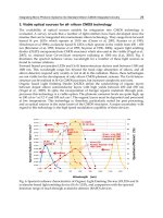
as negative and these chemotaxins are called chemorepellents. Energy sources usually attract
motile bacteria whereas bacteriotoxic agents act as repellents (Fig. 1). The finding, that bacteria
move actively towards or away from certain substances, was already made at the end of the
19th century by Engelmann (Engelmann, 1881) and Pfeffer (Pfeffer, 1884 & 1888). Thus, with
the help of chemotaxis bacteria direct their movement to find favourable niches with high
chemoattracor and low chemorepellent concentrations. This decision-making is based on
temporal sensing. As indicated above the overall motion of a bacterium is composed of
alternating phases of straight swimming and thumbling. In the presence of a chemical gradient
the straight swimming phases last longer, and if the bacterium is moving nat along this
gradient, it starts sooner to tumble and tries to reorientate depending on the chemotaxins
Motility and Energy Taxis of Salmonella spp.
31
concentration (Adler, 1975). The essential prerequisites for chemotaxis are, as already
mentioned, a flagella mediated motility, a variety of individual chemoreceptors and a highly
conserved chemosensory signal-transduction system.
2. Flagellar motility and chemotaxis
2.1 Experimental approaches
Before the mechanisms of flagellar motility and chemotaxis will be discussed, the most
common tools or experimental approaches to study and record bacterial motility and taxis
will be presented: microscopy and chemotaxis assay.
2.1.1 Microscopy
Conventional light microscopy is not sufficient to visualize flagellar filaments because of
their thinness and the swiftness. One very early approach to visualize flagella of living
bacterial cells is dark field microscopy (Macnab, 1976). Since light is scattered by dirt
particles reducing the contrast, it has to be considered that the medium and the specimen
slides must be remarkably clean. A great advance in this field is video-enhanced differential
interference-contrast microscopy (Block et al., 1991). Video microscopy combined with
computer based image processing made it possible to detect very small objects like
particular microtubules of ≈ 25 nm in diameter. Computerized image analysis offers the
option to estimate values like mean cell run speed and average tumbling frequency and
their variation in the presence or absence of attractants or repellents (Staropoli & Alon,
2000). Phase-contrast video microscopy combined with the analysis of superimposed image
series is a very useful tool, especially for the study of the taxis to and the motion near solid
surfaces (Lauga et al., 2006). A further helpful method, although not specifically associated
with flagellar motility and chemotaxis, is fluorescence microscopy, which can be used to
visualize protein-protein-interactions in the chemoreceptor signaltransduction pathway and
the fagellar motor, in combination with green fluorescence fusion proteins (Pierce et al.,
1999; Khan et al., 2000).
2.1.2 Chemotaxis assays
Another easy to handle experimental set of tools is composed from different kinds of
chemotaxis assays (Miller et al. 2009). One semiquantitative variant is based on changes of
the opalescence of a semi-solid agar due to the concentration of bacterial cells (Hugdahl et
al., 1988). In a first step, a phosphate buffered saline-agar solution is mixed with a bacterial
suspension of a specifiy optical density and poured into a petri dish. After solidification,
paper discs with the chemotaxins are placed onto the agar surface following incubation of
three to four hours. A more opaque zone can be seen in the surrounding of chemoattractants
(see Fig. 1A), whereas chemorepellents are girdled by a more transparent halo (see Fig. 1 B).
Other versions of the agar based chemotaxis assay deal with pure – bacteria free - agar
plates. After solidification of the agar small recessions are cut into the agar and are filled
with either a bacterial suspension or the test solution (Köhidai, 1995). A variation of this
assay uses parallel channels (PP-technique) cut from each of both recesses connected by a
third perpendicular channel between these two to facilitate diffusion of bacterial suspension
and test solution (Köhidai, 1995).
Salmonella – A Diversified Superbug
32
Fig. 1. Examples for chemotaxis: A: Attraction towards towards L-asparagine; B: Repulsion
from deoxycholic acid; C: Control (PBS)
A second method is a capillary assay. In this assay, heparinized glass capillaries bridge
between a bacterial suspension and a test solution (Koppelhus et al., 1994; Leick & Helle,
1983). Two-phase assays in spectrophotometer plastic cuvettes are suitable to monitor the
chemotaxis-mediated migration between the two fluid phases (Koppelhus et al., 1994).
The T-maze assay allows the quantification of the chemoresponse of two substances in
direct comparison, using a T-shaped experimental arrangement of three containers (Van
Houten et al., 1982).
The use of a so-called Boyden chamber is a third variant to study chemotaxis. Chambers
divided by filters are a third variant of chemotaxis assays. The suspension of motile cells is
placed into the upper vessel of the so-called Boyden chamber (Boyden, 1962). The test solution
with the chemoeffector is filled into the lower vessel. A filter membrane separates both parts
of the Boyden chamber. The pore diameter must be chosen according to the size of the
organism allowing its transmigration. To simulate in vivo conditions, the filter membranes
can be optionally covered with extracellular matrix proteins like collagen, elastin or fibrin.
Modifications of this technique connect the vessels either horizontally (Zigmond, 1988) or as
concentric rings (Zicha et al., 1991). Multiwell chambers make the parallel testing of
different substances in one occasion feasible.
2.2 Molecular structure of the flagella motor and chemoreceptors
2.2.1 Molecular structure and synthesis of the flagellar apparatus
Non-flagellar Type III secretory systems and the flagellar apparatus share a common basic
architecture. Thus, it seems apparently that both go back to a common evolutionary origin
(Toft & Fares, 2008).
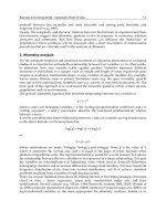
Basically, the flagellum is comprised of three parts: a helical filament, the hook and the basal
body (see Fig. 2). The filament of bacterial flagella is built up of multiple subunits of the
flagellin protein FliC (Samatey et al., 2004), which form so called protofilaments. Eleven
circular arranged protofilaments stacked into a left- or right-handed helix, according to the
direction of rotation (see above) comprise the filament (O’Brian & Bennet, 1972). It has a
length of about 10 µm and a diameter of about 20 nm. If a flagellum is virtually flattened, it
shows a constant interspace between adjacent turns, corresponding to the wavelength of a
Motility and Energy Taxis of Salmonella spp.
33
“sine wave” (Madigan & Martinko, 2006). This wavelength is specific for any bacterial
species and is determined by the structure of the flagellin protein and the rotational
direction of the filament (Madigan & Martinko, 2006).
The hook tethers the filament to the basal body. Electron microscopic studies demonstrated
that the hook of S. typhimurium has length of 55 6 nm (Hirano et al., 1994). The interdigitated
hook-subunits make up a bended tube with a 2 to 3 nm wide central channel, which continues
in the rod as well as in the filament (Shaik et al., 2005). The hook connects the filament to the
motor portion of the flagellar apparatus. The hooks flexibility permits the transmission of
torque from the motor to the helical propeller when both are not in a coaxial orientation to
each other (Berg & Anderson, 1973). A so-called gap compression/extension mechanism and
mutual sliding of the hook-subunits allows continuous structural change of the hook during
flagellar rotation at low energy cost (Furuta et al., 2006). The hook facilitates the
synchronization of several filaments bundled together at one cell pole (Macnab, 1977; Berg &
Anderson, 1973). The two hook-associated proteins (HAP 1 and 3) form a small hook-filament
junction, which acts as an adaptor for transition between the hook that is flexible in bending
but rigid against twisting and the much more stiff filament (Samatey et al., 2004).
Fig. 2. The flagellum of Gram-negative bacteria, like Salmonella sp., is a complex structure
consisting of the propeller like acting filament, the flexiple hook and the flagellar ATP-
driven motor, which is comprised of four ring-structures and the static motor complexes. An
integrated flagellar type III secretion system exports actively the proteins for flagellar
assembly but also some virulence associated factors. (Copyright: Wikimedia Commons,
public domain aviable from
Flagellum _base_diagram_en.svg)
Salmonella – A Diversified Superbug
34
The third part of the flagella – the basal body is comprised of a rod sticking in four ring-like
structures: the L-ring (associated with lipopolysaccharides) and the P-ring (associated with
peptidoglycanes) forming an outer cylinder embedded in the plasma membrane, the MS-
ring, building up a motor mounting plate; and the versatile C-ring (Macnab, 1999). Gram-
positive bacteria lacking an outer membrane lack consequentely the outer ring structures.
Overall more than 40 genes are involved in flagellar synthesis and subsequent motility in S.
typhimurium (Shaik et al. 2005). The MS- and the C-ring, the export apparatus as well as the
motor and switch are the first synthesized compounds of the flagella (Katayama et al., 1996;
Macnab, 2003). The following assembly steps utilize the type III export apparatus, while the
export substrates are supplied via an delivery apparatus located in a patch of membrane
near the center of the MS-ring to the channel (Suzuki et al., 1998; Macnab, 2003). It follows
the formation of the rod and the other two rings. The proteins of which the rod is comprised
are FlgB, FlgC, FlgF and FlgG (Homma et al., 1990). The bifunctional FlgJ protein, which has
a muraminidase activity to open the peptidoglycan layer for penetration of the sprouting
rod, is also able to bind other rod constituents (Hirano et al., 2001), and thus may act as a
rod capping protein promoting the assembly of the rod out of its four components (Nambu
et al.; 1999, Macnab, 2003). The assembly of the basal body is finished by the synthesis of the
periplasmic P-ring made out of FlgI (Homma et al., 1987) and the outer membrane L-ring
consisting of FlgH-subunits (Jones et al., 1990). In the following step, the rod cap is
dislodged, while the hook-cap, consisting of about 5 FlgD-subunits, is mounted (Macnab,
2003) and the hook is attached on the basal body. It consists, comparable to the flagellar
filament, of about 120 copies of a single kind of protein, FlgE – the so-called hook protein
(Samatey et al., 2004). Parallel, the L-ring is assembled (Kubori et al., 1992; Ohnishi et al.,
1987). After this, two junction protein zones, made either of FlgK or FlgL, are attached, and a
so-called filament-cap out of FliD-proteins is mounted on the hook (Homma et al., 1985;
Ikeda et al., 1987 & 1989). Cap proteins assist the organization of the flagellin proteins to
form a new filament (Ikeda et al., 1985). Between hook and cap, a junction zone is
synthesized before the protofilaments are assembled (Macnab, 2003). The flagellin molecules
pass the channel inside the hook and the filament and add on successively at the lower end.
The flagellar assembly starts beneath the cap and grows from its tip to its base. A mature
flagellum is composed of approximately 20 000 copies of flagellin protein. A broken flagella
can be repaired with newly synthesized flagellin units from the cytoplasm passing through
the filament channel (Homma & Iino, 1985). The proteins, which built up the flagellum, are
translocated to the distal part of the growing flagellum through the central channel by a
flagellar type III homologous protein secretion system (Ibuki et al., 2011). This secretion
system is comprised of two classes of proteins: soluble and membrane associated ones. The
essential soluble compounds of this ATP-consuming process are the soluble FliI-ATPase, its
regulator FliH and the FliJ-protein, that promotes the hexamerization of FliI-ATPases (Ibuki
et al., 2011). The remaining soluble components are specific chaperones: FlgN for the hook-
filament junction proteins, FliT for the filament cap protein, and FliS for flagellin (Macnab,
2003). The six membrane associated components FlhA, FlhB, FliO, FliP, FliQ, and FliR form a
complex within the MS-ring (Macnab, 2003).
A rotary motor is in principle built up of two functional components: the rotor and the
stator. The flagellar motor consists of the static Mot-complexes, which were affixed in the
inner cytoplasmatic membrane and the rotating C-ring. The Mot-complexes are
Motility and Energy Taxis of Salmonella spp.
35
transmembrane structures made from two proteins MotA and MotB (Macnab, 2003). The
cytoplasmic C-ring contains the motor/switch proteins – the Fli-proteins. The FliG-proteins
generate the torsional moment, while working against the Mot-complexes. The switch-
proteins, in Salmonella sp.: FliG, FliM and FliN can reverse the flagellar rotational direction
in response to intracellular signals (Francis et al., 1994; Yamaguchi et al. 1986). The FliM-
protein is the final effector of a sensory transduction chain (Bren et al., 1998; Sockett et al.,
1992). Thus, the stator is formed by the Mot-proteins, which mantle the C- and the MS-rings.
C- and MS-rings, as well as the rod, collectively form the rotor.
Driven by a transmembrane proton gradient, the flagellar motor is able to perform the
clockwise and a counterclockwise rotation of the filament, which is reversed stochastically
in the absence of any stimulus. The protons are pumped from outside across the
cytoplasmic membrane through the Mot-complexes. Calculations showed that about 1000
protons must be translocated to perform a single rotation of the flagellar filament. In the
proposed proton pump model, the protons flowing through the stator channels exert
electrostatic forces on helically arranged charge clusters on the rotor rings. Most probably,
the protons bind temporary to a specific aspartate residue of MotB, causing a change of
the stators conformation that drives the rotor through an elementary rotational step
(Kojima & Blair, 2001; Macnab, 2003). In the next step, the aspartate residue gets
deprotonated and the stator returns to its original conformation. These resulting series of
interactions between positive and negative charges generate a torsional moment as
protons flow through the Mot-complexes.
2.3 Chemoreceptor structure and signal transduction
Presence and concentration of chemotaxins are detected by a family of chemoreceptors
sharing a common two-component system architecture (Miller et al. 2009). Such two-
component systems are generally comprised of a membrane associated histidine autokinase
(CheA) and a cytoplasmic response regulator (CheY; Lux et al. 2004). Methyl-accepting
chemotaxis proteins (MCPs), embedded in the cytoplasmic membrane, sense the
environmental signals via their N-terminal periplasmic sensory domain to their C-terminal
cytoplasmic signaling domain. The MCP-monomers have a molecular mass of about 60 kDa
and form constitutively very stable homodimers, which are arranged in groups of three (Lux
et al. 2004). CheW linker proteins tether the CheA histidine kinase to the MCPs (Miller et al.
2009). This inhibits autophosphorylation of CheA, which in turn reduces the
phosphorylation of CheY response regulator at a conserved histidine residue.
Hypophosphorylated CheY can diffuse freely in the cytoplasm and can interact with FliM,
the switch protein of the flagellar motor (Mot), which is the final effector of sensory
transduction chain (Bren et al., 1998; Sockett et al., 1992). Thus, it triggers counterclockwise
rotation of the flagella, which leads to bacterial “running”. If a bacterium moves along a
gradient of a chemoattractant, the intracellular concentration of phosphorylated CheY
decreases. Consequently, the frequency of flagella switching decreases, and the number of
site directed “runs” along the gradient increases. Thus, addition of an attractant triggers a
counterclockwise rotation of the filament (Bren & Eisenbach, 2000).
In the opposite case, decreasing ligand occupancy of the MCPs leads to increased
autophoshorylation of CheA and in consequence to an amplified phosphorylation of CheY
Salmonella – A Diversified Superbug
36
and CheB. Phosphorylated CheY binds as well to the motor switch but triggers a clockwise
flagellar rotation resulting in bacterial “tumbling”.
CheB is a receptor-demethylating enzyme, which is also activated by phosphorylation. The
phosphatase CheZ is responsible for the dephosphorylation of phosphate-activated CheY
(Bourret & Stock, 2002).
The result of this chemosensing in three-dimensional spatial gradients of different
chemoattractors and chemorepellents is a stereoscopic “zigzag” path of motion (Berg &
Brown, 1972), until the bacterium reaches a niche with an equilibrium between the varying
chemoeffectors (Miller et al., 2009).
2.4 Sensory adaptation
Sensory adaption means reestablishment of the prestimulus state in the perpetual presence of
the stimulus. Adaptation to chemotactical stimuli is mostly due to modulation of the
methylation of certain sites of the MCP receptors. The central players in the process of sensory
adaption are the methyltransferase CheR, the methylesterase CheB, and the cytoplasmic
domains of the MCP-receptors that have adjacent to the CheA and CheW binding sites, sites
for methylation and demethylation of glutamyl side chains (Macnab, 2003).
CheR catalyzes in a S-adenosylmethionine consuming reaction the methylation of the
specific glutamate residues on the cytoplasmic domains of the MCPs (Bren & Eisenbach,
2000). This reaction enhances the CheA autophosphorylation favouring clockwise flagallar
rotation and is triggered by attracting stimuli (Borkovich et al., 1992; Ninfa et al., 1991). The
cytoplasmatic domains of the MCPs have a specific domain, which is methylated by CheR,
and a distinct CheR-binding site consisting of a pentapepetide that is only present in high-
abundance receptors. It was shown that CheR bound to the binding sites onto the high-
abundance receptors methylates the designated sites of the low-abundance receptors (Le
Moual et al., 1997; Li et al., 1997).
Its antagonist is the methylesterase CheB, which demethylates the MCPs during adaptation
to repelling stimuli. Additionally CheB has an amidase activity catalyzing the conversion of
glutamamine residues into glutamate on the MCPs (Djordjevic et al., 1998). The liberation of
glutamate residues inhibits the autophosphorylation of CheA favouring a counterclockwise
rotation of the flagellum. In addition CheB itself is regulated by CheA-mediated
phosphorylation (Hess et al., 1988; Lupas & Stock, 1989). Phosphorylation inhibits the
methylesterase activity, while the unphosphorylated enzyme has less methylesterase
activity. The binding sites on CheA for CheY and CheB are identical. Thus CheB competes
with CheY (Li et al., 1995).
Furthermore a high methylation rate decreases the receptors affinity to chemoattractants
(Bornhorst et al., 2000; Li et al., 2000). It was also suggested that a deferred activation of
CheZ, which is responsible for an enhanced CheY dephosphorylation, is involved in the
process of sensory adaptation (Blat et al., 1998).
These regulatory effects occur only after the initial chemotactic response and the steady state
of all these parallel-acting adaptational processes determines the extent of reaction to a
certain chemoeffector (Alon et al., 1999).
Motility and Energy Taxis of Salmonella spp.
37
2.5 Specific Salmonella chemoreceptors
Altogether, it is difficult to identify chemoreceptors specific for a certain taxin, because the
different MCPs can compensate each other in many cases, Thus, knockout mutants of
chemoreceptor genes show often no defects in their phenotype (Vegge et al., 2009; Tareen et
al. 2010). Up to now, four chemoreceptor specificities are identified for Salmonella spp.
The Tar chemoreceptor is specific for aspartate and initiates attractant signalling (Foster et
al. 1985; Milburn et al., 1991). The same receptor molecule interacts also with the periplasmic
maltose-binding protein, and senses in this way chemoattraction towards maltose
(Mowbray & Koshland 1987; Gardina et al., 1992). It was also demonstrated that this
receptor mediates attractant responses to phenol and repellent responses to glycerol and
nickel or cobalt ions in Escherichia coli as well as thermoresponses (Lee & Imae, 1990).
The ligand serine mediates positive taxis via the Tsr receptor, whereas Tsr sensing due to
leucine and glycerol results in a repulsion of the bacteria. (Lee & Imae, 1990; Jeffrey &
Koshland, 1993; Oosawa & Imae, 1984; Springer et al., 1977). Tsr functions also as
thermoreceptor. Temperature increase leads to smooth swimming of bacterial cells, whereas
temperature decrease induces tumbling (Lee et al., 1988).
S. typhimurium demonstrates attraction towards citrate and metal-citrate complexes, but
repulsion from phenol. This behavior gives the name to the third chemoreceptor in this
schedule – Tcp, that stands for taxis to citrate and away from phenol (Yamamoto & Imae, 1993).
The trg gene encodes a fourth chemoreceptor of the MCP family specific for
ribose/galactose (Blat & Eisenberg, 1995, Kasinkas et al. 2007).
2.6 Virulence factors secreted via the flagellar type III secretion system
As mentioned above, the flagellar apparatus is a homologue of a type III secretion system
that is able to secrete specific peptides and proteins in an ATP dependent mechanism into
the environment (Collazo & Galán, 1996; Eichelberg et al., 1994). Among these secreted
proteins are mostly structural components of the flagella, for example flagellin monomers,
and the hook protein, but also several virulence factors. It functions as a molecular syringe –
the so-called injectisome – that is used by bacteria to inject effector proteins directly into the
interior of host cells (Mota et al., 2005a+b; Arnold et al. 2009). Thus, these proteins play an
important role for host cell invasion and the pathogenesis of salmonellosis. It was shown,
that the N-terminal 30 residues of these effector proteins form a taxonomically universal,
type III specific secretion signal (Arnold et al. 2009; Samurdrala et al. 2009). About 65 type III
secretion system substrates are known for S. typhimurium (Samurdrala et al. 2009). Five well
described proteins involved in host cell invasion, typically the M-cells of the ileal Peyers’
Patches, are InvE, Sipa, Sipb, SipC, and SipD. InvE plays a pivotal role for triggering cellular
mechanisms, which lead to bacterial entry. It is required for translocation of other effector
proteins into the cytosol of host cells and forms complexes with SipA, SipB, and SipC.
(Kubori & Galán, 2002). Comparable to InvE the effector protein SipD, which has been
shown to be important for liver and ileum colonization, is suggested to modulate the
secretion of SipA, SipB, and SipC (Gong et al., 2010). Cell invasion occurs via a ruffle-
mediated mechanism, which is initiated by the activation of specific signal transduction
cascades and rearrangement of the actin cytoskeleton. The actin rearrangements are realized
Salmonella – A Diversified Superbug
38
by SipA interworking with SopE, a guanine-nucleotide exchange factor for Rho GTPases,
and SptP, a protein tyrosine phosphatase, (Brumell et al., 1999) as well as SipC that binds
and bundles F-actin (Myeni & Zhou, 2010).
SipB interacts after entering the cytosol of macrophages with cell signalling pathways to
induce apoptosis (Hersh et al. 1999). It associates with caspase-1 and promotes the
proteolytic activation of this protease.
Two further proteins entering macrophages are SrfN and PagK2, which were shown to be
essential for full virulence and are suggested to interact with host cellular components
(Yoon et al. 2011). These two effector proteins are translocated independently of the
injectisome. Thus, the flagellar type III secretion system is the only protein export
mechanism in Salmonella sp.
2.7 Role of chemotaxis and flagellar motility for the pathoegenesis of salmonelosis
The ability for directed movement and taxis towards and away from chemoeffectors plays a
crucial role for the pathogenesis of salmonellosis. Amongst others, Salmonella bacteria are able
to persist inside the inner leaf tissue of plants (Kroupitski et al., 2009; Goldberg et al., 2011). It
was shown that flagellar motility and chemotaxis towards nutrients produced by
photosynthetically active cells are crucial for entry into iceberg lettuce leaves via open stomata
and invasion into the plant tissue (Kroupitski et al., 2009). Enteropathogens have the ability to
adapt to the phyllosphere environment. They obviously interact with epiphytic bacteria
(Beuchat, 2002; Brandl, 2006; Heaton & Jones 2008) and become part of phylloplane biofilms,
where they gain protection from environmental stressors (Fett, 2000). Plants that might
become contaminated by the use of germ-containing water for irrigation or Salmonella-
containing liquid manure for fertilization might function as source of infection (Beuchat, &
Ryu, 1997; Brandl, 2006; Horby et al., 2003). Internal persistence after entering the plant tissue
explains the failure of lavation and sanitizers to eradicate Salmonella in leafy greens.
Furthermore, flagellar movement and chemotaxis are also pivotal for the intestinal
colonization of the different Salmonella hosts, especially for the competition for nutrients
with other bacteria of human microbiome (Stecher et al., 2008). Even the induction of colitis
depends on a functioning flagellar movement and chemotaxis (Stecher et al., 2004).
3. Conclusion
The flagellar apparatus is an evolutionary ancient multifunctional tool involved in motility,
bacterial cell aggregation, biofilm formation, protein export, and a virulence factor injection
via the injectisome. It is also the prototype of a sensing system, coupling energy taxis and
motility. The research on chemotaxis and flagellar motility is almost as old as bacteriology
itself, starting at the end of the 19th century. The research on Salmonella sp. plays here a
special role, as most of the knowledge about thermo- and chemotaxis, MCP-receptor signal
transduction, MCP-receptor sensory adaptation, structure, synthesis, and function of the
flagellar apparatus as well as effector protein secretion via a flagellar type III homologue
secretion system was made using Salmonella sp. and E. coli as model organism.
Thus, the flagellar apparatus regulated by energy taxis may be the most important structure
for intestinal colonization and pathogenesis of salmonellosis.
Motility and Energy Taxis of Salmonella spp.
39
4. Acknowledgement
The work of the author is funded by the Forschungsförderungsprogramm of the
Universitätsmedizin Göttingen, Germany and by the Deutsche Forschungsgemeinschaft
(PAK 400).
5. References
Adler, J. (1975) Chemotaxis in Bacteria. Annual Review of Biochemistry, Vol. 44, (1975), pp.
341-356, ISSN 0066-4154
Alon, U., Surette, M.G., Barkai, N. & Leibler, S. (1999). Robustness in bacterial chemotaxis.
Nature, Vol. 397, No. 6715, (January 1999), pp. 168-171, ISSN 0369-3392
Arnold, R., Brandmaier, S., Kleine, F., Tischler, P., Heinz, E., Behrens, S, Niinikoski, A., Mewes,
H.W., Horn, M. & Rattei, T. (2009) Sequence-based prediction of type III secreted
proteins. PLoS Pathogens. Vol. 5, No. 4, (April 2009), pp. e1000376, ISSN 1553-7366
Barnakov, A.N., Barnakova, L.A. & Hazelbauer, G.L. (1999). Efficient adaptational
demethylation of chemoreceptors requires the same enzyme-docking site as
efficient methylation. Proceedings of the National Academy of Sciences of the United
States of America. No. 96, Vol. 19, (September 1999), pp. 10667-10672, ISSN 0027-8424
Berg, H.C. & Anderson, R.A. (1973). Bacteria swim by rotating their flagellar filaments.
Nature. Vol. 245, No. 5425, (October 1973), pp. 380-282, ISSN 0028-0836
Berg, H.C. & Brown D.A. (1972). Chemotaxis in Escherichia coli analysed by three-dimensional
tracking. Nature. Vol. 239, No. 5374, (October 1972), pp 500- 504, ISSN 0028-0836
Beuchat L.R. (2002). Ecological factors influencing survival and growth of human pathogens
on raw fruits and vegetables. Microbes and infection / Institut Pasteur. Vol. 4, No. 4),
(April 2002), pp. 413-423, ISSN 1286-4579
Beuchat, L.R. & Ryu, J.H. (1997). Produce handling and processing practices. Emerging
infectious diseases. Vol. 3, No. 4, (October-December 1997), pp. 459-465., ISSN 1080-
6040
Blat, Y. & Eisenbach, M. (1995). Tar-dependent and -independent pattern formation by
Salmonella typhimurium. Journal of bacteriology. Vol. 177, No. 7, (April 1995), pp. 1683-
1691, ISSN 0021-9193
Blat, Y., Gillespie, B., Bren, A., Dahlquist, F.W. & Eisenbach, M. (1998). Regulation of
phosphatase activity in bacterial chemotaxis. Journal of molecular biology. Vol. 284,
No, 4, (December 1998), pp. 1191-1199, ISSN 0022-2836
Block, S.M., Fahrner, K.A. & Berg, H.C. (1991). Visualization of bacterial flagella by video-
enhanced light microscopy. Journal of Bacteriology, Vol. 173, No. 2, (January 1991),
pp. 933-936, ISSN 0021-9193
Borkovich, K.A., Alex, L.A. & Simon, M.I. (1992). Attenuation of sensory receptor signaling
by covalent modification. Proceedings of the National Academy of Sciences of the United
States of America. Vol. 89, No. 15, (August 1992), pp. 6756-6760, ISSN 0027-8424
Bornhorst, J.A. & Falke, J.J. (2000). Attractant regulation of the aspartate receptor-kinase
complex: limited cooperative interactions between receptors and effects of the
receptor modification state. Biochemistry. Vol. 39, No. 31, (August 2000), pp. 9486-
9493, ISSN 0006-2960
Salmonella – A Diversified Superbug
40
Bourret, R.B. & Stock, A.M. (2002). Molecular information processing: lessons from bacterial
chemotaxis. The Journal of biological chemistry. Vol. 277, No. 12, (March 2002), pp.
9625-9628, ISSN 0021-9258
Boyden, S.V. (1962). The chemotactic effect of mixtures of antibody and antigen on
polymorphonuclear leucocytes. The Journal of Experimental Medicine, (February
1962), Vol. 115, No. 3, pp. 453-466, ISSN 0022-1007
Brandl, M.T. (2006). Fitness of human enteric pathogens on plants and implications for food
safety. Annual review of phytopathology. Vol. 44, (2006), pp. 367-392, ISSN 0066-4286
Bren, A. & Eisenbach, M. (1998). The N terminus of the flagellar switch protein, FliM, is the
binding domain for the chemotactic response regulator, CheY. Journal of molecular
biology. Vol. 278, No. 3, (May 1998), pp. 507-514. ISSN 0022-2836
Bren, A. & Eisenbach, M. (2000). How signals are heard during bacterial chemotaxis:
protein-protein interactions in sensory signal propagation. Journal of bacteriology.
Vol. 182, No. 24, (December 2000), pp. 6865-6873, ISSN 0021-9193
Brumell, J.H., Steele-Mortimer, O. & Finlay, B.B. (1999). Bacterial invasion: Force feeding by
Salmonella. Current biology. Vol. 9, No. 8, (April 1999), pp. R277-80, ISSN 0960-9822
Collazo, C.M. & Galán, J.E. (1996). Requirement for exported proteins in secretion through
the invasion-associated type III system of Salmonella typhimurium. Infection and
immunity. Vol. 64, No. 9. (September 1996) pp. 3524-3531, ISSN 0019-9567
Djordjevic, S. & Stock, A.M. (1998). Structural analysis of bacterial chemotaxis proteins:
components of a dynamic signaling system. Journal of structural biology. Vol. 124,
No. 2-3, (December 1998), pp. 189-200, ISSN 1047-8477
Eichelberg, K., Ginocchio, C.C. & Galán, J.E. (1994). Molecular and functional
characterization of the Salmonella typhimurium invasion genes invB and invC:
homology of InvC to the F0F1 ATPase family of proteins. Journal of bacteriology. Vol.
176, No. 15, (August 1994), pp. 4501-4510, ISSN 0021-9193
Engelmann, T.W. (1881). Neue Methode zur Untersuchung der Sauerstoffausscheidung
pflanzlicher und thierischer Organismen. Pflügers Archiv für die Gesamte Physiolgie
der Menschen und Tiere, Vol. 25, (1881) pp. 285-292, ISSN 0365-267X
Erdem, A.L., Avelino, F., Xicohtencatl-Cortes. J. & Girón J.A. (2007). Host protein binding
and adhesive properties of H6 and H7 flagella of attaching and effacing Escherichia
coli. Journal of Bacteriology. Vol. 189, No. 20, (October 2007), pp. 7426-7435, ISSN
0021-9193
Fett W.F. (2000). Naturally occurring biofilms on alfalfa and other types of sprouts. Journal
of food protection. Vol. 63, No. 5, (May 2000), pp. 625-632, ISSN 0362-028X
Foster, D.L., Mowbray, S.L., Jap, B.K. & Koshland D.E. Jr. (1985). Purification and
characterization of the aspartate chemoreceptor. The Journal of biological chemistry.
Vol. 260, No. 21, (September 1985), pp. 11706-11710, ISSN 0021-9258
Francis, N.R., Sosinsky, G.E., Thomas, D. & DeRosier, D.J. (1994). Isolation, characterization
and structure of bacterial flagellar motors containing the switch complex. Journal of
molecular biology. Vol. 235, No. 4, (January 1994), pp. 1261-1270; ISSN 0022-2836
Frye, J., Karlinsey, J.E., Felise, H.R., Marzolf, B., Dowidar, N., McClelland, M. & Hughes,
K.T. (2006). Identification of new flagellar genes of Salmonella enterica serovar
Typhimurium. Journal of Bacteriology, Vol. 188, No. 6, (March 2006), pp. 2233-2243,
ISSN 0021-9193
Furuta, T., Samatey, F.A., Matsunami, H., Imada
, K., Namba, K. & Kitao, A. (2007). Gap
compression/extension mechanism of bacterial flagellar hook as the molecular
Motility and Energy Taxis of Salmonella spp.
41
universal joint. Journal of Structural Biology, Vol. 157, No. 3, (March 2007), pp. 481-
490, ISSN 1047-8477
Gardina, P., Conway, C., Kossman, M. & Manson, M. (1992). Aspartate and maltose-binding
protein interact with adjacent sites in the Tar chemotactic signal transducer of
Escherichia coli. Journal of bacteriology. Vol. 174, No. 5, (March 1992), pp.1528-1536,
ISSN 0021-9193
Gebremichael Y, Ayton GS, Voth GA. (2006). Mesoscopic modeling of bacterial flagellar
microhydrodynamics. Biophysical Journal. Vol. 91, No. 10, (November 2006), pp.
3640-3652, ISSN 0006-3495
Golberg, D., Kroupitski, Y., Belausov, E., Pinto, R. & Sela, S. (2011). Salmonella Typhimurium
internalization is variable in leafy vegetables and fresh herbs. International journal of
food microbiology. Vol. 145, No. 1, (January 2011), pp. 250-257, ISSN 0168-1605
Gong, H., Vu, G.P., Bai, Y., Yang, E., Liu, F. & Lu, S. (2010). Differential expression of
Salmonella type III secretion system factors InvJ, PrgJ, SipC, SipD, SopA and SopB
in cultures and in mice. Microbiology. Vol. 156, No. Pt 1, (January 2010), pp. 116-127,
ISSN 1350-0872
Hahn, H., Klein, P., Giesbrecht, R.E. & Streeck, R.E. (2004). Bakterien: Definition und
Aufbau, In: Medizinische Mikrobiologie und Infektologie, Hahn, H., Falke, D.,
Kaufmann, S.H.E. & Ullmann, U., pp. 169-179, Springer Medizin Verlag
Heidelberg, ISBN 3-540-21971-4, Berlin, Germany
Heaton, J.C. & Jones, K. (2008). Microbial contamination of fruit and vegetables and the
behaviour of enteropathogens in the phyllosphere: a review. Journal of applied
microbiology. Vol. 104, No. 3, (March 2008), pp. 613-626, ISSN 1364-5072
Hersh, D., Monack, D.M., Smith, M.R., Ghori, N., Falkow, S. & Zychlinsky, A. (1999). The
Salmonella invasin SipB induces macrophage apoptosis by binding to caspase-1.
Proceedings of the National Academy of Sciences of the United States of America. Vol. 96,
No. 5, (March 1999), pp. 2396-2401, ISSN 0027-8424
Hess, J.F., Oosawa, K., Kaplan, N. & Simon, M.I. (1988). Phosphorylation of three proteins in
the signaling pathway of bacterial chemotaxis. Cell. Vol. 53, No. 1, (April 1988), pp.
79-87, ISSN 0092-8674
Hirano, T., Minamino, T. & Macnab, R.M. (2001). The role in flagellar rod assembly of the N-
terminal domain of Salmonella FlgJ, a flagellum-specific muramidase. Journal of
molecular biology. Vol. 312, No. 2, (September 2001), pp. 359-369, ISSN 0022-2836
Hirano, T., Yamaguchi, S., Oosawa, K. & Aizawa, S. (1994). Roles of FliK and FlhB in
determination of flagellar hook length in Salmonella typhimurium. Journal of
bacteriology. Vol. 176, No. 17, (September 1994) pp. 5439-5449, ISSN 0021-9193
Homma, M. & Iino. T. Excretion of unassembled hook-associated proteins by Salmonella
typhimurium. (1985). Journal of bacteriology. Vol. 164, No. 3, (December 1985), pp.
1370-1372, ISSN 0021-9193
Homma, M., Komeda, Y., Iino, T. & Macnab, R.M. (1987). The flaFIX gene product of
Salmonella typhimurium is a flagellar basal body component with a signal peptide
for export. Journal of bacteriology. Vol. 169, No. 4, (April 1987), pp. 1493-1498, ISSN
0021-9193
Homma, M., Kutsukake, K. & Iino, T. (1985). Structural genes for flagellar
hook-associated
proteins in Salmonella typhimurium. Journal of bacteriology. Vol. 163, No. 2, (August
1985), pp. 464-471, ISSN 0021-9193