Báo cáo hóa học: " Are quantum dots ready for in vivo imaging in human subjects?" docx
Bạn đang xem bản rút gọn của tài liệu. Xem và tải ngay bản đầy đủ của tài liệu tại đây (625.04 KB, 17 trang )
NANO REVIEW
Are quantum dots ready for in vivo imaging in human subjects?
Weibo Cai Æ Andrew R. Hsu Æ Zi-Bo Li Æ
Xiaoyuan Chen
Received: 14 April 2007 / Accepted: 24 April 2007 / Published online: 30 May 2007
Ó to the authors 2007
Abstract Nanotechnology has the potential to profoundly
transform the nature of cancer diagnosis and cancer patient
management in the future. Over the past decade, quantum
dots (QDs) have become one of the fastest growing areas of
research in nanotechnology. QDs are fluorescent semi-
conductor nanoparticles suitable for multiplexed in vitro
and in vivo imaging. Numerous studies on QDs have re-
sulted in major advancements in QD surface modification,
coating, biocompatibility, sensitivity, multiplexing, target-
ing specificity, as well as important findings regarding
toxicity and applicability. For in vitro applications, QDs
can be used in place of traditional organic fluorescent dyes
in virtually any system, outperforming organic dyes in the
majority of cases. In vivo targeted tumor imaging with
biocompatible QDs has recently become possible in mouse
models. With new advances in QD technology such as
bioluminescence resonance energy transfer, synthesis of
smaller size non-Cd based QDs, improved surface coating
and conjugation, and multifunctional probes for multimo-
dality imaging, it is likely that human applications of QDs
will soon be possible in a clinical setting.
Keywords Quantum dot (QD) Á Nanoparticles Á
Nanotechnology Á Cancer Á Molecular imaging Á
Near-infrared fluorescence (NIRF) imaging Á
Nanomedicine
Introduction
To expedite the clinical application of nanotechnology, the
National Cancer Institute (NCI) is currently funding eight
Centers of Cancer Nanotechnology Excellence (CCNEs)
and twelve Cancer Nanotechnology Platform Partnerships
( It is believed that combining
development efforts in nanotechnology and cancer research
may quickly and effectively transform the prevention,
diagnosis, and treatment of cancer in the future. After
establishing an interdisciplinary nanotechnology work-
force, the goal was to have matured nanotechnology into a
clinically useful field by 2010. The NCI Alliance for
Nanotechnology in Cancer aims to develop research tools
to help identify new biological targets, agents to monitor
predictive molecular changes and prevent precancerous
cells from becoming malignant, imaging agents and diag-
nostics to detect cancer in the earliest pre-symptomatic
stage, multifunctional targeted devices to deliver multiple
therapeutic agents directly to the tumor, systems to provide
real-time assessment of therapeutic and surgical efficacy,
and novel methods to manage symptoms that reduce the
quality of life. The nanoparticles actively being pursued
include quantum dots (QDs) [1, 2], nanotubes [3], nano-
wires [4], nanoshells [5], and many others [6–9]. Among
these, QDs are the most widely studied and have many
potential clinical applications.
Organic fluorophores and dyes have been historically
used to label cells and tissues for both in vitro and in vivo
imaging [10]. However, due to their inherent photophysical
properties such as low photobleaching thresholds, broad
absorption/emission spectra, and small Stokes shifts, their
use is limited and they are not ideal agents for multiplex-
ing, long-term, or real-time imaging. On the other hand,
QDs are inorganic fluorescent semiconductor nanoparticles
W. Cai Á A. R. Hsu Á Z B. Li Á X. Chen (&)
The Molecular Imaging Program at Stanford (MIPS),
Department of Radiology and Bio-X Program, Stanford
University School of Medicine, 1201 Welch Rd, P095,
Stanford, CA 94305-5484, USA
e-mail:
123
Nanoscale Res Lett (2007) 2:265–281
DOI 10.1007/s11671-007-9061-9
with superior optical properties compared with organic
fluorophores [11, 12]. QDs have unique size- and compo-
sition-dependent optical and electrical properties due to
quantum confinement, hence their commonly used name of
quantum dots [13, 14]. QDs have many desirable properties
for biological imaging, such as high quantum yields, high
molar extinction coefficients (1–2 orders of magnitude
higher than organic dyes), strong resistance to photoble-
aching and chemical degradation, continuous absorption
spectra spanning UV to near-infrared (NIR; 700–900 nm),
long fluorescence lifetimes (>10 ns), narrow emission
spectra (typically 20–30 nm full width at half maximum),
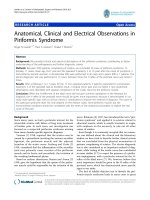
and large effective Stokes shifts [15–22]. Excitation-
emission matrix analysis has shown that QDs always emit
the same wavelength of light no matter what excitation
wavelength is used [23]. Therefore, multiple QDs with
different emission spectra can be simultaneously visualized
using a single excitation source (Fig. 1). Since the emission
spectrum of each QD is narrow, the fluorescence signal of
each QD can be readily separated and individually ana-
lyzed based on the emission spectrum in order to achieve
multiplexed imaging.
QDs and their advantageous photophysical properties
have given researchers new opportunities to explore ad-
vanced imaging techniques such as single molecule or
lifetime imaging while also providing new tools to revisit
traditional fluorescence imaging methodologies and extract
previously unobserved or inaccessible information. Given
their ability to cover nano, micro, and macro length scales,
QDs are particularly useful to study the wide range of di-
verse molecular and cellular events involved in the
pathology of diseases such as cancer. Since the first dem-
onstration of the biomedical potential of QDs in 1998 [1,
2], QD-based research has increased exponentially in re-
cent years. In less than a decade, QDs have overcome many
of the intrinsic limitations of traditional fluorophores and
become powerful tools in fields such as molecular biology,
cell biology, molecular imaging, and medical diagnostics.
The purpose of this review is to summarize and highlight
the biomedical applications of QDs to date and address
future research directions, obstacles, and potential uses of
QDs for clinical applications.
QD synthesis and conjugation strategies
QDs made directly in water often have a wide range of size
distributions while QDs synthesized at high temperature
(300 °C) in organic solvents are more monodisperse [21,
24–26]. Surface passivation by depositing an inorganic
capping layer (or shell) composed of a semiconductor
material with a wider band gap than the core material can
significantly increase the quantum yield, protect it from
oxidation, and prevent leaching of Cd or Se into the sur-
rounding solution [21, 27, 28]. Over the past decade, a
variety of procedures have been developed for synthesizing
high quality QDs, all of which are based on the initially
reported high-temperature pyrolytic reaction [25]. QDs
used in biomedical applications are colloidal nanocrystals
typically synthesized from periodic groups of II–VI (e.g.
CdSe, CdTe) or III–V (e.g. InP, InAs) including two- and
three-element systems [25–31]. Depending on the compo-
nent and size of the core, the emission peak can vary from
UV to NIR wavelengths (400–1350 nm). Over the years,
QD synthesis has become relatively simple, inexpensive,
and highly reproducible with minor complications.
QDs synthesized in organic solvents typically have
hydrophobic surface ligands [20, 21]. In order to make
them water soluble, surface functionalization with hydro-
phic ligands can be achieved in many ways [21, 32]. For a
comprehensive review, the readers are referred to ref. [21].
The first technique involves ligand exchange. The native
hydrophobic ligands are replaced by bifunctional ligands
which contain surface anchoring moieties (e.g. thiol) to
bind to the QD surface and hydrophilic end groups (e.g.
hydroxyl and carboxyl) to render water solubility [2, 33].
The second strategy employs polymerized silica shells
functionalized with polar groups to insulate the hydro-
phobic QDs [1]. While nearly all carboxy-terminated li-
gands limit QD dispersion to basic pHs [34], silica shell
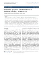
Fig. 1 (a) A series of QDs of
different core size and emission
wavelength can be excited
simultaneously by a single
excitation light source. (b)
Representative excitation (blue)
and emission (red) spectra of
QDs.
266 Nanoscale Res Lett (2007) 2:265–281
123
encapsulation provides stability over a much broader pH
range [35]. The third method maintains the native ligands
on the QDs and uses variants of amphiphilic diblock and
triblock copolymers and phospholipids to tightly interleave
the alkylphosphine ligands through hydrophobic interac-
tions [36–38]. Aside from rendering water solubility, these
surface ligands serve a critical role in insulating, passiv-
ating, and protecting the QD surface from deterioration in
biological media.
Water soluble QDs can be functionalized through a di-
verse array of conjugation strategies due to the large sur-
face area to volume ratio of QDs which provides numerous
surface attachment points for functional groups. First,
carboxylic acid groups on the QD surface can react with
amines via EDC coupling [39, 40]. This strategy has been
widely used to produce QD-streptavidin conjugates which
can then be used to attach biotinylated molecules [41, 42].
The versatility of QD-streptavidin conjugates makes them
attractive bioprobes, but the additive volume of QDs,
streptavidin, and extra layer(s) of functional molecules
limits their potential applications. The immunogenecity of
streptavidin is also a concern for applications in living
subjects [43]. EDC coupling can sometimes give interme-
diates which easily aggregate and can also make it difficult
to control the number of molecules attached to the surface
of a single QD. In an attempt to reduce the overall size of
the QD conjugate, researchers have used direct cross-
linking to attach ligands to the QD surface [44]. Second,
the amine groups on the QD surface can react with active
esters or they can be converted to maleimide (through a
heterobifunctional cross-linker) for Michael addition of a
sulfhydryl group in thiolated peptides, cysteine-tagged
proteins, or partially reduced antibodies [45]. Third, the
hydrophobic coating of QDs can be replaced with thiolated
peptides (to form thiol-bonding between sulfhydryl groups
and sulphur atoms on the QD surface) or polyhistidine-
containing proteins (histidine residues can coordinate to the
QD surface Zn atoms via metal complexation) thus en-
abling direct attachment of proteins/peptides to the QD
surface [46–50]. Finally, QD conjugation can also be
achieved via adsorption or non-covalent self-assembly
using engineered proteins [51–54].
Research has shown that a three-layer method using an
antibody against a specific target, a biotinylated secondary
antibody against the primary antibody, and a streptavidin
coated QD can effectively label target molecules with QDs
[38, 42]. This strategy is not limited to antibodies. QD-
streptavidin conjugates are commercially available and can
be used to attach virtually any biotinylated molecule to a
QD surface. Although the overall size of the resulting QD
conjugates are relatively large (>20 nm), this is not a major
concern for in vitro applications. Two of the most promi-
nent problems in QD functionalization are the lack of
homogeneity when attaching surface proteins to QDs and
the difficulty in precisely controlling the protein-to-QD
ratios. Both of these complications may result in QD
conjugates with misaligned protein orientations or large
aggregates of surface proteins which are not fully func-
tional or potentially nonfunctional. Although the biological
function of these molecules has not been severely affected
by QD conjugation in most reports, advances in conjuga-
tion strategy/chemistry are still needed in the future to
provide a robust platform for QD functionalization.
QDs for in vitro and cell-based applications
Numerous in vitro and cell-based uses have been discov-
ered for QDs because of their unique photophysical prop-
erties [22, 55–58]. QDs can be used in place of traditional
organic dyes in virtually any system and outperform dyes
in the majority of cases. The major advantage of QDs is
their strong resistance to photobleaching over long periods
of time (minutes to hours), allowing acquisition of images
with good contrast and signal intensity. Most QDs are
much brighter than organic dyes due to the combination of
higher extinction coefficients (0.5–5 · 10
6
M
–1
cm
–1
) and
higher quantum yields [20, 21]. QDs have been used in a
vast number of in vitro and cell-based applications.
Cellular labeling
In recent years, QDs have made the most progress and
drawn the greatest interest in the area of cellular labeling.
Numerous cellular components and proteins (in live or
fixed cells) have been labeled and visualized with func-
tionalized QDs, such as the nuclei, mitochondria, micro-
tubules, actin filaments, cytokeratin, endocytic
compartments, mortalin, and chaperonin proteins [51, 59–
62]. The cell membrane proteins and receptors that have
been labeled with QDs include prostate specific membrane
antigen (PSMA), HER kinases, glycine receptors, serotonin
transport proteins, p-glycoprotein, band 3 protein, and
many others [20, 21, 37, 38, 42, 44, 63–67]. The excellent
photostability of QDs is particularly useful for continuous
illumination of three dimensional (3D) optical sectioning
using confocal microscopy, where image reconstruction
and quality has been severely limited by photobleaching of
organic fluorophores [66, 68]. High sensitivity combined
with virtually an unlimited number of well-separated colors
all excitable by a single light source also makes QDs ideal
probes for multiplexed cellular imaging (a representative
example is shown in Fig. 2)[21, 68, 69].
One of the most interesting aspects of QDs for use in
immunofluorescence techniques is the small number of
QDs necessary to generate a detectable signal. A number of
Nanoscale Res Lett (2007) 2:265–281 267
123
studies have reported QD flickering in cellular specimens, a
phenomenon termed ‘‘blinking’’ [70, 71]. QD blinking has
shown that an individual QD can be observed with a sen-
sitivity limit of one QD per target molecule in immuno-
cytological conditions using current microscopy
technology. QD blinking can be overcome by passivating
the QD surface with thiol moieties [72] or by using QDs in
free suspension [73].
Cell tracking
As a result of their high photostability, QDs can be effec-
tively tracked over an extended period of time in order to
monitor cellular dynamics including movement, differen-
tiation, and fate [36, 42, 63, 74]. Large quantities of QDs
can be delivered into live cells using a variety of different
techniques such as microinjection [36], peptide-induced
transport [75], electroporation [76], and phagocytosis [63].
Once internalized, QDs can spread to daughter cells during
cell division. Lectin-coated QDs have been used to label
gram-positive bacteria and a single QD can be tracked for
several minutes as it diffuses into the membrane of live
cells and moves within the cytosol [77]. QD-peptide con-
jugates have been transfected and retained in living cells
for up to a week without detectable negative cellular ef-
fects [59]. Cellular endocytosis of QDs has also been
studied in which the endocytosis efficiency of 15 nm QD
conjugated sugar balls was compared with that of 5 nm and
50 nm particles and it was found that endocytosis was
highly size dependent [78]. All these cell tracking studies
would not have been possible to perform using traditional
organic dyes.
Fluorescent in situ hybridization (FISH)
FISH uses fluorescently labeled DNA probes for gene
mapping and identification of chromosomal abnormalities
[79, 80]. FISH allows for visualization and mapping of
cellular genetic material in order to quantify gene copy
numbers within tumor cells that have abnormal gene
amplification. DNA or oligonucleotides have been conju-
gated to QDs, and results from in vitro and cell-based as-
says have shown that these conjugates retain their ability to
form complementary sequences of Watson-Crick base pairs
[36, 81–88]. The significantly brighter and more photo-
stable fluorescence signals of QD over organic dyes can
allow for more stable and quantitative uses of FISH for
research and clinical applications (Fig. 3)[81]. It has re-
cently been reported that the fluorescence intensity of QD-
streptavidin based FISH probes varied according to the pH
of the final incubation buffer [89]. However, the exact
mechanism of this varying fluorescence has yet to be
clarified. Recently, direct multicolor imaging of multiple
subnuclear genetic sequences using QD-based FISH probes
was achieved in Escherichia coli [90].
Fluorescence resonance energy transfer (FRET)
FRET is a process in which energy is transferred from an
excited donor to an acceptor via a resonant, near-field di-
pole–dipole interaction [91]. FRET is sensitive to the dis-
tance between the donor and the acceptor on the 1–10 nm
range, a scale correlating to the size of biological macro-
molecules. FRET has been used with conventional organic
dyes and fluorescent proteins in order to monitor intracel-
lular interactions and binding events, but the results have
been suboptimal [92, 93]. QDs were first reported for
FRET applications in 1996 [94, 95], and since then,
Fig. 2 Pseudocolored fluorescence image depicting five-color QD
staining of fixed human epithelial cells. The nucleus, Ki-67 protein,
mitochondria, microtubules, actin filaments were each labeled with a
QD of different emission wavelength. From [21]
Fig. 3 Double labeling FISH using QD525 and QD585 oligonucleo-
tide probes. The same mRNA was detected with both QD525 (a) and
QD585 (b) probes. DAPI (c) staining and overlayed images (d) are
also shown. Scale bar = 20 lm. From [81]
268 Nanoscale Res Lett (2007) 2:265–281
123
numerous studies have demonstrated the use of QD-based
FRET in biological systems where QDs can be either en-
ergy donors or acceptors [48–50, 96–102].
There are two distinct advantages of using QDs as FRET
donors over organic fluorophores. First, QD emission can
be size-tuned to increase the spectral overlap with a spe-
cific acceptor dye. Second, FRET efficiency can be sig-
nificantly improved when several acceptor dyes interacting
with a single QD donor [48]. Using a 6 nm QD with a dye-
labeled protein attached to the QD surface, a FRET effi-
ciency of 22% can be obtained for a single donor–acceptor
pair [96]. Increasing the number of acceptors to five or
more can increase the FRET efficiency to 58% [48, 96].
Although FRET measurements using QDs can convey
qualitative molecular association information and appear to
have great potential as nanoscale biosensors, there are also
a number of limitations with QDs for FRET which should
be kept in mind. One major problem is the heterogeneity in
QD size which can affect the precision of single-molecule
FRET measurements unless the actual spectrum of each
individual QD can be measured. QD blinking, which is
strongly correlated with spectral jumping (changes in
emission peak position), can also significantly affect FRET
efficiency and accuracy [103]. Although QDs are superior
FRET donors compared with organic dyes, they are not
ideal FRET acceptors [104]. Red and NIR QDs are also not
optimal for FRET applications due to the long distance
between the donor and the acceptor, as well as the limited
choice of organic dyes that absorb in this region.
Additional applications of QDs
In addition to the abovementioned studies, QDs have also
been used for a variety of other purposes. Herein we
highlight some recent literature on novel uses of QDs. A
QD ‘‘peptide toolkit’’ has been constructed for the creation
of small, buffer soluble, mono-disperse peptide-coated
QDs with high colloidal stability [47]. QD-based probes
have been used for co-immunoprecipitation and Western
blot analysis, allowing for simpler and faster image
acquisition and quantification than traditional methods
(Fig. 4)[105–108]. Since QDs are both fluorescent and
electron dense, studies have investigated double- and tri-
ple-immunolabeling using light, electron, and correlated
microscopy in cells and rodent tissues [109, 110]. Cell-
penetrating QDs based on the use of multivalent and en-
dosome-disrupting surface coatings has been reported [111,
112]. Using live HeLa cells, the motion of individual ki-
nesin motors tagged with QDs has been successfully
demonstrated [113]. This study demonstrated the impor-
tance of single molecule experiments in the investigation of
intracellular transport. QD-based optical barcodes can de-
tect single nucleotide polymorpisms where the DNA se-
quences differ only at a single nucleotide [114, 115]. In
comparison with planar chips, bead-based multiplexing has
many distinct advantages such as greater statistical analy-
sis, faster assay time, and the flexibility to add additional
probes at lower costs [116]. DNA-driven QD arrays have
been investigated to utilize photogenerated currents for
optoelectronic photoelectrochemistry [117]. QDs have also
been used to track RNA interference [118], target surface
proteins in living cells [119], detect bacteria [41], and
couple with other nanoparticles such as carbon nanotubes
[120].
Over the last decade, QD-based probes have found
numerous applications where fluorescent dyes and proteins
were previously the only tools available. QDs have allowed
for complicated and difficult multiplexed cellular imaging
which was previously impossible given the limitations of
fluorescent dyes and proteins. Overall, QD-based probes
have almost completely outperformed traditional organic
dyes in in vitro and cell-based applications.
QDs for non-targeted imaging in living subjects
One of the primary goals of QD-based research is to
eventually translate QDs for use in clinical applications
such as in vivo imaging in human subjects. Modeling
studies have revealed that two spectral windows exist for
QD imaging in living subjects, one at 700–900 nm and
another at 1,200–1,600 nm [121]. QDs that emit in the NIR
region are suitable for biomedical applications because of
low tissue absorption, scattering, and autofluorescence in
this region which leads to high photon penetration in tis-
sues [122, 123]. Optically quenched NIR probes based on
fluorescent dyes have been employed to detect tumors and
have been shown to generate strong signals after enzyme
activation by tumor-associated proteases in vivo [124,
125]. NIR QDs provide a superior means to image disease
states due to their brightness and photostability in
Fig. 4 Western blot of two proteins (a & b) using two QD-antibody
conjugates. Overlay of the two images is shown in c. From [107]
Nanoscale Res Lett (2007) 2:265–281 269
123
comparison with commonly used fluorophores. For diag-
nostic purposes, the wavelength choice of NIR QDs can be
matched to the scatter of living tissue for optimal bio-
compatibility [121, 126]. Significant improvements in QD
synthesis, coating, and conjugation techniques combined
with their photostability and brightness have made QDs
invaluable tools for in vivo imaging. QDs have a large two-
photon cross-sectional efficiency 2–3 orders of magnitude
greater than that of organic dyes, thus making them well
suited for deep-tissue imaging in living subjects using two-
photon or time-gated low intensity excitation [73, 127].
Cell trafficking
Individual QDs have been encapsulated in phospholipid
block-copolymer micelles for embryo imaging [36]. Mi-
celle encapsulation resulted in great reductions in photo-
bleaching and low non-specific adsorption. After
conjugation with DNA, QDs were directly injected into
Xenopus embryos and QDs were found to be diffusely
distributed throughout the cell during early stages of
development while at later stages they mainly resided in
cell nuclei. The fluorescence signal of QDs could be fol-
lowed to the tadpole stage with little or no indication of
cytotoxicity. QDs were also reported to have high fluo-
rescent yield and robust photostability for successful
imaging of zebrafish embryos [128]. In both studies, QDs
were used as contrast agents in living organisms to dem-
onstrate the efficacy of QDs for long-term studies. These
findings have provided useful techniques in the fields of
embryology, cell biology, as well as disease phenotyping
and diagnosis.
QDs have been used as cell markers to study extrava-
sation in small animal models. QD-labeled tumor cells
were intravenously injected into live mice and there were
no distinguishable differences in behavior between the QD-
labeled tumor cells and unlabeled cells [127]. This report
successfully showed that QD-labeled tumor cells can per-
mit in vivo imaging despite tissue autofluorescence. These
QD-labeled cells could also be used to analyze the distri-
bution of tumor cells in organs and tissues and to track
different populations of cells. By using multiphoton laser
excitation, five different populations of cells have been
simultaneously identified.
Vasculature imaging
Two-photon imaging of vasculature through the skin of
living mice has been reported with water-soluble CdSe/
ZnS QDs [73]. QDs were dynamically observed in capil-
laries as deep as several hundred micrometers, and no
blinking in solution was observed on the nanosecond to
millisecond time scale. Compared to conventional methods
using 70-kD FITC-dextran, QDs provided significantly
more information at the same depth. In another report,
coronary vasculature was imaged in vivo and the effects of
tissue absorbance, scatter, and thickness on the perfor-
mance of QDs were analyzed when embedded in biological
tissue [121]. Theoretical modeling suggested that optimal
spectral windows for in vivo imaging exist at 700–900 nm
and 1200–1600 nm. Using multiphoton microscopy, QDs
can differentiate tumor vessels from perivascular cells and
matrix better than traditional fluorescently-labeled dextran
vessel markers (Fig. 5)[129]. Multiphoton microscopy
through gradient index lenses has also been used for min-
imally invasive, subcellular resolution imaging of cortical
layer V and hippocampus several millimeters deep in
anesthetized live animals [130].
NIR CdMnTe/Hg QDs have been used for deep-tissue
in vivo optical imaging [131]. QDs were grown in aqueous
solution and coated with bovine serum albumin. After ei-
ther subcutaneous or intravenous injection, these QDs were
used as angiographic contrast agents for vessels sur-
Fig. 5 Vasculature imaging with QDs. (a) Fluorescently labeled
dextran gave blurred images of tumor vessels. (b) QD imaging
yielded a sharp boundary between the vessel and interstitium. (c)
Concurrent imaging of both QD and GFP (green) provides clear
separation of the vessel from GFP-expressing perivascular cells. (d)
Vessels highlighted with QD (red) were imaged simultaneously with
the second harmonic generation signal from collagen (blue). Scale
bar = 50 lm. From [129]
270 Nanoscale Res Lett (2007) 2:265–281
123
rounding and penetrating murine squamous cell carcinoma
in mice. No significant photobleaching or degradation of
QDs was observed even after an hour of continuous exci-
tation. The stability of QDs combined with their time
resolution of optical detection makes them attractive can-
didates for pharmacokinetic imaging studies.
Visualization of blood vessels in the chick chorioallan-
toic membrane, a popular model for studying various as-
pects of blood vessel development such as angiogenesis,
was recently achieved with QDs [132]. Intravitally injected
QDs were biocompatible and stayed in circulation for over
four days without any observed deleterious effects. The
vascular residence time was adjustable through different
QD surface modifications. QDs with longer emission
wavelengths (>655 nm) virtually eliminated all chick-de-
rived autofluorescence. In comparison with FITC-dextran,
QDs were able to image vessels as well as or better than
FITC-dextran at 2–3 orders of magnitude lower concen-
tration. QDs were also fixable with low fluorescence loss,
allowing for further sample analysis when used in con-
junction with histological processing.
Lymph node mapping
Lymph node imaging with QDs has been reported in living
subjects. Type-II QDs, in which both the valence and
conduction bands in the core are lower (or higher) than
those in the shell, have tunable fluorescence emission while
preserving the absorption cross-section [133]. NIR type-II
CdTe/CdSe QDs (850 nm emission) were injected intra-
dermally into live mice and pigs [24]. These QDs rapidly
migrated to local sentinel lymph nodes (SLNs) and were
imaged virtually background-free, allowing image-guided
resection of a one centimeter deep lymph node in a pig
(Fig. 6). Imaging the lymph nodes one centimeter deep in
tissue required only 5 mW/cm
2
of excitation. This study is
the first demonstration of NIR QD-guided surgery, which
takes advantage of both the spectroscopic properties and
the relatively large size (>10 nm) of QDs. SLN mapping is
clinically important since these are the sites where meta-
static cancer cells are often found. Intraoperative SLN
mapping in various locations of the body has also been
reported in adult pigs [134–136], where only 200 pmol of
QDs was needed and these QDs quickly and accurately
mapped lymphatic drainage and SLNs. Many other SLN
mapping experiments have also been reported in mice [137,
138] and rats [139–141]. SLN mapping using QDs over-
comes the limitations of currently available methods and
provides highly sensitive, real-time image-guided dissec-
tion, which may permit potential mapping of SLNs and
lymphatic flow in patients.
Simultaneous two-color in vivo wavelength-resolved
spectral fluorescence lymphangiography using two NIR
QDs with different emission spectra has been reported
[142]. This study may provide insight into the mechanisms
of drainage from different lymphatic basins that may lead
to SLN detection of breast cancer as well as prevention of
complications such as lymphedema of the extremities.
Recently, it was demonstrated that QDs injected into model
tumors rapidly migrated to SLNs [143]. PEG-coated QDs
with terminal carboxyl, amino, or methoxyl groups all
similarly migrated from the tumor to surrounding lymph
nodes. Passage from the tumor through lymphatics to
adjacent nodes could be dynamically visualized through
the skin and at least two nodes could be typically defined.
Imaging during necropsy confirmed QD confinement to the
lymphatic system and demonstrated tagging of SLNs for
pathology. Examination of the SLNs identified by QD
localization showed that several of them contained meta-
static tumor foci.
Fig. 6 Sentinel lymph node
mapping using QDs. (a) Images
of a mouse injected
intradermally with type II NIR
QDs in the left paw. (b) Image
guided resection of a lymph
node in a pig. From [24]
Nanoscale Res Lett (2007) 2:265–281 271
123
Neural imaging
Diffusion within the extracellular space (ECS) of the brain
is necessary for chemical signaling and for neurons and
glia to access nutrients and therapeutic agents [144, 145].
Integrative optical imaging was employed to show that
water-soluble QDs diffuse within the ECS of adult rat
neocortex in vivo [146]. This report could improve the
modeling of neurotransmitter spread after spillover and
ectopic release while establishing size limits for diffusion
of drug delivery vectors such as viruses, liposomes, and
nanoparticles in brain ECS.
Intravenously injected QDs were shown to be taken up
by macrophages and localize to experimental glioma in a
rat model using optical detection [147]. Initial QD injec-
tions were performed at a concentration of 6.8 lMina
volume of 500 lL, which is more than 15 times the dose
injected for SLN mapping in a 35 kg adult pig (2,100 times
based on the animal body weight) [134–136]. It was
determined that an increase in concentration and volume
may help QDs avoid early sequestration by the reticulo-
endothelial system (RES; e.g. lymph nodes, liver, spleen,
and bone marrow [148]) and provide a better chance for
glioma uptake of QDs. Further increases in the injected QD
dose by three fold resulted in detectable fluorescence sig-
nals in the rat brain [149, 150]. Although it is suggested
that these techniques have the potential to be translated into
clinical use in humans allowing QDs to optically guide
brain tumor biopsies and resections, the enormous amount
of QDs needed to generate detectable signal in the tumor is
a major concern in terms of both toxicity and cost.
Surface coating and the in vivo behavior of QDs
Coating QDs with high molecular weight poly(ethylene
glycol) (PEG) molecules can reduce QD accumulation in
the liver and bone marrow [151]. QDs with different length
PEG coatings were tested using light and electron
microscopy on tissue sections and noninvasive whole-body
fluorescence imaging. QDs were found to remain stable in
the bone marrow and lymph nodes of animals after several
months, demonstrating their high stability without inter-
fering with normal cell physiology and cell differentiation.
Extensive study of different QD surface coatings re-
vealed important insights for future design of QD-based
probes and experimental setups [151, 152]. First, QDs are
easily visible through the skin of nude mice using NIR
QDs. The excitation wavelength is also important in
determining how deep QDs may be observed. Second,
carboxyl-coated QDs are rapidly taken up by the RES
while amino-terminal PEG-coated QDs have varying half-
lives in circulation depending on the molecular weight of
PEG. Third, when using neutral methoxy-terminated PEG
(mPEG) coating, results vary depending on the length of
the PEG and the degree of substitution. Highly substituted
QDs yielded half-lives in the 3- to 8-h range for mPEG-
5000 coated QDs (Fig. 7). Increasing the PEG size to
10 kD or 20 kD produced no further improvement in the
circulation half-life. Fourth, sites of deposition vary with
the QD surface coating. Amino-PEG, carboxy-PEG, and
mPEG-700 coated QDs are all deposited in the RES with
sites slightly varying. Deposition of uncharged PEG coated
QDs depended on the molecular size of the PEG and on the
density of substitution. Most importantly, the injected dose
of all types of QDs tested in these studies was excreted in
the feces within 1–2 days.
In vivo targeted imaging using QDs
In order to make QDs more useful for in vivo imaging and
other biomedical applications, QDs need to be effectively,
specifically, and reliably directed to a specific organ or
disease site without alteration. Specific targeting can be
obtained by attaching targeting molecules to the QD sur-
face. However, in vivo targeting and imaging is very
challenging due to the relatively large overall size (typi-
cally about 20 nm in diameter) and short circulation time
of QD conjugates. To date, there have been only a handful
of successful reports in the literature.
Peptide-conjugated QDs
The first report to demonstrate in vivo targeting of QD
conjugates employed peptides as the targeting ligands [46].
Peptide-conjugated QDs were injected intravenously into
Fig. 7 Different surface coating of QDs results in different in vivo
kinetics. mPEG-750 coated QDs circulates much shorter than mPEG-
5000 coated QDs. Even at 1 min, significant liver uptake of mPEG-
750 QDs is visible. At 1 h, mPEG-750 QDs completely cleared from
the circulation while mPEG-5000 QDs persisted. From [151]
272 Nanoscale Res Lett (2007) 2:265–281
123
MDA-MB-435 breast carcinoma xenograft-bearing nude
mice. Three peptides were tested: CGFECVRQCPERC
(denoted as GFE) which binds to membrane dipeptidase on
the endothelial cells [153, 154], KDEPQRRSARLSAK-
PAPPKPEPKPKKAPAKK (denoted as F3) which prefer-
entially binds to blood vessels and tumor cells in various
tumors [155], and CGNKRTRGC (denoted as LyP-1)
which recognizes lymphatic vessels and tumor cells in
certain tumors [156]. Since the QD used in this study emits
in the visible range which is not optimal for in vivo
imaging, ex vivo histological analysis were carried out to
show that QDs were specifically directed to the tumor
vasculature and organ targets by the surface peptide mol-
ecules. A high level of PEG substitution on the QDs was
found to be important to reduce non-selective accumulation
in the RES, thereby increasing the circulation half-life and
targeting efficiency. QD-F3 colocalizes with blood vessels
in tumor tissue and QD-LyP-1 also accumulated in tumor
tissue but did not colocalize with the blood vessel marker.
QD-F3 and QD-LyP-1 (of different emission wavelength)
injected into the same tumor mouse targeted different
structures in the tumor tissue, showing that QDs can be
targeted in vivo with a high level of specificity. This pio-
neering report first demonstrated the feasibility of specific
targeting of QD in vivo and opened up a new field of QD-
based research.
We reported the first in vivo targeted imaging of tumor
vasculature using peptide-conjugated QDs [45]. Cell
adhesion molecule integrin a
v
b
3
is highly expressed on
activated endothelial cells and tumor cells but is not readily
detectable in resting endothelial cells and most normal
organ systems [157, 158]. Previous reports have demon-
strated that integrin a
v
b
3
is an excellent tumor-related
target [157–165]. The fact that integrin a
v
b
3
is over-ex-
pressed on both tumor vasculature and tumor cells makes it
a prime target for in vivo targeted imaging using QDs, as
extravasation is not required to observe tumor signal.
Arginine–glycine–aspartic acid (RGD; potent integrin a
v
b
3
antagonist) containing peptides were conjugated to QD705
(emission maximum at 705 nm) and QD705-RGD exhib-
ited high affinity integrin a
v
b
3
specific binding in cell
culture and ex vivo. In vivo NIR fluorescence (NIRF)
imaging was carried out on athymic nude mice bearing
subcutaneous integrin a
v
b
3
-positive U87MG human glio-
blastoma tumors (Fig. 8)[45]. Tumor fluorescence inten-
sity reached a maximum at 6 h post-injection with good
contrast. The size of QD705-RGD (~20 nm) prevented
efficient extravasation, thus QD705-RGD mainly targeted
tumor vasculature instead of tumor cells. Immunofluores-
cence staining of the tumor vessels confirmed that the
majority of the QD fluorescence signal in the tumor colo-
calizes with the tumor vessels. Successful in vivo tumor
imaging using QD conjugates has introduced new per-
spectives for targeted NIRF imaging and may aid in cancer
detection and management including image-guided sur-
gery. This probe may also have great potential as a uni-
versal NIRF probe for detecting tumor vasculature in living
subjects.
Antibody-conjugated QDs
QD-based probes can be delivered to tumors through either
passive or active targeting mechanisms in living subjects.
In passive targeting, macromolecules and nanometer-sized
particles can accumulate in the tumor through enhanced
permeability and retention (EPR) effects [166, 167].
Angiogenic tumors produce vascular endothelial growth
factor [168–170], which hyperpermeabilizes tumor neo-
vasculature and causes leakage of circulating macromole-
cules and nanoparticles. Subsequent macromolecule or
nanoparticle accumulation occurs since tumors lack an
effective lymphatic drainage system.
ABC triblock copolymer-coated QDs for prostate cancer
targeting and imaging in live animals has been reported
[37]. Research has identified prostate-specific membrane
antigen (PSMA) as a cell-surface marker for both prostate
epithelial cells and neovascular endothelial cells [171].
Polymer-coated QDs were conjugated to PSMA-specific
monoclonal antibodies and it was estimated that there were
5–6 antibody molecules per QD. Using spectral imaging
techniques where fluorescence signals from QDs and
mouse autofluorescence can be separated based on the
emission spectra [172, 173], intravenously injected probe
were found to accumulate in the tumor site (Fig. 9)[37].
Multiplexed imaging was also demonstrated in live animals
using QD-labeled cancer cells. Since no histological anal-
ysis was carried out to investigate the expression level of
PSMA on the tumor cells and tumor vasculature, it was
unclear whether these QD conjugates targeted tumor vas-
culature or tumor cells. In addition, the QDs used in this
Fig. 8 RGD Peptide-conjugated QD705 successfully targets the
tumor vasculature in vivo. Mouse on the left was injected with
QD705-RGD and the mouse on the right was injected with QD705.
Arrows indicate tumors. From [45]
Nanoscale Res Lett (2007) 2:265–281 273
123
study were not optimized for tissue penetration or imaging
sensitivity because the emission wavelength was in the
visible region instead of the NIR region.
In a recent study, QDs were linked to anti-AFP (alpha-
fetoprotein, a marker for hepatocellular carcinoma cell
lines) antibody for in vivo tumor targeting and imaging
[174]. No in vitro validation of the QD probe was carried
out before the in vivo experiments. It was reported that
active tumor targeting and spectroscopic hepatoma imag-
ing was achieved using an integrated fluorescence imaging
system. The heterogeneous distribution of the QD-based
probe in the tumor was also evaluated by a site-by-site
measurement method. A major flaw of this study is that it
was not shown whether or not the anti-AFP antibody was
actually attached to the QD. Therefore, there is not enough
experimental evidence to support the conclusion that the
tumor contrast observed was from active rather than pas-
sive targeting.
Tracking a single QD conjugated with tumor-targeting
antibody in tumors of living mice was achieved using a
dorsal skinfold chamber and a high-speed confocal
microscope with a high-sensitivity camera [175]. QDs la-
beled with anti-HER2 monoclonal antibody were injected
into mice bearing HER2-overexpressing breast cancer to
analyze the molecular processes of its tumor delivery.
Movement of a single QD-antibody conjugate (total num-
ber of QD particles injected was ~1.2 · 10
14
) was observed
at 30 frames per second inside the tumor through the dorsal
skinfold chamber. QDs were observed during six processes
of delivery: in a blood vessel, during extravasation, in the
extracellular region, binding to HER2 on the cell mem-
brane, moving from the cell membrane to the perinuclear
region, and in the perinuclear region. The movement of the
QD-antibody conjugate at each stage followed a ‘‘stop and
go’’ pattern. Despite the technical difficulty of this exper-
iement, no information was obtained regarding the per-
centage of intravenously injected QDs that extravasated.
Therefore, little can be concluded about the overall
behavior of such QD-antibody conjugates in vivo. It was
unclear whether the ‘‘stop and go’’ pattern is typical for the
majority of injected QD conjugates or if it is only limited to
a small subset of QDs. It is likely that the majority of the
QD conjugates were taken up by the RES shortly after
injection and that only certain QD conjugates such as the
smallest particles actually extravasated.
Recent advances in QD technology
Bioluminescence resonance energy transfer (BRET)
QDs have shown great potential for molecular imaging and
cellular investigations of biological processes. However,
the requirement for external light excitation can partially
offset the favorable tissue penetration properties of NIR
QDs. This type of excitation also results in significantly
increased background autofluorescence. The use of direct
bioluminescence light to excite QDs has partially over-
come this problem [176]. Luciferases have been widely
used as reporter genes in biological research [177, 178].
However, the bioluminescence activity of commonly used
luciferases is too labile in serum. Specific mutations of
Renilla luciferase, selected using a consensus sequence
driven strategy, were screened for their ability to confer
stability of activity in serum as well as their light output
[179]. A mutant Renilla luciferase with eight mutations
(RLuc8) was selected with a 200-fold increase in resistance
to inactivation in murine serum and a 4-fold increase in
light output. Multiple molecules of RLuc8 were covalently
conjugated to a single fluorescent QD, forming a conjugate
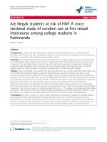
about 22 nm in hydrodynamic diameter (Fig. 10)[176].
When RLuc8 bound its substrate coelenterazine, it con-
verted chemical energy into photon energy and emitted
broad spectrum blue light peaking at 480 nm. Due to the
complete overlap of the RLuc8 emission and QD absorp-
tion spectra, QDs were efficiently excited in the absence of
external light. In vivo imaging showed greatly enhanced
signal-to-background ratio after injection of the QD-RLuc8
conjugate into the blood stream. RLuc8 can serve as a
Fig. 9 Antibody-conjugated QDs for in vivo cancer targeting and
imaging. Mouse on the left was a control. From [37]
Fig. 10 Self-illuminating QDs based on bioluminescence resonance
energy transfer. From [176]
274 Nanoscale Res Lett (2007) 2:265–281
123
BRET donor for virtually any QDs and these probes can be
used for multiplexed imaging. BRET has the potential to
greatly improve NIRF detection in living tissue and similar
QD conjugates can be obtained when RLuc8 is fused to
other proteins, thus enabling new possibilities for imaging
biological events [180]. One of the major goals BRET will
have to achieve before it can be widely used for in vivo
imaging is targeting specificity. Since there are many
RLuc8 molecules on the QD surface which cover the
majority of the QD surface area, it remains to be tested
whether there will be enough space left to attach enough
ligands for desirable targeting efficacy.
Non-Cd based QDs
There have been many serious questions and concerns
raised regarding the cytotoxicity of inorganic QDs con-
taining Cd, Se, Zn, Te, Hg, and Pb [181–184]. These
chemicals can be potent toxins, neurotoxins, and/or terat-
ogens depending on the dosage, complexation, and accu-
mulation in the liver and the nervous system. At very low
doses, these metals are bound by metallothionein proteins
and may be excreted slowly or sequestered in vivo in
adipose and other tissues [181, 185]. Cadmium has a half-
life of about 20 years in humans and it is a suspected
carcinogen that can accumulate in the liver, kidney, and
many other tissues since there is no known active mecha-
nism to excrete cadmium from the human body [186].
Although many studies have found no adverse effects of
QDs on cell viability, morphology, function, or develop-
ment over the duration of experiments (hours to days) at
concentrations optimized for labeling efficiency [36, 38,
63, 127], the cellular toxicity of QDs under extreme con-
ditions such as photo-oxidation and strong UV excitation
has been clearly demonstrated [185, 187]. In general, the
less protected the QD core or core/shell is, the sooner the
appearance of signs of interference with cell viability or
function as a result of Cd
2+
and/or Se
2–
release. Thick ZnS
overcoating (4–6 monolayers) in combination with effi-
cient surface capping has been shown to substantially re-
duce desorption of core ions and make QDs more
biologically inert [185]. Interestingly, the toxicity of QDs
has been utilized for photodynamic therapy applications
such as tumor ablation [188, 189]. As QD technology
evolves and brighter probes are created with improved
detection efficiency, the easiest way to decrease cytotoxic
effects would be to use lower quantities of QDs. In many
cases, the amount of free Cd
2+
ions released by QDs is far
below the dose needed to cause cadmium poisoning in
animal models.
InAs-based QDs can be a substitute for Cd-based QDs
with lower cytotoxicity [138, 190, 191]. The amount of As
used is estimated to be hundreds of times lower than the
dose of As
2
O
3
used to treat human leukemia. Mn- or Cu-
doped zinc chalcogenide QDs have been reported and can
cover a similar emission window as that of CdSe QDs [192,
193]. Besides the low toxicity by replacing Cd with Zn,
such QDs are also less sensitive to environmental changes
such as thermal, chemical, and photochemical distur-
bances. These doped QDs have color-tunability with good
quantum efficiency and are promising candidates for future
efforts to lower QD-based cytotoxicity. They also have
narrow emission spectra (45–65 nm full width at half
maximum) and can cover most of the visible spectral
window. In the near future, it is expected that doped QDs
that emit in the NIR region will be developed. Extensive
scrutiny and research into the toxicity profiles will be
needed before QDs can be employed in any medical pro-
cedures. In addition, further studies are also needed to
investigate the clearance mechanism of QDs from living
systems.
Moving towards smaller QDs
For inorganic nanoparticles such as QDs, the particle size
and shape is relatively rigid compared to other organic
nanoparticles such as dendrimers. To date, most of the QDs
evaluated in vivo are 15 nm or more in hydrodynamic
diameter. Although tumor vasculature is typically quite
leaky, such size does not permit efficient extravasation. It is
expected that with smaller sizes, QDs will extravasate more
efficiently and give more efficient in vivo targeting of both
tumor vasculature and tumor cells. Smaller sized QDs are
also expected to have lower RES uptake which will
translate into better image quality. Different core/shell
structures and thinner polymer coating has been reported to
reduce the overall size of QDs [137, 193], and unusually
small, water soluble QDs composed of InAs/ZnSe (core
diameter < 2 nm) have been developed [137]. Although
these QDs have lower quantum yield (<10%), the smaller
size is attractive for imaging applications. These unusually
small QDs were not trapped in SLNs, bur rather they mi-
grated into the lymphatic system and the channels between
the nodes. In addition, these small QDs could also migrate
out of the blood vessels and into the interstitial fluid.
Dendron-coated QDs have high stability, versatility, and
chemical/biochemical proccessibility [194, 195]. Unlike
the typical polymer coating, dendron-ligands are tight and
small in radial dimension, resulting in an overall smaller
size of QDs (Fig. 11). The surface density and length of the
PEG units on the outer surface of the resulting dendron-
coated QDs can be varied by synthesizing dendron ligands
with different terminal structures. A ‘‘peptide toolkit’’ has
been reported which can provide a straightforward means
for improving biocompatibility for cell biology and in vivo
applications [47]. In the future, it is likely that small
Nanoscale Res Lett (2007) 2:265–281 275
123
molecule or peptide-coated QDs will have better opportu-
nities for development and expansion in in vivo applica-
tions than protein or antibody-conjugated QDs.
Multifunctional probes
Among all of the molecular imaging modalities currently
available, no single modality is perfect and sufficient to
obtain all the necessary information [196]. Due to the
current obstacles in fluorescence tomography [197–199], it
is difficult to adequately quantify QD signal in living
subjects based on fluorescence intensity alone, particularly
in deep tissues. Combining QD-based imaging with 3D
tomography techniques such as positron emission tomog-
raphy (PET), single photon emission computed tomogra-
phy (SPECT), and magnetic resonance imaging (MRI) can
permit the elucidation of targeting mechanisms, biodistri-
bution, and dynamics in living animals with higher sensi-
tivity and/or accuracy. One of the most promising
applications for QDs is the development of multifunctional
QD-based probes for multimodality molecular imaging
in vitro and in vivo. A multimodality approach would
make it possible to image targeted QDs at all scales, from
whole-body down to nanometer resolution, using a single
probe.
A series of core/shell CdSe/Zn
1–x
Mn
x
S nanoparticles
have been synthesized for use in both optical imaging and
MRI [200]. Mn
2+
content was in the range of 0.6–6.2% and
varies with the thickness of the shell or amount of Mn
2+
introduced to the reaction. The quantum yield and Mn
2+
concentration in the nanoparticles were sufficient to pro-
duce contrast for both modalities at a relatively low con-
centration. Bifunctional nanocomposite systems consisting
of Fe
2
O
3
magnetic nanoparticles and CdSe QDs have been
synthesized [201]. QDs can be coated with paramagnetic
and pegylated lipids for use as detectable and targeted
probes with MRI [202]. These QDs are useful as dualmo-
dality contrast agents due to their high relaxivity and
ability to retain their optical properties. Several other QD-
based probes for both fluorescence imaging and MRI have
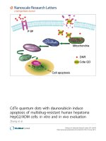
also been reported (Fig. 12)[202–204]. Polymer-coated
Fe
2
O
3
cores overcoated with a CdSe-ZnS QD shell and
functionalized with antibodies have been used to magnet-
ically capture breast cancer cells and view them with flu-
orescence imaging [205]. Magnetic QDs composed of
CdS-FePt have also been synthesized [206].
QDs have relatively large surface areas which can be
conjugated with more than one targeting ligand. Novel
tumor-specific antibody fragments, growth factors, pep-
tides, and small molecules can be attached to QDs for the
delivery of QDs to tumors in vivo for multi-parameter
imaging of biomarkers, with the ultimate goal of guiding
therapy selection and predicting response to therapy. This
nano-platform approach will enable detection and mea-
surement of many biomarkers simultaneously which may
lead to better signal/contrast than QDs modified with only
one type of targeting ligand. The ability to accurately as-
sess the pharmacokinetics and tumor targeting efficacy of
the biologically modified QDs is of crucial importance to
assess future multitargeting (to target multiple targets with
the same QD) and eventually multiplexing (to target mul-
tiple targets simultaneously using QDs of different emis-
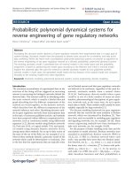
sion wavelengths) studies. Dualmodality PET/NIRF
imaging probe offers synergistic advantages over the single
modality imaging probe by overcoming the difficulty of
quantifying fluorescence intensity in vivo and ex vivo. For
the first time, we quantitatively evaluated the tumor tar-
geting efficacy of dualfunctional QD-based probes using
both NIRF and PET imaging (Fig. 13)[207]. Both RGD
peptides and macrocyclic chelator DOTA were conjugated
to QD705. RGD peptides can allow for integrin a
v
b
3
tar-
geting and DOTA can complex
64
Cu (a positron emitter
with 12.7 h half-life) to enable PET imaging [160, 208,
209]. Non-invasive PET imaging using radiolabeled QD
conjugates can provide a robust and reliable measure of the
in vivo biodistribution of QDs. With further improvement
in QD technology, it is expected that accurate evaluation of
the in vivo tumor targeting efficacy using quantitative
imaging modalities (e.g. PET) will greatly facilitate future
biomedical applications of QDs. Such information will also
be critical for fluorescence-guided surgery by sensitive,
Fig. 11 Schematic illustration of the formation of dendron-coated
QDs. From [195]
Fig. 12 A QD-based probe for both fluorescence imaging and MRI.
From [204]
276 Nanoscale Res Lett (2007) 2:265–281
123
specific, and real-time intraoperative visualization of
molecular features of normal and disease processes.
Conclusion and perspectives
QDs as biological probes have lived up to much of their
initially promoted potential for in vitro and in vivo imag-
ing. Since the first demonstration of QDs for biological
applications [1, 2], numerous breakthroughs in QD tech-
nology have led to the recent success of in vivo targeted
imaging of QDs in live animals [37, 45]. Future develop-
ment of improved QD-based biological probes for in vivo
optical imaging is promising for both basic science and
clinical applications.
Nanotechnology has the potential to significantly impact
cancer diagnosis and cancer patient management. QD-
based ex vivo protein nanosensors (e.g. FISH, FRET) and
in vivo imaging are both critical for future optimization in
cancer management. Ex vivo diagnostics in combination
with in vivo diagnostics can markedly impact future cancer
patient management by providing a synergistic approach
that neither strategy can provide alone. After further
development and validation, QD-based approaches (both
ex vivo nanosensor and in vivo imaging) will eventually be
able to predict which patients will likely respond to a
specific anticancer therapy and monitor their response to
personalized therapy (Fig. 14). With their capacity to
provide enormous sensitivity, throughput, and flexibility,
QDs have the potential to profoundly impact cancer patient
management in the future.
QD-based tumor imaging in mice can not be directly
scaled up to in vivo imaging in human applications due to
limited optical signal penetration depth. In clinical settings,
optical imaging is relevant for tissues close to the surface
of the skin, tissues accessible by endoscopy, and intraop-
erative visualization. NIR optical imaging devices for
detecting and diagnosing breast cancer have been tested in
patients and the initial results are encouraging [210, 211].
Multiple wavelength QDs emitting in the NIR region can
Fig. 13 Dualfunctional QD-
based probe for both PET and
NIRF imaging. (a) PET image
of harvested major organs/
tissues at 5 h post-injection of
the dualfunctional probe. (b)
NIRF image of harvested major
organs/tissues at 5 h post-
injection of the probe. (c)
Immunofluorescence staining of
the tumor tissue revealed that
QDs are targeting the tumor
vasculature. From [207]
Fig. 14 Patients can have their tumors biopsied and blood samples
drawn for protein profiling by ex vivo nanosensors to predict their
response to a given therapy. In addition, they will also be imaged with
molecular imaging probes of different types to predict their response.
Post-treatment and potentially during treatment, patient response will
be evaluated by blood analysis and molecular imaging to ensure the
accurate differentiation of responders from non-responders.
Nanoscale Res Lett (2007) 2:265–281 277
123
allow for multiplexed imaging of deeper tissues, thus sig-
nificantly extending potential human applications. QD-
based multitarget imaging can also play an important role
in optically guided surgery in the future. Overall, the major
roadblocks for clinical translation of QDs are inefficient
delivery, toxicity, and lack of quantification. However,
with the development of smaller non-Cd based multifunc-
tional QDs and further improvement on conjugation strat-
egy, it is expected that QDs will achieve optimal tumor
targeting efficacy with acceptable toxicity profile for clin-
ical translation in the near future using either NIRF
imaging alone or multimodality imaging.
Acknowledgements The authors would like to thank the National
Institute of Biomedical Imaging and Bioengineering (NIBIB) (R21
EB001785), NCI (R21 CA102123, P50 CA114747, U54 CA119367,
R24 CA93862), Department of Defense (DOD) (W81XWH-04–1-
0697, W81XWH-06-1-0665, W81XWH-06-1-0042, W81XWH-07-1-
0374, DAMD17-03-1-0143), Benedict Cassen Postdoctoral Fellow-
ship from the Education and Research Foundation of the Society of
Nuclear Medicine (to W.C.), and Stanford University School of
Medicine Medical Scholars Program (to A.R.H.).
References
1. M. Bruchez Jr., M. Moronne, P. Gin, S. Weiss, A.P. Alivisatos,
Science 281, 2013 (1998)
2. W.C. Chan, S. Nie, Science 281, 2016 (1998)
3. Z. Liu, W. Cai, L. He, N. Nakayama, K. Chen, X. Sun, X. Chen,
H. Dai, Nat. Nanotechnol. 2, 47 (2007)
4. Y. Cui, C.M. Lieber, Science 291, 851 (2001)
5. L.R. Hirsch, R.J. Stafford, J.A. Bankson, S.R. Sershen, B. Ri-
vera, R.E. Price, J.D. Hazle, N.J. Halas, J.L. West, Proc. Natl.
Acad. Sci. USA 100, 13549 (2003)
6. P. Grodzinski, M. Silver, L.K. Molnar, Expert Rev. Mol. Diagn.
6, 307 (2006)
7. A.G. Cuenca, H. Jiang, S.N. Hochwald, M. Delano, W.G.
Cance, S.R. Grobmyer, Cancer 107, 459 (2006)
8. M. Ferrari, Nat. Rev. Cancer 5, 161 (2005)
9. E.S. Kawasaki, A. Player, Nanomedicine 1, 101 (2005)
10. A. Miyawaki, A. Sawano, T. Kogure, Nat. Cell Biol. Suppl, S1
(2003)
11. Al.L. Efros, A.L. Efros, Sov. Phys. Semicond. 16, 772 (1982)
12. A.I. Ekimov, A.A. Onushchenko, Sov. Phys. Semicond. 16, 775
(1982)
13. V.V. Milanovic, Z. Ikonic, Phys. Rev. B 39, 7982 (1989)
14. W.C. Chan, D.J. Maxwell, X. Gao, R.E. Bailey, M. Han, S. Nie,
Curr. Opin. Biotechnol. 13, 40 (2002)
15. B.O. Dabbousi, J. RodriguezViejo, F.V. Mikulec, J.R. Heine, H.
Mattoussi, R. Ober, K.F. Jensen, M.G. Bawendi, J. Phys. Chem.
B 101, 9463 (1997)
16. M. Dahan, T. Laurence, F. Pinaud, D.S. Chemla, A.P. Alivisa-
tos, M. Sauer, S. Weiss, Optics Lett. 26, 825 (2001)
17. X. Michalet, F. Pinaud, T.D. Lacoste, M. Dahan, M.P. Bruchez,
A.P. Alivisatos, S. Weiss, Single Mol. 2, 261 (2001)
18. P. Alivisatos, Nat. Biotechnol. 22, 47 (2004)
19. C.J. Murphy, Anal. Chem. 74, 520A (2002)
20. X. Michalet, F.F. Pinaud, L.A. Bentolila, J.M. Tsay, S. Doose,
J.J. Li, G. Sundaresan, A.M. Wu, S.S. Gambhir, S. Weiss, Sci-
ence 307, 538 (2005)
21. I.L. Medintz, H.T. Uyeda, E.R. Goldman, H. Mattoussi, Nat.
Mater. 4, 435 (2005)
22. Z.B. Li, W. Cai, X. Chen, J. Nanosci. Nanotechnol. 7, in press
(2007)
23. A.J. Nozik, Annu. Rev. Phys. Chem. 52, 193 (2001)
24. S. Kim, Y.T. Lim, E.G. Soltesz, A.M. De Grand, J. Lee, A.
Nakayama, J.A. Parker, T. Mihaljevic, R.G. Laurence, D.M.
Dor, L.H. Cohn, M.G. Bawendi, J.V. Frangioni, Nat. Biotech-
nol. 22, 93 (2004)
25. C.B. Murray, D.J. Norris, M.G. Bawendi, J. Am. Chem. Soc.
115, 8706 (1993)
26. Z.A. Peng, X. Peng, J. Am. Chem. Soc. 123, 183 (2001)
27. M.A. Hines, P. Guyotsionnest, J. Phys. Chem. 100, 468 (1996)
28. X.G. Peng, M.C. Schlamp, A.V. Kadavanich, A.P. Alivisatos, J.
Am. Chem. Soc. 119, 7019 (1997)
29. R.E. Bailey, S. Nie, J. Am. Chem. Soc. 125
, 7100 (2003)
30. J.M. Tsay, M. Pflughoefft, L.A. Bentolila, S. Weiss, J. Am.
Chem. Soc. 126, 1926 (2004)
31. X. Peng, L. Manna, W. Yang, J. Wickham, E. Scher, A. Kad-
avanich, A.P. Alivisatos, Nature 404, 59 (2000)
32. W.W. Yu, E. Chang, R. Drezek, V.L. Colvin, Biochem. Bio-
phys. Res. Commun. 348, 781 (2006)
33. H.T. Uyeda, I.L. Medintz, J.K. Jaiswal, S.M. Simon, H. Mat-
toussi, J. Am. Chem. Soc. 127, 3870 (2005)
34. W.J. Parak, Nanotechnology 14, R15 (2003)
35. T. Nann, P. Mulvaney, Angew. Chem. Int. Ed. Engl. 43, 5393
(2004)
36. B. Dubertret, P. Skourides, D.J. Norris, V. Noireaux, A.H.
Brivanlou, A. Libchaber, Science 298, 1759 (2002)
37. X. Gao, Y. Cui, R.M. Levenson, L.W.K. Chung, S. Nie, Nat.
Biotechnol. 22, 969 (2004)
38. X. Wu, H. Liu, J. Liu, K.N. Haley, J.A. Treadway, J.P. Larson,
N. Ge, F. Peale, M.P. Bruchez, Nat. Biotechnol. 21, 41 (2003)
39. J.C. Sheehan, P.A. Cruickshank, G.L. Boshart, J. Org. Chem. 26,
2525 (1961)
40. M. Bodanszky, Pept. Res. 5, 134 (1992)
41. R. Edgar, M. McKinstry, J. Hwang, A.B. Oppenheim, R.A.
Fekete, G. Giulian, C. Merril, K. Nagashima, S. Adhya, Proc.
Natl. Acad. Sci. USA 103, 4841 (2006)
42. M. Dahan, S. Levi, C. Luccardini, P. Rostaing, B. Riveau, A.
Triller, Science 302, 442 (2003)
43. D. Marshall, R.B. Pedley, J.A. Boden, R. Boden, R.G. Melton,
R.H. Begent, Br. J. Cancer 73, 565 (1996)
44. S.J. Rosenthal, I. Tomlinson, E.M. Adkins, S. Schroeter, S.
Adams, L. Swafford, J. McBride, Y. Wang, L.J. DeFelice, R.D.
Blakely, J. Am. Chem. Soc. 124, 4586 (2002)
45. W. Cai, D.W. Shin, K. Chen, O. Gheysens, Q. Cao, S.X. Wang,
S.S. Gambhir, X. Chen, Nano Lett. 6, 669 (2006)
46. M.E. Akerman, W.C.W. Chan, P. Laakkonen, S.N. Bhatia, E.
Ruoslahti, Proc. Natl. Acad. Sci. USA 99, 12617 (2002)
47. F. Pinaud, D. King, H P. Moore, S. Weiss, J. Am. Chem. Soc.
126, 6115 (2004)
48. A.R. Clapp, I.L. Medintz, J.M. Mauro, B.R. Fisher, M.G.
Bawendi, H. Mattoussi, J. Am. Chem. Soc. 126, 301 (2004)
49. I.L. Medintz, S.A. Trammell, H. Mattoussi, J.M. Mauro, J. Am.
Chem. Soc. 126, 30 (2004)
50. I.L. Medintz, J.H. Konnert, A.R. Clapp, I. Stanish, M.E. Twigg,
H. Mattoussi, J.M. Mauro, J.R. Deschamps, Proc. Natl. Acad.
Sci. USA 101, 9612 (2004)
51. K. Hanaki, A. Momo, T. Oku, A. Komoto, S. Maenosono, Y.
Yamaguchi, K. Yamamoto, Biochem. Biophys. Res. Commun.
302, 496 (2003)
52. E.R. Goldman, G.P. Anderson, P.T. Tran, H. Mattoussi, P.T.
Charles, J.M. Mauro, Anal. Chem. 74, 841 (2002)
53. H. Mattoussi, J. Am. Chem. Soc. 122, 12142 (2000)
278 Nanoscale Res Lett (2007) 2:265–281
123
54. H. Mattoussi, J.M. Mauro, E.R. Goldman, T.M. Green, G.P.
Anderson, Phys. Status Solidi. B-Basic Res. 224, 277 (2001)
55. A.P. Alivisatos, W. Gu, C. Larabell, Annu. Rev. Biomed. Eng.
7, 55 (2005)
56. F. Pinaud, X. Michalet, L.A. Bentolila, J.M. Tsay, S. Doose, J.J.
Li, G. Iyer, S. Weiss, Biomaterials 27, 1679 (2006)
57. A.M. Smith, G. Ruan, M.N. Rhyner, S. Nie, Ann. Biomed. Eng.
34, 3 (2006)
58. M.P. Bruchez, Curr. Opin. Chem. Biol. 9, 533 (2005)
59. F. Chen, D. Gerion, Nano Lett. 4, 1827 (2004)
60. Z. Kaul, T. Yaguchi, S.C. Kaul, T. Hirano, R. Wadhwa, K.
Taira, Cell Res. 13, 503 (2003)
61. A. Mansson, M. Sundberg, M. Balaz, R. Bunk, I.A. Nicholls, P.
Omling, S. Tagerud, L. Montelius, Biochem. Biophys. Res.
Commun. 314, 529 (2004)
62. D. Ishii, K. Kinbara, Y. Ishida, N. Ishii, M. Okochi, M. Yohda,
T. Aida, Nature 423, 628 (2003)
63. J.K. Jaiswal, H. Mattoussi, J.M. Mauro, S.M. Simon, Nat. Bio-
technol. 21, 47 (2003)
64. D.S. Lidke, P. Nagy, R. Heintzmann, D.J. Arndt-Jovin, J.N.
Post, H.E. Grecco, E.A. Jares-Erijman, T.M. Jovin, Nat. Bio-
technol. 22, 198 (2004)
65. A. Sukhanova, J. Devy, L. Venteo, H. Kaplan, M. Artemyev, V.
Oleinikov, D. Klinov, M. Pluot, J.H. Cohen, I. Nabiev, Anal.
Biochem. 324, 60 (2004)
66. F. Tokumasu, J. Dvorak, J. Microsc. 211, 256 (2003)
67. O. Minet, C. Dressler, J. Beuthan, J. Fluoresc. 14, 241 (2004)
68. T.D. Lacoste, X. Michalet, F. Pinaud, D.S. Chemla, A.P. Ali-
visatos, S. Weiss, Proc. Natl. Acad. Sci. USA 97, 9461 (2000)
69. A.M. Smith, S. Dave, S. Nie, L. True, X. Gao, Expert Rev. Mol.
Diagn. 6, 231 (2006)
70. R.G. Neuhauser, K.T. Shimizu, W.K. Woo, S.A. Empedocles,
M.G. Bawendi, Phys. Rev. Lett. 85, 3301 (2000)
71. J. Yao, D.R. Larson, H.D. Vishwasrao, W.R. Zipfel, W.W.
Webb, Proc. Natl. Acad. Sci. USA 102, 14284 (2005)
72. S. Hohng, T. Ha, J. Am. Chem. Soc. 126, 1324 (2004)
73. D.R. Larson, W.R. Zipfel, R.M. Williams, S.W. Clark, M.P.
Bruchez, F.W. Wise, W.W. Webb, Science 300, 1434 (2003)
74. T. Pellegrino, W.J. Parak, R. Boudreau, M.A. Le Gros, D. Ge-
rion, A.P. Alivisatos, C.A. Larabell, Differentiation 71, 542
(2003)
75. L.C. Mattheakis, J.M. Dias, Y.J. Choi, J. Gong, M.P. Bruchez, J.
Liu, E. Wang, Anal. Biochem. 327, 200 (2004)
76. S. Ramachandran, N.E. Merrill, R.H. Blick, D.W. van der We-
ide, Biosens. Bioelectron. 20, 2173 (2005)
77. J.A. Kloepfer, R.E. Mielke, M.S. Wong, K.H. Nealson, G.
Stucky, J.L. Nadeau, Appl. Environ. Microbiol. 69, 4205 (2003)
78. F. Osaki, T. Kanamori, S. Sando, T. Sera, Y. Aoyama, J. Am.
Chem. Soc. 126, 6520 (2004)
79. J. Nath, K.L. Johnson, Biotech. Histochem. 73, 6 (1998)
80. J. Nath, K.L. Johnson, Biotech. Histochem. 75, 54 (2000)
81. P. Chan, T. Yuen, F. Ruf, J. Gonzalez-Maeso, S.C. Sealfon,
Nucleic Acids Res. 33, e161 (2005)
82. D. Gerion, W.J. Parak, S.C. Williams, D. Zanchet, C.M. Mic-
heel, A.P. Alivisatos, J. Am. Chem. Soc. 124, 7070 (2002)
83. J.R. Lakowicz, I. Gryczynski, Z. Gryczynski, K. Nowaczyk, C.J.
Murphy, Anal. Biochem. 280, 128 (2000)
84. R. Mahtab, H.H. Harden, C.J. Murphy, J. Am. Chem. Soc. 122,
14 (2000)
85. G.P. Mitchell, C.A. Mirkin, R.L. Letsinger, J. Am. Chem. Soc.
121, 8122 (1999)
86. W.J. Parak, R. Boudreau, M. Le Gros, D. Gerion, D. Zanchet,
C.M. Micheel, S.C. Williams, A.P. Alivisatos, C. Larabell, Adv.
Mater. 14, 882 (2002)
87. S. Pathak, S.K. Choi, N. Arnheim, M.E. Thompson, J Am Chem
Soc 123, 4103 (2001)
88. Y. Xiao, P.E. Barker, Nucleic Acids Res. 32, e28 (2004)
89. Y. Xiao, W.G. Telford, J.C. Ball, L.E. Locascio, P.E. Barker,
Nat. Methods 2, 723 (2005)
90. S.M. Wu, X. Zhao, Z.L. Zhang, H.Y. Xie, Z.Q. Tian, J. Peng,
Z.X. Lu, D.W. Pang, Z.X. Xie, Chemphyschem 7, 1062 (2006)
91. L. Stryer, Annu. Rev. Biochem. 47, 819 (1978)
92. E.A. Jares-Erijman, T.M. Jovin, Nat. Biotechnol. 21, 1387
(2003)
93. E.A. Jares-Erijman, T.M. Jovin, Curr. Opin. Chem. Biol. 10, 409
(2006)
94. C.R. Kagan, C.B. Murray, M.G. Bawendi, Phys. Rev. B 54,
8633 (1996)
95. C.R. Kagan, C.B. Murray, M. Nirmal, M.G. Bawendi, Phys.
Rev. Lett. 76, 1517 (1996)
96. I.L. Medintz, A.R. Clapp, H. Mattoussi, E.R. Goldman, B.
Fisher, J.M. Mauro, Nat. Mater. 2, 630 (2003)
97. Y. Nagasaki, T. Ishii, Y. Sunaga, Y. Watanabe, H. Otsuka, K.
Kataoka, Langmuir 20, 6396 (2004)
98. E. Oh, M.Y. Hong, D. Lee, S.H. Nam, H.C. Yoon, H.S. Kim, J.
Am. Chem. Soc. 127, 3270 (2005)
99. F. Patolsky, R. Gill, Y. Weizmann, T. Mokari, U. Banin, I.
Willner, J. Am. Chem. Soc. 125, 13918 (2003)
100. D.M. Willard, L.L. Carillo, J. Jung, A. van Orden, Nano Lett. 1,
469 (2001)
101. A.R. Clapp, I.L. Medintz, H. Mattoussi, Chemphyschem 7,47
(2006)
102. I.L. Medintz, A.R. Clapp, F.M. Brunel, T. Tiefenbrunn, H. Te-
tsuo Uyeda, E.L. Chang, J.R. Deschamps, P.E. Dawson, H.
Mattoussi, Nat. Mater. 5, 581 (2006)
103. S. Hohng, T. Ha, Chemphyschem 6, 956 (2005)
104. A.R. Clapp, I.L. Medintz, B.R. Fisher, G.P. Anderson, H.
Mattoussi, J. Am. Chem. Soc. 127, 1242 (2005)
105. H.Y. Liu, T.Q. Vu, Nano Lett. 7, 1044 (2007)
106. R. Bakalova, Z. Zhelev, H. Ohba, Y. Baba, J. Am. Chem. Soc.
127, 9328 (2005)
107. R.L. Ornberg, T.F. Harper, H. Liu, Nat. Methods 2
, 79 (2005)
108. S.C. Makrides, C. Gasbarro, J.M. Bello, Biotechniques 39, 501
(2005)
109. B.N. Giepmans, T.J. Deerinck, B.L. Smarr, Y.Z. Jones, M.H.
Ellisman, Nat. Methods 2, 743 (2005)
110. R. Nisman, G. Dellaire, Y. Ren, R. Li, D.P. Bazett-Jones, J.
Histochem. Cytochem. 52, 13 (2004)
111. Y. Zhang, M.K. So, J. Rao, Nano Lett. 6, 1988 (2006)
112. H. Duan, S. Nie, J. Am. Chem. Soc. 129, 3333 (2007)
113. S. Courty, C. Luccardini, Y. Bellaiche, G. Cappello, M. Dahan,
Nano Lett. 6, 1491 (2006)
114. M. Han, X. Gao, J.Z. Su, S. Nie, Nat. Biotechnol. 19, 631
(2001)
115. H. Xu, M.Y. Sha, E.Y. Wong, J. Uphoff, Y. Xu, J.A. Treadway,
A. Truong, E. O’Brien, S. Asquith, M. Stubbins, N.K. Spurr,
E.H. Lai, W. Mahoney, Nucleic Acids Res. 31, e43 (2003)
116. S.J. Rosenthal, Nat. Biotechnol. 19, 621 (2001)
117. E. Katz, I. Willner, Angew. Chem. Int. Ed. Engl. 43, 6042
(2004)
118. A.A. Chen, A.M. Derfus, S.R. Khetani, S.N. Bhatia, Nucleic
Acids Res. 33, e190 (2005)
119. M. Howarth, K. Takao, Y. Hayashi, A.Y. Ting, Proc. Natl.
Acad. Sci. USA 102, 7583 (2005)
120. M. Olek, T. Busgen, M. Hilgendorff, M. Giersig, J. Phys. Chem.
B Condens. Matter Mater. Surf. Interfaces Biophys. 110, 12901
(2006)
121. Y.T. Lim, S. Kim, A. Nakayama, N.E. Stott, M.G. Bawendi, J.V.
Frangioni, Mol. Imaging 2, 50 (2003)
122. J.V. Frangioni, Curr. Opin. Chem. Biol. 7, 626 (2003)
123. G. Reich, Adv. Drug. Deliv. Rev. 57, 1109 (2005)
124. C. Bremer, C.H. Tung, R. Weissleder, Nat. Med. 7, 743 (2001)
Nanoscale Res Lett (2007) 2:265–281 279
123
125. R. Weissleder, C.H. Tung, U. Mahmood, A. Bogdanov Jr., Nat.
Biotechnol. 17, 375 (1999)
126. A.E. Cerussi, A.J. Berger, F. Bevilacqua, N. Shah, D. Jaku-
bowski, J. Butler, R.F. Holcombe, B.J. Tromberg, Acad. Radiol.
8, 211 (2001)
127. E.B. Voura, J.K. Jaiswal, H. Mattoussi, S.M. Simon, Nat. Med.
10, 993 (2004)
128. S. Rieger, R.P. Kulkarni, D. Darcy, S.E. Fraser, R.W. Koster,
Dev. Dyn. 234, 670 (2005)
129. M. Stroh, J.P. Zimmer, D.G. Duda, T.S. Levchenko, K.S. Co-
hen, E.B. Brown, D.T. Scadden, V.P. Torchilin, M.G. Bawendi,
D. Fukumura, R.K. Jain, Nat. Med. 11, 678 (2005)
130. M.J. Levene, D.A. Dombeck, K.A. Kasischke, R.P. Molloy,
W.W. Webb, J. Neurophysiol. 91, 1908 (2004)
131. N.Y. Morgan, S. English, W. Chen, V. Chernomordik, A. Russo,
P.D. Smith, A. Gandjbakhche, Acad. Radiol. 12, 313 (2005)
132. J.D. Smith, G.W. Fisher, A.S. Waggoner, P.G. Campbell, Mi-
crovasc. Res. 73, 75 (2007)
133. S. Kim, B. Fisher, H.J. Eisler, M. Bawendi, J. Am. Chem. Soc.
125, 11466 (2003)
134. E.G. Soltesz, S. Kim, R.G. Laurence, A.M. DeGrand, C.P.
Parungo, D.M. Dor, L.H. Cohn, M.G. Bawendi, J.V. Frangioni,
T. Mihaljevic, Ann. Thorac. Surg. 79, 269 (2005)
135. C.P. Parungo, S. Ohnishi, S.W. Kim, S. Kim, R.G. Laurence,
E.G. Soltesz, F.Y. Chen, Y.L. Colson, L.H. Cohn, M.G. Baw-
endi, J.V. Frangioni, J. Thorac. Cardiovasc. Surg. 129, 844
(2005)
136. E.G. Soltesz, S. Kim, S.W. Kim, R.G. Laurence, A.M. De
Grand, C.P. Parungo, L.H. Cohn, M.G. Bawendi, J.V. Frangioni,
Ann. Surg. Oncol. 13, 386 (2006)
137. J.P. Zimmer, S.W. Kim, S. Ohnishi, E. Tanaka, J.V. Frangioni,
M.G. Bawendi, J. Am. Chem. Soc. 128, 2526 (2006)
138. S.W. Kim, J.P. Zimmer, S. Ohnishi, J.B. Tracy, J.V. Frangioni,
M.G. Bawendi, J. Am. Chem. Soc. 127, 10526 (2005)
139. C.P. Parungo, Y.L. Colson, S.W. Kim, S. Kim, L.H. Cohn, M.G.
Bawendi, J.V. Frangioni, Chest 127, 1799 (2005)
140. C.P. Parungo, D.I. Soybel, Y.L. Colson, S.W. Kim, S. Ohnishi,
A.M. De Grand, R.G. Laurence, E.G. Soltesz, F.Y. Chen, L.H.
Cohn, M.G. Bawendi, J.V. Frangioni, Ann. Surg. Oncol. 14, 286
(2007)
141. J.V. Frangioni, S.W. Kim, S. Ohnishi, S. Kim, M.G. Bawendi,
Methods Mol. Biol. 374, 147 (2007)
142. Y. Hama, Y. Koyama, Y. Urano, P.L. Choyke, H. Kobayashi,
Breast Cancer Res. Treat. 103, 23 (2007)
143. B. Ballou, L.A. Ernst, S. Andreko, T. Harper, J.A. Fitzpatrick,
A.S. Waggoner, M.P. Bruchez, Bioconjug. Chem. 18, 389
(2007)
144. C. Nicholson, J. Neural. Transm. 112, 29 (2005)
145. C. Nicholson, K.C. Chen, S. Hrabetova, L. Tao, Prog. Brain Res.
125, 129 (2000)
146. R.G. Thorne, C. Nicholson, Proc. Natl. Acad. Sci. USA 103,
5567 (2006)
147. O. Muhammad, A. Popescu, S.A. Toms, Methods Mol. Biol.
374, 161 (2007)
148. M.D. Chavanpatil, A. Khdair, J. Panyam, J. Nanosci. Nano-
technol. 6, 2651 (2006)
149. M.A. Popescu, S.A. Toms, Expert Rev. Mol. Diagn. 6, 879
(2006)
150. H. Jackson, O. Muhammad, H. Daneshvar, J. Nelms, A. Pope-
scu, M.A. Vogelbaum, M. Bruchez, S.A. Toms, Neurosurgery
60, 524 (2007)
151. B. Ballou, B.C. Lagerholm, L.A. Ernst, M.P. Bruchez, A.S.
Waggoner, Bioconjug. Chem. 15, 79 (2004)
152. B. Ballou, Curr. Top. Dev. Biol. 70, 103 (2005)
153. D. Rajotte, E. Ruoslahti, J. Biol. Chem. 274, 11593 (1999)
154. D. Rajotte, W. Arap, M. Hagedorn, E. Koivunen, R. Pasqualini,
E. Ruoslahti, J. Clin. Invest. 102, 430 (1998)
155. K. Porkka, P. Laakkonen, J.A. Hoffman, M. Bernasconi, E.
Ruoslahti, Proc. Natl. Acad. Sci. USA 99, 7444 (2002)
156. P. Laakkonen, K. Porkka, J.A. Hoffman, E. Ruoslahti, Nat. Med.
8, 751 (2002)
157. W. Cai, X. Chen, Anti-Cancer Agents Med. Chem. 6, 407
(2006)
158. W. Cai, J. Rao, S.S. Gambhir, X. Chen, Mol. Cancer Ther. 5,
2624 (2006)
159. W. Cai, S.S. Gambhir, X. Chen, Biotechniques 39, S6 (2005)
160. W. Cai, Y. Wu, K. Chen, Q. Cao, D.A. Tice, X. Chen, Cancer
Res. 66, 9673 (2006)
161. W. Cai, X. Zhang, Y. Wu, X. Chen, J. Nucl. Med. 47, 1172
(2006)
162. X. Chen, P.S. Conti, R.A. Moats, Cancer Res. 64, 8009 (2004)
163. Y. Wu, X. Zhang, Z. Xiong, Z. Cheng, D.R. Fisher, S. Liu, X.
Chen, J. Nucl. Med. 46, 1707 (2005)
164. Z. Xiong, Z. Cheng, X. Zhang, M. Patel, J.C. Wu, S.S. Gambhir,
X. Chen, J. Nucl. Med. 47, 130 (2006)
165. X. Zhang, Z. Xiong, X. Wu, W. Cai, J.R. Tseng, S.S. Gambhir,
X. Chen, J. Nucl. Med. 47, 113 (2006)
166. H. Maeda, J. Wu, T. Sawa, Y. Matsumura, K. Hori, J. Control.
Release 65, 271 (2000)
167. T. Tanaka, S. Shiramoto, M. Miyashita, Y. Fujishima, Y. Kaneo,
Int. J. Pharm. 277, 39 (2004)
168. W. Cai, K. Chen, K.A. Mohamedali, Q. Cao, S.S. Gambhir,
M.G. Rosenblum, X. Chen, J. Nucl. Med. 47, 2048 (2006)
169. W. Cai, X. Chen, Front. Biosci. 12, 4267 (2007)
170. N. Ferrara, Endocr. Rev. 25, 581 (2004)
171. S.S. Chang, V.E. Reuter, W.D. Heston, P.B. Gaudin, Urology
57, 801 (2001)
172. R.M. Levenson, Lab. Med. 35, 244 (2004)
173. J.R. Mansfield, K.W. Gossage, C.C. Hoyt, R.M. Levenson, J.
Biomed. Opt. 10, 41207 (2005)
174. X. Yu, L. Chen, K. Li, Y. Li, S. Xiao, X. Luo, J. Liu, L. Zhou,
Y. Deng, D. Pang, Q. Wang, J. Biomed. Opt. 12, 014008 (2007)
175. H. Tada, H. Higuchi, T.M. Wanatabe, N. Ohuchi, Cancer Res.
67, 1138 (2007)
176. M.K. So, C. Xu, A.M. Loening, S.S. Gambhir, J. Rao, Nat.
Biotechnol. 24, 339 (2006)
177. C.H. Contag, M.H. Bachmann, Annu. Rev. Biomed. Eng. 4, 235
(2002)
178. R.S. Negrin, C.H. Contag, Nat. Rev. Immunol.
6, 484 (2006)
179. A.M. Loening, T.D. Fenn, A.M. Wu, S.S. Gambhir, Protein Eng.
Des. Sel. 19, 391 (2006)
180. Y. Zhang, M.K. So, A.M. Loening, H. Yao, S.S. Gambhir, J.
Rao, Angew. Chem. Int. Ed. Engl. 45, 4936 (2006)
181. V.L. Colvin, Nat. Biotechnol. 21, 1166 (2003)
182. P.H. Hoet, I. Bruske-Hohlfeld, O.V. Salata, J. Nanobiotechnol-
ogy 2, 12 (2004)
183. R. Hardman, Environ. Health Perspect. 114, 165 (2006)
184. J.M. Tsay, X. Michalet, Chem. Biol. 12, 1159 (2005)
185. A.M. Derfus, W.C.W. Chan, S.N. Bhatia, Nano Lett. 4, 11 (2004)
186. R. Nath, R. Prasad, V.K. Palinal, R.K. Chopra, Prog. Food Nutr.
Sci. 8, 109 (1984)
187. C. Kirchner, T. Liedl, S. Kudera, T. Pellegrino, A.M. Javier,
H.E. Gaub, S. Stoelzle, N. Fertig, W.J. Parak, Nano Lett. 5, 331
(2005)
188. R. Bakalova, H. Ohba, Z. Zhelev, M. Ishikawa, Y. Baba, Nat.
Biotechnol. 22, 1360 (2004)
189. A.C. Samia, X. Chen, C. Burda, J. Am. Chem. Soc. 125, 15736
(2003)
190. L. Landin, M.S. Miller, M. Pistol, C.E. Pryor, L. Samuelson,
Science 280, 262 (1998)
280 Nanoscale Res Lett (2007) 2:265–281
123
191. N. Tanaka, J. Yamasaki, S. Fuchi, Y. Takeda, Microsc. Micro-
anal. 10, 139 (2004)
192. N. Pradhan, D. Goorskey, J. Thessing, X. Peng, J. Am. Chem.
Soc. 127, 17586 (2005)
193. N. Pradhan, D.M. Battaglia, Y. Liu, X. Peng, Nano Lett. 7, 312
(2007)
194. W. Guo, J.J. Li, Y.A. Wang, X. Peng, J. Am. Chem. Soc. 125,
3901 (2003)
195. Y. Liu, M. Kim, Y. Wang, Y.A. Wang, X. Peng, Langmuir 22,
6341 (2006)
196. T.F. Massoud, S.S. Gambhir, Genes Dev. 17, 545 (2003)
197. X. Montet, V. Ntziachristos, J. Grimm, R. Weissleder, Cancer
Res. 65, 6330 (2005)
198. X. Montet, J.L. Figueiredo, H. Alencar, V. Ntziachristos, U.
Mahmood, R. Weissleder, Radiology 242, 751 (2007)
199. V. Ntziachristos, C.H. Tung, C. Bremer, R. Weissleder, Nat.
Med. 8, 757 (2002)
200. S. Wang, B.R. Jarrett, S.M. Kauzlarich, A.Y. Louie, J. Am.
Chem. Soc. 129, 3848 (2007)
201. S.T. Selvan, P.K. Patra, C.Y. Ang, J.Y. Ying, Angew. Chem. Int.
Ed. Engl. 46, 2448 (2007)
202. W.J. Mulder, R. Koole, R.J. Brandwijk, G. Storm, P.T. Chin,
G.J. Strijkers, C. de Mello Donega, K. Nicolay, A.W. Griffioen,
Nano Lett. 6, 1 (2006)
203. G.A. van Tilborg, W.J. Mulder, P.T. Chin, G. Storm, C.P. Re-
utelingsperger, K. Nicolay, G.J. Strijkers, Bioconjug. Chem. 17,
865 (2006)
204. L. Prinzen, R.J. Miserus, A. Dirksen, T.M. Hackeng, N. Dec-
kers, N.J. Bitsch, R.T. Megens, K. Douma, J.W. Heemskerk,
M.E. Kooi, P.M. Frederik, D.W. Slaaf, M.A. van Zandvoort,
C.P. Reutelingsperger, Nano Lett. 7, 93 (2007)
205. D. Wang, J. He, N. Rosensweig, Z. Rosenzweig, Nano Lett. 4,
409 (2004)
206. H. Gu, R. Zheng, X. Zhang, B. Xu, J. Am. Chem. Soc. 126, 5664
(2004)
207. W. Cai, K. Chen, Z.B. Li, S.S. Gambhir, X. Chen, J. Nucl. Med.
submitted (2007)
208. W. Cai, K. Chen, L. He, Q. Cao, A. Koong, X. Chen, Eur. J.
Nucl. Med. Mol. Imaging Epub (2007)
209. A.R. Hsu, W. Cai, A. Veeravagu, K.A. Mohamedali, K. Chen, S.
Kim, H. Vogel, L.C. Hou, V. Tse, M.G. Rosenblum, X. Chen, J.
Nucl. Med. 48, 445 (2007)
210. P. Taroni, G. Danesini, A. Torricelli, A. Pifferi, L. Spinelli, R.
Cubeddu, J. Biomed. Opt. 9, 464 (2004)
211. X. Intes, Acad. Radiol. 12, 934 (2005)
Nanoscale Res Lett (2007) 2:265–281 281
123