Báo cáo hóa học: " Research Article Adaptive Transmission of Medical Image and Video Using Scalable Coding and Context-Aware Wireless Medical Networks" potx
Bạn đang xem bản rút gọn của tài liệu. Xem và tải ngay bản đầy đủ của tài liệu tại đây (1.63 MB, 12 trang )
Hindawi Publishing Corporation
EURASIP Journal on Wireless Communications and Networking
Volume 2008, Article ID 428397, 12 pages
doi:10.1155/2008/428397
Research Article
Adaptive Transmission of Medical Image and
Video Using Scalable Coding and Context-Aware
Wireless Medical Networks
Charalampos Doukas and Ilias Maglogiannis
Department of Information and Communication Systems Engineering, School of Sciences, University of the Aegean,
83200 Karlovasi, Samos, Greece
Correspondence should be addressed to Ilias Maglogiannis,
Received 15 June 2007; Accepted 25 September 2007
Recommended by Yang Xiao
The aim of this paper is to present a novel platform for advanced transmission of medical image and video, introducing context
awareness in telemedicine systems. Proper scalable image and video compression schemes are applied to the content according to
environmental properties (i.e., the underlying network status, content type, and the patient status). The transmission of medical
images and video for telemedicine purposes is optimized since better content delivery is achieved even in the case of low-bandwidth
networks. An evaluation platform has been developed based on scalable wavelet compression with region-of-interest support for
images and adaptive H.264 coding for video. Corresponding results of content transmission over wireless networks (i.e., IEEE
802.11e, WiMAX, and UMTS) have proved the effectiveness and efficiency of the platform.
Copyright © 2008 C. Doukas and I. Maglogiannis. This is an open access article distributed under the Creative Commons
Attribution License, which permits unrestricted use, distribution, and reproduction in any medium, provided the original work is
properly cited.
1.
INTRODUCTION
A number of telemedicine applications exist nowadays,
providing remote medical action systems (e.g., remote
surgery systems), patient remote telemonitoring facilities
(e.g., homecare of chronic disease patients), and transmission of medical content for remote assessment [1–5]. Such
platforms have been proved to be significant tools for the
optimization of patient treatment offering better possibilities for managing chronic care, controlling health delivery
costs, and increasing quality of life and quality of health services in underserved populations. Collaborative applications
that allow for the exchange of medical content (e.g., a patient
health record) between medical experts for educational purposes or for assessment assistance are also considered to be of
great significance [6–8]. Due to the remote locations of the
involved actuators, a network infrastructure (wired and/or
wireless) is needed to enable the transmission of the medical data. The majority of the latter data are usually medical images and/or medical video related to the patient. Thus,
telemedicine systems cannot always perform in a success-
ful and efficient manner. Issues like large data volumes (e.g.,
video sequences or high-quality medical images), unnecessary data transmission occurrence, and limited network resources can cause inefficient usage of such systems [9, 10]. In
addition, wired and/or wireless network infrastructures often
fail to deliver the required quality of service (e.g., bandwidth
requirements, minimum delay, and jitter requirements) due
to network congestion and/or limited network resources. Appropriate content coding techniques (e.g., video and image
compression) have been introduced in order to assess such
issues [11–13]; however, the latter are highly associated with
specific content type and cannot be applied in general. Additionally, they do not consider the underlying network status
for appropriate coding and still cannot resolve the case of unnecessary data transmission.
Scalable coding and context-aware medical networks can
overcome the aforementioned issues, through performing
appropriate content adaptation. This paper presents an improved patient state and network-aware telemedicine framework. The scope of the framework is to allow for medical
image and video transmissions, only when determined to
2
EURASIP Journal on Wireless Communications and Networking
be necessary, and to encode the transmitted data properly
according to the network availability and quality, the user
preferences, and the patient status. The framework’s architecture is open and does not depend on the monitoring applications used, the underlying networks, or any other issues
regarding the telemedicine system used. A prototype evaluation platform has been developed in order to validate the
efficiency and the performance of the proposed framework.
WiMAX [14], UMTS, and 802.11e network infrastructures
have been selected for the networking of the involved entities. The latter wireless technologies provide wide area network connectivity and quality of service (QoS) for specified
types of applications. They are considered thus to be suitable for delivering scalable coded medical video services since
the QoS classes can be associated with scalable compression schemes. Through the concomitance of the advanced
scalable video and image coding and the context-awareness
framework, medical video and image delivery can be optimized in terms of better resources utilization and best perceived quality. For example, in the case of patient monitoring, where constant video transmission is required, highercompression schemes in conjunction with lower QoS network classes might be selected for the majority of content
transmission, whereas in case of an emergency event, lowercompression and high QoS classes provide better content delivery for proper assessment. In addition, when a limited resource network is detected (e.g., due to low-bandwidth or
high-congestion conditions), video can be replaced by still
images transmission. Different compression and transmission schemes may also apply depending on the severity of the
case, for example, content transmission for educational purposes versus a case of telesurgery. A scalable wavelet-based
compression scheme with region-of-interest (ROI) support
[13] has been developed and used for the coding of still medical images, whereas in the case of video, an implementation
of scalable H.264 [15] coding has been adopted.
The rest of the paper is organized as follows. Section 2
presents related work in the context of scalable coding and
adaptive image and video telemedicine systems. Section 3 describes the proposed scalable image coding scheme, whereas
Section 4 deals with the scalable H.264 video coding.
Section 5 introduces the proposed context-awareness framework. Performance aspects using a prototype evaluation platform are discussed in Section 6. Finally, Section 7 concludes
the article and discusses future work.
2.
RELATED WORK IN SCALABLE
CODING AND ADAPTIVE IMAGE AND
VIDEO TELEMEDICINE SYSTEMS
Scalable image and video coding has attracted recently the
interest of several networking research groups from both the
academia and the industry since it is the technology that
enables the seamless and dynamic adaptation of content to
network and terminal characteristics and user requirements.
More specifically, scalable coding refers to the creation of
a bitstream containing different subsets of the same media
(image or video). These subsets consist of a basic layer that
provides a basic approximation of the media using an effi-
cient compression scheme and additional datasets, which include additional information of the original image or video
increasing the media resolution or decreasing the distortion.
The key advantage of scalable coding is that the target bitrate
or reconstruction resolution does not need to be known during coding and that the media do not need to be compressed
multiple times in order to achieve several bitrates for transmission over various network interfaces. Another key issue is
that in scalable coding, the user may determine regions of interest (ROIs) and compress/code them at different resolution
or quality levels. This feature is extremely desired in medical
images and videos transmitted through telemedicine systems
with limited bandwidth since it allows at the same time for
zero loss of useful diagnostic information in ROIs and significant compression ratios which result in lower transmission
times.
The concept of applying scalable coding in medical images is not quite new. The JPEG2000 imaging standard [16]
has been tested in previous published works on medical
images [17]. The standard uses the general scaling method
which scales (shifts) coefficients so that the bits associated
with the ROI are placed in higher bitplanes than the bits associated with the background. Then, during the embedded
coding process, the most significant ROI bitplanes are placed
in the bitstream before any background bitplanes of the image. The scaling value is computed using the MAXSHIFT
method, also defined within the JPEG2000 standard. In this
method, the scaling value is computed in such a way that it is
possible to have arbitrary shaped ROIs without the need for
transmitting shape information to the decoder. The mapping
of the ROI from the spatial domain to the wavelet domain
is dependent on the used wavelet filters and it is simplified
for rectangular and circular regions. The encoder scans the
quantized coefficients and chooses a scaling value S such that
the minimum coefficient belonging to the ROI is larger than
the maximum coefficient of the background (non-ROI area).
A major drawback, however, of the JPEG2000 standard is
the fact that it does not support lossy-to-lossless ROI compression. Lossless compression is required in telemedicine
systems when the remote diagnosis is based solely on the
medical image assessment. In [18], a lossy-to-lossless ROI
compression scheme based on set partitioning in hierarchical trees (SPIHTs) [19] and embedded block coding with
optimized truncation (EBCOT) [20] is proposed. The input images are segmented into the object of interest and the
background, and a chain code-based shape coding scheme
[21] is used to code the ROI’s shape information. Then,
the critically sampled shape-adaptive integer wavelet transforms [22] are performed on the object and background images separately to facilitate lossy-to-lossless coding. Two alternative ROI wavelet-based coding methods with application to digital mammography are proposed by Penedo et
al. in [24]. In both methods, after breast region segmentation, the region-based discrete wavelet transform (RBDWT)
[23] is applied. Then, in the first method, an object-based
extension of the set partitioning in hierarchical trees (OBSPIHTs) [19] coding algorithm is used, while the second
method uses an object-based extension of the set partitioned
embedded block (OB-SPECK) [25] coding algorithm. Using
C. Doukas and I. Maglogiannis
3
Compression
Scanning
Wavelet transform
Wavelet
domain
image
data
Spatial
image
data
Decompression
Inverse wavelet transform
Statistical coding
Binary scanning
decisions and
bits of the
coefficients
Scanning using
precalculated decisions
Compressed
image
Statistical decoding
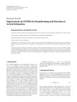
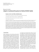
Figure 1: The structure of the DLWIC compression algorithm.
S
S A2
A1
B2 C2
B1
A2
B2 C2
A0
B1
C1
20
A1
C1
A0
RMS error versus compression factor for different
image sets
18
16
14
12
10
B0
C0
8
B0
C0
6
4
2
0
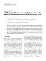
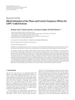
Figure 2: Octave band composition produced by recursive wavelet
transform is illustrated on the left and the pyramid structure inside
the coefficient matrix is shown on the right.
RBDWT, it is possible to efficiently perform wavelet subband
decomposition of an arbitrary shape region, while maintaining the same number of wavelet coefficients. Both OB-SPIHT
and OB-SPECK algorithms are embedded techniques; that is,
the coding method produces an embedded bitstream which
can be truncated at any point, equivalent to stopping the
compression process at a desired quality. The wavelet coefficients that have larger magnitude are those with larger information content. In a comparison, with full-image compression methods as SPIHT and JPEG2000, OB-SPIHT and
OB-SPECK exhibited much higher quality in the breast region at the same compression factor [24]. A different approach is presented in [26], where the embedded zerotree
wavelets (EZWs) coding technique is adopted for ROI coding in progressive image transmission (PIT). The method
uses subband decomposition and image wavelet transform
to reduce the correlation in the subimages at different resolutions. Thus, the whole frequency band of the original image is divided into different subbands at different resolutions.
The EZW algorithm is applied to the resulting wavelet coefficients to refine and encode the most significant ones.
Scalable video coding (SVC) has been a very active working area in the research community and in international standardizations as well. Video scalability may be handled in dif-
0.01
0.2
0.4
0.6
0.8
Compression factor
1
Skin lesion image
MRI
Medical video image
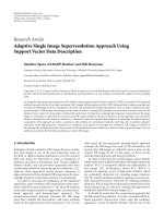
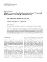
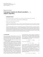
Figure 3: RMS error for different medical images according to quality factors.
ferent ways: a video can be spatially scalable and can accommodate a range of resolutions according to the network capabilities and the users’ viewing screens; it can be temporally scalable and can offer different frame rates (i.e., low
frame rates for slow networks); it can be scalable in terms
of quality or signal-to-noise ratio (SNR) including different quality levels. In all cases, the available bandwidth of
the transmission channel and the user preferences determine
resolution, frame rate, and quality of the video sequence. A
project on SVC standardization was originally started by the
ISO/IEC Moving Picture Experts Group (MPEG). Based on
an evaluation of the submitted proposals, the MPEG and
the ITU-T Video Coding Experts Group (VCEG) agreed to
jointly finalize the SVC project as an amendment of their
H.264/MPEG4-AVC standard [15], for which the scalable extension of H.264/MPEG4-AVC, as proposed in [34], was selected as the first working draft. As an important feature of
the SVC design, most components of H.264/MPEG4-AVC
4
EURASIP Journal on Wireless Communications and Networking
are used according to their specification in the standard. This
includes the intra- and motion-compensated predictions, the
transform and entropy coding, the deblocking, as well as the
NAL unit packetization (network abstraction layer (NAL)).
The base layer of an SVC bitstream is generally coded in
compliance with the H.264/MPEG4-AVC Standard, and each
H.264/MPEG-4 AVC Standard-conforming decoder is capable of decoding this base layer representation when it is provided with an SVC bitstream. New tools are only added for
supporting spatial and signal-to-noise ratio (SNR) scalability.
Regarding context awareness, despite the numerous implementations and proposals of telemedicine and e-health
platforms found in the literature (an indicative reference collection can be found in [1–8]), only a few systems seem to
be context-aware. The main goal of context-aware computing is to acquire and utilize information about the context
of a device to provide services that are appropriate to particular people, places, times, events, and so forth [40]. According to the latter, the work presented in [41] describes a
context-aware mobile system for interhospital communication taking into account the patient’s and physician’s physical
locations for instant and efficient messaging regarding medical events. Bardram presents in [42] additional use cases of
context awareness within treatment centers and provides design principles of such systems. The project “AWARENESS”
(presented in [43]) provides a more general framework for
enhanced telemedicine and telediagnosis services depending
on the patient’s status and location. To the best of our knowledge, there is no other work exploiting context awareness for
optimizing network utilization and efficiency within the context of medical networks and telemedicine services. A more
detailed description of the context-aware medical framework
is provided in Section 5 along with the proposed implementation.
3.
THE PROPOSED SCALABLE IMAGE
CODING SCHEME
The proposed methodology adopts the distortion-limited
wavelet image codec (DLWIC) algorithm [27]. In DLWIC,
the image to be compressed is firstly converted to the wavelet
domain using the orthonormal Daubechies wavelet transform [28]. The transformed data are then coded by bitlevels
and the output is coded using QM-coder [29], an advanced
binary arithmetic coder. The algorithm processes the bits
of the wavelet transformed image data in decreasing order
concerning their significance in terms of mean square error (MSE). This produces a progressive output stream enabling the algorithm to be stopped at any phase of the coding.
The already coded output can be used to construct an approximation of the original image. The latter feature is considered to be useful especially when a user browses medical
images using slow bandwidth connections, where the image
can be viewed immediately after only few bits have been received; the subsequent bits then make it more accurate. DLWIC uses the progressivism by stopping the coding when the
quality of the reconstruction exceeds a threshold given as an
input parameter to the algorithm. The presented approach
solves the problem of distortion limiting (DL) allowing the
user to specify the MSE of the decompressed image. Furthermore, this technique is designed to be as simple as possible consuming less amount of memory in the compressiondecompression procedure, thus being suitable for usage on
mobile devices.
Figure 1 represents the structure of the DLWIC compression algorithm consisting of three basic steps: (1) the wavelet
transform, (2) the scanning of the wavelet coefficients by
bitlevels, and (3) the coding of the binary decisions made by
the scanning algorithm and the coefficients bits by the entropy encoder. The decoding procedure is almost identical:
(1) binary decisions and coefficient bits are decoded; (2) the
coefficient data are generated using the same scanning algorithm as in the coding phase, but using the previously coded
decision information; (3) the coefficient matrix is converted
to a spatial image with the inverse wavelet transform.
The transform is applied recursively to the rows and
columns of the matrix representing the original spatial domain image. This operation gives us an octave band composition (see Figure 2). The left side (B) of the resulting coefficient matrix contains horizontal components of the spatial
domain image, the vertical components of the image are on
the top (A), and the diagonal components are along the diagonal axis (C). Each orientation pyramid is divided into levels;
for example, the horizontal orientation pyramid (B) consists
of three levels (B0, B1, and B2). Each level contains details
of different size; the lowest level (B0), for example, contains
the smallest horizontal details of the spatial image. The three
orientation pyramids have one shared top level (S), which
contains scaling coefficients of the image, representing essentially the average intensity of the corresponding region in the
image. Usually, the coefficients in the wavelet transform of a
natural image are small on the lower levels and bigger on the
upper levels. This property is very important for the compression; the coefficients of this highly skewed distribution
can be thus coded using fewer bits.
The coefficient matrix of size W × H is scanned by
bitlevels beginning from the highest bitlevel nmax required
for coding the biggest coefficient in the matrix (i.e., the number of the significant bits in the biggest coefficient):
nmax = log 2 max
ci, j
0 ≤ i < W0 ≤ j < H
+1 ,
(1)
where the coefficient in (i, j) is marked with ci,j. The coefficients are represented using positive integers as well as the
sign bits that are stored separately. The coder first codes all
the bits on the bitlevel nmax of all coefficients, then all the
bits on bitlevel nmax −1 , and so on until the least significant
bitlevel 1 is reached or the scanning algorithm is stopped.
The sign is coded together with the most significant bit (the
first 1 bit) of a coefficient.
Figure 3 depicts the root mean square (RMS) error results concerning the application of DLWIC algorithm for
both lossless (quality factor equal to one) and lossy (quality
factor smaller than one) compressions to three different test
image sets. The latter consisted of 10 skin lesion images, 10
magnetic resonance images (MRIs), and 10 snapshot images
C. Doukas and I. Maglogiannis
5
Quantization
x
Wavelet
transform
RONI
(background)
X
Entropy
encoder
MUX
Bit
allocation
Step size
Bitstream
ROI
Source image
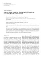
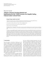
Figure 4: ROI coding system.
Table 1: Average Structural SIMilarity (SSIM) index for three different test image sets using different compression factors. The SSIM index
provides an indication of perceptual image similarity between original and compressed images.
Test image
Compression factor
Skin lesion images
MRI images
Medical video snapshots
Average SSIM index (%)
0.2
0.4
86.4475
93.0545
84.8253
94.1986
90.2179
94.5156
taken from a medical video (see Figure 5 for corresponding
images from the aforementioned datasets). With respect to
the acquired metrics from the test images, the discussed compression method produces acceptable image quality degradation (RMS value is less than 4 in the case of lossy compression with factor 0.6). For a closer inspection of the compression performance, the Structural SIMilarity (SSIM) index found in [30] is also used as an image quality indicator of
the compressed images. The specific metric provides a means
of quantifying the perceptual similarity between two images.
Perceptual image quality methods are traditionally based on
the error difference between a distorted image and a reference image, and they attempt to quantify the error by incorporating a variety of known properties of the human visual
system. In the case of SSIM index, the structural information
in an image is considered as an attribute for reflecting the
structure of objects, independently of the average luminance
and contrast, and thus the image quality is assessed based on
the degradation of the structural information. A brief literature review [31–33] has shown clearly the advantages of the
SSIM index against traditional RMS and peak signal-to-noise
ratio (PSNR) metrics and the high adoption by researchers
in the field of image and video processing. Average SSIM
index values for different compression factors are presented
in Table 1. As derived by the conducted similarity comparison experiments using SSIM, the quality degradation even in
high compression ratios is not major (i.e., 90.2% and 9.69%
for compression factors 0.2 and 0.8, resp., in case of medical
video images). This fact proves the efficiency of the proposed
algorithm.
In this point, it should be noted that concerning lossy
compression, DLWIC performs better in case of medical images of large sizes. Lossy compression is performed by multiplexing a small number of wavelet coefficients (composing the base layer and a few additional layers for enhance-
0.6
97.0601
97.2828
97.4221
0.8
99.4466
99.6702
99.6969
ment). Thus, a large number of layers are discarded, resulting in statistically higher compression results concerning the
file size. However, lossy medical image compression is considered to be unacceptable for performing diagnosis in most
of the imaging applications, due to quality degradation that,
even minor, can affect the assessment. Therefore, in order
to improve the diagnostic value of lossy compressed images,
the ROI (region of interest) coding concept is introduced in
the proposed application. ROI coding is used to improve the
quality in specific regions of interest only by applying lossless
or low compression in these regions, maintaining the high
compression in regions of noninterest. The wavelet-based
ROI coding algorithm implemented in the proposed application is depicted in Figure 4. An octave decomposition is used
which repeatedly divides the lower subband into 4 subbands.
Let D denote the number of decomposition level, then the
number of subbands M is equal to 4+3(D−1). Assuming that
the ROI shape is given by the client as a binary mask form on
the source image, the wavelet coefficients on the ROI and on
the region of noninterest (RONI) are quantized with different step sizes. For this purpose, a corresponding binary mask
is obtained, called WT mask, on the transform domain. The
whole coding procedure can be summarized in the following
steps.
(i) The ROI mask is set on the source image x.
(ii) The mask and the requested image x are transferred to
the application server.
(iii) The corresponding WT mask B is obtained.
(iv) The DWT coefficient X is calculated.
(v) Bit allocations for the ROI and RONI areas are obtained.
(vi) The X is quantized with the bit allocation from the previous step for each subband of each region.
(vii) The resulting quantized coefficient is encoded.
6
EURASIP Journal on Wireless Communications and Networking
(a)
(b)
(c)
(d)
(e)
(f)
Figure 5: Image samples compressed at different scaling factors and region of interest (ROI) coding. (a) Skin lesion image, (b) MRI image,
and (c) medical video image (snapshot) compressed at 0.5 scale factor, respectively. (d)–(f) The same images with background compressed
at 0.1 scale factor and ROI at 0.5.
Table 2: Patient data and data levels indicating an urgent status.
Acquired patient data
ECG (electrocardiogram, 3 leads)
BP (noninvasive blood pressure)
PR (pulse rate)
HR (heart rate)
SpO2 (hemoglobin oxygen saturation)
Data levels indicating an urgent state
ST wave elevation and depression T-wave inversion
90 mm Hg > systolic > 170 mm Hg
50/min > PR > 110/min
50/min > HR > 110/min
<90 (%)
Group of pictures (GOP)
Key
frame
Key
frame
Figure 6: Hierarchical prediction structure of H.264/MPEG-4AVC.
(viii) The WT mask B is encoded.
(ix) The entropy coded coefficient and WT mask are multiplexed in order to create the bitstream.
The decoding process follows the reverse order at the
client side. The major advantage of the proposed ROI coding method is that it produces a progressive output stream,
and thus the ROI is decoded progressively at the receiver. The
user has the capability to stop the transmission at any phase
of the coding, while the already transmitted output can be
used to construct an approximation of the original image.
The specific feature is especially desired for browsing medical
images in low-bandwidth mobile networks. In comparison
to the JPEG2000 standard, the proposed scheme is preferable
since it supports lossy-to-lossless ROI compression. The simplicity of the latter ROI coding requires low computational
complexity allowing the usage of the method in real time and
even on mobile devices as well.
Figure 5 visualizes the compression effect on image samples from the three different datasets used, using different
compression factors and ROI coding. Images (a)–(c) are
compressed at 0.5 factor, whereas images (d)–(f) have their
background compressed at 0.1 and the ROI at 0.5, respectively. The implementation of the scalable coder has been
performed in C++, whereas the encoding part has been developed in Java enabling its usage in both standalone and
Web applications (Java applets).
4.
H.264 SCALABLE VIDEO CODING
In contrast to older video coding standards as MPEG2, the coding and display order of pictures are completely decoupled in H.264/MPEG4-AVC standard. Any picture can be marked as reference picture and used for
motion-compensated prediction of the following pictures
C. Doukas and I. Maglogiannis
7
Biosignals
C
Patient video/
image
Collect
patient data
Patient
data
Determine
patient status
and proper
coding
scheme
Patient status
monitoring module
Network status
monitoring module
Medical broker
Threshold
parameters
Data coding
module
Underlying network
infrastructure
Properly coded medical
content
Figure 7: Architecture of the proposed context-aware medical video services framework.
independently of the corresponding slice coding types. These
features allow for the coding of picture sequences with arbitrary temporal dependencies.
Temporal scalable bitstreams can be generated by using
hierarchical prediction structures as illustrated in Figure 6
without any changes to H.264/MPEG4-AVC. The so-called
key pictures are coded in regular intervals by using only previous key pictures as references. The pictures between two
key pictures are hierarchically predicted as shown in Figure 6.
It is obvious that the sequence of key pictures represents the
coarsest supported temporal resolution, which can be refined
by adding pictures of the following temporal prediction levels. In addition to enabling temporal scalability, the hierarchical prediction structures also provide an improved coding efficiency compared to classical IBBP coding (named after the corresponding frame sequence) on the cost of an increased encoding-decoding delay [35]. Furthermore, the efficiency of the tools for supporting spatial and SNR scalability
is improved as it will be proven in the following sections. It
should also be noted that the delay of hierarchical prediction structures can be controlled by restricting the motioncompensated prediction in pictures of the future.
Spatial scalability is achieved by an oversampled pyramid
approach. The pictures of different spatial layers are indepen-
dently coded with layer-specific motion parameters as illustrated in Figure 6. However, in order to improve the coding
efficiency of the enhancement layers in comparison to simulcast, additional interlayer prediction mechanisms have been
introduced. These prediction mechanisms have been made
switchable so that an encoder can freely choose which base
layer information should be exploited for an efficient enhancement layer coding. Since the incorporated interlayer
prediction concepts include techniques for motion parameter and residual prediction, the temporal prediction structures of the spatial layers should be temporally aligned for an
efficient use of the interlayer prediction. It should be noted
that all NAL units for a time instant form an access unit and
thus have to follow each other inside an SVC bitstream.
5.
INTRODUCING THE CONTEXT-AWARENESS
FRAMEWORK
This section discusses in detail the proposed contextawareness framework that enables the monitoring of network and patient statuses and determines the appropriate
coding of medical video and images using the aforementioned coding techniques. The architecture of the discussed
framework is illustrated in Figure 7. The major modules are
8
EURASIP Journal on Wireless Communications and Networking
Table 3: Video frame, H.264 layer, and WiMAX classes correlation for each scenario.
Video sequences
I
rtPS
rtPS
A
B
Frame types
P
BE
nrtPS
B
BE
BE
BL
128 Kbps
128 Kbps
H.264 layers
EL1
—
256 Kbps
EL2
—
256 Kbps
Table 4: Received packet and frame statistics for the evaluation experiments.
Frame type
Packet delay (ms)
Frame delay (ms)
I
P
B
302.85
339.87
973.86
323.45
322.89
962.32
I
P
B
302.85
340.81
942.43
323.32
323.59
969.36
Packet jitter (ms)
Video sequence A
6.23
7.14
9.31
Video sequence B
6.71
7.29
7.62
(a) the network status monitoring module that determines
the current network interface used and the corresponding
status, (b) the patient status monitoring module that collects
patient data and determines the patient status, and (c) the
data coding module which is responsible for properly coding
(i.e., compressing) the transmitted video or image, according to instructions given by (d) the medical broker (i.e., usually a repository containing predefined or dynamically defined threshold values for determining patient and network
statuses).
The patient state can be determined through a number
of biosensors (i.e., heart rate and body temperature sensors)
and corresponding vital signals. Defined threshold values in
the latter signals determine the case of an immediate video
data transmission with better quality (alarm event) to the
monitoring unit. In case of normal patient status, periodical video transmission might occur at lower video quality,
or alternatively video can be replaced by highly compressed
images (suitable for low-bandwidth networks). Video and
image coding and transmission can also vary according to
network availability and quality. The framework can be also
used in cases of remote assessment or telesurgery; according
to the network interface used, appropriate video coding is applied to the transmitted medical data, thus avoiding possible
transmission delays and optimizing the whole telemedicine
procedure. The image and video compression factors are automatically selected based on the current patient status and
the underlying network type. Further modification of the latter factors can be performed by the users (i.e., physicians)
when the perceived image/video quality is considered to be
inappropriate for assessment due to the network conditions.
The framework’s architecture is open and does not depend on the monitoring applications used, the underlying
networks, or any other issues regarding the telemedicine system used. For this purpose, Web services [36, 37] have been
used as a communication mechanism between the major
framework components and the external patient monitoring applications used. The message exchange has been imple-
Frame jitter (ms)
Packet loss (%)
Frame loss (%)
7.19
8.09
9.43
3.2
12.4
47.6
0.1
11.1
47.1
7.27
8.17
8.27
3.2
11.9
43.7
0.1
11.1
43.3
mented through SOAP [38], a simple yet very effective and
flexible XML-based communication mechanism. The latter
involves the session initialization (which more precisely includes user authentication and service discovery) and the exchange of status and control messages. The status messages
include information regarding the patient data as generated
from the monitoring sensors and the underlying network
status and quality, whereas the control messages contain instructions regarding the proper coding of the transmitted
data (see Figure 9). It should be noted that the involved modules for the aforementioned communication (see Figure 8)
can all reside at the patient’s site, or alternatively the medical
broker can reside at the remote treatment site for the direct
collection of medical data and the reactive instruction’s provision.
The following section provides information regarding
the evaluation of the proposed platform using H.264 and
wavelet scalable coding for image and video data.
6.
EVALUATION PLATFORM
In order to validate the adaptive transmission of medical
video and image data using context-aware medical networks,
an evaluation platform has been implemented based on the
concept described in Section 5. H.264 [15, 34] has been
used for video coding and scalable wavelet for image coding [13], respectively. The main components of which the
platform consists are the attached biosensors to the patient,
the software modules responsible for collecting the corresponding signals and determining the appropriate video coding depending on the patient and network statuses, and
the simulated network infrastructures (i.e., IEEE 802.11g
(WLAN), UMTS, and WiMAX) for data transmission to
the monitoring units (e.g., a treatment center, an ambulance, or a physician at a remote site). Two patient states
have been defined: normal and urgent. The patient data
that are monitored through corresponding sensors are ECG,
C. Doukas and I. Maglogiannis
9
Table 5: Response time for UMTS and WLAN radio segments.
Response time TR (s) WLAN | UMTS
Compression scheme
No compression
JPEG
Skin lesion (520 Kb)
8.6 | 35.2
8.1 | 27
8.3 | 28
MRI (525 Kb)
8.8 | 41.3
Image type (file size)
Video snapshot (1 Mb)
14.2 | 54
8.4 | 29.3
Wavelet (lossless)
3.6 | 20
4.3 | 21.3
5.1 | 22
Wavelet (lossy)
3 | 19.6
3.9 | 19.9
4 | 17
Table 6: ROI transmission time for UMTS, WLAN, and WiMAX emulated radio segments.
ROI coding
Image type (file size)
CR (262 Kb)
CT (525 Kb)
MR (1 Mb)
Patient status
monitoring module
Network monitoring
module
ROI transmission time (s)
WLAN
1.5
1.55
2.1
UMTS
5
5.7
6.2
Data coding
module
Medical
broker
Remote monitoring
medical unit
Authentication and service discovery
Network and patient status notification
Proper coding notification
Patient (coded) data transmission
Figure 8: Message exchange between the framework’s modules for the case of remote patient status monitoring.
Figure 9: XML instance of an SOAP message containing information about the patient status.
WiMAX
1.42
1.5
1.9
10
EURASIP Journal on Wireless Communications and Networking
BP (noninvasive blood pressure), PR (pulse rate), HR (heart
rate), and SpO2 (hemoglobin oxygen saturation).
6.1. Evaluation results from H.264 video compression
For[RS10] the evaluation of H.264 video coding, two video
sequences have been used for transmission corresponding
to the two defined patient statuses. The media access control (MAC) layer of 802.16 enables the differentiation among
traffic categories with different multimedia requirements.
The standard [44] supports four quality-of-service scheduling types: (1) unsolicited grant service (UGS) for the constant bitrate (CBR) service, (2) real-time polling service
(rtPS) for the variable bitrate (VBR) service, (3) nonrealtime polling service (nrtPS) for nonreal-time VBR, and (4)
best effort (BE) service for service with no rate or delay requirements. For the specific scenario, a simulated WiMAX
wireless network of 1 Mbps has been used; the following rates
for the supported traffic classes have been allocated: 200 Kbps
for the UGS class, 300 Kbps for the rtPS class, 200 Kbps for
the nrtPS classes, and 200 Kbps for the best BE class. Each
group of pictures (GOP) is consisted of I, P, and B frames
structured by repeating sequences of the period IBBPBBPBB.
The GOP contains 25 frames per second, and the maximum
UDP packet size is at 1000 bytes (payload only). The NS2
simulator [45] and a WiMAX module presented in [46] have
been used for this purpose. A number of 11 nodes randomly
distributed at a surface of 1000 m2 using omnidirectional antenna models provided by NS2 have been simulated.
A scalable extension of H.264/AVC encoder and decoder
was used, provided by [39]. A number of background flows
are also transmitted in the simulated network in order to fill
in the respective WiMAX class capacity in the link. The background traffic is increased from 210 Kbps to 768 Kbps leading the system in congestion. For evaluation purposes, we
adopt a simpler QoS mapping policy, by using direct mapping of packets to WiMAX classes. All packets are formed
into three groups, according to the type of context that they
contain, and each group of packets is mapped to one WiMAX
class.
The first simulation scenario refers to the normal patient status. The corresponding video sequence has been used
with a single layer H.264 transmission; rtPS for transmitting I frames and nrtPS and BE for transmitting P and B
frames are used, respectively. The second simulation scenario
refers to the urgent patient status and it considers a scalable H.264 stream transmission consisting of two layers; the
base layer (BL) packets are encoded using the scalable extension H.264/AVC codec at 128 kbps and two enhancement layers (ELs) (i.e., EL1 and EL2) are encoded each at 256 kbps,
respectively. The correlation between the video frames, the
H.264 layers, and the network classes is presented in Table 3.
The experimental results prove better network resources
utilization in case of the normal patient status, and acceptable video quality in case of the urgent patient status. PSNR
and Structural SIMilarity (SSIM) index [31] have been used
as quality metrics. In the case of normal patient status (video
sequence A, higher compression, and low network quality
class used), PSNR and SSIM were calculated at the receiver
at 23.456 and 0.762, respectively. In the case of the urgent
patient status (video sequence B, better video coding, and
higher network quality class used), PSNR and SSIM were calculated at 29.012 and 0.918, respectively.
Table 4 presents corresponding results regarding the
packet and frame statistics at the receiver side for each frame
type (i.e., I, P, and B). There is a decrease in frame delay,
loss, and jitter for the second video sequence despite the
fact that video is encoded in higher quality. The latter is
translated into both better network resource utilization and
proper video quality when context awareness indicates the
proper video coding and transmission schemes.
6.2.
Evaluation results from wavelet
image compression
Regarding the wavelet image coding, the first set of measurements concerns the framework’s response time (i.e., the
time to transmit a compressed image) for different image
types (skin lesion, MRI, and video snapshot with sizes of 520,
525 Kb, and 1.3 Mb, resp.) for different types of compression
(no compression, JPEG compression with a quality factor of
0.75, and lossless and lossy discrete wavelet compression), for
the cases where either UMTS or WLAN is the access network.
The corresponding results are depicted in Table 5.
The lossless compression can be selected for cases where
the underlying network infrastructure has the means (i.e.,
high bandwidth, limited jitter, and delay) to support transmission of larger data size or in cases where the context
of the transmission demands high perceived image quality
(e.g., a patient emergency event). In correspondence to the
latter, lossy image compression can be used in the case of
patient monitoring through still images using a resourcelimited wireless network (e.g., UMTS).
With respect to the evaluation results, discrete wavelet
compression reduces the actual medical image downloading
time improving in this way the response time for the proposed application. An additional performance metric of the
proposed medical application concerns the ROI transmission
time for the same image dataset for three emulated radio access networks (i.e., UMTS, WLAN, and WiMAX). The corresponding results are depicted in Table 6.
7.
CONCLUSION
Medical video and image transmission is a key issue for the
successful deployment and usage of telemedicine applications especially when wireless network infrastructures are
used. Adaptive and scalable coding on the other hand is considered to be quite important since it is the technology that
enables the seamless and dynamic adaptation of content according to network or patient status and their requirements.
This paper introduces the concept of adaptive transmission
of medical video and image data in telemedicine systems using context-aware medical networks. Adaptive transmission
is achieved through scalable video coding using H.264 and
wavelet-based scalable image compression with ROI coding
support. The simplicity of the latter ROI coding requires
C. Doukas and I. Maglogiannis
low computational complexity allowing for the usage of the
method in real time and even on mobile devices as well.
Context awareness is achieved through the monitoring of
the patient status, the context of the data transmission, and
the network status. Evaluation results using different wireless
networks (i.e., IEEE 802.11e, WiMAX, and UMTS) indicate
the effectiveness of the platform in the context of both efficient data compression with acceptable quality degradation
and proper data transmission over wireless networks. What
remains as future work is the establishment of the innovative
context-aware medical video and image coding platform into
a real patient-care environment, providing helpful information regarding the assessment of the platform in use.
REFERENCES
[1] J. C. Lin, “Applying telecommunication technology to healthcare delivery,” IEEE Engineering in Medicine and Biology Magazine, vol. 18, no. 4, pp. 28–31, 1999.
[2] S. Pavlopoulos, E. Kyriacou, A. Berler, S. Dembeyiotis, and
D. Koutsouris, “A novel emergency telemedicine system based
on wireless communication technology-AMBULANCE,” IEEE
Transactions on Information Technology in Biomedicine, vol. 2,
no. 4, pp. 261–267, 1998.
[3] S. Deb, S. Ghoshal, V. N. Malepati, and D. L. Kleinman, “Telediagnosis: remote monitoring of large-scale systems,” in Proceedings of IEEE Aerospace Conference, vol. 6, pp. 31–42, Big
Sky, Mo, USA, March 2000.
[4] Y. B. Choi, J. S. Krause, H. Seo, K. E. Capitan, and K. Chung,
“Telemedicine in the USA: standardization through information management and technical applications,” IEEE Communications Magazine, vol. 44, no. 4, pp. 41–48, 2006.
[5] C. S. Pattichis, E. Kyriacou, S. Voskarides, M. S. Pattichis,
R. Istepanian, and C. N. Schizas, “Wireless telemedicine systems: an overview,” IEEE Antennas and Propagation Magazine,
vol. 44, no. 2, pp. 143–153, 2002.
[6] M. Akay, I. Marsic, A. Medl, and G. Bu, “A system for
medical consultation and education using multimodal human/machine communication,” IEEE Transactions on Information Technology in Biomedicine, vol. 2, no. 4, pp. 282–291,
1998.
[7] J. Zhou, X. Shen, and N. D. Georganas, “Haptic tele-surgery
simulation,” in Proceedings of the 3rd IEEE International Workshop on Haptic, Audio and Visual Environments and their Applications (HAVE ’04), pp. 99–104, Ottawa, Canada, October
2004.
[8] P. Fontelo, E. DiNino, K. Johansen, A. Khan, and M. Ackerman, “Virtual microscopy: potential applications in medical education and telemedicine in countries with developing
economies,” in Proceedings of the 38th Annual Hawaii International Conference on System Sciences (HICSS ’05), p. 153, Big
Island, Hawaii, USA, January 2005.
[9] A.-L. Lage, J. Martins, J. Oliveira, and W. Cunha, “A quality of
service approach for managing tele-medicine multimedia applications requirements,” in Proceedings of the 4th IEEE International Workshop on IP Operations and Management (IPOM
’04), pp. 186–190, Beijing, China, October 2004.
[10] C. LeRouge, M. J. Garfield, and A. R. Hevner, “Quality attributes in telemedicine video conferencing,” in Proceedings
of the 35th Annual Hawaii International Conference on System
Sciences (HICSS ’02), pp. 2050–2059, Big Island, Hawaii, USA,
January 2002.
11
[11] H. Yu, Z. Lin, and F. Pan, “Applications and improvement of
H.264 in medical video compression,” IEEE Transactions on
Circuits and Systems, vol. 52, no. 12, pp. 2707–2716, 2005.
[12] G. Bernabe, J. Gonzalez, J. M. Garcia, and J. Duato, “A new
lossy 3-D wavelet transform for high-quality compression of
medical video,” in Proceedings of the IEEE EMBS International Conference on Information Technology Applications in
Biomedicine (ITAB ’00), pp. 226–231, Arlington, Va, USA,
November 2000.
[13] C. N. Doukas, I. Maglogiannis, and G. Kormentzas, “Medical
image compression using wavelet transform on mobile devices
with ROI coding support,” in Proceedings of the 27th Annual
International Conference of the IEEE Engineering in Medicine
and Biology Society (EMBC ’05), vol. 7, pp. 3779–3784, Shanghai, China, September 2005.
[14] IEEE 802.16 WG, “IEEE Standard for Local and Metropolitan Area Networks—Part 16: Air Interface for Fixed Broadband Wireless Access Systems,” IEEE Std. 802.16-2004, October 2004.
[15] ITU-T Rec. & ISO/IEC 14496-10 AVC, “Advanced Video Coding for Generic Audiovisual Services,” version 3, 2005.
[16] ISO/IEC JTC 1/SC 29/WG 1 (ITU-T SG8), “JPEG 2000 Final
Committee Version 1.0,” March 2000.
[17] G. Anastassopoulos and A. Skodras, “JPEG2000 ROI coding in medical imaging applications,” in Proceedings of the
2nd IASTED International Conference on Visualisation, Imaging and Image Processing (VIIP ’02), pp. 783–788, Palma de
Mallorca, Spain, August 2002.
[18] Z. Liu, J. Ha, Z. Xiong, Q. Wu, and K. Castleman, “Lossyto-lossless ROI coding of chromosome images using modified
SPIHT and EBCOT,” in Proceedings of IEEE International Symposium on Biomedical Imaging, pp. 317–320, Washington, DC,
USA, July 2002.
[19] A. Said and W. A. Pearlman, “A new, fast, and efficient image codec based on set partitioning in hierarchical trees,”
IEEE Transactions on Circuits and Systems for Video Technology, vol. 6, no. 3, pp. 243–250, 1996.
[20] D. Taubman, “High performance scalable image compression
with EBCOT,” IEEE Transactions on Image Processing, vol. 9,
no. 7, pp. 1158–1170, 2000.
[21] Z. Liu, Z. Xiong, Q. Wu, Y.-P. Wang, and K. Castleman, “Cascaded differential and wavelet compression of chromosome
images,” IEEE Transactions on Biomedical Engineering, vol. 49,
no. 4, pp. 372–383, 2002.
[22] G. Minami, Z. Xiong, A. Wang, and S. Mehrotra, “3-D wavelet
coding of video with arbitrary regions of support,” IEEE Transactions on Circuits and Systems for Video Technology, vol. 11,
no. 9, pp. 1063–1068, 2001.
[23] S. Li and W. Li, “Shape-adaptive discrete wavelet transforms
for arbitrarily shapedvisual object coding,” IEEE Transactions
on Circuits and Systems for Video Technology, vol. 10, no. 5, pp.
725–743, 2000.
[24] M. Penedo, W. A. Pearlman, P. G. Tahoces, M. Souto, and
J. J. Vidal, “Region-Based Wavelet Coding Methods for Digital Mammography,” IEEE Transactions on Medical Imaging,
vol. 22, no. 10, pp. 1288–1296, 2003.
[25] A. Islam and W. A. Pearlman, “Embedded and efficient lowcomplexity hierarchical image coder,” in Visual Communications and Image Processing, vol. 3653 of Proceedings of SPIE,
pp. 294–305, San Jose, Calif, USA, January 1999.
[26] R. S. Dilmaghani, A. Ahmadian, M. Ghavami, M. Oghabian,
and H. Aghvami, “Multi rate/resolution control in progressive
medical image transmission for the region of interest (ROI)
using EZW,” in Proceedings of the 25th Annual International
12
[27]
[28]
[29]
[30]
[31]
[32]
[33]
[34]
[35]
[36]
[37]
[38]
[39]
[40]
[41]
[42]
[43]
EURASIP Journal on Wireless Communications and Networking
Conference of the IEEE Engineering in Medicine and Biology
Society (EMBS ’03), vol. 1, pp. 818–820, Cancun, Mexico,
September 2003.
J. M. Shapiro, “Embedded image coding using zerotrees of
wavelet coefficients,” IEEE Transactions on Signal Processing,
vol. 41, no. 12, pp. 3445–3462, 1993.
A. Said and W. A. Pearlman, “A new, fast, and efficient image codec based on set partitioning in hierarchical trees,”
IEEE Transactions on Circuits and Systems for Video Technology, vol. 6, no. 3, pp. 243–250, 1996.
J. Lehtinen, “Limiting distortion of a wavelet image codec,”
Acta Cybernetica, vol. 14, no. 2, pp. 341–354, 1999.
Z. Wang, A. C. Bovik, H. R. Sheikh, and E. P. Simoncelli, “Image quality assessment: from error visibility to structural similarity,” IEEE Transactions on Image Processing, vol. 13, no. 4,
pp. 600–612, 2004.
Y.-B. Tong, Q. Chang, and Q.-S. Zhang, “Image quality assessing by using NN and SVM,” in Proceedings of the 5th International Conference on Machine Learning and Cybernetics
(ICMLC ’06), vol. 2006, pp. 3987–3990, Dalian, China, August
2006.
G.-H. Chen, C.-L. Yang, L.-M. Po, and S.-L. Xie, “Edge-based
structural similarity for image quality assessment,” in Proceedings of IEEE International Conference on Acoustics, Speech
and Signal Processing (ICASSP ’06), vol. 2, pp. II933–II936,
Toulouse, France, May 2006.
Z.-Y. Mai, C.-L. Yang, and S.-L. Xie, “Improved best prediction
mode(s) selection methods based on structural similarity in
H.264 I-frame encoder,” in Proceedings of IEEE International
Conference on Systems, Man and Cybernetics (SMC ’05), vol. 3,
pp. 2673–2678, Waikoloa, Hawaii, USA, October 2005.
H. Schwarz, et al., “Technical Description of the HHI proposal
for SVC CE1,” ISO/IEC JTC1/WG11, Doc. m11244, Palma de
Mallorca, Spain, October 2004.
H. Schwarz, D. Marpe, and T. Wiegand, “Hierarchical B pictures,” Joint Video Team, Doc. JVT-P014, Poznan, Poland, July
2005.
M. Hori and M. Ohashi, “Applying XML Web services into
health care management,” in Proceedings of the 38th Annual
Hawaii International Conference on System Sciences (HICSS
’05), p. 155, Big Island, Hawaii, USA, January 2005.
Y. Lee, C. Patel, S. A. Chun, and J. Geller, “Towards intelligent
Web services for automating medical service composition,” in
Proceedings of IEEE International Conference on Web Services,
pp. 384–391, 2004.
SOAP specifications, />JSVM 0 software, G1/savce/index
.htm.
T. P. Moran and P. Dourish, “Introduction to this special issue
on context-aware computing,” Human-Computer Interaction,
vol. 16, no. 2–4, pp. 87–95, 2001.
M. A. Munoz, M. Rodriguez, J. Favela, A. I. Martinez-Garcia,
and V. M. Gonzalez, “Context-aware mobile communication
in hospitals,” Computer, vol. 36, no. 9, pp. 38–46, 2003.
J. E. Bardram, “Applications of context-aware computing in
hospital work: examples and design principles,” in Proceedings of the ACM Symposium on Applied Computing (SAC ’04),
vol. 2, pp. 1574–1579, Nicosia, Cyprus, March 2004.
T. Broens, A. van Halteren, M. van Sinderen, and K. Wac, “Towards an application framework for context-aware m-health
applications,” in Proceedings of the 11th Open European Summer School (EUNICE ’05), Madrid, Spain, July 2005.
[44] IEEE 802.16 Working Group, “IEEE Standard for Local and
Metropolitan Area Networks—Part 16: Air Interface for Fixed
Broadband Wireless Access Systems,” IEEE Std. 802.16-2004,
October 2004.
[45] The Network Simulator—ns2, />[46] J. Chen, C.-C. Wang, F. C.-D. Tsai, et al., “Simulating wireless
networks: the design and implementation of WiMAX module
for ns-2 simulator,” in Proceedings of ACM Workshop on ns2: The IP Network Simulator (WNS2 ’06), Pisa, Italy, October
2006.