Immune and metabolic interactions of human erythrocytes a molecular perspective
Bạn đang xem bản rút gọn của tài liệu. Xem và tải ngay bản đầy đủ của tài liệu tại đây (2.66 MB, 11 trang )
<span class="text_page_counter">Trang 1</span><div class="page_container" data-page="1">
<i>Endocrine, Metabolic & Immune Disorders -Drug Targets</i>
<b><small>ISSN: 1871-5303eISSN: 2212-3873</small></b>
<small>ImpactFactor:1.973</small>
<i><b><small>The official journal of the ITALIAN ASSOCIATION OF CLINICALENDOCRINOLOGISTS, shortly defined AME</small></b></i>
<i><b><small>(Associazione Medici Endocrinologi)</small></b></i>
<i><b><small>Endocrine, Metabolic & Immune Disorders - Drug Targets, 2021, 21, 843-853</small></b></i>
<b><small>Abstract: Apart from their main function as oxygen carriers in vertebrates, erythrocytes are also </small></b>
<small>in-volved in immune regulation. By circulating throughout the body, the erythrocytes are exposed andinteract with tissues that are damaged as a result of a disease. In this study, we summarize the litera-ture regarding the contribution of erythrocytes to immune regulation and metabolism. Under the cir-cumstances of a disease state, the erythrocytes may lose their antioxidant capacity and release Dam-age Associated Molecular Patterns, resulting in the regulation of innate and adaptive immunity. Inaddition, the erythrocytes scavenge and affect the levels of chemokines, circulating cell-free mtD-NA, and C3b attached immune complexes. Furthermore, through surface molecules, erythrocytescontrol the function of T lymphocytes, macrophages, and dendritic cells. Through an array of en-zymes, red blood cells contribute to the pool of blood’s bioactive lipids. Finally, the erythrocytescontribute to reverse cholesterol transport through various mechanisms. Our study is highlightingoverlooked molecular interactions between erythrocytes and immunity and metabolism, whichcould lead to the discovery of potent therapeutic targets for immunometabolic diseases.</small>
<b>Keywords: Erythrocytes, immunity, metabolism, lipid signaling, reverse cholesterol transport, cytokine signaling, DAMP </b>
bind-ing, DAMP release, cellular interactions.
<b>1. INTRODUCTION</b>
Inflammation represents a highly evolutionarily conserved set of cellular and molecular events aiming to re-duce the tissue damage or infection that initiated inflamma-tion. The main purpose is to clear necrotic cells, rehabilitate the tissue damage, and elicit a tissue repair mechanism. How-ever, unless the process of inflammation resolution starts, chronic inflammation drives further tissue damage and fibro-sis. The main molecular players of inflammation are damage
(lipopolysaccharide, RNA, mitochondrial DNA, histones etc), cytokines, chemokines, bioactive lipid mediators, recep-tors on both innate and adaptive immunity cells for the above molecules, and reactive oxygen species.
Epithelial cells, neutrophils, monocytes, macrophages, dendritic cells, Natural Killer Cells, mast cells, T and B lym-phocytes constitute the group of cells implicated in the pro-cess [1].
<small>* Address correspondence to this author at the Department of Medicine,Faculty of Life Sciences, Democritus University of Thrace, 68100 Alexan-droupolis, Greece; Tel/Fax: ++30-25510-30502;</small>
<small>E-mail: </small>
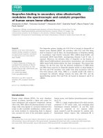
Apart from the various types of cells mentioned above, emerging data highlight a potential immune-modulatory role of erythro-cytes. Indeed, within the context of evolutionary biology, non-mammalian vertebrate erythro-cytes have an important immunomodulatory function [2]. However, in th-ese animals, erythrocytes are nucleated in contrast to human ones. Nevertheless, human erythron-cytes retain a
<b>consider-able immunomodulatory capacity [2, 3] (Fig. 1).</b>
The normal functioning of red blood cells requires the presence of certain micro- and macromolecules. These molecules ensure the appropriate energy metabolism priori-ties, help to maintain the proper antioxidant capacity and af-fect normal cell death and clearance. How-ever, under condi-tions of tissue injury, the release of some of these molecules, which normally are not available in the extracellular environ-ment, leads to their recognition as DAMP. Others, induce oxidative stress.
It also appears that red blood cells possess several macro-molecules, through which they can scavenge pro-inflamma-tory agents from the site of inflammation. However, due to acquired or genetic differences in the red blood cells ability to remove these factors, this function may be negligible to re-duce inflammation and / or increase it.
<b><small> 2212-3873/21 $65.00+.00 © 2021 Bentham Science Publishers</small></b>
</div><span class="text_page_counter">Trang 2</span><div class="page_container" data-page="2"><i><b><small>Fig. (1). Immunomodulatory functions of human erythrocytes. This figure was created using Servier Medical Art templates, which are </small></b></i>
<i><small>li-censed under a Creative Commons Attribution 3.0 Unported License; .</small></i>
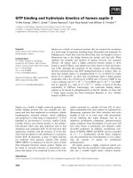
<i><b><small>Fig. (2). Potential molecular mechanisms for the biosynthesis of signalling lipids in human erythrocytes. This figure was created using </small></b></i>
<i><small>Servi-er Medical Art templates, which are licensed undServi-er a Creative Commons Attribution 3.0 Unported License; .</small></i>
Lipid signaling, through interfering with signaling net-works, regulates a range of cellular functions, such as metabolism, inflammation-related signaling, cell prolifera-tion, growth, motility, apoptosis and autophagy [4]. Thus, lipids are implicated in the molecular basis of human dis-ease, including those affecting metabolism, immune system, nervous system, and cancer. The cellular source of these lipids includes an array of cells. However, the role of ery-throcytes is usually underestimated, leading to the possible
<b>ignorance of many molecular therapeutic targets (Fig. 2).</b>
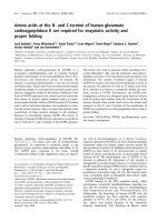
Erythrocytes comprise 45% of the total blood volume, while lipids make up 40% of the red blood cell mass [5]. Fur-thermore, the concentration of cholesterol in erythrocytes is similar to that of lipoproteins [6]. Red blood cells exchanges cholesterol and phospholipids with lipoproteins [7, 8]. In ad-dition, erythrocytes have been shown to participate in
macrophages to the liver [9]. Therefore, the erythrocyte has characteristics that make it an ideal lipid carrier in the blood
<b>(Fig. 3). However, contrary to lipoproteins, erythrocytes,</b>
</div><span class="text_page_counter">Trang 3</span><div class="page_container" data-page="3"><i><b><small>Fig. (3). Contribution of erythrocytes to reverse cholesterol transport. This figure was created using Servier Medical Art templates, which are</small></b></i>
<i><small>licensed under a Creative Commons Attribution 3.0 Unported License; .</small></i>
despite the absence of intracellular organelles, can metabol-ize membrane lipids, resulting in the formation of bioactive lipids.
In this review, we attempt to bring light to the possible role of red blood cells in inflammation and im-munometabolism, to reveal novel therapeutic targets.
<b>2. ERYTHROCYTES IN OXIDATIVE STRESS</b>
Under normal conditions, the erythrocyte can function as an antioxidant cellular agent as it passes through the tissues. This is due to its antioxidant properties: GSH, NADH and NADPH and its antioxidant enzymes: peroxide dismutase, catalase, peroxidase, glutathione peroxidase, peroxidizing 2 and glutaredoxin 2 [10]. As the age of the erythrocyte in-creases, so does the concentration of oxidized proteins (Bar-tosz, 1981), but also the oxidation of hemoglobin [11]. Among the antioxidants of the erythrocytes (GSH, NADH and NADPH), GSH plays a particularly important role, since its biosynthesis [12] is the only way for its production. GSH production, however, decreases with increasing ery-throcyte age due to reduced glucose entry [13], decreased pyruvate kinase activity, dehydrogenase 6-phosphate glu-cose, and aminotransferase aspartic.
In addition, passing through tissue with oxidative stress leads to converting the erythrocyte from an antioxidant to a pro-oxidative cell [10].
Red blood cells with reduced antioxidant potential are likely to be actively involved in inflammation. The interac-tion of oxidized erythrocytes with monocytes in the pres-ence of LPS or zymosan, led to an increase in TNF-α and
<i>IL-10 production [14]. Aoshiba et al., showed that </i>
ex-tracellular levels, which could diffuse into the cell and cause neutrophil apoptosis. However, the suppression of erythro-cyte’s glutathione metabolism did not allow the protective
properties of erythrocyte versus neutrophil apoptosis [15]. Fi-nally, erythrocytes’ antioxidant capacity is implicated in the inhibition of apoptosis and the cell death-associated oxida-tive stress of T lymphocytes; this requires intact erythro-cytes to come into contact with T lymphoerythro-cytes [16].
<b>3. DAMP RELEASE</b>
<b>3.1. Heme and Hemoglobin Release</b>
Hemoglobin, while inside the red blood cell, is protected from oxidation. However, its release makes it prone to oxida-tion. Oxidized hemoglobin triggers the formation of com-plexes that are sources of free radicals such as methemo-globin. In fact, oxidation of hemoglobin can also be caused by peroxidized lipids [17].
Heme and oxidized hemoglobin can lead to the induc-tion of expression of various adhesion molecules in endothe-lial cells such as ICAM1, VCAM1 and E-Selectin [18, 19]. In fact, oxidized hemoglobin has led to the re-organization of cell cytoskeleton resulting in increased permeability of the endothelial gaps [19]. In addition, a study has shown that heme causes chemotaxis in neutrophils [20]. The effect of heme in inflammation seems to require TLR4 and NFKβ [21].
<i>A study by Buttari et al., shows that heme causes </i>
chemo-tactic activity in monocytes but also in dendritic cells de-rived from monocytes. Endothelial cell attachment and en-dothelial migration have also been implicated. This action appears to require the attachment of hemoglobin to CD163, signaling via ERK and MAPK, and cell cycle rearrange-ment. In fact, the study suggests that oxidized hemoglobin may have the same effect since the administration of amino acetylcysteine reduced the incidence of chemotaxis [22].
</div><span class="text_page_counter">Trang 4</span><div class="page_container" data-page="4"><b>3.2. ATP Release</b>
<i>Sikora et al., showed that erythrocytes release ATP after</i>
hemolysis and that this release is not subject to regulation by mechanical pressure, hypoxia or cAMP [23]. The ATP re-leased could have immunomodulatory effects. ATP causes chemotaxis in neutrophils, macrophages, and dendritic cells [24], regulates the function of T lymphocytes depending on its concentration [25] and affects the concentrations of IL-1β cytokines, TNF-α and IL-10 [26].
<b>3.3. Vesicle Release</b>
As erythrocytes age, vesicles are produced, which ap-pear to require or at least be associated with the hydrolysis of phosphoinositides to diacylglycerol, but also the external-ization of PSer. In addition, the hydrolysis of SM by the acidic sphingomyelinase of the erythrocyte can lead to the production of microparticles [27].
<i>Sadallah et al. [28] found that erythrocytes release </i>
vesi-cles which, due to their size, the presence of lipid membrane and specific proteins, can be characterized as ectosomes. In fact, these ectosomes expressed PSer on their surface and were phagocytosed by macrophages and led to a reduced ex-pression of TNF-α and CXCL8 (IL-8) by macrophages stim-ulated by LPS or Zymozan A. Therefore, it appears that th-ese ectosomes have immunosuppressive properties.
Microparticles released by erythrocytes attach to mono-cytes, triggering the release of pro-inflammatory cytokines, resulting in the proliferation of CD4 + and CD8 + T
<i>lympho-cytes [29]. A study by Belizaire et al. [30] found that </i>
micro-particles released by stored red blood cells activate neu-trophils as shown by CD11b expression, oxidative stress and phagocytic capacity
<b>3.4. Cytokine Release</b>
IL-33 is a cytokine, mainly located in the nucleus. Its re-lease can function as a DAMP. In addition, IL-33 possess pleiotropic immune function, since it can regulate the devel-opment and/or function of Innate Lymphoid Cells 2, T
T
<i>lymphocytes and Natural Killer Cells [31]. Wei et al. [32],</i>
showed that IL-33 is expressed in erythroid cells and is re-leased by the mature erythrocyte through hemolysis; in fact IL-33 levels are released by erythrocytes significantly affect and relate to cytokine levels. blood. The same investigators also reported that IL-33 could induce the expression of IL 8 in airway epithelial cells.
Evidently, the release of cytokines from erythrocytes has
<i>been validated ex vivo. Karsten et al., showed that </i>
erythro-cytes of healthy volunteers could release, during culture in PBS, more than 40 cytokines and chemokines; the same study showed that erythrocytes, when incubated with recom-binant cyto-kines and chemokines, reduce their content in the conditioned medium [33].
<b>4.1. Duffy Blood Group and Chemokine Scavenging</b>
<i>Darbonne et al. [34], were the first to show that </i>
erythro-cytes, through the Duffy Blood group antigen, bind the chemokines CXCL8 (IL-8) and CCL2 (MCP1). In fact, the same study showed that the binding of chemokines to the erythrocyte receptor did not cause its internalization, while the chemokines that remained bound to the receptor did not exert their action on other cells. Subsequent studies show that this receptor binds, in addition to CXCL8 and CCL2 and CCL5 (RANTES), MGSA, NAP-2 and others [35].
<i>However, Yamamoto et al. [36], argue that DARC together</i>
with bound cytokines can be internalized and therefore re-tain cytokines.
Of particular importance is the fact that the loss of DARC from erythrocytes leads to inflammation of the lungs [37]. In addition, binding of chemokines CXCL1 and CX-CL2/3 to DARC has been found to protect against neu-trophil migration and airway inflammation [38]. In fact, it ap-pears that the expression of DARC in non-hematopoietic cells does not affect the above events, thus demonstrating the role of this receptor in the red blood cell [38]. Erythro-cytes, possibly through the Duffy blood group and the bind-ing of CCL5/RANTES, affect the endothelial migration of eosinophils, which is particularly important in inflammation during allergy [39]. Finally, it is possible that despite the par-ticipation of DARC in scavenging chemokine in circulation, it could at the same time increase their half-life.
<b>4.2. Erythropoietin Receptor and IL-2 Scavenging</b>
<i>Kirtch et al. [40], showed that incubation of IL-2 with</i>
erythrocytes leads to the binding of a considerable number of IL-2 molecules to the erythropoietin receptor expressed in immature and new erythrocytes, even after multiple washes. In fact, it appears that this is due to the similarity between the erythropoietin receptor and the IL-2 receptor. In addi-tion, reticulocytes bind 400% more IL-2 than mature cytes [41]. Subsequently, another study showed that erythro-cytes could be used as carriers of IL-2, which could release and enhance cytotoxicity [42]
<b>4.3. TLR9 and Mitochondrial DNA Scavenging</b>
TLR9 is a member of the toll-like receptor family, and is expressed mainly by innate immunity cells. Its function is in recognition of bacterial, viral and mitochondrial DNA [43].
<i>Hotz et al. [44], showed that erythrocytes express TLR9</i>
and under normal conditions impart a significant amount of mitochondrial DNA. However, in the case of systemic in-flammation, most mtDNA is not associated with red blood cell TLR9. In fact, the lack of TLR9 from erythrocytes led
<i>to inflammation in vivo. The same study revealed important</i>
inter- and intra-individual variation regarding erythrocyte TLR9 levels.
</div><span class="text_page_counter">Trang 5</span><div class="page_container" data-page="5">As in the case of DARC, TLR9 could increase the half-life of cell-free mitochondrial DNA, releasing it when or where the concentration of cell-free mitochondrial DNA is low [2].
<b>4.4. Complement Receptor 1 and Complement Scaven-ging</b>
The erythrocytes possess the complement receptor 1 (CR1) which binds immune complexes that have attached the protein to the C3b supplement and then the erythrocyte carries these immune complexes to remove liver phagocytes [45]. The CR1 genotype is likely to affect C3b binding ca-pacity [46]. Acquired CR1 reductions include release through ectosomes, but mainly through proteolysis that is
<i>likely to be induced in the in vivo disease environment [45].</i>
<b>5. IMMUNE REGULATION THROUGH PHYSICALINTERACTION</b>
<b>5.1. CD47</b>
CD47 plays an important role in the removal of erythro-cytes. CD47 is recognized by the SIPR receptor and acts as a self-marker and as a “do not eat me” signal to the
<i>phago-cytes that express the receptor [47]. However, Burger et al.[48], showed that in vitro-aging red blood cells underwent a</i>
change in CD47 steroid formation, thereby binding to TSP-1. Also, peptide administration of TSP-1 led to ery-throphagocytosis. The change in CD47 structure and its asso-ciation with TSP-1 was also observed in red blood cells that had been stored for a long time. These results led the re-searchers in the above study to classify CD47 as a “molecu-lar switch”.
<i>Schakel et al., showed that erythrocytes, through the </i>
in-teraction of CD47 with SIPR, prevent the maturation of a subtype of dendritic cells characterized by the modification of 6-sulfo LacNac of PSGL-1 and differentiate from leuko-cytes. In addition, the same study showed that erythrocytes prevented the release of IL-12 and TNF-α [49].
<i>Subsequently, Buttari et al. [50], showed again that </i>
ery-throcytes from healthy people prevent the maturation of den-dritic cells exposed to LPS and this was accompanied by re-duced amounts of IL-12, IL-6, TNF-α and increased amounts of IL-10. What is remarkable about this study, how-ever, is that erythrocytes from patients with arteriosclerosis did not prevent the maturation of dendritic cells from healthy controls. This was accompanied by an increase in the amount of IL-6, IL-12, TNF-α and reduced amounts of IL-10. The erythrocytes of these patients showed increased oxidative stress, reduced amounts of glycophorin A, CD47 and increased PSer on the extracellular side of the lipid bilay-er of bilay-erythrocytes. The above data led the authors to assume that the reduced amount of CD47 in erythrocytes causes the inability of erythrocytes to inhibit the maturation of dendrit-ic cells
<b>5.2. Glycophorin A</b>
Erythrocytes suppress the activation of neutrophils through the binding of glycophorin A to Siglec-9 of
neu-trophils, as shown by oxidative stress, chemotaxis, produc-tion of extracellular traps, apoptosis, etc. Inhibiproduc-tion of this in-teraction is associated with neutrophil activation [51]. De-creased glycophorin A has been found in aged red blood cells [52], indicating that aging of red blood cells reduces an-tioxidant capacity, lowering glycophorin A and increasing production of microvesicles, leads to the activation of neu-trophils.
<b>5.3. Externalisation of Phosphatidylserine</b>
The externalization of PSer in erythrocytes has been found to be a feature of the aging of these cells. In a study
<i>by Boas et al. [53], the externalization of PSer was found to</i>
be increased in aged erythrocytes and was associated with the removal of these erythrocytes from circulation.
<i>Howev-er, Franco et al., observed, after re-administration of </i>
erythro-cytes, that the externalization of PSer was not observed in aged erythrocytes [54]. What is likely to be the case is that the externalization of PSer occurs more easily in aged ery-throcytes after eryptosis (erythrocyte apoptosis) induction [13]. However, inflammation can cause the externalization of PSer by increasing the number of sphingomyelinases in the blood that could subsequently act on the erythrocyte [55]. Furthermore, cholesterol loading in the erythrocyte membrane inhibits the externalization of PSer [56]. In any case, externalized phosphatidylserine can be recognized by macrophage receptors as well as Tim-1, Tim-4 and Stabilin-i-2. In addition, lactaderin, GAS6, and protein S act as a
AXL family receptors [57].
<i>A study shows that both in vitro and in vivo </i>
accumula-tion of erythrocytes in the liver occurs during hepatic steato-sis. These erythrocytes, due to oxidative stress externalize the PSer, which is recognized by the Kupffer cells and then erythrophagocytosis occurs. This has been linked to in-creased hepatic oxidative stress and inflammation [58].
<b>6. LIPID SIGNALING6.1. S1P Release</b>
S1P is bioactive lysophospholipid. It creates a gradient in blood; a mechanism necessary for lymphocyte egression from lymphoid organs. In addition S1P contributes to
<i>inflam-mation and vascular development [59]. Hanel et al. [60]</i>
found that erythrocytes constitute a particularly important source of plasma S1P, mainly through their plasma mem-brane. In fact, erythrocytes contain 54% of total S1P in blood. In each case, erythrocytes determine S1P levels in the blood, as transfusion of erythrocytes into experimental ani-mals lacking sphingosine kinase 1/2 restored S1P plasma lev-els [61].
<i>Hanel et al. also showed that erythrocytes do not </i>
synthe-size S1P, since an inverse relationship was observed be-tween the levels of this lipid metabolite in plasma and ery-throcyte. Finally, a dynamic equilibrium between the two
<i>compartments was observed in vivo [60].</i>
However, in the erythrocyte, there is an active form of acid sphingomyelinase - an enzyme that hydrolyzes
</div><span class="text_page_counter">Trang 6</span><div class="page_container" data-page="6">sphin-gomyelin to produce ceramide. This enzyme is increased un-der hypoxia in erythrocytes of sickle cell anemia [27]. In ad-dition, erythrocytes contain ceramidase - an enzyme that cleaves ceramide to sphingosine. Inhibition of this enzyme decreased the levels of sphingosine and S1P in erythrocytes [62]. It is particularly important that erythrocytes also con-tain active sphingosine kinase. In fact, its activity increases during periods of hypoxia through adenosine binding to the A2B receptor in the red blood cell, leading to ERK1 / 2 sig-naling. The end result is an increase in S1P levels [63]. Fur-thermore, the red blood cell can respond to low plasma S1P levels by increasing the activity of sphingosine kinase 1 [64].
<i>Subsequently, studies by Bode et al. [65], found that the</i>
red cell releases S1P to high-density lipoproteins and plasma albumin. In fact, HDLs carry greater amounts of S1P than
<i>al-bumin. A study by Christensen et al. [66] showed that the</i>
majority of HDL- bound S1P molecules are associated with apolipoprotein M (apoM). This was observed even in the absence of the remaining molecules of the HDL complex. Fi-nally, the exchange of S1P from erythrocytes to apoM was found to be sensitive to inhibition of ABCC1 but not of
<i>ABCB1. In contrast to the above study, Vu et al. [67]</i>
showed that the Mfsd2b transporter is particularly important for the release of S1P from erythrocytes and platelets. Final-ly, another study showed that Band 3 also participates in the release of S1P from erythrocytes [68]. Regardless of the car-rier responsible for the transport of S1P from the erythrocyte to plasma / serum, it appears to be S1P-specific, as lipids with similar structure did not inhibit S1P transport [69].
<b>6.2. LPC and LPA Release</b>
LPA.is another bioactive lysophospholipid that acts through the activation of five receptors. Its role in
<i>inflamma-tion is well recognized [70]. Cripps et al. [71], have shown</i>
that erythrocytes contain lysophosphatidic acid acyltrans-ferase (LPAAT) enzyme in their membrane, which converts plasma LPA to phosphatidic acid (PA). In fact, it was found that the red blood cell regulates serum LPA levels. In addi-tion, Phosphatidic acid in erythrocytes with PSer external-ized appears to be hydrolyzed by phospholipase A2, an en-zyme which increases during inflammation, and results in
<i>in-creased LPA in erythrocytes [72]. Subsequently, Cripps et</i>
<i>al. showed that erythrocyte storage reduces the activity of</i>
the LPA acetyltransferase enzyme, thereby increasing the amount of LPA in the red blood cell [73], which can then ex-ert its pharmacological actions.
Alternatively, the red blood cells may release a substrate for lysophospholipase D, LPC, which can further be
<i>convert-ed to LPA. This is at least indicative of a study by Aoki et</i>
<i>al.; LPC was found in the erythrocyte incubation </i>
superna-tant and the addition of exogenous lysophospholipase D in-creased LPA levels [74]. Erythrocytes also contain the en-zyme cytosolic phospholipase A2 [75]. In fact, its levels in-crease under hypoxia via ERK signaling and regulate LPC levels in the red blood cell [76].
<b>6.3. LTB4 Release</b>
The red blood cells appear to possess the ability to synth-esize some eicosanoids. In particular, red blood cells can me-tabolize leukotriene LTA4 to LTB4 as they contain the en-zyme LTA4 hydrolase [77].
<b>6.4. PGE1 and PGE2 Synthesis</b>
In addition, red blood cells can also synthesize prostag-landins PGE1 and PGE2 [78]. The metabolic pathway has not been extensively studied, but cyclooxygenase 1 (COX-1), which acts in the initial steps of synthesizing these bioactive lipids, has been found [78].
<b>6.5. PAF Release</b>
<i>According to Lang et al., the erythrocyte, in response to</i>
the hyperosmotic shock, synthesizes PAF, which is released by the cell, acts in autocrine signaling, and activates its re-ceptor in the erythrocyte surface, causing sphingomyelinase activation and ceramide production. This pathway is possib-ly involved in eryptosis [79]. Whether this circuit has paracrine action merits further examination.
<b>6.6. EET Release</b>
EET is anti-inflammatory bioactive lipids, whose metabolism has been explored as potential therapeutic tar-gets [80]. Erythrocytes can synthesize and release EET after specific stimuli. It has been found that the stimulation of the P2X7 receptor by ATP promotes the release of EETs [81]. The pathway probably starts with the release of AAs from the erythrocyte membrane phospholipids after phospholi-pase activity [82]. The action of the hemoglobin monooxyge-nase follows by the aid of nicotinic acid or riboflavin [83]. Indeed, the release of EETs from erythrocytes determines plasma EET levels [84]. Finally, the erythrocyte contains an active epoxide hydrolase enzyme that hydrolyzes the EETs. Inhibition of this enzyme increases plasma EETs levels [85].
<b>6.7. 12(S)-HETE Release</b>
A study by Kobayashi and Levine [86] showed that ery-throcytes, after an increase in intracellular calcium concen-tration, induced the production of 12(S)-HETE, which is im-munologically active. The source of arachidonate was PC and PE, indicating that phospholipase A2 is involved in AA mobilization and not phospholipase C. Then, the activity of lipoxygenase is followed [87]. 12(S)-HETE was found in the erythrocyte supernatant, indicating that red cells release these bioactive lipids [86].
<b>7. CONTRIBUTION TO REVERSE CHOLESTEROLTRANSPORT</b>
Erythrocytes make up 45% of total blood volume, while lipids comprise 40% of red blood cell mass. Also, the con-centration of cholesterol in erythrocytes is similar to that of lipoproteins [6].
In humans, erythrocytes exchange phospholipids and cholesterol with lipoproteins (7, 8 Kinetic studies show that
</div><span class="text_page_counter">Trang 7</span><div class="page_container" data-page="7">cholesterol moves between red blood cells and lipoproteins
<i>via aqueous diffusion [7].</i>
About 50% of blood cholesterol is found in erythrocytes, indicating that the flow of cholesterol to the erythrocyte is similar to the efflux of free cholesterol from the tissues [88]. A study found that in mice lacking the apoAI gene, the reverse transport of cholesterol occurs to a large extent from the red blood cell. In mice that did not lack apoAI, the ery-throcyte was found to be involved, but to a lesser extent. The model suggested by the authors is that red blood cells re-ceive cholesterol from LDL and HDL and transfer it either directly to the liver cells or to the endothelial cells [9].
However, the transfer of cholesterol from red blood cells to the liver could be done through very-low-density
<i>lipopro-teins/chylomicrons. A study by Chung et al.. [89], found</i>
that incubating erythrocytes with autologous plasma after fasting containing LCAT and CETP proteins for 18 hours at 37 degrees led to an increase in plasma cholesterol through a concomitant increase in esterified cholesterol of HDL, LDL and VLDL. The postprandial plasma containing chylomi-crons led to a greater rise in plasma cholesterol, leading to a 356% elevation of cholesterol levels in chylomicrons. The ability of plasma to receive cholesterol from red blood cells was positively correlated with plasma cholesterol, triglyc-erides, VLDL, LDL. These results were confirmed in a sub-sequent study [90]. Therefore, chylomicrons accept free cholesterol from red blood cells and can then transport it to the liver through their remnants. In the above mechanism, apolipoprotein B, which is associated with erythrocytes, may be involved. The amount of apoB in red blood cells is not related to serum apoB levels [91].
Others showed that erythrocytes contribute to reverse cholesterol transport by providing PC regeneration from LCAT-formed LPC [92]. Finally, a recent study showed that red blood cells indirectly accept cholesterol from macrophages. The same study also showed that erythrocytes possibly donate cholesterol to albumin and apoA1, but do not accept cholesterol from apoA1 [93]. These data imply that the erythrocyte constitutes an important mediator of cholesterol transport in the circulation.
It is now evident that erythrocytes not only contribute to innate immunity regulation, but they are also capable of de-termining important components of adaptive immunity. Nev-ertheless, erythrocyte immunobiology remains elusive. Vari-ous receptors implicated in the binding of inflammatory agents are found in erythrocyte membranes. However, a lot of questions merit investigation: kinetics, interactions, mech-anisms of surface-level reductions. Furthermore, the crosstalk between aging, induction of eryptosis and release of DAMPs contains a lot of unknowns.
The molecular mechanisms for the function of erythro-cyte-derived lipid mediators remain unexplored. Despite the progress regarding the understanding of lipid biology, many questions need to be addressed. Which transporters permit
the release of the lipid mediators by the erythrocytes? Which signaling pathways regulate the activity of the enzymes re-sponsible for the biosynthesis of these lipids? Is the release of these lipid mediators affected during immune-metabolic and neurometabolic diseases such as non-alcoholic fatty liv-er disease, athliv-erosclliv-erosis and Alzheimliv-er’s disease? Is the synthesis and release of these lipids a group of therapeutic targets?
Finally, red blood cells constitute a neglected “lipopro-tein” capable of influencing cholesterol trafficking in tis-sues. Exploration of the implicated mechanisms shall unveil novel therapeutic targets.
It is our opinion that the answers to these questions could plausibly reveal cellular and a handful of molecular novel therapeutic targets.
<b>LIST OF ABBREVIATIONS</b>
ADORA2B = Adenosine A2B Receptor
2b
</div><span class="text_page_counter">Trang 8</span><div class="page_container" data-page="8">NADPH = Nicotinamide Adenine Dinucleotide Phos-phate
of activated B cells
<b>CONSENT FOR PUBLICATION</b>
Not applicable.
The research work was supported by the Hellenic Foun-dation for Research and Innovation (HFRI) under the HFRI Ph.D Fellowship grant (Fellowship Number: 1343).
<small>tory responses and inflammation-associated diseases in organs.</small>
<i><b><small>Vol. 9. Oncotarget. Impact Journals, 2018, LLC, 7204-7218.</small></b></i>
<small>cells and the immune system and its impact on atherosclerosis.</small>
<i><b><small>BioMed Res. Int., 2015, 2015616834</small></b></i>
<small>chemical characteristics of hemoglobin-free ghosts of human </small>
<i><b><small>ery-throcytes. Arch. Biochem. Biophys., 1963, 100(1), 119-130.</small></b></i>
<small> PMID:14028302</small>
<small>Nikolić, M.; Stanić, D.; Antonijević, N.; Niketić, V. Cholesterol[6]</small>
<small>bound to hemoglobin in normal human erythrocytes: a new form</small>
<i><b><small>of cholesterol in circulation? Clin. Biochem., 2004, 37(1), 22-26.</small></b></i>
<small> PMID:14675558</small>
<small>Dmitry, Y. Lipids that directly regulate innate immune signal[7]</small>
<i><b><small>transduction Innate Immunity , 2018, 26 (1 ), 4 -14 .</small></b></i>
<small>Dushianthan, A; Cusack, R; Koster , G.; Grocott, M.; Postle, A. In-[8]</small>
<small>sight into erythrocyte phospholipid molecular flux in healthy hu-mans and in patients with acute respiratory distress syndrome</small>
<i><small>Blood Cells Play a Role in Reverse Cholesterol Transport </small></i>
<i><b><small>Arterios-cler Thromb Vasc Biol., 2012, 32(6), 1460-5.</small></b></i>
<small>Minetti, M.; Agati, L.; Malorni, W. The microenvironment can[10]</small>
<small>shift erythrocytes from a friendly to a harmful behavior: </small>
<i><b><small>pathoge-netic implications for vascular diseases. Cardiovasc. Res., 2007,</small></b></i>
<small>Toulany, M.; Köberle, M.; Skabytska, Y.; Saki, M.; Biedermann,T.; Duszenko, M.; Lang, F.; Wieder, T.; Bosman, G.J. The impact</small>
<i><b><small>of erythrocyte age on eryptosis. Br. J. Haematol., 2012, 157(5),</small></b></i>
<small>Augmented TNF-alpha and IL-10 production by primed humanmonocytes following interaction with oxidatively modified </small>
<i><b><small>autolo-gous erythrocytes. J. Leukoc. Biol., 2001, 70(2), 289-296.</small></b></i>
<small>inhibit activation-induced cell death and oxidative stress in human</small>
<i><b><small>peripheral blood T lymphocytes. Blood, 2001, 97(10), 3152-3160.</small></b></i>
<small>Heme induces the expression of adhesion molecules ICAM-1,</small>
<i><small>VCAM-1, and E selectin in vascular endothelial cells. Proc. Soc.</small></i>
<i><b><small>Exp. Biol. Med., 1997, 216(3), 456-463.</small></b></i>
<small> PMID: 9402154Silva, G.; Jeney, V.; Chora, A.; Larsen, R.; Balla, J.; Soares, M.P.[19]</small>
<small>Oxidized hemoglobin is an endogenous proinflammatory agonist</small>
<i><b><small>that targets vascular endothelial cells. J. Biol. Chem., 2009,</small></b></i>
<i><small>284(43), 29582-29595.</small></i>
<small> PMID: 19700768Monteiro, A.P.; Pinheiro, C.S.; Luna-Gomes, T.; Alves, L.R.;[20]</small>
<small>Maya-Monteiro, C.M.; Porto, B.N.; Barja-Fidalgo, C.; Benjamim,C.F.; Peters-Golden, M.; Bandeira-Melo, C.; Bozza, M.T.; Canet-ti, C. Leukotriene B4 mediates neutrophil migration induced by</small>
<i><b><small>heme. J. Immunol., 2011, </small></b><small>186(11), 6562-6567.</small></i>
<small> PMID: 21536805</small>
</div><span class="text_page_counter">Trang 9</span><div class="page_container" data-page="9"><small>Belcher, J.D.; Chen, C.; Nguyen, J.; Milbauer, L.; Abdulla, F.;[21]</small>
<small>Alayash, A.I.; Smith, A.; Nath, K.A.; Hebbel, R.P.; Vercellotti,G.M. Heme triggers TLR4 signaling leading to endothelial cell </small>
<i><small>ac-tivation and vaso-occlusion in murine sickle cell disease. Blood,</small></i>
<small>ti, V.; Capoano, R.; Salvati, B.; Businaro, R.; Di Giammarco, G.;Riganò, R. Haemoglobin triggers chemotaxis of human mono-cyte-derived dendritic cells: possible role in atherosclerotic lesion</small>
<small>Duménil, A-M.; Sáez, J.C. ATP promotes the fast migration ofdendritic cells through the activity of pannexin 1 channels andP2X7</small><i><b><small> receptors. Sci. Signal., 2017, 10(506)eaah7107</small></b></i>
<small> PMID: 29162744Trabanelli, S.; Ocadlíková, D.; Gulinelli, S.; Curti, A.; Salvestrini,[25]</small>
<small>V.; Vieira, R.P.; Idzko, M.; Di Virgilio, F.; Ferrari, D.; Lemoli,R.M. Extracellular ATP exerts opposite effects on activated and</small>
<i><small>regulatory CD4+ T cells via purinergic P2 receptor activation. J.</small></i>
<i><b><small>Immunol., 2012, 189(3), 1303-1310.</small></b></i>
<small> PMID: 22753942Cauwels, A.; Rogge, E.; Vandendriessche, B.; Shiva, S.; Brouck-[26]</small>
<small>aert, P. Extracellular ATP drives systemic inflammation, tissue </small>
<i><b><small>da-mage and mortality. Cell Death Dis., 2014, 5(3), e1102-e1102.</small></b></i>
<small> PMID: 24603330Awojoodu, A.O.; Keegan, P.M.; Lane, A.R.; Zhang, Y.; Lynch,[27]</small>
<small>K.R.; Platt, M.O.; Botchwey, E.A. Acid sphingomyelinase is acti-vated in sickle cell erythrocytes and contributes to inflammatory</small>
<i><b><small>microparticle generation in SCD. Blood, 2014, 124(12),</small></b></i>
<small>Muench, M.O.; Heitman, J.W.; Norris, P.J. Exosomes from redblood cell units bind to monocytes and induce proinflammatory </small>
<i><b><small>cy-tokines, boosting T-cell responses in vitro. Blood, 2014, 123(5),</small></b></i>
<small>wards, M.J.; Caldwell, C.C.; Lentsch, A.B.; Pritts, T.A. Micropar-ticles from stored red blood cells activate neutrophils and cause</small>
<i><small>lung injury after hemorrhage and resuscitation. J. Am. Coll. Surg.,</small></i>
<small>C.A.; Valente, A.J.; Baker, J.B. Red blood cells are a sink for </small>
<i><b><small>inter-leukin 8, a leukocyte chemotaxin. J. Clin. Invest., 1991, 88(4),</small></b></i>
<small>Itoga, M. Intracellular Storage of Duffy Antigen-Binding</small>
<i><b><small>Chemokines by Duffy-Positive Red Blood Cells. Clin Lab, 2017,</small></b></i>
<i><small>63(4), 717-23.</small></i>
<small>Mangalmurti, N.S.; Xiong, Z.; Hulver, M.; Ranganathan, M.; Liu,[37]</small>
<small>X.H.; Oriss, T.; Fitzpatrick, M.; Rubin, M.; Triulzi, D.; Choi, A.;Lee, J.S. Loss of red cell chemokine scavenging promotes </small>
<i><b><small>transfu-sion-related lung inflammation. Blood, 2009, 113(5), 1158-1166.</small></b></i>
<small> PMID:19064726</small>
<small>Reutershan, J.; Harry, B.; Chang, D.; Bagby, G.J.; Ley, K. DARC[38]</small>
<small>on RBC limits lung injury by balancing compartmental </small>
<i><b><small>distribu-tion of CXC chemokines. Eur. J. Immunol., 2009, 39(6),</small></b></i>
<small> PMID: 19499525Kanda, A.; Adachi, T.; Kayaba, H.; Yamada, Y.; Ueki, S.; Yam-[39]</small>
<small>aguchi, K.; Hamada, K.; Fujita, M.; Chihara, J. Red blood cellsregulate eosinophil chemotaxis by scavenging RANTES secreted</small>
<i><b><small>from endothelial cells. Clin. Exp. Allergy, 2004, 34(10),</small></b></i>
<small>targeted erythrocytes coated with recombinant human interleukin</small>
<i><small>2 on T-lymphocyte proliferation in vitro. Biotechnol. Appl.</small></i>
<i><b><small>Biochem., 1994, 19(3), 331-340.</small></b></i>
<small>PMID: 8031507</small>
<small>Moyes, R.B.; DeLoach, J.R. Binding of human recombinant inter-[41]</small>
<small>leukin 2 to murine erythrocytes is erythropoietin receptor </small>
<i><b><small>mediat-ed. Comp. Haematol. Int., 1996, 6(3), 134-140.</small></b></i>
<small>Moyes, R.B.; Kirch, H.; DeLoach, J.R. Enhanced biological activi-[42]</small>
<small>ty of human recombinant interleukin 2 coupled to mouse redblood cells as evaluated using the mouse Meth A sarcoma model.</small>
<i><b><small>Biotechnol. Appl. Biochem., 1996, 23(1), 29-36.</small></b></i>
<small>Sondheimer, N.; Rivella, S.; Worthen, G.S.; Mangalmurti, N.S.Red Blood Cells Homeostatically Bind Mitochondrial DNAthrough TLR9 to Maintain Quiescence and to Prevent Lung </small>
<i><b><small>In-jury. Am. J. Respir. Crit. Care Med., 2018, 197(4), 470-480.</small></b></i>
<small>Taniguchi, H.; Koide, N.; Matsuura, K.; Ogura, T.; Tobe, K.; Tsu-ji, T. Regulation of circulating immune complexes by complement</small>
<i><small>receptor type 1 on erythrocytes in chronic viral liver diseases. Gut,</small></i>
<i><b><small>2002, 51(4), 591-596.</small></b></i>
<small> PMID: 12235086Oldenborg, P-A. CD47: A Cell Surface Glycoprotein Which Regu-[47]</small>
<small>lates Multiple Functions of Hematopoietic Cells in Health and </small>
<i><b><small>Dis-ease. ISRN Hematol., 2013, 2013614619</small></b></i>
<small> PMID: 23401787Burger, P.; Hilarius-Stokman, P.; de Korte, D.; van den Berg,[48]</small>
<small>T.K.; van Bruggen, R. CD47 functions as a molecular switch for</small>
<i><b><small>erythrocyte phagocytosis. Blood, 2012, 119(23), 5512-5521.</small></b></i>
<small> PMID:22427202</small>
<small>Schäkel, K.; von Kietzell, M.; Hänsel, A.; Ebling, A.; Schulze, L.;[49]</small>
<small>Haase, M.; Semmler, C.; Sarfati, M.; Barclay, A.N.; Randolph,G.J.; Meurer, M.; Rieber, E.P. Human 6-sulfo LacNAc-expressingdendritic cells are principal producers of early interleukin-12 and</small>
<i><b><small>are controlled by erythrocytes. Immunity, 2006, 24(6), 767-777.</small></b></i>
</div><span class="text_page_counter">Trang 10</span><div class="page_container" data-page="10"><small> PMID: 16782032Buttari, B.; Profumo, E.; Cuccu, B.; Straface, E.; Gambardella, L.;[50]</small>
<small>Malorni, W.; Genuini, I.; Capoano, R.; Salvati, B.; Riganò, R. Ery-throcytes from patients with carotid atherosclerosis fail to control</small>
<i><b><small>dendritic cell maturation. Int. J. Cardiol., 2012, 155(3), 484-486.</small></b></i>
<small> PMID: 22265585Lizcano, A.; Secundino, I.; Döhrmann, S.; Corriden, R.; Rohena,[51]</small>
<small>C.; Diaz, S.; Ghosh, P.; Deng, L.; Nizet, V.; Varki, A. Erythrocytesialoglycoproteins engage Siglec-9 on neutrophils to suppress </small>
<small>and red cell viability in red cell aging and in hemolytic anemia.</small>
<i><b><small>Proc. Natl. Acad. Sci. USA, 1998, 95(6), 3077-3081.</small></b></i>
<small> PMID: 9501218Franco, R.S.; Puchulu-Campanella, M.E.; Barber, L.A.; Palascak,[54]</small>
<small>M.B.; Joiner, C.H.; Low, P.S.; Cohen, R.M. Changes in the </small>
<i><small>proper-ties of normal human red blood cells during in vivo aging. Am. J.</small></i>
<i><b><small>Hematol., 2013, 88(1), 44-51.</small></b></i>
<small> PMID: 23115087</small>
<small>Dinkla, S.; Wessels, K.; Verdurmen, W.P.R.; Tomelleri, C.; Cluit-[55]</small>
<small>mans, J.C.A.; Fransen, J.; Fuchs, B.; Schiller, J.; Joosten, I.;Brock, R.; Bosman, G.J. Functional consequences of sphin-gomyelinase-induced changes in erythrocyte membrane structure.</small>
<i><b><small>Cell Death Dis., 2012, 3(10), e410-e410.</small></b></i>
<small> PMID: 23076218van Zwieten, R.; Bochem, A.E.; Hilarius, P.M.; van Bruggen, R.;[56]</small>
<small>Bergkamp, F.; Hovingh, G.K.; Verhoeven, A.J. The cholesterolcontent of the erythrocyte membrane is an important determinant</small>
<i><b><small>of phosphatidylserine exposure. Biochim. Biophys. Acta, 2012,</small></b></i>
<small>Nakatani, K.; Ikeda, K.; Nakajima, Y.; Ikura, Y.; Ueda, M.;Arakawa, T.; Hato, F.; Kawada, N. Erythrophagocytosis by livermacrophages (Kupffer cells) promotes oxidative stress, inflamma-tion, and fibrosis in a rabbit model of steatohepatitis: implications</small>
<i><small>for the pathogenesis of human nonalcoholic steatohepatitis. Am. J.</small></i>
<i><b><small>Pathol., 2007, 170(3), 967-980.</small></b></i>
<small> PMID: 17322381Nagahashi, M.; Abe, M.; Sakimura, K.; Takabe, K.; Wakai, T.[59]</small>
<small>The role of sphingosine-1-phosphate in inflammation and cancer</small>
<i><b><small>progressionCancer Science; Blackwell Publishing Ltd, 2018, 109,</small></b></i>
<small>J.B.; Xu, Y.; Camerer, E.; Zheng, Y.W.; Huang, Y.; Cyster, J.G.;Coughlin, S.R. Promotion of lymphocyte egress into blood and</small>
<i><small>lymph by distinct sources of sphingosine-1-phosphate. Science,</small></i>
<small>Dowhan, W.; Idowu, M.; Juneja, H.S.; Molina, J.G.; Blackburn,M.R.; Kellems, R.E.; Xia, Y. Elevated adenosine signaling viaadenosine A2B receptor induces normal and sickle erythrocyte</small>
<i><b><small>sphingosine kinase 1 activity. Blood, 2015, 125(10), 1643-1652.</small></b></i>
<small> PMID:</small>
<small>Knapp, M.; Lisowska, A.; Zabielski, P.; Musiał, W.; Baranowski,[64]</small>
<small>M. Sustained decrease in plasma sphingosine-1-phosphate concen-tration and its accumulation in blood cells in acute myocardial </small>
<i><b><small>in-farction. Prostaglandins Other Lipid Mediat., 2013, 106, 53-61.</small></b></i>
<small> PMID:24120760</small>
<small>Bode, C.; Sensken, S-C.; Peest, U.; Beutel, G.; Thol, F.; Levkau,[65]</small>
<small>B.; Li, Z.; Bittman, R.; Huang, T.; Tölle, M.; van der Giet, M.;Gräler, M.H. Erythrocytes serve as a reservoir for cellular and </small>
<i><b><small>ex-tracellular sphingosine 1-phosphate. J. Cell. Biochem., 2010,</small></b></i>
<i><small>109(6), 1232-1243.</small></i>
<small> PMID: 20186882</small>
<small>Christensen, P.M.; Bosteen, M.H.; Hajny, S.; Nielsen, L.B.;[66]</small>
<small>Christoffersen, C. Apolipoprotein M mediates </small>
<i><b><small>sphingosine-1-phos-phate efflux from erythrocytes. Sci. Rep., 2017, 7(1), 14983.</small></b></i>
<small> PMID: 29118354Vu, T.M.; Ishizu, A-N.; Foo, J.C.; Toh, X.R.; Zhang, F.; Whee,[67]</small>
<small>D.M.; Torta, F.; Cazenave-Gassiot, A.; Matsumura, T.; Kim, S.;Toh, S.E.S.; Suda, T.; Silver, D.L.; Wenk, M.R.; Nguyen, L.N.Mfsd2b is essential for the sphingosine-1-phosphate export in </small>
<i><b><small>ery-throcytes and platelets. Nature, 2017, 550(7677), 524-528.</small></b></i>
<small> PMID: 29045386Kurano, M.; Nishikawa, M.; Kuma, H.; Jona, M.; Yatomi, Y. In-[68]</small>
<small>volvement of Band3 in the efflux of sphingosine 1-phosphate</small>
<i><b><small>from erythrocytes.PLoS One; Gerós H, editor, 2017, 12, pp.</small></b></i>
<small>Kobayashi, N.; Kobayashi, N.; Yamaguchi, A.; Nishi, T. Charac-[69]</small>
<small>terization of the ATP-dependent sphingosine 1-phosphate </small>
<i><b><small>trans-porter in rat erythrocytes. J. Biol. Chem., 2009, 284(32),</small></b></i>
<small>contain a membrane lysophosphatidic acid acyltransferase that </small>
<i><small>mo-dulates serum lysophosphatidic acid concentration. J. Am. Coll.</small></i>
<i><b><small>Surg., 2007, 205(3), S34.</small></b></i>
<small>Neidlinger, N.A.; Larkin, S.K.; Bhagat, A.; Victorino, G.P.;[72]</small>
<small>Kuypers, F.A. Hydrolysis of phosphatidylserine-exposing redblood cells by secretory phospholipase A2 generates </small>
<i><small>lysophospha-tidic acid and results in vascular dysfunction. J. Biol. Chem.,</small></i>
<i><b><small>2006, 281(2), 775-781.</small></b></i>
<small> PMID: 16278219Cripps, M.W.; Ereso, A.Q.; Victorino, G.P.; Harken, A.H.; Sou-[73]</small>
<small>pene, E.; Kuypers, F. Lysophosphatidic acid formation in old</small>
<i><small>packed red blood cells causes post transfusion vascular leak. J.</small></i>
<i><b><small>Am. Coll. Surg., 2008, 207(3), S37.</small></b></i>
<small>Aoki, J.; Taira, A.; Takanezawa, Y.; Kishi, Y.; Hama, K.; Kishi-[74]</small>
<small>moto, T.; Mizuno, K.; Saku, K.; Taguchi, R.; Arai, H. Serum ly-sophosphatidic acid is produced through diverse phospholipase</small>
<i><b><small>pathways. J. Biol. Chem., 2002, 277(50), 48737-48744.</small></b></i>
<small>Luo, R.; Parchim, N.F.; Liu, H.; Huang, A.; Adebiyi, M.G.; Jin, J.;Alexander, D.C.; Milburn, M.V.; Idowu, M.; Juneja, H.S.;Kellems, R.E.; Dowhan, W.; Xia, Y. Hypoxia-mediated impaired</small>
<i><small>erythrocyte Lands’ Cycle is pathogenic for sickle cell disease. Sci.</small></i>
<i><b><small>Rep., 2016, 6, 29637.</small></b></i>
<small> PMID: 27436223</small>
<small>McGee, J.E.; Fitzpatrick, F.A. Erythrocyte-Neutrophil Interac-[77]</small>
<small>tions: Formation of Leukotriene B4 by Transcellular Biosynthesis</small>
<i><b><small>Proceedings of the National Academy of Sciences, 1986, 83, pp.</small></b></i>
<small>Oonishi, T.; Sakashita, K.; Ishioka, N.; Suematsu, N.; Shio, H.;[78]</small>
</div>