A TUMOR OF THE THORACIC CAVITY IS DETECTED BY AN ABDOMINAL ULTRASOUND
Bạn đang xem bản rút gọn của tài liệu. Xem và tải ngay bản đầy đủ của tài liệu tại đây (3.38 MB, 19 trang )
<span class="text_page_counter">Trang 2</span><div class="page_container" data-page="2">
HISTORY OF THE PATIENT
• A 53-year-old female patient went to the ultrasound department for an annual routine check-up. At that time, she only felt tired sometimes. She likely
had no symptoms
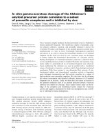
</div><span class="text_page_counter">Trang 3</span><div class="page_container" data-page="3">THE ULTRASOUND IMAGING SHOWED THAT SHE HAD AN ABNORMAL MASS IN THE BOTTOM OF HER RIGHT LUNG. WE COULD SEE IT BY THE ULTRASOUND SECTION THROUGH THE RIGHT LIVER AND THE DIAGRAM. AT FIRST, IT WAS HARD TO HAVE THE RIGHT DIAGNOSIS BECAUSE WE WONDERED IF IT WAS A LIVER MIRROR ARTIFACT IMAGE
</div><span class="text_page_counter">Trang 4</span><div class="page_container" data-page="4">VIDEO DIAMETER OF THE MASS ARE 55X35
MM
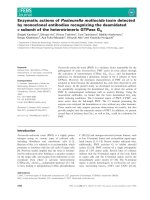
</div><span class="text_page_counter">Trang 5</span><div class="page_container" data-page="5">THEN WE TRIED TO USE OTHER SECTIONS, WITH THE ULTRASOUND SECTION THROUGH FROM HER BACK, THE MASS WAS STILL DETECTED SO THAT IS A REAL
LESSON. THE LESSON IS 53CM 35 CM IN DIAMETER, AND IT APPEARS DIFFERENT FROM THE LIVER IN TERMS OF DENSITY, IT HAD NO AIR BRONCHOGRAM IMAGE, AND THE DOPPLER MODE DID NOT SHOW ANY SIGNAL DOPPLER CLEARLY. SO WITH THESE ULTRASOUND IMAGES, AND THE PATIENT WAS WITHOUT SYMPTOMS, I THOUGHT IT WAS HARDLY A LUNG CONSOLIDATION, IT WAS MORE LIKELY A TUMOR
</div><span class="text_page_counter">Trang 6</span><div class="page_container" data-page="6">ULTRASOUND SECTION THROUGH THE BACK OF THE PATIENT
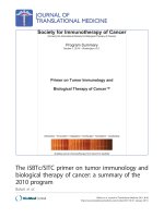
</div><span class="text_page_counter">Trang 7</span><div class="page_container" data-page="7">THEN, IN THE COMPUTED TOMOGRAPHY IMAGING (CT SCAN), IT IS LIKELY A TUMOR OF THE LUNG.
</div><span class="text_page_counter">Trang 9</span><div class="page_container" data-page="9">THE PATIENT WAS UNDERGOING SURGERY TO CUT THE TUMOR. THE PATHOLOGY SHOWS IT IS A BENIGN TUMOR OF THE LUNG - PULMONARY
LEIOMYOMA.
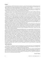
</div><span class="text_page_counter">Trang 10</span><div class="page_container" data-page="10">LUNG ULTRASOUND SECTIONS
• An illustration showing the technique of the lung and intercostal upper abdomen ultrasound examination
(arrows indicate directions of probe application).
</div><span class="text_page_counter">Trang 11</span><div class="page_container" data-page="11">CASE REFERENCE 1 : LUNG ULTRASONOGRAMS OF LESIONS ON THE
PLEURAL DIAPHRAGMATIC SURFACE
<b>• (2a) nodules on the right diaphragmaticpleural surface and pleural effusions; (2b)</b>
pleural nodules and pleural effusions on the
<b>left side; and (2c) (2d) Bulky tumors on the</b>
right diaphragmatic pleural (and abdominal) surface and pleural effusions. Abbreviations: (*) diaphragm; (A) ascites; (L) liver; (PE) pleural effusions; (S) spleen; (T) tumor. Comment: The diaphragm is seen as a “bright line” and indicates the reflection between the air-filled lung and adjacent tissues. A normal diaphragm is 3– 10 mm thick in the costal part and in the crus, respectively.
•
</div><span class="text_page_counter">Trang 12</span><div class="page_container" data-page="12">CASE REFERENCE 2:INTERCOSTAL UPPER ABDOMEN ULTRASONOGRAMS OF LESIONS ON THE ABDOMINAL DIAPHRAGMATIC
<b>• : (3a) bulky tumors between the liver</b>
anddiaphragm;<b>(3b)</b>solid-cystic tumorsbetweentheliverand
<b>diaphragm; (3c) tumor on the spleensurface; and (3d) plaque lesion on the</b>
right posterior abdominal surface of the diaphragm and pleural effusions. Abbreviations: (*) diaphragm; (L) liver; (PE) pleural effusions; (S) spleen; (T) tumor.
•
</div><span class="text_page_counter">Trang 13</span><div class="page_container" data-page="13">CASE REFERENCE 3 : LUNG ULTRASONOGRAPHY AND CHEST COMPUTED TOMOGRAPHY (CT) OF THE LOWER PARTS OF THE PLEURAL SPACE AND LUNGS
<b>• (4a) ultrasound presentation of lung </b>
consolidation, a sonographic air
bronchogram with inflammation, and a metastatic parenchymal lung lesion (arrow, FL), pleural effusions, and
<b>diaphragm thickening; (4b) chest CT </b>
presentation of lung consolidation in the
<b>right and left lower lobes; and (4c) (4d) </b>
enlarged cardiophrenic lymph nodes (hyperechoic round lesions) on
ultrasonography (3c) and chest CT (4d). Abbreviations: (CPLN) cardiophrenic
lymph nodes; (DT) diaphragm thickening; (FL) focal lesion in the lung; (H) heart; (L) liver; (LA) lung with atelectasis; (T) tumor.
</div><span class="text_page_counter">Trang 14</span><div class="page_container" data-page="14">