IRON DEFICIENCY ANEMIA WITH HIGH FERRITIN LEVEL Nguyễn Thị Hồng Anh , MD
Bạn đang xem bản rút gọn của tài liệu. Xem và tải ngay bản đầy đủ của tài liệu tại đây (5.25 MB, 28 trang )
<span class="text_page_counter">Trang 1</span><div class="page_container" data-page="1">
IRON DEFICIENCY ANEMIA WITH HIGH FERRITIN LEVEL
<b>Nguyễn Thị Hồng Anh , MD</b>
</div><span class="text_page_counter">Trang 13</span><div class="page_container" data-page="13"><b>Iron functions</b>
• Iron has multiple biochemical and physiological functions other than <b>erythropoiesis</b> and iron deficiency exerts various adverse effects that may arise either before or after the onset of anemia. • As well as being critical for erythropoiesis, iron is essential for the
<b>function of key enzymes </b>in the mitochondrial electron transport system , which may explain the fatigue that can develop in
nonanemic iron-deficient individuals.
• Iron deficiency has also been associated with poor <b>immune function</b>. There is a clear need for systematic diagnosis and correction of iron deficiency in inflammatory conditions.
All too often, investigation and treatment of iron deficiency are only triggered by the onset of (iron deficiency) anemia, at which point iron deficiency has become severe enough to exhaust iron stores and
restrict erythropoiesis.
</div><span class="text_page_counter">Trang 14</span><div class="page_container" data-page="14"><b>Ferritin roles</b>
•<b>Ferritin (</b>in the liver, spleen, heart and kidney,
serum<b>) is the ubiquitous iron storage protein, as </b>
a component of the iron regulatory System in Healthy Individuals.
•<b>Ferritin is an important marker of inflammation </b>
in the body, so ferritin levels can be elevated anytime you have an inflammatory condition, such as an underlying infection.
serum ferritin levels no longer correlate with iron availability in the presence of inflammation.
</div><span class="text_page_counter">Trang 15</span><div class="page_container" data-page="15"><b>Systemic iron homeostasis</b>
• Systemic iron homeostasis is usually maintained in the face of fluctuating dietary iron intake and varying levels of demand by regulatory
mechanisms coordinated by the hepatic hormone <b>hepcidin</b>. Hepcidin
binds to and leads to internalization and degradation of the iron exporter
<b>ferroportin. This reduces the mobilization of iron into the circulation </b>
from enterocytes and from iron stores in hepatocytes and macrophages. • In healthy individuals, increasing levels of transferrin-bound iron and
elevated iron stores stimulate hepcidin upregulation, which suppresses iron export and thus lowers circulating levels of iron.
• Conversely, hepcidin production is inhibited in the presence of declining levels of iron in the circulation and in tissues or in response to other
stimuli such as hypoxia and intensified erythropoiesis after blood loss . In this situation, reduced levels of hepcidin stimulate increased iron
acquisition and release by the enterocytes in the duodenum and efflux of ferritin-bound iron from storage sites to normalize iron availability and meet increased erythroid needs.
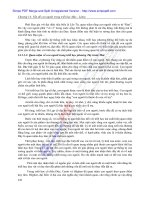
</div><span class="text_page_counter">Trang 17</span><div class="page_container" data-page="17"><small>Normal iron homeostasis in the reticuloendothelial macrophage. Macrophages phagocytoseaged or damaged red blood cells, using heme oxygenase 1 to release iron from heme, a </small>
<small>recycling process that accounts for approximately 90% of the body's daily iron needs. Iron is rapidly released to circulating transferrin or, when present in excess, stored in ferritin. When required, ferritin is degraded in the lysosomes via a process called ferritinophagy and the iron is released. Iron(II) is exported from the macrophage via ferroportin in the cell </small>
<small>membrane in a process coupled to reoxidation from iron(II) to iron(III) by membrane-bound ceruloplasmin. Iron(III) is then loaded onto transferrin for transport in the plasma.</small>
</div><span class="text_page_counter">Trang 18</span><div class="page_container" data-page="18"><b>The Effect of Inflammation on Iron Homeostasis</b>
•Patients with inflammatory conditions may have diminished iron stores, a situation described as “absolute iron deficiency.” As in patients without inflammation, this can arise due to low dietary iron intake, poor iron absorption, and/or blood loss .
•In some cases, however, there may be adequate iron stores, with normal levels of serum ferritin, but insufficient iron is delivered by transferrin to meet cells' demand, a situation termed “<b>functional iron deficiency</b>” .
•<b>Functional iron deficiency (or iron-restricted erythropoiesis) in inflammatory conditions is caused by elevated hepcidin levels</b>, triggered by inflammatory cytokines such as IL-6 . The consequent internalization and degradation of ferroportin lowers the amount of iron available for binding to transferrin. Accordingly, TSAT is
reduced .
</div><span class="text_page_counter">Trang 19</span><div class="page_container" data-page="19">•The increase in hepcidin levels in the presence of
inflammation can be profound (98ng/mL in patients with mild CHF , 270 ng/mL in CKD stages 2–4, and
577 ng/mL in active IBD). There is evidence that<b>levels of hepcidin correlate with the inflammatory marker CRP</b>but the relation between hepcidin levels and the severity of inflammatory diseases is complex, with
factors such as levels of stored iron and anemia playing a role.
•Other mechanisms can also affect iron homeostasis in the presence of inflammation . These include
<b>downregulation of transferrin expression by hepatocytes in response to increased levels of </b>
<b>circulating IL-6 and other proinflammatory cytokines and suppression of ferroportin mRNA .</b>
</div><span class="text_page_counter">Trang 20</span><div class="page_container" data-page="20"><b>Special Situations Affecting Serum Ferritin Levels</b>
<small>•</small> <b><small>Obese </small></b><small>patients : adiposity-related inflammation </small> <b><small>serum Ferritin , hepcidin</small></b>
<small>•</small> <b><small>Older</small></b> <small>patients : Low-grade inflammation </small> <b><small>serum Ferritin , hepcidin</small></b>
<small>•</small> <b><small>Cancer</small></b> <small>patients: chronic inflammatory </small> <b><small>serum Ferritin , IL-6 , CRP , hepcidin</small></b>
<small>•Hepatitis patients: </small><b><small>serum Ferritin </small></b>
<small>•</small> <b><small>Liver disease </small></b><small>patients: severity expression of ferroportiniron export from hepatocytes iron deposits in the liver hepcidin production </small>
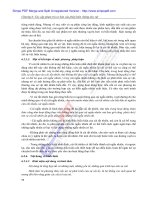
</div><span class="text_page_counter">Trang 21</span><div class="page_container" data-page="21"><b>Limitations of Serum Ferritin in Diagnosing Iron Deficiency in Inflammatory Conditions</b>
<small>•Patients with inflammatory conditions such as inflammatory bowel disease (45% of IBD), chronic heart failure (50% of CHF), and chronic kidney disease (24–85% of CKD) have high rates of iron deficiency with adverse clinical consequences. Under normal circumstances, serum ferritin levels are a sensitive marker for iron status but ferritin is an acute-phase reactant that becomes elevated in response to </small>
<small>inflammation, complicating the diagnosis. </small>
<small>•Proinflammatory cytokines also trigger an increase in hepcidin, which restricts uptake of dietary iron and promotes sequestration of iron by ferritin within storage sites. Patients with inflammatory conditions may thus have restricted availability of iron for erythropoiesis and other cell functions due to increased hepcidin expression, despite normal or high levels of serum ferritin. The standard </small>
<i><small>threshold for iron deficiency (<30 μg/L) therefore does not apply and </small></i><b><small>transferrin saturation (TSAT)</small></b><small>, a marker of iron availability, should also be assessed. A serum </small>
<i><small>ferritin threshold of <100 μg/L </small></i><b><small>or TSAT < 20% can be considered diagnostic for iron deficiency in CHF, CKD, and IBD.</small></b> <i><small>If serum ferritin is 100–300 μg/L,</small></i> <b><small>TSAT < 20% is required to confirm iron deficiency</small></b><small>. Routine surveillance of serum ferritin and TSAT in these at-risk groups is advisable so that iron deficiency can be </small>
<small>detected and managed.</small>
</div><span class="text_page_counter">Trang 22</span><div class="page_container" data-page="22"><b>Other Diagnostic Tests for Iron Deficiency</b>
<small>Where inflammation is present and serum ferritin with TSAT testing is inconclusive, other tests may be necessary </small>
</div><span class="text_page_counter">Trang 23</span><div class="page_container" data-page="23"><b>Thrombocytosis And Anemia: What Is The Link?</b>
<b>Causes of reactive thrombocytosis</b>
•Chronic inflammatory conditions
•<b>Iron deficiency anemia</b>
•Trauma or surgical removal of the spleen •Certain medications
•After surgery
•Some blood disorders
•Many other factors could contribute to the development of secondary thrombocytosis.
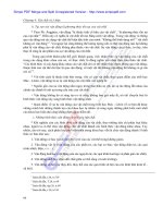
</div><span class="text_page_counter">Trang 24</span><div class="page_container" data-page="24"><b>Can iron deficiency anemia cause thrombocytosis?</b>
IDA can lead to increased platelet production, which can cause
thrombocytosis. The exact pathway of how IDA can lead to thrombocytosis is not fully mapped, but research studies suggest the following:
• The mother cells of platelets are called megakaryocytes, and the mother cells of red blood cells are called erythroid. Both megakaryocytes and erythrocytes are sensitive to the amount of iron in the body and share the same precursor progenitor cells (founder of the family). When
someone develops iron deficiency, these progenitor cells prefer to produce megakaryocytes (platelets) rather than erythroid (red blood
cells). Hence, the individual becomes anemic (fewer red blood cells) and develops thrombocytosis (more platelets).
• One additional theory has also been proposed. The development of thrombocytosis in IDA individuals may result from an evolutionary
adaptation to an iron deficiency caused by blood loss from an injury. The increase in platelet count would boost hemostasis, which aids blood
coagulation to stop the bleeding and helps the body heal from the injury.
</div><span class="text_page_counter">Trang 25</span><div class="page_container" data-page="25">