COMPUTATIONAL DISCOVERY OF VIRUSES AND THEIR HOSTS
Bạn đang xem bản rút gọn của tài liệu. Xem và tải ngay bản đầy đủ của tài liệu tại đây (11.21 MB, 167 trang )
<span class="text_page_counter">Trang 1</span><div class="page_container" data-page="1">
<small>UvA-DARE is a service provided by the library of the University of Amsterdam (http</small><i><small>s</small></i><small>://dare.uva.nl)</small>
Computational discovery of viruses and their hosts
Citation for published version (APA):
Kinsella, C. M. (2023). Computational discovery of viruses and their hosts. [Thesis, fully internal, Universiteit van Amsterdam].
General rights
It is not permitted to download or to forward/distribute the text or part of it without the consent of the author(s) and/or copyright holder(s), other than for strictly personal, individual use, unless the work is under an open content license (like Creative Commons).
Disclaimer/Complaints regulations
If you believe that digital publication of certain material infringes any of your rights or (privacy) interests, please let the Library know, stating your reasons. In case of a legitimate complaint, the Library will make the material inaccessible and/or remove it from the website. Please Ask the Library: or a letter to: Library of the University of Amsterdam, Secretariat, Singel 425, 1012 WP Amsterdam, The Netherlands. You will be contacted as soon as possible.
</div><span class="text_page_counter">Trang 4</span><div class="page_container" data-page="4"><b>Computational discovery of viruses and their hosts </b>
Cormac M. Kinsella
</div><span class="text_page_counter">Trang 5</span><div class="page_container" data-page="5">ISBN: 978-94-6483-273-0 © 2023 Cormac M. Kinsella
Layout and cover design: Cormac M. Kinsella
Chapter facing art: Kristel Parv Kinsella, inspired by the works of J. R. R. Tolkien Printing: Ridderprint, the Netherlands
The research reported in this doctoral thesis received financial assistance from the European Union’s Horizon 2020 research and innovation programme, under the Marie Skłodowska-Curie Actions grant agreement no. 721367 (HONOURs). Financial support for the printing of this thesis was kindly provided by the Amsterdam UMC.
</div><span class="text_page_counter">Trang 6</span><div class="page_container" data-page="6">Computational discovery of viruses and their hosts
ACADEMISCH PROEFSCHRIFT
ter verkrijging van de graad van doctor aan de Universiteit van Amsterdam op gezag van de Rector Magnificus
prof. dr. ir. P.P.C.C. Verbeek
ten overstaan van een door het College voor Promoties ingestelde commissie, in het openbaar te verdedigen in de Agnietenkapel
op maandag 11 september 2023, te 14.00 uur
door Cormac Michael Kinsella geboren te Harrow
</div><span class="text_page_counter">Trang 7</span><div class="page_container" data-page="7"><i><small>Promotor: </small></i> <small>dr. C.M. van der Hoek AMC-UvA </small>
<small>dr. A. Bart </small>
<small>AMC-UvA Tergooi Ziekenhuis </small>
<small>prof. dr. C.A. Russell </small>
</div><span class="text_page_counter">Trang 8</span><div class="page_container" data-page="8"><b>Chapter 1 </b>General introduction and scope of this thesis 7
<b>2 </b>Enhanced bioinformatic profiling of VIDISCA libraries
<i>for virus detection and discovery (Virus Research, 2019) </i>
19
<b>3 </b><i>Entamoeba and Giardia parasites implicated as hosts of CRESS viruses (Nature Communications, 2020) </i>
33
<b>4 </b>Host prediction for disease-associated gastrointestinal
<i>cressdnaviruses (Virus Evolution, 2022) </i>
57
<b>5 </b>Vertebrate-tropism of a cressdnavirus lineage implicated
<i>by poxvirus gene capture (PNAS, 2023) </i>
85
<b>6 </b>Human clinical isolates of pathogenic fungi are host to
<i>diverse mycoviruses (Microbiology Spectrum, 2022) </i>
</div><span class="text_page_counter">Trang 10</span><div class="page_container" data-page="10"><b>Chapter 1 </b>
<b>General introduction and scope of this thesis </b>
</div><span class="text_page_counter">Trang 11</span><div class="page_container" data-page="11"><b>The discovery of viruses, a distinct class of disease agents </b>
‘Virus’, derived from a Latin word meaning poison, has been used to non-specifically describe infectious disease agents for centuries<small>1</small>. When scientists in the 1800s came to understand that some microbes could cause disease, a flurry of cellular pathogens were isolated in pure culture by growing them on nutrient-rich matrices, allowing their
associations to disease to be directly tested under experimental conditions<small>2</small>. An assumption that culturable bacteria, fungi, and protists caused all infectious diseases took root. Usage of the term ‘virus’ remained non-specific into the early 1900s, with apparent oxymorons such as ‘bacterial viruses’ appearing<small>3</small> – meaning ‘bacterial agents of disease’ – not ‘viruses infecting bacteria’ as we might now understand it. However, in 1898 a key conceptual leap was made that would shape the modern conception of viruses, namely that a category of disease agents distinct from bacteria existed. First, work by Friedrich Loeffler and Paul Frosch showed that the causative agent of foot and mouth disease could pass through filters capable of holding back all known bacterial cells<small>4</small>. They postulated a very small, particulate agent of disease that was capable of replication (i.e., not a toxin). Secondly, Dutch
microbiologist Martinus Beijerinck showed that the agent causing tobacco mosaic disease could also pass filters<small>5</small>. Beijerinck proposed a non-bacterial identity for the agent, though he considered it to be liquid-like, or as he called it: “contagious living fluid”. A new class of agents known as ‘filterable viruses’ were thus recognised, and over the following decades non-specific usage of the terminology faded, until ‘filterable’ was also eventually dropped.
<b>What defines a virus? </b>
We now understand that viruses are not liquid-like, instead they are made up of infectious
<b>particles called virions. The small size of most virions explains why they can pass fine </b>
filters, though size does not define them. In fact, so-called ‘giant viruses’ have been found that are larger than the smallest bacteria<small>6,7</small><b>. More fundamentally, viruses are acellular but </b>
require cells to replicate, as they lack some of the necessary machinery for producing
<b>further generations. They are thus obligate intracellular parasites of host replication machinery, and must transmit between host cells to gain access to this. Virions represent </b>
individual virus units, such that in some cases a single virion can produce a new infection.
<b>At the least, virions possess a genome or genome segment of RNA or DNA, and some </b>
<b>proteins encoded by that genome. While these features define most known viruses, </b>
biological discoveries regularly complicate attempts at an all-encompassing yet restrictive definition. For example, one definition<small>8</small> splits biological entities into either ribosome-encoding or capsid-ribosome-encoding forms, i.e., cellular life and viruses respectively. However, viruses that lack capsids and encode other proteins are now known<small>9</small>, excluding them from this definition, and also from the viroids (virus-like elements that do not encode protein). Dropping the capsid requirement of the definition opens the door to other selfish genetic elements usually considered distinct from viruses, such as some transposons or plasmids. A clean definition is likely elusive, and given that viruses are a polyphyletic group (i.e., they did not all evolve from a single common ancestor) this should be expected. Individual
</div><span class="text_page_counter">Trang 12</span><div class="page_container" data-page="12">discoveries should therefore be evaluated in terms of how much their genetic relationships and biological behaviours overlap with those considered typically viral.
<b>The development of virus discovery techniques </b>
The visible effects of viruses have long been readily apparent to humans<small>10,11</small>, likely since our origin<small>12</small>. Experimentation with viruses also began before their nature was understood, for example Edward Jenner’s work on smallpox vaccination in the 1700s<small>13</small>. Virus discovery as a field arguably began with Loeffler, Frosch, and Beijerinck’s conclusions regarding filterable viruses<small>4,5</small>. By 1912, application of filtration techniques resulted in the discovery of at least 17 distinct viruses<small>14,15</small>, though detection and study was only possible via the diseases they induced. The subsequent development of virus discovery was tied to technological innovations enabling deeper characterisation and thus categorisation of filterable agents. Key early advances were the 1935 crystallisation of tobacco mosaic virus (TMV)<small>16</small>, the 1937 discovery of viral nucleic acids<small>17</small>, the 1939 electron microscope analysis of TMV<small>18</small>, and the 1941 application of X-ray crystallography techniques<small>19</small>. These enabled analysis of virus biochemistry and morphology.
Viruses only replicate in host cells, so early attempts to produce pure virus cultures in nutrient media were unsuccessful. Early propagation was done in whole organisms or eggs, and this had multiple drawbacks including bacterial contamination of stocks<small>20</small>. It was during a negative experiment aiming to grow pure vaccinia virus that Frederick Twort inadvertently established the first virus culture, though it was not vaccinia. Reporting in 1915<small>21</small>, Twort noticed that colonies of growing bacterial contaminants were killed off by a filterable, dilutable, infectious agent that could be propagated between colonies. Subsequent work from 1917 by Félix d'Hérelle named the ‘bacteriophages’ and properly established virus culture in bacterial cells, and specifically the plaque assay, as vital tools in virus research and discovery<small>22</small>. As eukaryotic tissue and cell culture techniques developed later in the 1900s, many viruses were discovered by inoculating cultures with infectious material and isolating agents<small>23–25</small>. Cell, tissue, or host tropism could also be tested using panels of different cell cultures<small>25</small>, something that Twort already comprehended in 1915 when testing bacteriophage host tropism<small>21</small>. With advances in immunology, the possibility to characterise isolated viruses by their antigenic or serological properties also developed<small>26</small>, and with this came the ability to test for viruses using immunoassays<small>25,27</small>. While two agents may share similar morphology and cytopathic effects, different responses to antibodies could distinguish ‘serotypes’.
By the 1970s scientists already had powerful tools to find and characterise new pathogenic viruses, but a revolution in molecular biology was underway. Restriction enzymes that cut DNA in specific locations had been isolated<small>28</small>, vital components of molecular cloning techniques that enabled amplification of specific nucleic acids<small>29</small>. In 1977 Frederick Sanger refined a technique for DNA sequencing and the first ever virus genome sequence was published, φX174<small>30,31</small>. This would eventually allow determination of comparative virus
</div><span class="text_page_counter">Trang 13</span><div class="page_container" data-page="13">relationships, but did not immediately overhaul virus discovery methods, as it required pure input DNA at high copy number, and was therefore limited to viruses established in culture or cloned fragments. In the 1980s the polymerase chain reaction (PCR) method was developed<small>32,33</small>, which enabled amplification of specific DNA sequences via multiple cycles
<i>of in vitro reactions. Because PCR utilises ‘primer’ sequences that match sections of a </i>
target, it could also be used to detect closely related targets<small>34</small>. Primers designed to target sequences highly conserved across an entire viral lineage have often been used to detect unknown members of the group<small>35</small>. However, detection range is limited by design, and more divergent viruses will not be found.
To solve this, advanced molecular biology techniques agnostic to virus sequence were applied. These included shotgun cloning, wherein total DNA from a sample was randomly sheared, and fragments were then cloned and Sanger sequenced<small>36,37</small>. As this could be applied to mixed samples containing nucleic acids from multiple organisms, it became known as ‘metagenomics’<small>37</small>. Representational difference analysis was another approach<small>38</small>, which disproportionately amplified nucleic acids found in one sample but not another (i.e., a virus found in a test sample, but not in a control sample). Similarly, techniques such as sequence-independent single primer amplification (SISPA) and virus discovery based on cDNA-amplified fragment length polymorphism (VIDISCA) used restriction enzymes to digest nucleic acids in control and test samples before amplification, with different nucleic acid fragments then visualised by gel electrophoresis<small>39,40</small>. Samples containing a new virus displayed unique nucleic acid fragments, which were then excised from the gel, cloned, and sequenced. Inclusion of a reverse transcription step converting RNA virus genomes to DNA enabled detection of either genome type, and further laboratory techniques could non-specifically enrich virus nucleic acids relative to background. These included centrifugation of samples to remove heavier cell debris, filtration of supernatants to remove other large particles, treatment with nucleases such as DNase to digest naked host chromosomal DNA, and use of selective primers during reverse transcription to reduce host ribosomal RNA levels<small>39–42</small>.
<b>Virus discovery with high-throughput sequencing </b>
Despite the maturation of virology during the 1900s, key issues remained at the turn of the millennium. One of these, discussed by Twort even in 1915<small>21</small>, was efficient identification of viruses that do not cause visible disease or cytopathic effect, and relatedly, how to find viruses infecting host species difficult to isolate in cell culture. While molecular techniques offered promising solutions, they remained low-throughput and logistically complex<small>36,38–40</small>. It would be the development of high-throughput sequencing (HTS) platforms in the 2000s<small>43</small>
that precipitated a major leap forward for virus discovery. Also known as massively parallel sequencing or next-generation sequencing, HTS techniques allow simultaneous sequencing of millions of DNA fragments in a processed sample known as a ‘library’. As the fragments overlap in their sequence content, they can be computationally ‘assembled’ together into longer sequences<small>44</small>, including whole virus genomes. Using sequence similarity detection
</div><span class="text_page_counter">Trang 14</span><div class="page_container" data-page="14">algorithms such as the basic local alignment search tool (BLAST)<small>45</small>, novel virus genomes can be identified. Because HTS requires no prior knowledge of target sequences and no cloning, it was readily integrated with metagenomic approaches<small>46</small> (i.e., metagenomic HTS), enabling discovery of apathogenic or unculturable viruses from any environment<small>47</small>. Complicating this, sequenced genomes can remain undetected if they are highly divergent from known viruses. While fast and sensitive protein similarity detection algorithms<small>48–50</small>
and even protein structure-based comparison tools<small>51</small> have pushed the limits of remote homology detection, scientists have not yet charted all virus sequence ‘dark matter’. Today, virus discovery techniques such as VIDISCA have been updated to take advantage of HTS technology (i.e., VIDISCA-NGS<small>42</small>), while further techniques have been
developed<small>52–54</small>. Overall, the importance of metagenomic HTS is such that it spawned the age of ‘viromic’ studies, aiming to sequence all viral genomes in a particular individual, community, or environment. The vast increase in data processing requirements drove advances in computational algorithms used in sequence analysis, and together these technologies have enabled discovery of hundreds to hundreds of thousands of virus genomes even within single reports<small>55–57</small>. With virus genome discovery now far outpacing the ability to characterise individual viruses in the laboratory, the International Committee on Taxonomy of Viruses (ICTV) recently took the step of allowing assignment of virus taxonomy to sequences acquired using metagenomic HTS alone<small>58</small>. Further, moving away from traditional characterisation metrics such as phenotype, taxonomy is now
recommended to centre around monophyletic evolutionary relationships, in effect prioritising genomic sequence information<small>59</small>.
<b>The host identity problem </b>
Over most of the history of virology, the identity of host species has been self-evident, because virus discovery efforts began with a host disease. With the metagenomic HTS revolution, this ‘host first’ identification order is reversed for most new viruses<small>58,60</small>. Many viruses today have a known genome sequence but an unknown host, referred to in parts of this thesis as ‘stray viruses’. At first glance this problem might appear simple; for example, we may conclude a novel virus discovered in the intestines of a person is a human-infecting virus. However, this is not always true. Microbe cells outnumber mammal cells in
humans<small>61</small>, and all of these can suffer virus infections. Many eukaryotic parasites live in mammalian guts<small>62</small>, and food contains numerous viruses capable of transiting the digestive system<small>63</small>. Most environments are analogous, in that the potential host diversity is high, and links between individual viruses and their specific hosts are obscured. This is an important challenge to solve, as without host information we cannot clearly conclude the medical or veterinary importance of stray viruses, and cannot contextualise their evolution.
Laboratory approaches to solve host identities vary in their utility. Attempting to isolate a stray virus in cell culture may be suitable when a specific host is suspected<small>64</small>, but is otherwise low-throughput and unlikely to succeed. Many potential host taxa have never
</div><span class="text_page_counter">Trang 15</span><div class="page_container" data-page="15">been isolated in culture, and no single laboratory maintains all established culture systems. More promisingly, library preparation techniques that compartmentalise samples at the level of single cells before sequencing allow capture of viruses inside specific identifiable organisms<small>65</small>. Other approaches such as proximity ligation link physically close nucleic acids<small>66</small> and can thus show which organism a virus is in. Methodologies include hybridisation of viral mRNA to host rRNA before sequencing<small>67</small>, and Hi-C<small>64</small>. As these techniques are done upstream of sequencing, they do not offer a solution for stray viruses identified using conventional HTS, i.e., the majority.
For stray viruses, computational methods of host identification are currently the most appropriate. Phylogenetic analysis is often used to find the most closely related virus with a known host, as host tropism is generally a conserved feature of viruses, allowing educated predictions<small>60</small>. Viruses often coevolve with their hosts, resulting in similar evolutionary branching patterns that may hold for millions of years<small>68</small>. However, accuracy of inferences depends on the degree of host switching in the lineage, the viral host range, and the degree of relatedness to viruses with determined hosts. Furthermore, it requires prior knowledge of some host identities across the viral lineage, information which is often absent. Many other approaches utilise similar prior knowledge<small>69,70</small>. For example, machine learning approaches train algorithms by analysing many genome sequences of viruses with known hosts, and then apply this to predict hosts in unknown cases<small>71</small>. This can be effective for lineages in which many host relationships are already known<small>72</small>, but it will never predict a host that does not occur in the training data. If available, host genome assemblies can partly solve these issues. Viruses occasionally leave genomic traces in host genomes, and detecting these can directly link virus lineages to hosts. In prokaryotic hosts, bacteriophage sequences are sometimes incorporated into clustered regularly interspaced short palindromic repeats (CRISPRs) for use in antiviral defence. Detecting CRISPR similarity to exogenous bacteriophages allows host inference<small>73</small>. In eukaryotic hosts that lack CRISPR, endogenous viral elements (EVEs) may offer an equivalent line of evidence. EVEs are occasionally generated upon infection of host germline cells, and can be vertically inherited as part of the genome for millions of years, allowing investigation of virus host ranges<small>74</small>.
<i><b>A host inference study system: the Cressdnaviricota </b></i>
As mentioned above, the first virus sequenced was φX174, which has a circular genome of single-stranded (ss)DNA and infects a prokaryote. This genomic arrangement was
previously thought extremely rare for viruses infecting eukaryotes. During the 1970s and 1980s two plant-pathogenic lineages were identified, the geminiviruses and
nanoviruses<small>75,76</small>. Both were notable for their small virion sizes, between 15 and 20 nanometers in diameter. Upon genome sequencing the two lineages were found to share a
<i>homologous Rep gene, indicating common ancestry between them</i><small>77</small>. In 1974 the only lineage known to infect vertebrates was found, the circoviruses<small>78,79</small>. Considerable interest in the group was raised when a globally important disease of pigs (postweaning multisystemic wasting syndrome) was found to be circovirus-induced<small>80</small>. In 2005 and 2010 additional
</div><span class="text_page_counter">Trang 16</span><div class="page_container" data-page="16">lineages causing cell lysis of diatoms and debilitation of a fungus were found, the bacilladnaviruses and genomoviruses respectively<small>81,82</small>. United by a similar genome
<i>organisation and a homologous Rep gene encoding a protein with both an endonuclease and </i>
a helicase domain, the acronym CRESS DNA (circular Rep-encoding single-stranded DNA) virus was coined to refer to them collectively<small>83</small>. Application of rolling circle amplification to enrich circular DNAs and metagenomic analysis gradually revealed CRESS viruses were widespread and diverse<small>54,83–88</small>, and numerous stray CRESS viruses have been found, including in association to disease<small>89–92</small>. At the outset of this thesis in November 2017, the five lineages mentioned above were all officially accepted families
<i>(named Geminiviridae, Nanoviridae, Circoviridae, Bacilladnaviridae, and Genomoviridae), and the unofficial family Kirkoviridae was proposed in the literature</i><small>89</small>. During work on this
<i>thesis, the Smacoviridae</i><small>93,94</small><i>, Redondoviridae</i><small>90</small><i>, and Metaxyviridae</i><small>95</small> were described by other authors and accepted as official families, while the unofficial lineages CRESSV1 to CRESSV6 were reported<small>96</small>, and likely represent further family-level clusters. In recognition
<i>of this rapidly expanding diversity, the virus phylum Cressdnaviricota was recently </i>
established<small>97</small>. Housing many stray virus lineages – including some associated to disease – the phylum represents an appropriate study system to develop host inference techniques.
<b>Scope of this thesis </b>
The aims of this thesis were to develop and apply computational approaches to both the
<i>discovery of viruses and the identification of their hosts. While the Cressdnaviricota were a </i>
major focus of this work, the overarching goal was to address challenges common across the virus discovery field. The intention is that this thesis will contribute to understanding the evolutionary history and biology of additional virus groups, and their current roles in disease.
Previous work in our laboratory established the library-preparation method VIDISCA-NGS as a powerful tool for enrichment and discovery of viruses. We developed a novel
<b>computational workflow for analysis of VIDISCA-NGS data, reported in chapter 2. In </b>
addition to field-standard sequence-similarity based approaches, the workflow was designed to leverage the reproducible production of specific restriction fragments from a given DNA template. The resulting ‘cluster-profiling analysis’ enabled identification of virus-like sequences even in the absence of detectable sequence similarity.
Application of the resulting computational workflow led to the discovery of previously
<b>unknown cressdnaviruses in human stool, reported in chapter 3. Determination of their </b>
<i>genetic relationships revealed three families, which we named Naryaviridae, Nenyaviridae, and Vilyaviridae, now officially recognised by the ICTV</i><small>98</small>. To identify their hosts, we applied case-control analyses of human stool samples, alongside analyses of host EVEs and small RNAs, and virus recombination. Hosts were identified as members of the important
<i>human parasite genera Entamoeba and Giardia. </i>
</div><span class="text_page_counter">Trang 17</span><div class="page_container" data-page="17">Building upon this work, we aimed to develop a computational workflow that required no training data and was capable of virus host prediction in the absence of host genome
<b>assemblies, reported in chapter 4. Focusing on cressdnaviruses, we first phylogenetically </b>
characterised additional unclassified lineages, resolving lineages CRESSV7 to CRESSV39. Examining disease-associated lineages found in the gastrointestinal tracts of humans and
<i>pigs, we predicted hosts of four, namely the Redondoviridae with Entamoeba gingivalis, Kirkoviridae with parabasalids including Dientamoeba, CRESSV1 with Blastocystis, and CRESSV19 with Endolimax. </i>
Horizontal gene transfer from viruses to hosts occasionally generates EVEs, which are
<b>useful for determination of virus host relationships. In chapter 5, we extended this concept </b>
to horizontal gene transfer between viruses, in a case where the host of one virus lineage
<i>was already known. We showed the cressdnavirus lineage CRESSV3 donated Rep genes to </i>
avipoxviruses, large dsDNA pathogens of birds and other saurians. This implied saurian
<i>hosts for CRESSV3, only the second cressdnavirus lineage after the Circoviridae </i>
recognised to infect vertebrates. We renamed this unofficial lineage as the family
<i>Draupnirviridae, and provided evidence that they first infected saurian hosts over 100 </i>
million years ago.
Some cressdnaviruses infecting fungi can induce debilitation and hypovirulence effects. In
<b>chapter 6, we carried out a virus discovery project on isolates of human-pathogenic fungi </b>
looking for further new species. While we did not identify cressdnaviruses infecting fungi, we did find a wide diversity of new RNA viruses in the cultures, including one from a lineage never previously confirmed as fungus-infecting.
<b>In chapter 7, the results are evaluated and possibilities for future work are discussed. </b>
</div><span class="text_page_counter">Trang 18</span><div class="page_container" data-page="18"><b>References </b>
<small>1. Horzinek, M. C. The birth of virology. Antonie Van Leeuwenhoek 71, 15–20 (1997). </small>
<small>2. Blevins, S. M. & Bronze, M. S. Robert Koch and the ‘golden age’ of bacteriology. Int. J. Infect. Dis. 14, e744–e751 (2010). </small>
<small>3. Rosenau, M. J. The inefficiency of bacterial viruses in the extermination of rats. in The rat and its relation to the public health (Public Health and Marine-Hospital Service of the United States, 1910). </small>
<small>4. Witz, J. A reappraisal of the contribution of Friedrich Loeffler to the development of the modern concept of virus. Arch. Virol. 143, 2261–2263 </small>
<small>7. La Scola, B. et al. A giant virus in amoebae. Science 299, 2033 (2003). </small>
<small>8. Raoult, D. & Forterre, P. Redefining viruses: Lessons from Mimivirus. Nat. Rev. Microbiol. 6, 315–319 (2008). 9. Ayllón, M. A. et al. ICTV virus taxonomy profile: Botourmiaviridae. J. Gen. Virol. 101, 454 (2020). </small>
<small>10. Saunders, K., Bedford, I. D., Yahara, T. & Stanley, J. The earliest recorded plant virus disease. Nature 422, 831–831 (2003). 11. Strouhal, E. Traces of a smallpox epidemic in the family of Ramesses V of the Egyptian 20th dynasty. Anthropologie 34, 315–319 (1996). 12. Enard, D., Cai, L., Gwennap, C. & Petrov, D. A. Viruses are a dominant driver of protein adaptation in mammals. Elife 5, e12469 (2016). 13. Jenner, E. An inquiry into the causes and effects of the variolæ vaccinæ, a disease discovered in some of the western counties of England, particularly Gloucestershire, and known by the name of the cow pox. (Sampson Low, 1798). </small>
<small>14. Flexner, S. Some problems in infection and its control. Science 36, 685–702 (1912). 15. Wolbach, S. B. The filterable viruses, a summary. Bost. Med. Surg. J. 167, 419–427 (1912). </small>
<small>16. Stanley, W. M. Isolation of a crystalline protein possessing the properties of tobacco-mosaic virus. Science 81, 644–645 (1935). </small>
<small>17. Bawden, F. C. & Pirie, N. W. The isolation and some properties of liquid crystalline substances from solanaceous plants infected with three strains of tobacco mosaic virus. Proc. R. Soc. London. Ser. B - Biol. Sci. 123, 274–320 (1937). </small>
<small>18. Kausche, G. A., Pfankuch, E. & Ruska, H. Die sichtbarmachung von pflanzlichem virus im übermikroskop. Naturwissenschaften 27, 292–299 (1939). </small>
<small>19. Bernal, J. D. & Fankuchen, I. X-ray and crystallographic studies of plant virus preparations. J. Gen. Physiol. 25, 111–165 (1941). 20. Noguchi, H. Pure cultivation in vivo of vaccine virus free from bacteria. J. Exp. Med. 21, 539–570 (1915). </small>
<small>21. Twort, F. W. An investigation on the nature of ultra-microscopic viruses. Lancet 186, 1241–1243 (1915). </small>
<small>22. D’Hérelle, F. Bacteriophage as a treatment in acute medical and surgical infections. Bull. N. Y. Acad. Med. 7, 329–348 (1931). 23. Hematian, A. et al. Traditional and modern cell culture in virus diagnosis. Osong Public Heal. Res. Perspect. 7, 77–82 (2016). </small>
<small>24. Enders, J. F., Weller, T. H. & Robbins, F. C. Cultivation of the lansing strain of poliomyelitis virus in cultures of various human embryonic tissues. Science 109, 85–87 (1949). </small>
<small>25. Hsiung, G. D. Diagnostic virology: From animals to automation. Yale J. Biol. Med. 57, 727–733 (1984). </small>
<small>26. Rowe, W. P., Huebner, R. J., Hartley, J. W., Ward, T. G. & Parrott, R. H. Studies of the adenoidal-pharyngeal-conjunctival (APC) group of viruses. </small>
<small>30. Sanger, F., Nicklen, S. & Coulson, A. R. DNA sequencing with chain-terminating inhibitors. Proc. Natl. Acad. Sci. 74, 5463–5467 (1977). 31. Sanger, F. et al. Nucleotide sequence of bacteriophage φX174 DNA. Nature 265, 687–695 (1977). </small>
<small>32. Saiki, R. K. et al. Enzymatic amplification of β-globin genomic sequences and restriction site analysis for diagnosis of sickle cell anemia. Science 230, 1350–1354 (1985). </small>
<small>33. Mullis, K. B. & Faloona, F. A. Specific synthesis of DNA in vitro via a polymerase-catalyzed chain reaction. Methods Enzymol. 155, 335–350 (1987). </small>
<small>34. Lane, D. J. et al. Rapid determination of 16S ribosomal RNA sequences for phylogenetic analyses. Proc. Natl. Acad. Sci. 82, 6955–6959 (1985). 35. Zhu, N. et al. A novel coronavirus from patients with pneumonia in China, 2019. N. Engl. J. Med. 382, 727–733 (2020). </small>
<small>36. Breitbart, M. et al. Genomic analysis of uncultured marine viral communities. Proc. Natl. Acad. Sci. 99, 14250–14255 (2002). </small>
<small>37. Rondon, M. R. et al. Cloning the soil metagenome: A strategy for accessing the genetic and functional diversity of uncultured microorganisms. Appl. Environ. Microbiol. 66, 2541–2547 (2000). </small>
<small>38. Nishizawa, T. et al. A novel DNA virus (TTV) associated with elevated transaminase levels in posttransfusion hepatitis of unknown etiology. Biochem. Biophys. Res. Commun. 241, 92–97 (1997). </small>
<small>39. Hoek, L. van der et al. Identification of a new human coronavirus. Nat. Med. 10, 368 (2004). </small>
<small>40. Allander, T., Emerson, S. U., Engle, R. E., Purcell, R. H. & Bukh, J. A virus discovery method incorporating DNase treatment and its application to the identification of two bovine parvovirus species. Proc. Natl. Acad. Sci. 98, 11609–11614 (2001). </small>
<small>41. Endoh, D. et al. Species-independent detection of RNA virus by representational difference analysis using non-ribosomal hexanucleotides for reverse transcription. Nucleic Acids Res. 33, e65 (2005). </small>
<small>42. de Vries, M. et al. A sensitive assay for virus discovery in respiratory clinical samples. PLoS One 6, e16118 (2011). </small>
</div><span class="text_page_counter">Trang 19</span><div class="page_container" data-page="19"><small>43. Margulies, M. et al. Genome sequencing in microfabricated high-density picolitre reactors. Nature 437, 376–380 (2005). 44. Myers, E. W. et al. A whole-genome assembly of Drosophila. Science 287, 2196–2204 (2000). </small>
<small>45. Altschul, S. F., Gish, W., Miller, W., Myers, E. W. & Lipman, D. J. Basic Local Alignment Search Tool. J. Mol. Biol. 215, 403–410 (1990). 46. Edwards, R. A. et al. Using pyrosequencing to shed light on deep mine microbial ecology. BMC Genomics 7, 1–13 (2006). </small>
<small>47. Angly, F. E. et al. The marine viromes of four oceanic regions. PLOS Biol. 4, e368 (2006). </small>
<small>48. Edgar, R. C. Search and clustering orders of magnitude faster than BLAST. Bioinformatics 26, 2460–2461 (2010). </small>
<small>49. Buchfink, B., Reuter, K. & Drost, H. G. Sensitive protein alignments at tree-of-life scale using DIAMOND. Nat. Methods 18, 366–368 (2021). 50. Karplus, K., Barrett, C. & Hughey, R. Hidden Markov models for detecting remote protein homologies. Bioinformatics 14, 846–856 (1998). 51. Söding, J., Biegert, A. & Lupas, A. N. The HHpred interactive server for protein homology detection and structure prediction. Nucleic Acids Res. 33, W244–W248 (2005). </small>
<small>52. Wylezich, C., Papa, A., Beer, M. & Höper, D. A versatile sample processing workflow for metagenomic pathogen detection. Sci. Rep. 8, 13108 (2018). </small>
<small>53. Conceiỗóo-Neto, N. et al. Modular approach to customise sample preparation procedures for viral metagenomics: A reproducible protocol for virome analysis. Sci. Rep. 5, 16532 (2015). </small>
<small>54. Tisza, M. J. et al. Discovery of several thousand highly diverse circular DNA viruses. Elife 9, e51971 (2020). 55. Shi, M. et al. Redefining the invertebrate RNA virosphere. Nature 540, 539–543 (2016). </small>
<small>56. Tisza, M. J. & Buck, C. B. A catalog of tens of thousands of viruses from human metagenomes reveals hidden associations with chronic diseases. Proc. Natl. Acad. Sci. 118, e2023202118 (2021). </small>
<small>57. Edgar, R. C. et al. Petabase-scale sequence alignment catalyses viral discovery. Nature 602, 142–147 (2022). 58. Simmonds, P. et al. Virus taxonomy in the age of metagenomics. Nat. Rev. Microbiol. 15, 161–168 (2017). 59. Simmonds, P. et al. Four principles to establish a universal virus taxonomy. PLOS Biol. 21, e3001922 (2023). </small>
<small>60. Wolf, Y. I. et al. Doubling of the known set of RNA viruses by metagenomic analysis of an aquatic virome. Nat. Microbiol. 5, 1262–1270 (2020). 61. Sleator, R. D. The human superorganism – of microbes and men. Med. Hypotheses 74, 214–215 (2010). </small>
<small>62. Patterson, Q. M. et al. Circoviruses and cycloviruses identified in Weddell seal fecal samples from McMurdo Sound, Antarctica. Infect. Genet. Evol. 95, 105070 (2021). </small>
<small>63. Victoria, J. G. et al. Metagenomic analyses of viruses in stool samples from children with acute flaccid paralysis. J. Virol. 83, 4642–4651 (2009). 64. Keeler, E. L. et al. Widespread, human-associated redondoviruses infect the commensal protozoan Entamoeba gingivalis. Cell Host Microbe 31, 58-68.e5 (2023). </small>
<small>65. Yoon, H. S. et al. Single-cell genomics reveals organismal interactions in uncultivated marine protists. Science 332, 714–717 (2011). 66. Marbouty, M., Baudry, L., Cournac, A. & Koszul, R. Scaffolding bacterial genomes and probing host-virus interactions in gut microbiome by proximity ligation (chromosome capture) assay. Sci. Adv. 3, e1602105 (2017). </small>
<small>67. Ignacio-Espinoza, J. C. et al. Ribosome-linked mRNA-rRNA chimeras reveal active novel virus host associations. bioRxiv (2020). 68. Aiewsakun, P. & Katzourakis, A. Marine origin of retroviruses in the early Palaeozoic Era. Nat. Commun. 8, 1–12 (2017). </small>
<small>69. Kapoor, A., Simmonds, P., Lipkin, W. I., Zaidi, S. & Delwart, E. Use of nucleotide composition analysis to infer hosts for three novel picorna-like viruses. J. Virol. 84, 10322–10328 (2010). </small>
<small>70. Ahlgren, N. A., Ren, J., Lu, Y. Y., Fuhrman, J. A. & Sun, F. Alignment-free d2* oligonucleotide frequency dissimilarity measure improves prediction of hosts from metagenomically-derived viral sequences. Nucleic Acids Res. 45, 39–53 (2017). </small>
<small>71. Mock, F., Viehweger, A., Barth, E. & Marz, M. VIDHOP, viral host prediction with deep learning. Bioinformatics 37, 318–325 (2021). 72. Eng, C. L. P., Tong, J. C. & Tan, T. W. Predicting host tropism of influenza A virus proteins using random forest. BMC Med. Genomics 7, S1 (2014). </small>
<small>73. Dion, M. B. et al. Streamlining CRISPR spacer-based bacterial host predictions to decipher the viral dark matter. Nucleic Acids Res. 49, 3127–3138 (2021). </small>
<small>74. Katzourakis, A. & Gifford, R. J. Endogenous viral elements in animal genomes. PLoS Genet. 6, e1001191 (2010). 75. Harrison, B. D. et al. Plant viruses with circular single-stranded DNA. Nature 270, 760–762 (1977). </small>
<small>76. Chu, P. W. G. & Helms, K. Novel virus-like particles containing circular single-stranded DNAs associated with subterranean clover stunt disease. Virology 167, 38–49 (1988). </small>
<small>77. Boevink, P., Chu, P. W. G. & Keese, P. Sequence of subterranean clover stunt virus DNA: Affinities with the geminiviruses. Virology 207, 354–361 (1995). </small>
<small>78. Ritchie, B. W., Niagro, F. D., Lukert, P. D., Steffens, W. L. & Latimer, K. S. Characterization of a new virus from cockatoos with psittacine beak and feather disease. Virology 171, 83–88 (1989). </small>
<small>79. Tischer, I., Rasch, R. & Tochtermann, G. Characterization of papovavirus and picornavirus-like particles in permanent pig kidney cell lines. Zenibl. Bukt. 226, 153–167 (1974). </small>
<small>80. Ellis, J. et al. Isolation of circovirus from lesions of pigs with postweaning multisystemic wasting syndrome. Can. Vet. J. 39, 44–51 (1998). 81. Nagasaki, K. et al. Previously unknown virus infects marine diatom. Appl. Environ. Microbiol. 71, 3528–3535 (2005). </small>
<small>82. Yu, X. et al. A geminivirus-related DNA mycovirus that confers hypovirulence to a plant pathogenic fungus. Proc. Natl. Acad. Sci. 107, 8387–8392 (2010). </small>
<small>83. Rosario, K. et al. Diverse circular ssDNA viruses discovered in dragonflies (Odonata: Epiprocta). J. Gen. Virol. 93, 2668–2681 (2012). 84. Rosario, K. & Breitbart, M. Exploring the viral world through metagenomics. Curr. Opin. Virol. 1, 289–297 (2011). </small>
<small>85. Rosario, K., Duffy, S. & Breitbart, M. Diverse circovirus-like genome architectures revealed by environmental metagenomics. J. Gen. Virol. 90, 2418–2424 (2009). </small>
<small>86. Siqueira, J. D. et al. Complex virome in feces from Amerindian children in isolated Amazonian villages. Nat. Commun. 9, 4270 (2018). 87. Blinkova, O. et al. Novel circular DNA viruses in stool samples of wild-living chimpanzees. J. Gen. Virol. 91, 74–86 (2010). </small>
</div><span class="text_page_counter">Trang 20</span><div class="page_container" data-page="20"><small>88. Breitbart, M. & Rohwer, F. Method for discovering novel DNA viruses in blood using viral particle selection and shotgun sequencing. Biotechniques 39, 729–736 (2005). </small>
<small>89. Li, L. et al. Exploring the virome of diseased horses. J. Gen. Virol. 96, 2721–2733 (2015). </small>
<small>90. Abbas, A. A. et al. Redondoviridae, a family of small, circular DNA viruses of the human oro-respiratory tract that are associated with periodontitis and critical illness. Cell Host Microbe 25, 719–729 (2019). </small>
<small>91. Phan, T. G. et al. The fecal virome of South and Central American children with diarrhea includes small circular DNA viral genomes of unknown origin. Arch. Virol. 161, 959–966 (2016). </small>
<small>92. Zhao, G. et al. Intestinal virome changes precede autoimmunity in type I diabetes-susceptible children. Proc. Natl. Acad. Sci. 114, E6166–E6175 (2017). </small>
<small>93. Varsani, A. & Krupovic, M. Smacoviridae: a new family of animal-associated single-stranded DNA viruses. Arch. Virol. 163, 2005–2015 (2018). 94. Ng, T. F. F. et al. A diverse group of small circular ssDNA viral genomes in human and non-human primate stools. Virus Evol. 1, vev017 (2015). 95. Gronenborn, B., Randles, J., HJ, V. & Thomas, J. Create one new family (Metaxyviridae) with one new genus (Cofodevirus) and one species (Coconut foliar decay virus) moved from the family Nanoviridae (Mulpavirales). Int. Comm. Taxon. Viruses Propos. number 2020.022P (2021). 96. Kazlauskas, D., Varsani, A. & Krupovic, M. Pervasive chimerism in the replication-associated proteins of uncultured single-stranded DNA viruses. Viruses 10, v10040187 (2018). </small>
<small>97. Krupovic, M. et al. Cressdnaviricota: A virus phylum unifying seven families of Rep-encoding viruses with single-stranded, circular DNA genomes. J. Virol. 94, e00582-20 (2020). </small>
<small>98. Krupovic, M. & Varsani, A. Naryaviridae, Nenyaviridae, and Vilyaviridae: Three new families of single-stranded DNA viruses in the phylum Cressdnaviricota. Arch. Virol. 167, 2907–2921 (2022). </small>
</div><span class="text_page_counter">Trang 22</span><div class="page_container" data-page="22"><b>Chapter 2 </b>
<b>Enhanced bioinformatic profiling of VIDISCA libraries for virus detection and discovery </b>
Cormac M. Kinsella, Martin Deijs, Lia van der Hoek
<i>Virus Research, 2019 </i>
</div><span class="text_page_counter">Trang 23</span><div class="page_container" data-page="23">
<b>Abstract </b>
VIDISCA is a next-generation sequencing (NGS) library preparation method designed to enrich viral nucleic acids from samples before highly-multiplexed low depth sequencing. Reliable detection of known viruses and discovery of novel divergent viruses from NGS data require dedicated analysis tools that are both sensitive and accurate. Existing software was utilised to design a new bioinformatic workflow for high-throughput detection and discovery of viruses from VIDISCA data. The workflow leverages the VIDISCA library preparation molecular biology, specifically the use of Mse1 restriction enzyme which produces biological replicate library inserts from identical genomes. The workflow performs total metagenomic analysis for classification of non-viral sequence including parasites and host, and separately carries out virus specific analyses. Ribosomal RNA sequence is removed to increase downstream analysis speed and remaining reads are clustered at 100% identity. Known and novel viruses are sensitively detected via alignment to a virus-only protein database, and false positives are removed. A new cluster-profiling analysis takes advantage of the viral biological replicates produced by Mse1 digestion, using read clustering to flag the presence of short genomes at very high copy number. Importantly, this analysis ensures that highly repeated sequences are identified even if no homology is detected, as is shown here with the detection of a novel gokushovirus genome from human faecal matter. The workflow was validated using read data derived from serum and faeces samples taken from HIV-1 positive adults, and serum samples from pigs that were infected with atypical porcine pestivirus.
<b>Highlights </b>
• A sensitive bioinformatic workflow for virus detection in VIDISCA data. • Flagging of possible novel viruses in unclassified reads using clustering. • Cluster-profiling analysis for reproducible sample comparison.
• Multiple analysis approaches provide extra utility to the user.
<b>Introduction </b>
The host range expansion of viral pathogens and emergence of novel species can pose substantial threats to human health (Parrish et al., 2008). Viruses evolve rapidly, possess high molecular diversity, and are found in relatively low concentration alongside host nucleic acids in most sample types. These factors complicate detection of novel viral genetic material and necessitate specific virus discovery methods to achieve sufficient detection sensitivity. Next-generation sequencing (NGS) and metagenomics have greatly accelerated the discovery of novel viruses when contrasted with traditional wet-lab virological techniques such as isolation in cell culture, as they can be performed on any
</div><span class="text_page_counter">Trang 24</span><div class="page_container" data-page="24">virus directly from biological or environmental samples, in a high-throughput way (Shi et al., 2018, 2016). Approaches that prioritise an unbiased metagenomic profile require high sequencing depth to ensure pathogen detection, and are therefore relatively expensive per viral nucleotide. The incorporation of virus enrichment techniques prior to sequencing reduces the required depth for detection (Conceiỗóo-Neto et al., 2015; de Vries et al., 2011), and may be desirable when processing tens to hundreds of samples.
VIDISCA is a virus discovery NGS library preparation method that enriches viral nucleic acids in samples before low depth Ion Torrent sequencing, allowing processing of 140 samples per week. The wet-lab procedure, described in detail elsewhere (de Vries et al., 2011; Edridge et al., 2018), is summarised here in order to highlight advantages for bioinformatic analysis. First, cells and debris are pelleted, and virus-containing supernatant is DNase treated to reduce residual cellular DNA. Virion proteins are linearised to release nucleic acid, which is extracted using the Boom method (Boom et al., 1990). RNA viruses are reverse transcribed using non-ribosomal RNA (rRNA) hexamer primers (Endoh et al., 2005), which reduce the proportion of rRNA transcribed into DNA. After second-strand synthesis, double-stranded DNA products are digested using the frequent cutting Mse1 restriction enzyme, an important feature unique to VIDISCA library preparation. Sequencing primers are ligated onto the two sticky ends of a restriction fragment, before size selection against both long and short fragments, amplification with PCR, and
sequencing with the Ion Torrent PGM platform (Thermo Fisher Scientific, Waltham, MA, USA).
The inclusion of Mse1 digestion during library preparation has advantageous implications for virus discovery bioinformatics. Viral genomes are short compared to their host, and can be at high copy number during infection. Since Mse1 reproducibly cuts homologous restriction fragments from genomes of the same type, high numbers of viral biological replicates with identical start and end sites are expected in library inserts prior to PCR. This is in contrast with a randomly fragmented library in which identical start and end sites are relatively rare. The VIDISCA insert redundancy is not expected from background or host nucleic acid, except that with ‘virus-like’ characteristics, i.e. high copy number, such as mitochondrial DNA. The virus replicates should result in characteristic redundancy in sequencing data, which can be identified via read clustering. Additionally, since Mse1 cuts TTAA sites, it cuts more rarely in GC rich rRNA (de Vries et al., 2011). Viable rRNA VIDISCA fragments are generally longer as a result, and can be disproportionately reduced during size selection, contributing to a high sensitivity that enables lower sequencing depth and analysis time. Recently VIDISCA was used to discover the suspected human pathogen Ntwetwe virus with 2 reads from 6,947, whereas an in-house Illumina workflow optimised for virus detection found only 8 reads among the 2,741,915 obtained (Edridge et al., 2018). Here we present a new bioinformatic workflow designed to process VIDISCA data. The core task is sensitive virus detection including false positive reduction. The workflow includes metagenomic analysis for identification of host background and non-viral
</div><span class="text_page_counter">Trang 25</span><div class="page_container" data-page="25">organisms including parasites, and collects descriptive metrics in order to flag unusual properties of samples, such as high rRNA content. It outputs text and interactive HTML results for detailed investigation of samples, and includes a new cluster-profiling analysis used to flag the presence of sequences at high copy number (e.g. virus infections). This analysis also provides an informative profile of sample content in different classification bins, including known and novel viruses, mitochondrial DNA, and background sequence. Notably, the flagging of highly repetitive reads does not rely on identity searches, ensuring that abundant unknown sequences can be identified. The utility of the workflow is
presented with examples.
<b>Materials and methods </b>
<b>2.1. Bioinformatic workflow for VIDISCA next-generation sequencing data </b>
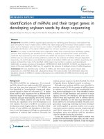
The new bioinformatic workflow for VIDISCA NGS data is summarised graphically (Fig. 1) and described in detail below. As input, the workflow takes FASTA formatted
sequences. Eukaryotic and prokaryotic virus protein databases used by the workflow were constructed in advance from respective NCBI Identical Protein Groups datasets, followed by clustering at 95% identity using CD-HIT v4.7 (Fu et al., 2012). First, metagenomic analysis of raw reads is carried out using Centrifuge v1.0.3 (Kim et al., 2016) against the pre-built NCBI non-redundant nucleotide Centrifuge index including known viruses, eukaryotes, and prokaryotes (February 2018). Centrifuge classification tables are visualised as interactive HTML charts using Recentrifuge (Martí, 2018).
Fig. 1. Schematic overview of the bioinformatic workflow for VIDISCA data, showing the main virus detection and discovery steps (orange), the metagenomic analysis (green), and visualisation processes (blue).
</div><span class="text_page_counter">Trang 26</span><div class="page_container" data-page="26">Next, the main virus detection steps are run. Reads from rRNA are separated from raw reads using SortMeRNA v2.1 (Kopylova et al., 2012). Non-rRNA reads are sorted by length and clustered at 100% identity using CD-HIT v4.7, and ‘clstr’ files are retained for later processing. Clustered non-rRNA reads are queried against the eukaryotic virus protein database using the UBLAST algorithm provided as part of the USEARCH v10 software package, with -mincodons set to 15, -accel to 0.8, and -evalue to 1e-4 (Edgar, 2010). Unmatched reads from this step are queried against the prokaryotic virus protein database, and those remaining unclassified are mapped to human, pig, and chicken mitochondrial DNA sequences using the BWA-MEM algorithm of BWA v0.7.17 (Li, 2013). Reads matching the eukaryotic virus protein database are treated as putatively viral, and are next queried against the NCBI nt. database (April 2018) using BLASTn v2.4.0 (Camacho et al., 2009). Those classified by BLASTn as viral are regarded as confident viral reads (classified as viral twice), those classified as non-viral are regarded as false positives, and those that remain unclassified are regarded as possible unknown viruses (classified as viral once). This information is used to split the UBLAST protein classification tables into the three categories, each of which are visualised separately as interactive HTML charts using KronaTools v2.7 (Ondov et al., 2011). The BLASTn classification of false positives is also visualised for inspection and comparison to the original viral classification.
Cluster-profiling outputs are produced using the CD-HIT ‘clstr’ files, which are converted into a table reporting the representative sequences, the number of reads clustered per representative, and the proportion of the original non-rRNA that each represents in a sample. The classification bin (such as ‘confident virus’, or ‘mitochondrial DNA’) of each representative read is then added to the table, including a bin for unclassified sequences. This output is plotted as a bar chart using ggplot2, with separate bars for classification bins, and representative reads stacked according to proportional amount of clustering (Wickham, 2016). The classification bins are ‘Virus (aa + nt)’ including reads classified as viral twice, ‘Virus (aa)’ including reads classified as viral once, ‘False pos. (nt)’ including reads removed as probable false positives, ‘Phage (aa)’ including reads aligning to our prokaryotic virus database, ‘MitoDNA’ including reads mapped to mitochondrial DNA references, ‘Centrifuge’ including reads identified by the metagenomic tool Centrifuge, and ‘No hit’ including reads with no assigned classification. The bar chart output provides a visual overview of the proportion of reads from a sample that were classified in a particular bin. Furthermore, reads that represent many other reads are visually identifiable due to their higher relative proportion. This allows the presence of clustering to be identified in each bin separately. Most repetitive non-viral sequences are accounted for via removal of rRNA and binning of mitochondrial DNA, however unclassified sequences putatively from viruses require manual inspection or full-length sequencing in order to establish their likely provenance.
For each classification bin, the 10 representative sequences accounting for the largest proportion of reads are automatically extracted as FASTA files for inspection, for example with BLASTx. All text tables and sample-specific files produced by the analysis are
</div><span class="text_page_counter">Trang 27</span><div class="page_container" data-page="27">packaged into sample folders, and descriptive metrics about the run time and classification performance for each sample are reported to a log file for later examination.
<b>2.2. Data selection and workflow testing </b>
Three VIDISCA datasets were selected and analysed using the new bioinformatic workflow, in order to assess specific aspects of workflow performance and utility. First, VIDISCA reads from 194 serum samples collected in 1994–1995 from HIV-1 infected adults were run. The aim was to determine whether the bioinformatic workflow outputs could be used to troubleshoot the likely causes of pathogen detection failure. This was done by comparison of HIV-1 detection by VIDISCA with pre-existing HIV-1 load data obtained using nucleic acid sequence based amplification (NASBA). Outputs from samples in which HIV-1 was unexpectedly not detected were manually inspected to determine the cause of failure.
Second, VIDISCA reads from 194 faecal samples from the above mentioned cohort were run (Oude Munnink et al., 2014). The aim was to test the prediction that cluster-profiling could be used to flag virus-like characteristics in unclassified reads, and therefore identify novel viruses at high load missed by classification algorithms. Cluster-profiling outputs were examined for evidence of clustering among unclassified reads and a single sample (F115) was selected for follow up. Illumina reads from a randomly fragmented library of the sample were downloaded from the European Nucleotide Archive (accession
ERR233419), cleaned of adapters, quality trimmed (minimum 50bp, sliding window trim < Q20) with Trimmomatic v0.38 (Bolger et al., 2014), and assembled using SPAdes v3.12 (Bankevich et al., 2012). The 10 unclassified VIDISCA representative sequences accounting for the most clustering were BLAST queried against the contigs, and the most common target sequence was extracted and manually curated.
Third, VIDISCA reads from 13 serum samples taken from sows experimentally infected with atypical porcine pestivirus (APPV) and 16 serum samples taken from the
transplacentally-infected piglets of the sows were run (de Groof et al., 2016). In this case, sequencing was carried out on an Ion Proton instrument (Thermo Fisher Scientific, Waltham, MA, USA). The aims were to statistically test support for the assumption that a higher viral load would result in higher clustering among viral reads, and to explore whether such an association was strongly influenced by PCR bias toward abundant templates. Since the dataset included individuals infected with the same virus strain at a large range of viral loads, this was carried out as a reliability test of the main assumption underlying cluster-profiling analysis, that VIDISCA library preparation selects for
biological replicates from identical genomes, resulting in read clustering associated with the biological load of a sequence.
</div><span class="text_page_counter">Trang 28</span><div class="page_container" data-page="28"><b>3. Results and discussion </b>
<b>3.1. Bioinformatic workflow design </b>
The new VIDISCA bioinformatic workflow has been designed to prioritise sensitivity to viruses, however non-virus metagenomics and the efficiency of analysis have also been
<i>considered. K-mer based metagenomic tools such as Kraken (Wood and Salzberg, 2014) </i>
are commonly used for pathogen detection, since they provide very rapid classification of
<i>reads via exact matches of length k between reads and reference indexes. Metagenomic </i>
samples often contain species with variable nucleotide identity to their most related
<i>reference sequence. Since k must be set in advance, high k decreases classification sensitivity for distantly related species, and low k decreases precision to well represented </i>
taxa. To circumvent this, the metagenomic software tool Centrifuge was selected for the
<i>workflow since it uses FM-indexed reference sequences, allowing k to be optimal for each </i>
individual read in a metagenomic sample, maximising both sensitivity and precision while simultaneously minimising index size and memory requirements (Kim et al., 2016). Detection of novel viruses is normally achieved via local alignment of reads to viral proteins, a computationally intensive operation. High speed algorithms are available to decrease analysis time, for example UBLAST (Edgar, 2010), DIAMOND (Buchfink et al., 2015), or Kaiju (Menzel et al., 2016). Minimisation of query reads and database size can provide additional gains. The VIDISCA workflow incorporates several of these speed-ups, including rRNA removal to reduce query reads, and redundancy removal in non-rRNA using clustering. Clustering information is retained for retrospective classification of redundant reads and cluster-profiling analysis. These steps reduced average protein query counts by 31% and 45% in the 194 faecal and 194 serum datasets respectively. A virus-only protein database was constructed and clustered for a size reduction of 81%. Alignment of reads to a taxonomically restricted database raises the likelihood of spurious hits due to chance similarity, therefore false positive removal via BLAST analysis against the NCBI nucleotide database is required. Due to the prior selection steps mentioned above, a minority of reads require this querying, for example an average of 1.5% and 2.4% of reads from the above faecal and serum datasets were queried.
<b>3.2. Assessment of the bioinformatic workflow performance </b>
The VIDISCA bioinformatic workflow was used to identify the causes of HIV-1 detection failure in data generated from archival serum samples collected from HIV-1 positive adults. Bioinformatic analysis detected the pathogen in 128 of 194 samples (66%) with an average of 42,124 total reads per sample. Of the VIDISCA negative samples, 23 (35%) had undetectable HIV-1 loads when specifically tested with NASBA, while 9 (7%) VIDISCA positive samples did. There was a median value of 84 HIV-1 copies/μl in VIDISCA positive samples and 14 in negative (Fig. 2A), suggesting detection failure was mostly attributable to viral load. Viral load was positively associated with the proportion of HIV-1 reads (Spearman’s rho = 0.61, p < .001), however the variance was poorly described by a
</div><span class="text_page_counter">Trang 29</span><div class="page_container" data-page="29">linear regression model (Fig. 2B), showing that sample dependent factors crucially impact the metagenomic profile. Notably, rRNA proportion was weakly but positively associated with HIV-1 proportion (Spearman’s rho = 0.34, p < .001), while the proportion of non-rRNA identified as human (including residual genomic DNA and cellular RNA) was found to have a weak negative association with the HIV-1 proportion (Spearman’s rho = -0.17, p = .017). Together these observations imply sample-specific biases against integrity or representation of the RNA fraction. Contributing factors could include higher degradation susceptibility during freeze-thaw cycles, high host DNA content with only partial degradation during DNase treatment, high intrinsic RNase activity in certain samples, or sample-specific inhibition of reverse transcription. An additional explanation could be that rRNA acts as a carrier for low concentrations of viral RNA.
Fig. 2. A: HIV-1 viral RNA load in serum and VIDISCA outcome. HIV-1 detection in sequence reads is indicated with HIV-1 (+), and lack of detection is indication with HIV-1 (-). On the x-axis the HIV-1 RNA load per μl of serum is plotted. B: Linear regression model fitted to HIV-1 viral load against HIV-1 reads as a percentage of total reads, F(1,192) = 56.68, p < .001, R<small>2</small> = 0.228. A low 23% of variance in proportion is explained by viral load when assuming a linear relationship.
HIV-1 was not detected in 11 outlier samples with over 50 HIV-1 copies/μl and an average read count of 40,290. In 3 of these, cluster-profiling showed that 78–90% of processed (non-rRNA) reads belonged to Hepatitis B virus, which commonly dominates VIDISCA metagenomic profiles if present. One sample also showed possible competition with Torque Teno virus which represented 30% of processed reads. A further 6 samples had
approximately 80–95% of processed reads classified by Centrifuge as host or bacterial sequence with very low read clustering, suggesting a highly diverse library insert
distribution probably derived from cell lysis. In the final sample an unusually high 75% of processed reads were not classified by any analysis. Manual BLAST analysis on some of
</div><span class="text_page_counter">Trang 30</span><div class="page_container" data-page="30">these unclassified reads gave bacterial hits or weak alignment scores suspected to originate from unknown bacteriophages, suggesting bacterial growth in the stored material.
<b>3.3. Cluster-profiling for virus discovery </b>
A cluster-profiling analysis was incorporated in the workflow based on the prediction that short viral genomes at high load would result in distinctive read clustering characteristics, since VIDISCA library preparation produces homologous library inserts from each genome based on its Mse1 restriction sites. The analysis uses read clustering and classification information generated as part of the workflow to generate a visual output, and therefore does not require significant additional computational time. Importantly, the clustering signal generated by high copy number sequences does not require identity-based
classification. This could potentially allow detection of highly divergent viruses with low protein identity to relatives represented in databases.
Cluster-profiling images generated using VIDISCA data from 194 faecal samples were analysed and sample F115 was selected for follow-up due to a high degree of clustering among unclassified reads – 12% of the 16,160 processed reads were clustered into only 100 unclassified representative sequences (Fig. 3), suggesting an unknown entity at high copy number. Available Illumina data from a randomly fragmented library of this sample were assembled into 9157 contigs. Ten unclassified representative VIDISCA sequences accounting for the most reads, which were automatically extracted by the workflow, were aligned to the contigs using BLAST. Of the 10, 8 aligned to a single contig, suggesting that they were part of a genome of a novel virus present at high copy number. Manual curation of this 5 kb sequence showed that it is a novel gokushovirus (circular ssDNA
bacteriophage, NCBI accession number MK263179) with 72% nucleotide identity to its closest relative. The sequences of this virus were not identified by the classification components of the workflow since the related viral proteins were not part of the reference set. Mapping of complete read-sets revealed that 6.83% of Illumina read-pairs from the sample were derived from the virus and 17.27% of VIDISCA reads were. The result confirms the expectation that viruses at high load produce characteristic clusters in VIDISCA data, ensuring that those missed by identity searches can still be detected.
</div><span class="text_page_counter">Trang 31</span><div class="page_container" data-page="31">Fig. 3. Cluster-profiling bar chart from sample F115. Representative sequences produced by read clustering are plotted according to their final classification bin (x-axis) and stacked in order of their relative abundance with respect to the original non-rRNA read set (i.e. the proportion of identical reads, y-axis). Coloured bars therefore signify those sequences representing many identical reads, while many singleton reads make up black regions. Classification bins on the x-axis are those described in section 2.1. Read clustering can be seen in the phage (‘Phage’, red), metagenomically identified (‘Centrifuge’, blue), and unclassified (‘No hit’, yellow) read bins.
<b>3.4. Association between viral read clustering and viral load </b>
Cluster-profiling analysis for discovery of viruses, as shown in Fig. 3, relies on a high level of sequence redundancy in order to generate a visible signal that can be investigated. A strong association between viral load and the level of clustering observed in viral reads is expected, an effect that would underlie application of the analysis to the discovery of novel
</div><span class="text_page_counter">Trang 32</span><div class="page_container" data-page="32">viruses. To test this assumption VIDISCA reads from 29 serum samples taken from pigs infected with APPV were analysed. The workflow detected APPV reads in 27 of these, and a strong linear association between viral load and the proportion of APPV reads was observed after removal of a single outlier (linear regression, F(1,26) = 70.57, p < .001, R<small>2</small> = 0.73). As expected, there was a strong association between viral load and the average number of reads clustered per APPV representative sequence (Spearman’s rho = 0.81, p < .001). To account for the possibility that this effect was due to stochastic PCR bias disproportionately amplifying abundant templates (Kebschull and Zador, 2015), an association between viral load and the proportion of all APPV reads that were represented by the top APPV sequence cluster was tested for. Since viral load should correspond to the abundance of replicate templates prior to PCR, PCR bias would be expected to occur in samples with the highest loads. No such relationship existed (Spearman’s rho = 0.17, p = 0.41).
Together the observations show that the degree of clustering among viral reads corresponds well with true biological load, and does not suffer from significant PCR bias toward abundant templates. While the analysis therefore can be applied to detection of novel viruses in unclassified reads, it is important to note that only infections with a high load and a high proportional amount of reads are likely to be observed. For example, it is unlikely that the analysis would have successfully flagged the presence of HIV-1 reads in the human serum samples analysed above, had they not been successfully classified using alignment tools. Nonetheless, it does provide an additional approach to both virus detection and the graphical representation of sample content, which are useful supplements to the more sensitive approaches utilised by the bioinformatic workflow.
<b>3.5. Conclusions </b>
A new bioinformatic workflow for sensitive virus detection and discovery in VIDISCA sequence data has been presented, which includes false positive removal and total metagenomic analysis. The workflow has been validated for virus detection in samples derived from individuals infected with known pathogens. The new cluster-profiling analysis, based on the VIDISCA library preparation molecular biology, has been used to flag a novel virus in unclassified reads, serving as a proof of concept for discovery of more divergent viruses.
<b>Data availability </b>
Code is available upon request. For example outputs from the pipeline, see the GitHub repository at:
</div><span class="text_page_counter">Trang 33</span><div class="page_container" data-page="33"><b>Acknowledgements </b>
This research has received funding from the European Union’s Horizon 2020 research and innovation programme, under the Marie Skłodowska-Curie Actions grant agreement no. 721367 (HONOURs). We would like to thank Dr. Ad de Groof of Intervet International BV for sharing APPV RT-qPCR data and Arthur W.D. Edridge for helpful feedback on the manuscript.
</div><span class="text_page_counter">Trang 34</span><div class="page_container" data-page="34"><b>References </b>
<small>Bankevich, A. et al., 2012. SPAdes: A new genome assembly algorithm and its applications to single-cell sequencing. J. Comput. Biol. 19, 455–77. Bolger, A.M., Lohse, M., Usadel, B., 2014. Trimmomatic: A flexible trimmer for Illumina sequence data. Bioinformatics 30, 2114–2120. Boom, R. et al., 1990. Rapid and simple method for purification of nucleic acids. J. Clin. Microbiol. 28, 495–503. </small>
<small>Buchfink, B., Xie, C., Huson, D.H., 2015. Fast and sensitive protein alignment using DIAMOND. Nat. Methods 12, 59–60. Camacho, C. et al., 2009. BLAST+: Architecture and applications. BMC Bioinformatics 10, 421. </small>
<small>Conceiỗóo-Neto, N. et al., 2015. Modular approach to customise sample preparation procedures for viral metagenomics: A reproducible protocol for virome analysis. Sci. Rep. 5, 16532. </small>
<small>de Groof, A. et al., 2016. Atypical porcine pestivirus: A possible cause of congenital tremor type A‐II in newborn piglets. Viruses 8, 271. de Vries, M. et al., 2011. A sensitive assay for virus discovery in respiratory clinical samples. PLoS One 6, e16118. </small>
<small>Edgar, R.C., 2010. Search and clustering orders of magnitude faster than BLAST. Bioinformatics 26, 2460–2461. </small>
<small>Edridge, A.W.D. et al., 2018. Novel orthobunyavirus identified in the cerebrospinal fluid of a Ugandan child with severe encephalopathy. Clin. Infect. Dis. </small>
<small>Endoh, D. et al., 2005. Species-independent detection of RNA virus by representational difference analysis using non-ribosomal hexanucleotides for reverse transcription. Nucleic Acids Res. 33, e65. </small>
<small>Fu, L., Niu, B., Zhu, Z., Wu, S., Li, W., 2012. CD-HIT: Accelerated for clustering the next-generation sequencing data. Bioinformatics 28, 3150–3152. Kebschull, J.M., Zador, A.M., 2015. Sources of PCR-induced distortions in high-throughput sequencing data sets. Nucleic Acids Res. 43, e143. Kim, D., Song, L., Breitwieser, F.P., Salzberg, S.L., 2016. Centrifuge: Rapid and sensitive classification of metagenomic sequences. Genome Res. 26, 1721–1729. </small>
<small>Kopylova, E., Noé, L., Touzet, H., 2012. SortMeRNA: Fast and accurate filtering of ribosomal RNAs in metatranscriptomic data. Bioinformatics 28, 3211–3217. </small>
<small>Li, H., 2013. Aligning sequence reads, clone sequences and assembly contigs with BWA-MEM. arXiv:1303.3997v1 [q-bio.GN]. Martí, J.M., 2018. Recentrifuge: Robust comparative analysis and contamination removal for metagenomic data. bioRxiv 190934. Menzel, P., Ng, K.L., Krogh, A., 2016. Fast and sensitive taxonomic classification for metagenomics with Kaiju. Nat. Commun. 7, 11257. Ondov, B.D., Bergman, N.H., Phillippy, A.M., 2011. Interactive metagenomic visualization in a Web browser. BMC Bioinformatics 12, 385. Oude Munnink, B.B. et al., 2014. Unexplained diarrhoea in HIV-1 infected individuals. BMC Infect. Dis. 14, 22. </small>
<small>Parrish, C.R. et al., 2008. Cross-species virus transmission and the emergence of new epidemic diseases. Microbiol. Mol. Biol. Rev. 72, 457–70. Shi, M. et al., 2018. The evolutionary history of vertebrate RNA viruses. Nature 556, 197–202. </small>
<small>Shi, M. et al., 2016. Redefining the invertebrate RNA virosphere. Nature 540, 1–12. Wickham, H., 2016. ggplot2: Elegant Graphics for Data Analysis. Springer-Verlag, New York. </small>
<small>Wood, D.E., Salzberg, S.L., 2014. Kraken: Ultrafast metagenomic sequence classification using exact alignments. Genome Biol. 15, R46. </small>
</div><span class="text_page_counter">Trang 36</span><div class="page_container" data-page="36"><b>Chapter 3 </b>
<i><b>Entamoeba and Giardia parasites implicated as </b></i>
<b>hosts of CRESS viruses </b>
Cormac M. Kinsella, Aldert Bart, Martin Deijs, Patricia Broekhuizen, Joanna Kaczorowska, Maarten F. Jebbink, Tom
van Gool, Matthew Cotton, Lia van der Hoek
<i>Nature Communications, 2020 </i>
</div><span class="text_page_counter">Trang 37</span><div class="page_container" data-page="37">
<b>Abstract </b>
Metagenomic techniques have enabled genome sequencing of unknown viruses without isolation in cell culture, but information on the virus host is often lacking, preventing viral characterisation. High-throughput methods capable of identifying virus hosts based on genomic data alone would aid evaluation of their medical or biological relevance. Here, we address this by linking metagenomic discovery of three virus families in human stool samples with determination of probable hosts. Recombination between viruses provides evidence of a shared host, in which genetic exchange occurs. We utilise networks of viral recombination to delimit virus-host clusters, which are then anchored to specific hosts using (1) statistical association to a host organism in clinical samples, (2) endogenous viral elements in host genomes, and (3) evidence of host small RNA responses to these elements.
<i>This analysis suggests two CRESS virus families (Naryaviridae and Nenyaviridae) infect Entamoeba parasites, while a third (Vilyaviridae) infects Giardia duodenalis. The trio </i>
supplements five CRESS virus families already known to infect eukaryotes, extending the CRESS virus host range to protozoa. Phylogenetic analysis implies CRESS viruses infecting multicellular life have evolved independently on at least three occasions.
<b>Introduction </b>
Determining hosts of viruses is integral to understanding their medical or ecological impact. This is particularly challenging for virus species discovered using metagenomic
sequencing, since samples such as stool or environmental matrices contain diverse potential hosts<small>1,2</small>. A decade of metagenomic studies have shown that viruses with circular Rep-encoding single-stranded DNA genomes (CRESS viruses) are highly diverse and pervasively distributed<small>3,4</small>, yet currently, the majority of known CRESS virus genetic diversity falls outside established families with characterised hosts<small>5</small>. Five CRESS virus
<i>families have experimentally confirmed eukaryotic hosts: Bacilladnaviridae, Circoviridae, Geminiviridae, Genomoviridae, and Nanoviridae</i><small>6</small>, respectively infecting diatoms<small>7</small>, vertebrates<small>8,9</small>, plants<small>10</small>, fungi<small>11</small> and plants<small>12</small>. Unclassified lineages of metagenomically identified CRESS diversity exist in at least six further clusters labelled CRESSV1 through CRESSV6, and a multitude of chimeric species difficult to place phylogenetically<small>13</small>. Unclassified CRESS viruses are frequently found in human and non-human primate stool samples, generating interest into their host specificity and potential impact on
health<small>14,15,16,17</small>. Classically, virus–host relationships are determined via recognition of host disease, followed by virus isolation in cell culture. Since this is impractical for
metagenomically identified viruses, case-control studies are used to reveal associations between viruses and disease. Importantly though, this does not confirm the host; for
<i>example, the CRESS virus family Redondoviridae is associated with human periodontal </i>
disease and critical illness<small>18</small>, but it remains unknown whether the viruses infect humans or a separate host, itself associated with or causing the observed clinical outcomes.
</div><span class="text_page_counter">Trang 38</span><div class="page_container" data-page="38">Genomic evidence of virus–host interactions can directly establish links between species.
<i>For instance, the Smacoviridae, a CRESS virus family previously assumed to infect </i>
eukaryotes, were recently suggested to infect archaea<small>19</small> on the basis of CRISPR spacer sequences matching a smacovirus inside the genome of an archaeon. Similarly, virus genomes can integrate into host genomes, leaving endogenous viral elements, identification of which reveals historical infections<small>20,21</small>. Searches for endogenous viral elements related to CRESS viruses have revealed integrations into the genomes of eukaryotes, for instance,
<i>sequences related to the replication-associated protein (Rep) of Geminiviridae, major global </i>
crop pathogens, are integrated in the tobacco genome<small>22</small>.
<i>Rep-like sequences are found in the genomes of the protozoan gut parasites Entamoeba histolytica and Giardia duodenalis</i><small>23</small>, important human pathogens belonging to distantly related genera<small>24</small>. The Rep-like elements could imply that the parasites host CRESS viruses, however, the sequences do not belong to a known family<small>3</small>. One proposed alternative hypothesis is that that they were gained from bacterial plasmids directly<small>23</small>, which are
<i>thought to be the ancestors of CRESS virus Rep genes</i><small>25</small>. Compatible with this, no sequence
<i>related to a capsid protein (Cap) has been found integrated in Entamoeba or Giardia </i>
genomes. While several studies have discussed or attempted to identify an association between CRESS viruses and gut parasites<small>3,26,27,28</small>—none has been found to date—and indeed no CRESS virus is known to infect any protozoan. Here we provide evidence that
<i>the parasite genera Entamoeba and Giardia are hosts of CRESS viruses, introducing a </i>
framework for host determination of metagenomically sequenced viruses that can be widely applied.
<b>Results </b>
<b>Unclassified CRESS viruses are associated to parasites in human stool </b>
Stool samples from 374 individuals (belonging to two independent cohorts, see "Methods") were enriched for viruses using the VIDISCA method, metagenomically sequenced, and bioinformatically analysed to identify unknown CRESS viruses. We used sequence assembly of short reads in combination with inverse PCR and Sanger sequencing to determine 20 full-length CRESS virus coding sequences (accessions MT293410.1– MT293429.1). The 20 sequences included 18 complete genomes covering all untranslated regions, and these had a genome organisation akin to known CRESS viruses, with a conserved nonanucleotide motif at an apparent replication origin, and open reading frames
<i>that aligned to viral Rep and Cap genes (Supplementary Table 1). Using PCR or mapping </i>
of sequencing reads to the assembled genomes, we determined that 21 of 374 samples were positive for the viruses.
<i>All 374 samples were also analysed for the presence of Entamoeba and Giardia parasites </i>
using either microscopy, sequencing-based approaches, PCR targeting the 18S ribosomal
</div><span class="text_page_counter">Trang 39</span><div class="page_container" data-page="39">RNA, or a combination thereof (see “Methods”). We observed that all 21 of the samples
<i>containing one of the CRESS viruses were also positive for either Entamoeba or Giardia </i>
(Table 1 and Supplementary Table 2). Across the 374 samples, presence of any of the 20
<i>viruses was significantly associated with Entamoeba or Giardia infection using Pearson’s chi-squared test (χ</i><small>2</small><i> = 36.77, p < 0.001), therefore we hypothesised that the viruses infected </i>
one or both of the parasites. To test the possible host role of other gut protozoa (including
<i>Blastocystis, Dientamoeba, Cryptosporidium and Endolimax among others), we carried out </i>
further parasitological typing on the 21 virus-positive samples (see “Methods”). We found these taxa were absent from all, or a majority of the 21 samples—implying they are not hosts of the viruses (Supplementary Table 2).
<i><b>Table 1: Entamoeba and Giardia status of human samples positive for any of the </b></i>
<b>CRESS viruses identified in this study. </b>
<b>Parasite status <sup>Number of samples </sup>(N = 374) </b>
<b>Positive for CRESS viruses identified in this study </b>
<i>Entamoeba positive only </i> 130 18
<i>Giardia positive only </i> 3 0
<i>Entamoeba and Giardia </i>
<i>Entamoeba and Giardia </i>
<b>Whole CRESS virus genomes are integrated into parasite genomes </b>
In order to identify endogenous viral elements related to the identified CRESS viruses, we aligned all 20 coding sequences to GenBank databases, namely the non-redundant
nucleotide (BLASTn, Supplementary Table 3), protein (BLASTx, Supplementary Table 4),
<i>and whole-genome shotgun contigs of Entamoeba and Giardia (BLASTn, Supplementary </i>
Table 5). Viral queries aligned with high identity and coverage to nucleotides and predicted proteins from parasite genomes, suggesting the presence of CRESS virus-derived
endogenous viral elements. The 20 viruses were not uniform in their database hits, showing genetic variation among them; each virus strongly aligned to sequences from either
<i>Entamoeba or Giardia, but not both, suggesting the presence of distinct viral lineages with </i>
independent virus–host relationships. Among viruses aligning to sequences from the
<i>Entamoeba genus, variability was also observed in the parasite species—queries either hit E. histolytica, E. dispar, E. nuttalli, or E. invadens. Among viruses aligning to sequences from Giardia duodenalis, alignments were found against major genotypes infecting </i>
humans, specifically A2 and B. Importantly, alignment to parasite genomes revealed
</div><span class="text_page_counter">Trang 40</span><div class="page_container" data-page="40">evidence of whole virus genome integrations. For example, one virus genome (accession
<i>MT293413.1) aligned inside an 11.6 kilobase (kb) contig from E. dispar </i>
(AANV02000527.1) with 100% query coverage and 84% nucleotide identity (Fig. 1a),
<i>while another (accession MT293421.1) aligned inside a 15.2 kb contig from G. duodenalis </i>
(AHGT01000120.1) with 99% query coverage and 73% nucleotide identity. As the only
<i>known examples of parasite endogenous viral elements containing both the Rep and Cap </i>
viral genes, they cast doubt on the hypothesis that Rep-like elements in protozoal genomes were derived from bacteria<small>23</small>. Since CRESS virus integration is likely mediated by the Rep protein during viral genome replication in the host nucleus<small>29</small>, the elements directly
<i>implicate Entamoeba and Giardia as hosts. </i>
<b>Fig. 1: Whole CRESS virus genomes are integrated in Entamoeba genomes. a Cropped </b>
<i>nucleotide alignment between Entamoeba dispar contig (AANV02000527.1) containing a complete virus integration and the genome of Entamoeba-associated CRESS DNA virus 1, </i>
isolate 84-AMS-03 (accession MT293413.1); also see Supplementary Fig. 2. Coloured vertical bars denote single nucleotide variations between the sequences (adenine = green, guanine = red, thymine = blue, cytosine = orange), with conservation across the alignment
<b>displayed below. b Dotplot of BLAT generated nucleotide alignment between endogenous </b>
<i>viral elements and flanking sequence from two closely related Entamoeba species (x-axis </i>
<b>sequence reverse complemented). c Example of the circular genome organisation of identified CRESS viruses. d Exogenous virus DNA is protected by a viral capsid, as it can </b>
be PCR-amplified after filtration and treatment with DNase (one independent experiment).
We next considered and eliminated potential sources of error, firstly, that parasite genomes did not truly contain CRESS endogenous viral elements, but rather that the assemblies were contaminated with virus genome sequences found in the original sample or reagents. To
<i>eliminate this possibility, we compared independently generated genome assemblies of E. histolytica and G. duodenalis, which were derived from parasite stocks in different </i>
laboratories or biobanks, and included strains isolated from patients across multiple countries and years. We could identify the same endogenous viral elements in several of the
</div>