Báo cáo Y học: Selection of effective antisense oligodeoxynucleotides with a green fluorescent protein-based assay Discovery of selective and potent inhibitors of glutathione S-transferase Mu expression doc
Bạn đang xem bản rút gọn của tài liệu. Xem và tải ngay bản đầy đủ của tài liệu tại đây (507.69 KB, 10 trang )
Selection of effective antisense oligodeoxynucleotides with a green
fluorescent protein-based assay
Discovery of selective and potent inhibitors of glutathione
S
-transferase Mu expression
Peter A. C. ¢t Hoen
1,2
, Bram-Sieben Rosema
1
, Jan N. M. Commandeur
2
, Nico P. E. Vermeulen
2
,
Muthiah Manoharan
3
, Theo J. C. van Berkel
1
, Eric A. L. Biessen
1
and Martin K. Bijsterbosch
1
1
Division of Biopharmaceutics, Leiden/Amsterdam Center for Drug Research, the Netherlands;
2
Division of Molecular Toxicology,
Leiden/Amsterdam Center for Drug Research, the Netherlands;
3
ISIS Pharmaceuticals, Carlsbad, California, USA
Antisense oligodeoxynucleotides (AS-ODNs) are frequently
used for the down-regulation of protein expression. Because
the majority of potential antisense sequences lacks effect-
iveness, fast screening methods for the selection of effective
AS-ODNs are needed. We describe a new cellular screening
assay for the evaluation of the potency and specificity of new
antisense sequences. Fusion constructs of the gene of interest
and the gene encoding the enhanced green fluorescent pro-
tein (EGFP) are cotransfected with AS-ODNs to COS-7
cells. Subsequently, cells are analysed for expression of the
EGFP fusion protein by flow cytometry. With the assay, we
tested the effectiveness of a set of 15 phosphorothioate
ODNs against rat glutathione S-transferase Mu1 (GSTM1)
and/or Mu2 (GSTM2). We found several AS-ODNs that
demonstrated potent, sequence-specific, and concentration-
dependent inhibition of fusion protein expression. At 0.5 l
M
,
AS-6 and AS-8 inhibited EGFP–GSTM1 expression by
95 ± 4% and 81 ± 6%, respectively. AS-5 and AS-10 were
selective for GSTM2 (82 ± 4% and 85 ± 0.4% decrease,
respectively). AS-2 and AS-3, targeted at homologous
regions in GSTM1 and GSTM2, inhibited both isoforms
(77–95% decrease). Other AS-ODNs were not effective or
displayed non-target-specific inhibition of protein expres-
sion. The observed decrease in EGFP expression was
accompanied by a decrease in GSTM enzyme activity. As
isoform-selective, chemical inhibitors of GSTM and GSTM
knock-out mice are presently unavailable, the selected
AS-ODNs constitute important tools for the study of the
role of GSTM in detoxification of xenobiotics and protec-
tion against chemical-induced carcinogenesis.
Keywords: antisense oligodeoxynucleotide; carcinogenesis;
genetic polymorphism; glutathione S-transferase; green
fluorescent protein.
Antisense oligodeoxynucleotides (AS-ODNs) are frequently
used for the down-regulation of gene expression, both
in vitro and in vivo [1–4]. Due to the low stability of
phosphodiester ODNs (PO-ODNs) in biological systems,
more stable oligonucleotide analogues with a variety of
chemical modifications have been developed [5,6]. ODNs
with a phosphorothioate-modified backbone (PS-ODNs)
are the most commonly used AS-ODNs. As AS-ODNs act
via Watson–Crick base pairing with their target mRNAs,
the nucleotide sequence of the target gene is in principle
sufficient information for the design of AS-ODNs. It
appears, however, that not all AS-ODNs are potent
inhibitors of protein expression. In studies where large sets
of PS-ODNs, directed against a single target gene, were
tested for their ability to down-regulate their target mRNA
and protein in cell culture [7,8], only 5–10% of the sequences
tested appeared to be effective. Thus there is a need for rapid
and accurate screening assays for the selection of effective
and specifically acting AS-ODNs.
Screening for effective antisense sequences is usually
performed in cell-free systems or in cell culture. Several cell-
free assay systems have been described [9]. These assays are
fast, but not always reliable predictors for activity in
biological systems. Use of differentiated cells generates more
relevant information on the effectiveness of AS-ODNs in
physiological systems. However, cellular assays are fre-
quently hampered by low or irreproducible transfection of
oligonucleotides. Furthermore, each new target requires the
set-up and optimization of target-specific assays. Therefore,
we developed a new assay that uitilizes fusion constructs of a
particular gene with the gene encoding enhanced green
fluorescent protein (EGFP) as reporter. Because the
screening is based on flow cytometric detection of EGFP
expression, there is no need for development of target-
specific assays. Reproducible transfections are achieved by
using an easy transfectable cell line and the cotransfection of
antisense PS-ODNs directed against the gene of interest and
plasmids encoding the chimeric gene. In the present assay,
Correspondence to M. K. Bijsterbosch, Leiden/Amsterdam Center for
Drug Research, Division of Biopharmaceutics, PO Box 9502,
2300 RA Leiden, the Netherlands.
Fax: +31 71 5276032, Tel.: +31 71 5276038,
E-mail:
Abbreviations: ODN, oligodeoxynucleotide; AS, antisense; PO:
phosphodiester; PS, phosphorothioate; EGFP: enhanced green
fluorescent protein; GST, glutathione S-transferase; GSTM, gluta-
thione S-transferase Mu; CDNB, 1-chloro-2,4-dinitrobenzene; GSH,
glutathione; DOTAP, N-[1-(2,3-dioleoyloxy)propyl]-N,N,N-
trimethylammonium; PMSF, phenylmethanesulfonyl fluoride;
TRITC, tetramethylrhodamine isothiocyanate; DMEM, Dulbecco’s
modified Eagle’s medium; FACS: fluorescence activated cell sorter.
Enzymes: glutathione S-transferase (EC 2.5.1.18).
(Received 13 December 2001, revised 4 April 2002,
accepted 9 April 2002)
Eur. J. Biochem. 269, 2574–2583 (2002) Ó FEBS 2002 doi:10.1046/j.1432-1033.2002.02924.x
only transfected cells are analysed, thus eliminating the
background of expression in untransfected cells.
With this assay, we determined the effectiveness of a set of
antisense PS-ODNs directed against the glutathione
S-transferase Mu1 (GSTM1) and Mu2 (GSTM2) isoforms
of the rat. GSTs play an important role in the detoxification
of DNA- and/or protein-reactive compounds by catalyzing
the conjugation of electrophilic groups with the tripeptide
glutathione [10]. Approximately 50% of the Caucasian
population is deficient for GSTM1, the human orthologue
of rat GSTM1 and GSTM2 [11]. Meta-analyses of epide-
miological studies reveal that this deficiency is associated
with an increased risk of lung and colorectal cancer,
especially when the GSTM-null genotype is combined with
high-inducibility of cytochrome P450 1A1 [12–18]. It would,
however, be very important to demonstrate directly the
effect of differences in GSTM expression levels on the
prevalence of cancer biomarkers in in vitro andinanimal
models. As GSTM knock-out mice are still unavailable,
temporal modulation of the expression of GSTM isoforms
by AS-ODNs in relevant in vitro and in vivo models is an
attractive possibility. In the current paper, several target-
specific AS-ODNs are selected from a set of 15 PS-ODNs.
These ODNs selectively inhibit the expression of GSTM1
and/or GSTM2 and can be used to study the influence of
reduced GSTM expression on the detoxification of xeno-
biotics and protection against chemical-induced carcino-
genesis.
MATERIALS AND METHODS
Materials
PCR primers were from Eurogentec, Seraing, Belgium.
PS-ODNs were synthesized according to standard phos-
phoramidite chemistry. The pEGFP-C1 plasmid and the
rabbit anti-EGFP Living Colors Peptide antibody were
from Clontech. VentÒ DNA Polymerase was from New
England Biolabs. 1-chloro-2,4-dinitrobenzene (CDNB),
glutathione (GSH), dithiothreitol, N-[1-(2,3-dioleoyl-
oxy)propyl]-N,N,N-trimethylammonium salt (DOTAP),
and propidium iodide were from Sigma. Phenyl-
methanesulfonyl fluoride (PMSF) and Tween-20 were from
Merck. Tetramethylrhodamine isothiocyanate 5,6-mixed
isomers (TRITC) was from Molecular Probes. Cell culture
agents were from BioWhittaker. Milkpowder was from
Campina Melkunie (Eindhoven, the Netherlands). A
horseradish peroxidase-conjugated antirabbit IgG antibody
and an enhanced chemiluminescence assay were purchased
from Amersham Pharmacia Biotech. All other chemicals
were of analytical grade.
Cloning of GSTM cDNAs into pEGFP-C1
Full-length cDNAs encoding GSTM1 (bases )21 to
+1039; GenBank accession no. X04229, cloned in the PstI
site of pBR322) and GSTM2 (bases )2 to +1036; GenBank
accession no. J03914, derived mRNA sequence, cloned in
the EcoRI site of pUC18) were kindly provided by D. Tu,
Pennsylvania State University, PA, USA. The cDNA
inserts were amplified and isolated by the sticky-end PCR
method [19]. Briefly, the cDNAs were amplified with two
sets of PCR primers (forA + revA and forB + revB) for
each plasmid using Vent DNA polymerase (Table 1). The
two PCR products were subjected to melting and cooling.
Four different double-stranded products were obtained, one
of which had the correct 5¢-EcoRI and 3¢-BamHI over-
hanging ends. The PCR products were cloned into the
EcoRI- and BamHI-digested pEGFP-C1 plasmid to gener-
ate the C-terminal fusion constructs pEGFP-M1 and
pEGFP-M2. Sequencing of the plasmids confirmed the
in-frame ligation of the GSTM cDNAs and the absence of
any PCR-induced mistakes in the inserts.
TRITC-labelling of ODN
A 24-mer PO-ODN, provided with three PS-linkages at the
5¢-end and a 3¢-end primary amino group (sequence:
T*A*A*GCTGTCCCGGGGTCTACGGCC), was label-
led with TRITC by incubating 15 nmol ODN in 500 lL
0.1
M
Na-carbonate buffer (pH 9.0) with 10 molar equiv-
alents of TRITC (dissolved in dimethylformamide at
2mgÆmL
)1
). The mixture was incubated overnight with
shaking at room temperature. The TRITC-labelled ODN
was separated from unreacted TRITC by gel filtration on a
Sephadex G-25 column (20 · 0.4 cm), eluted with water.
The TRITC-ODN was precipitated from the eluent by
adding 0.01 vols 1
M
MgCl
2
,0.1vols3
M
NaAc pH 5.2,
and 3 vols cold ethanol. The precipitate was formed by
overnight incubation at )20 °C and centrifugation for
30 min at 13000 g at 4 °C. The pellet was washed three times
with 80% EtOH and subsequently dissolved in deionized
water. The purity and identity of the TRITC-labelled ODN
were checked by PAGE under denaturing conditions.
Cell culture and transfection
COS-7 cells (European Collection of Cell Cultures, Salis-
bury, UK) were grown at 37 °Cina5%CO
2
atmosphere in
Dulbecco’s modified Eagle’s medium (DMEM) containing
10% (v/v) fetal bovine serum, 2 m
ML
-glutamine,
100 UÆmL
)1
penicillin and 100 lgÆmL
)1
streptomycin. At
24 h before transfection, cells were seeded in 12-wells plates
Table 1. Primers used for sticky-end PCR. Separate PCR reactions
were carried out with the following primer sets: GSTM1-forA and
GSTM1-revA; GSTM1-forB and GSTM1-revB; GSTM2-forA and
GSTM2-revA; GSTM2-forB and GSTM2-revB. Products from the
first two PCR reactions were combined, melted and reannealed to give
four GSTM1 cDNA products, of which one has the right EcoRI/
BamHI overhanging ends (underlined) to enable ligation in EcoRI and
BamHI restricted pEGFP-C1. Combining the last two PCR reactions
results in the formation of GSTM2 cDNA with EcoRI/BamHI over-
hanging ends (underlined) which was cloned in a similar way into
pEGFP-C1.
Primer Sequence
GSTM1-forA 5¢-AATTCCATGCCTATGATACTGGGAT-3¢
GSTM1-forB 5¢- CCATGCCTATGATACTGGGAT-3¢
GSTM1-revA 5¢- CTAAAGATGAGACAGGCCTGG-3¢
GSTM1-revB 5¢-GATCCTAAAGATGAGACAGGCCTGG-3¢
GSTM2-forA 5¢-AATTCGATGCCTATGACACTGGGTTAC-3¢
GSTM2-forB 5¢- CGATGCCTATGACACTGGGTTAC-3¢
GSTM2-revA 5¢- CGTGGTTCACACTTTATTGCAAATC-3¢
GSTM2-revB 5¢-GATCCGTGGTTCACACTTTATTGCAAATC-3¢
Ó FEBS 2002 EGFP-based selection of antisense sequences (Eur. J. Biochem. 269) 2575
at a density of 1 · 10
5
cells/well,whichresultedincultures
that were approximately 50% confluent at the day of
transfection. For cotransfections of plasmid and ODN, the
appropriate amount of plasmid [diluted to a concentration
of 0.2 lgÆlL
)1
in HBS (0.15
M
NaCl in 20 m
M
Hepes,
pH 7.4)] was mixed with the appropriate amount of ODN
(diluted to a concentration of 0.2 lgÆlL
)1
in HBS). Subse-
quently, a transfection mixture was prepared by slowly
adding the plasmid/ODN mixture to a solution of DOTAP
in HBS (1 lgÆlL
)1
; charge ratio DNA : DOTAP ¼ 1:5).
The DNA and DOTAP solutions were mixed by repeated
pipetting. The transfection mixture was diluted with HBS to
a total volume of 100 lL, and incubated for 15 min at room
temperature. Then, the culture medium was taken from the
cells and replaced by 400 lL of DMEM without serum or
antibiotics, and 100 lL of the transfection mixture was
slowly added. After 4 h, the transfection mixture was
removed from the cells, and serum-containing medium
was added. All analyses were performed after culture in the
serum-containing medium for a further 18 h.
Flow cytometry
Cells were detached from the culture plates with trypsin,
centrifuged for 5 min at 400 g, washed once with 1 mL
NaCl/P
i
, and dispersed in 1 mL NaCl/P
i
. Immediately
before FACS analysis, 3 lL1l
M
propidium iodide was
added. Cellular fluorescence of approximately 3000 cells was
determined in a Becton Dickinson FACS Calibur flow
cytometer. The EGFP signal was detected in the FL-1
channel; TRITC and propidium iodide signals were detected
in the FL-3 channel. Only single cells were gated in forward/
sideward scatter plots; dead cells were excluded from the
analysis by gating of propidium iodide-positive cells.
GST activity assay
COS-7 cells were transfected with either pEGFP, pEGFP-
M1 or pEGFP-M2 as described above. Then, the cells were
washed twice with NaCl/P
i
and lysed in 300 lL10m
M
sodium phosphate buffer (pH 7.4) containing 2 m
M
dithio-
threitol, 1 m
M
EDTA, and 50 l
M
PMSF. The lysates were
homogenized by short sonication. Total GST activity was
analysed in a CDNB conjugation assay, essentially as
described before [20]. The assay makes use of the GST-
catalyzed addition of GSH to CDNB. The CDNB–GSH
conjugate formed can be measured spectrophotometrically.
To this end, 50 lL of protein lysate ( 10 lgprotein,
concentration determined with the Bradford protein assay
[21]) was incubated with 150 lL of a solution of 1.67 m
M
CDNB in 0.1
M
potassium phosphate buffer pH 6.5. Lysis
buffer, instead of lysate, was used as a blank. The reaction
was started by the addition of 50 lL5m
M
GSH dissolved
in potassium phosphate buffer pH 6.5. CDNB–GSH con-
jugate formation was monitored over time with a Perkin-
Elmer HTS7000 bioassay plate reader at 340 nm and 37 °C.
The rate of conjugate formation was constant 15–45 min
after the addition of GSH.
Western blotting
Lysates of COS-7 cells transfected with pEGFP, pEGFP-
M1 or pEGFP-M2, prepared as described above, were
analysed for GFP expression by Western blotting. Five lg
total cellular protein, dissolved in denaturing loading buffer
(62 m
M
Tris/HCl pH 6.8, 12.5% v/v glycerol, 1.25% w/v
SDS, 2.5% v/v 2-mercaptoethanol, and 0.25% w/v Bromo-
phenol blue) were heated for 4 min at 96 °C, and subjected
to gel electrophoresis in an SDS/15% polyacrylamide gel.
Proteins were blotted overnight at 4 °C onto a nitrocellulose
membrane at a current of 76 mA. Thereafter, the nitrocel-
lulose membrane was incubated for 1 h in blocking buffer,
consisting of 10 m
M
Tris/HCl pH 8.0, 150 m
M
NaCl,
0.5 m
M
CaCl
2
, 5% w/v milkpowder, 1% w/v BSA, 0.25%
v/v Tween-20. Then, the membrane was incubated for 1 h at
room temperature with the primary anti-EGFP antibody
(100 · diluted in blocking buffer without milk powder,
containing 0.5% v/v Tween-20). The membrane was
washed 10 times with NaCl/P
i
containing 0.02% v/v
Tween-20, and incubated with a horseradish peroxidase-
conjugated donkey antirabbit IgG (10 · dilutedin10·
diluted blocking buffer). EGFP was detected by an
enhanced chemiluminescence assay, according to the
manufacturer’s protocol.
Statistical analysis
Data were analysed statistically for significance with a one
or two sample student t-test.
GRAPHPAD INSTAT
Software
version 3.00, GraphPad Software Inc. (San Diego, CA,
USA), was used for this purpose.
RESULTS
Cloning of EGFP–GSTM fusion constructs
Cellular screening of antisense sequences for their potential
to inhibit gene expression is often complicated by irrepro-
ducible transfection procedures and lack of good quantita-
tive assays for monitoring of gene expression. To
circumvent these problems, we developed a screening assay,
based on fusion proteins of the target protein with EGFP,
that enables accurate determination of the effects of
AS-ODNs by flow cytometry. C-terminal fusion constructs
of GSTM1 and GSTM2 with EGFP (named pEGFP-M1
and pEGFP-M2, respectively) were made by ligating PCR-
amplified cDNAs, coding for GSTM1 and GSTM2 (PCR
primers in Table 1), into the multiple-cloning site of the
pEGFP-C1 vector. Sequence analysis confirmed a correct
in-frame ligation of the two cDNAs and the absence of any
sequence errors in the inserts. By flow cytometric analysis, it
was shown that transfection of COS-7 cells with pEGFP,
pEGFP-M1, or pEGFP-M2 proceeded with equal efficien-
cies (30 ± 2%, 31 ± 1%, and 29 ± 1%, respectively).
The average intensity of the fluorescent signal of the EGFP–
M1 and EGFP–M2 fusion proteins was only slightly lower
than that of EGFP itself [2.1 ± 0.1, 1.7 ± 0.1, and
1.8 ± 0.1 (· 10
3
arbitrary units) for EGFP, EGFP–M1,
and EGFP–M2, respectively], indicating that the EGFP
moiety of the fusion proteins retained its activity. Fluores-
cent microscopy revealed that EGFP and the EGFP–M1
and EGFP–M2 fusion proteins localized in the cytosol.
GST activity in lysates of the transfected COS-7 cells was
assayed by measuring GSH–CDNB conjugate formation.
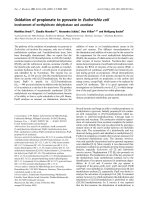
As shown in Fig. 1A, total GST activity in COS-7 cells was
increased 3.4- and 1.9-fold after transfection with pEGFP–
2576 P. A. ’t Hoen et al. (Eur. J. Biochem. 269) Ó FEBS 2002
M1 and pEGFP–M2, respectively. The observed differences
in CDNB conjugation between pEGFP–M1- and pEGFP–
M2-transfected cells can be explained by the lower catalytic
activity of the GSTM2-2 protein towards CDNB compared
with the activity of the GSTM1-1 protein [22]. The size of
the fusion proteins was 50 kDa, as determined by
Western blotting with an EGFP-specific primary antibody
(Fig. 1B). This value is in close agreement with the expected
size, calculated by summation of the molecular weights of
EGFP (25 kDa) and GSTM (27 kDa).
Colocalization of ODN and pEGFP
For proper evaluation of antisense effects, it is important
that the AS-ODNs and the EGFP-expressing plasmids are
transfected into the same cells. This was accomplished by
the cotransfection of plasmid and AS-ODN. By FACS
analysis, it was shown that after cotransfection of COS-7
cells with a fluorescently labelled ODN and pEGFP, ODN
and plasmid colocalized in the same target cells as > 90% of
the EGFP-positive cells were also positive for the TRITC-
labelled ODN (Fig. 2). The observations suggest that the
uptake of ODN is far more efficient than the uptake of the
EGFP plasmid, because almost all cells were positive for
TRITC-labelled ODNs, whereas only 33% of the cells
were expressing EGFP.
Screening of ODNs for their antisense activity
To identify AS-ODNs that are potent and sequence-specific
inhibitors of GSTM1 and/or GSTM2 expression, 15 PS-
ODNs were screened for their ability to inhibit EGFP–
GSTM fusion protein expression. The ODNs were targeted
against different regions in the mRNA of GSTM1 and
GSTM2 (Table 2). Some of the ODNs (i.e. AS-1, AS-6,
AS-7, AS-8, AS-12 and AS-15) were designed to inhibit
selectively GSTM1 expression, whereas others (i.e. AS-5,
AS-10, AS-11, AS-13 and AS-14) were designed to inhibit
selectively GSTM2 expression. A third group of ODNs (i.e.
AS-2, AS-3, AS-4 and AS-9) was directed against homol-
ogous regions in the GSTM1 and GSTM2 mRNAs, and
should therefore inhibit the expression of both isoforms. An
unrelated AS-ODN (AS-ctrl) with no sequence homology
with EGFP, GSTM1 or GSTM2 was taken as a negative
control. An EGFP-specific PO-ODN (AS-GFP), with two
PS-linkages at either end for protection against nuclease
activity, was taken as a positive control. It has been reported
that this ODN inhibits GFP expression in HeLa cells
transiently transfected with pEGFP [23].
Initially, COS-7 cells were transfected with 1.6 lgofthe
AS-ODNs (final concentration in the medium: 0.5 l
M
)and
0.5 lg of pEGFP, pEGFP-M1 or pEGFP-M2. After a 4-h
transfection period and culture for a further 18 h, cells
were analysed for EGFP expression by flow cytometry.
Propidium iodide was added to the cell suspensions to
exclude nonviable cells from the analysis. The percentage
of propidium iodide-positive cells increased from 3% in
cell cultures that were transfected with plasmid only, to
9% in cell cultures cotransfected with ODN and
plasmid. This is probably due to cytotoxicity of DOTAP,
as a larger amount of DOTAP was used for cotransfection
than for transfection of plasmid alone. The number of
propidium-iodide positive cells was the same for all
cotransfected PS-ODNs.
Clear differences were found in the ability of the various
AS-ODNs to inhibit EGFP expression. The control
Fig. 1. Expression of EGFP–GSTM fusion proteins in COS-7 cells.
COS-7 cells were transfected with pEGFP-M1, pEGFP-M2, or
pEGFP (0.5 lg DNA per well). After a further 18 h of culture, the cells
were lysed. (A) Total GST activity in 50 lL of protein lysate was
measured by following CDNB–GSH conjugate formation over time.
The increase in absorption at 340 nm was recorded with lysis buffer as
a blank. The GST activity is expressed as a percentage of the activity in
pEGFP-transfected cells (0.46 DA
340
Æmin
)1
Æmg protein
)1
). Means of
12 determinations in three separate experiments ± SEM are shown.
**P < 0.0001 (unpaired student t-test). (B) A Western blot was per-
formed on 5 lg protein lysate of pEGFP-M1 (lane 1), pEGFP-M2
(lane 2) and pEGFP (lane 3) transfected cells. The samples were
denatured, and separated by SDS/15% PAGE, together with a
Bio-Rad prestained kaleidoscope protein marker. Subsequently,
proteins were blotted onto a nitrocellulose membrane, and the blot was
incubated consecutively with a rabbit anti-EGFP antibody and a
peroxidase-labelled goat anti-rabbit secondary antibody. EGFP-
containing proteins were visualized with enhanced chemiluminescence.
The positions and molecular weights of the marker proteins are indi-
cated in the left margin. In the right margin, the estimated sizes of the
protein bands are shown.
Ó FEBS 2002 EGFP-based selection of antisense sequences (Eur. J. Biochem. 269) 2577
AS-ODN did not have any effect on the expression of
EGFP, EGFP–M1 or EGFP–M2, indicating that cotrans-
fection of PS-ODNs per se does not influence EGFP
expression. AS-6 and AS-8, directed against GSTM1,
inhibited EGFP–M1 expression by 95 ± 1% and
81 ± 6%, respectively (Fig. 3A). The expression of EGFP
and the other isoform, EGFP–M2, were also affected, but
the inhibitory effect on expression of pEGFP–M1 was
significantly greater (P < 0.05) than the effect on expres-
sion of EGFP or EGFP–M2. AS-1, however, inhibited the
expression of all three proteins, and the expression of EGFP
even by > 95%. This is probably not caused by sequence-
specific hybridization with the EGFP mRNA, because the
maximal continuous homologous region with the EGFP
sequence was eight nucleotides long. AS-7 also displayed
some nonspecific inhibition of protein synthesis: its effect on
EGFP–M1 expression, although greater, was not signifi-
cantly different from its effect on the expression of EGFP or
EGFP–M2. Two other AS-ODNs against GSTM1, AS-12
and AS-15, were completely ineffective in the down-
regulation of protein synthesis.
Similar results were found for AS-ODNs targeted at
GSTM2. AS-5 and AS-10 inhibited EGFP–M2 expression
by 82 ± 4% and 85 ± 0.4%, respectively, and affected
the expression of the control proteins EGFP and EGFP–
M1 by < 10% (Fig. 3B). For AS-10, the isoform-specif-
icity was remarkably good, as this AS-ODN contains only
three mismatches with respect to the sequence of GSTM1.
Again, one AS-ODN, AS-11, displayed nonspecific effects
on the expression of all three proteins, whereas two other
ODNs, AS-13 and AS-14, were not able to affect protein
expression.
AS-2 and AS-3, targeted against the coding sequence of
both GSTM1 and GSTM2, inhibited the expression of the
EGFP–GSTM isoforms by 95% (AS-2) and 80%
(AS-3), while expression of the control EGFP was inhibited
by 45 ± 12% and 18 ± 13%, respectively (Fig. 3C). AS-9
demonstrated severe nonspecific effects on EGFP expres-
sion, as the expression of EGFP, alone or in a fusion
construct, was inhibited by > 95%. The effects on EGFP
expression were not due to sequence-specific hybridization
because a significant homology with the sequence of EGFP
was not found. AS-4 showed less severe, but significant,
nonspecific effects on EGFP expression. Surprisingly, the
AS-GFP, which was reported to down-regulate EGFP
expression [23], did not have any effect on the expression of
either of the EGFP proteins at the tested concentration of
0.5 l
M
.
Fig. 2. FACS analysis of COS-7 cells
cotransfected with TRITC-ODN and pEGFP.
Untransfected COS-7 cells (A), cells trans-
fected with 0.2 l
M
TRITC-ODN (B), cells
transfected with 0.5 lg pEGFP (C), and cells
transfected with 0.2 l
M
TRITC-ODN and
0.5 lg pEGFP (D) were analysed by flow
cytometry for EGFP expression (FL-1, x-axis)
and TRITC-ODN uptake (FL-3, y-axis).
Single cells were gated in the forward–side-
ward scatter plot (gate R1, not shown). The
following gates were applied: R2, nontrans-
fected; R3, TRITC-positive; R4, GFP-posit-
ive; R5, TRITC-positive and GFP-positive.
(A–D) provide representive examples of
multiple FACS analyses. The table gives the
amounts of cells (expressed as percentage of
the total amount of cells) counted in each gate
under the different incubation conditions.
2578 P. A. ’t Hoen et al. (Eur. J. Biochem. 269) Ó FEBS 2002
Determination of the concentration/activity profile
of the AS-ODNs
To compare the potency and specificity of some of the
effective AS-ODNs, COS-7 cells were transfected with
different concentrations of AS-1, AS-2, AS-5, AS-6, and
AS-9. Figure 4 shows the effects of cotransfection with 0.01,
0.1 and 0.5 l
M
AS-ODN on the expression of pEGFP,
pEGFP–M1, and pEGFP–M2. Both the true antisense
effects and the non-target-specific effects appeared to be
highly concentration-dependent. AS-5 was a potent and
selective inhibitor of EGFP–M2 (IC
50
value 0.2 l
M
), and
did not have any effect on the expression of EGFP–M1 or
EGFP at the highest concentration tested. AS-2, directed
against both GSTM isoforms, and AS-6, directed at
GSTM1, potently inhibited the expression of their respect-
ive targets with IC
50
values slightly above 0.1 l
M
. At
0.1 l
M
, the inhibition was specific. However, at the highest
concentration tested also the expression of EGFP was
affected, indicating that sequence-specific antisense effects
occur at lower concentrations than non-target-specific
effects. AS-9 was the most potent inhibitor of both
EGFP–M1 and EGFP–M2 expression with estimated
IC
50
values < 0.1 l
M
. However, the EGFP expression
was also inhibited, although to a slightly lesser extent. AS-1,
targeted to GSTM1, was another nonspecific inhibitor of
EGFP expression as the IC
50
values for inhibition of EGFP
expression and of EGFP–M1 were both in the same range.
Analysis of the effect of AS-ODNs on GST activity
To examine whether the inhibitory effects of the AS-ODNs
on EGFP–GSTM fusion protein expression were associated
with a decrease in GST activity, we determined the effect of
cotransfection with AS-ODNs on the GST activity in
lysates of COS-7 cells transfected with pEGFP, pEGFP-
M1, or pEGFP-M2. In the GST activity assay, we tested
AS-5 and AS-10, directed against GSTM2, and AS-6,
directed against GSTM1, which were found to be specific
inhibitors of either GSTM1 or GSTM2 expression in the
EGFP assay. The results are shown in Table 3. None of
these ODNs affected the CDNB conjugation in cells
transfected with pEGFP, indicating that the AS-ODNs
were specific for the rat GSTM isoforms, and did not inhibit
the activity of endogenous GSTs present in lysates of COS-7
cells. The rates of CDNB conjugation in the lysates of
pEGFP-M1 and pEGFP-M2 transfected cells were correc-
ted for the endogenous GST activity, determined in COS-7
cells transfected with pEGFP. AS-6 appeared to be a very
potent inhibitor of GSTM1-1 enzyme activity (95 ± 2%
decrease). AS-5 and AS-10 were somewhat less potent
inhibitors of GSTM2-2 enzyme activity (77 ± 6%, and
70 ± 6% decrease, respectively). However, conjugation by
the nontargeted isoform was also affected by 40–50%.
Nonetheless, the effects of the different AS-ODNs on the
activity of the targeted GSTM isoform were significantly
greater than on the nontargeted GSTM isoform
(P < 0.002 for all tested AS-ODNs).
DISCUSSION
Most of currently available screening assays for the selection
of effective AS-ODNs are based on cell-free assays, e.g.
RNAse H digestion screens and oligonucleotide scanning
arrays [9]. As activity in cell-free assay may not always
correlate with activity in cellular systems, we developed in the
present study a novel cellular screening assay for the
selection of effective AS-ODNs with a sequence-specific
Table 2. Antisense ODN sequences.
Target site
b
Mismatch
(number of bases)
c
Name Sequence
a
Region
b
GSTM1 GSTM2
AS-ctrl TGAGAGCTGAAAGCAGGTCCAT Unrelated – –
AS-GFP G*A*GCTGCACGCTGCCG*T*C GFP–CDS – –
AS-1 GGCGG
ATCGGGTGTGTCAGC CDS 36–55 – 5
AS-2 CCACTGGCTTCTGTCATAGT CDS 119–138 119–138 0
AS-3 GAAGTCCAGGCCCAGTTTGA CDS 152–171 152–171 0
AS-4 TCAATTAAGTAGGGCAGATT CDS 175–194 175–194 0
AS-5 TCTCCA
AAACGTCCACACGA CDS – 285–304 4
AS-6 ACAAAGCATGATGAGCTGCA CDS 326–345 – 8
AS-7 GAGTA
GAGCTTCATCTTCTC CDS 397–426 – 1
AS-8
ACTGGTCAAGAATGTCATAA CDS 480–499 – 7
AS-9 CAGGTTTGGGAAGGCGTCCA CDS 524–543 524–543 0
AS-10 CAGGCCCTC
AAACCGAGCCA CDS – 554–573 3
AS-11
GTCTGGACTTTGTGGTGCTA STOP – 655–674 13
AS-12 GGCATGACTGGGGTGAGGTT 3¢-UTR 786–805 – 5
AS-13 AA
AATCAGTGAGGGAAGGGT 3¢-UTR – 870–889 8
AS-14 TCTAATCTCTCAGGCCAGGC 3¢-UTR – 921–940 10
AS-15
GCAGCTCCCCCACCAGGAAC 3¢-UTR 978–997 – 12
a
All sequences were PS-ODNs except for AS-GFP. The sequence of AS-GFP is taken from literature [23]: it is a PO-ODN with PS-modified
internucleotide linkages at the 3¢- and 5¢-ends, indicated by asterisks.
b
The region in the mRNA against which the ODNs are indicated as
follows: CDS, coding sequence; STOP, STOP codon; 3¢-UTR, 3¢-untranslated region. The target sites in the GSTM1 or GSTM2 mRNAs
are indicated, nucleotide 1 being the ATG start site.
c
The number of mismatches in the corresponding region of the nontargeted isoform are
given. Mismatches are underlined in the sequence.
Ó FEBS 2002 EGFP-based selection of antisense sequences (Eur. J. Biochem. 269) 2579
mode of action. In the present assay, antisense activity is
directly correlated with EGFP-derived fluorescence by
constructing fusion proteins of the target protein and EGFP.
Unlike in conventional target-specific screens, in the current
assay specific antibodies need not be available and isoform-
specific assays for the determination of enzyme activity need
not be developed. Furthermore, the measurement of
EGFP-derived fluorescence by flow cytometry has excellent
quantitative properties and offers good reproducibility. This
is probably due to the elimination of variation in transfection
efficiencies as a complicating factor in the assessment of
antisense effectiveness. Our experiments in which an EGFP-
containing plasmid was cotransfected with fluorescently
labelled ODNs, suggest that all EGFP-positive cells had
taken up ODNs. Therefore, in the present assay the antisense
effects are determined in the whole population of cells that
express the target gene. In other cellular assays, including a
luciferase reporter gene-based assay [24], antisense effects
may be underestimated because not all cells that express the
gene of interest are transfected with AS-ODNs. An EGFP-
based approach has been used previously for the selection of
ribozymes against the c-erbB-2 oncogene [25]. However, in
this earlier study the plasmid coding for the c-erb-B-2 EGFP
fusion protein, was cotransfected with a ribozyme expressing
plasmid and not with an exogenously added antisense
molecule. Cotransfection with the ribozyme-expressing
plasmid resulted in a reduction of EGFP expression to a
maximum of 70%, whereas we observed a > 90% reduction
with our most potent ODNs. Possibly, a significant part of
the c-erbB-2-EGFP transfected cells had not taken up a
ribozyme construct.
A C-terminal fusion construct and not an N-terminal
fusion construct, was used because AS-ODNs against the
3¢-untranslated region of the mRNA of the gene of interest,
which has been shown to be a favourable region for
antisense action [7], can only be tested in C-terminal fusion
constructs.
The newly developed screening assay was used for the
selection of effective AS-ODNs against rat GSTM1 and
GSTM2 out of a set of 15 PS-ODNs. Some ODNs were
designed to specifically inhibit either GSTM1 or GSTM2
expression, which show a sequence identity of 80% at the
DNA level. For these ODNs, the nontargeted isoform
served as a mismatch target control with 1–13 mismatches.
Other ODNs were targeted against homologous regions in
both isoforms. As a control for the true antisense nature of
the observed effects on protein expression, the effects of the
ODNs on the expression of EGFP without a fusion
construct were evaluated. Three ODNs (AS-3, AS-5 and
AS-10) were found to inhibit gene expression with very high
sequence specificity. These ODNs reduced at 0.5 l
M
the
expression of their target isoforms by > 80%, whereas the
nontargeted isoform and/or EGFP control were not
affected significantly. Other ODNs (AS-2, AS-6, AS-7 and
AS-8) displayed a combination of target sequence-specific
and nontarget-specific inhibitory effects on EGFP levels.
These ODNs inhibited the targeted isoform to a signifi-
cantly greater extent than the nontargeted isoform and/or
EGFP control, but also attenuated the expression of the
controls by 40–60%. Three ODNs (AS-1, AS-9 and AS-11)
had severe non-sequence-specific effects on EGFP expres-
sion. In these cases, the expression of EGFP without a
GSTM fusion was affected to a similar extent as the
expression of the targeted proteins. Five ODNs (AS-4,
AS-12, AS-13, AS-14 and AS-15) did not demonstrate
major effects on EGFP–GSTM fusion protein expression.
Our results indicate once again that, when evaluating
antisense effects, identification of false-positives is common.
As stated before by others [26], it is crucial to analyse the
effects on the expression of target-related control proteins,
which is easily accomplished in our screening assay.
Fig. 3. Effects of AS-ODNs on EGFP and EGFP–GSTM fusion
protein expression. COS-7 cells were transfected with 0.5 lg pEGFP
(open bars), pEGFP-M1 (hatched bars) or pEGFP-M2 (closed bars),
together with 0.5 l
M
of the indicated AS-ODNs. The ODNs were
directed against GSTM1 (A), GSTM2 (B), or both GSTM isoforms
(C). AS-ctrl is a control ODN without sequence homology with
GSTM or EGFP. AS-GFP is an AS-ODN against EGFP, taken from
[23]. At 22 h after transfection, cells were analysed for EGFP expres-
sion (FL-1) and propidium iodide uptake (FL-3) by flow cytometry.
The number of living (i.e. propidium iodide-negative), EGFP-positive
cells was counted and is expressed as the percentage of EGFP-positive
cells in cultures transfected with plasmid, but without AS-ODN.
Means of three independent experiments ± SEM are shown.
*P < 0.05; **P < 0.005 (one group student t-test compared to con-
trol without AS-ODN).
2580 P. A. ’t Hoen et al. (Eur. J. Biochem. 269) Ó FEBS 2002
Table 3. Effects of AS-ODNs on GST activity. COS-7 cells were transfected with 0.5 lg pEGFP, pEGFP-M1 or pEGFP-M2, together with 0.1 l
M
of the indicated AS-ODNs. AS-5 and AS-10 are directed against GSTM2; AS-6 is directed against GSTM1. At 22 h after transfection, GST activity
in the protein lysates was determined by assaying CDNB conjugation over time. Conjugation rates are expressed as percentages of EGFP controls
(column 2, 3 and 5), or percentages of the additional EGFP-M1-dependent (column 4) or EGFP-M2-dependent (column 6) GST activity,
calculated by subtraction of the endogenous GST activity, which was determined in pEGFP-transfected cultures. Means of 10–12 determinations in
three separate experiments ± SEM are shown. Statistical significance of the difference between AS-ODN-treated and untreated cultures are
indicated:
a
P < 0.005,
b
P < 0.0001. Statistical significance of the difference between the effect of the AS-ODNs on EGFP–M1 and EGFP–M2
expression are indicated:
c
P ¼ 0.0013 (AS-5),
d
P < 0.0001 (AS-6),
e
P ¼ 0.0002 (AS-10).
CDNB conjugation rate
EGFP EGFP-M1 EGFP-M2
AS-ODN
% of EGFP
control
% of EGFP
control
% of EGFP-M1
control
% of EGFP
control
% of EGFP-M2
control
– 100 ± 6 340 ± 16 100 ± 6 190 ± 11 100 ± 7
AS-5 93 ± 7 207 ± 4
b
51 ± 3
c
118 ± 6
b
23 ± 6
c
AS-6 99 ± 6 110 ± 4
b
5±2
d
146 ± 6
a
57 ± 6
d
AS-10 95 ± 5 244 ± 9
b
61 ± 4
e
125 ± 7
a
30 ± 6
e
Fig. 4. Concentration-dependent inhibition of
EGFP and EGFP–GSTM fusion protein
expression by AS-ODNs. COS-7 cells were
transfected with 0.5 lgofpEGFP(n), pEG-
FP-M1 (j)orpEGFP-M2(d), together with
the indicated concentrations of AS-1 (A),
AS-2 (B), AS-5 (C), AS-6 (D) or AS-9 (E). AS-
1 and AS-6 are directed against GSTM1, AS-5
is directed against GSTM2, whereas AS-2 and
AS-9 are complementary to both GSTM1 and
GSTM2. At 22 h after transfection, cells were
analysed for GFP expression (FL-1) and
propidium iodide uptake (FL-3) by flow
cytometry. The number of living (i.e. propi-
dium iodide-negative), EGFP-positive cells
was counted and is expressed as the percentage
of EGFP-positive cells in cultures transfected
with plasmid, but without AS-ODN. Means
of three independent experiments ± SEM are
shown. An unpaired student t-test was used to
determine whether the effect on EGFP–M1 or
EGFP–M2 expression was significantly dif-
ferent from the effect on EGFP expression:
*P <0.05;**P < 0.005.
Ó FEBS 2002 EGFP-based selection of antisense sequences (Eur. J. Biochem. 269) 2581
The sensitivity of the inhibition towards mismatches in
the target sequences appeared to be high. One mismatch
(AS-7) was not sufficient to achieve complete isoform-
specificity (the expression of the targeted and nontargeted
isoform was reduced by 77 ± 6% and 45 ± 15%, respect-
ively). The presence of three mismatches (AS-10), however,
resulted in isoform-specifc inhibition of EGFP–GSTM
fusion protein expression (85 ± 0.4% and 10 ± 0.3%
reduction of targeted and nontargeted isoform, respect-
ively). Interestingly, an AS-ODN against EGFP, described
to be effective in HeLa cells [23], was totally ineffective in
inhibiting the expression of either of the EGFP-containing
proteins in our study. This may be attributed to the fact that
the ODN was a phosphorothioate-capped PO-ODN. The
sensitivity of these chimeras towards nucleolytic degrada-
tion is higher than that of PS-ODNs, and depends on the
cell-type used [27,28].
True antisense effects and nonantisense effects elicited by
the ODNs were both found to be concentration-dependent.
The IC
50
values of the most potent, specifically acting
AS-ODNs were 0.2 l
M
. It should be noted that for most
AS-ODNs, with the exception of AS-5, the concentration
window where sequence-specific antisense effects were
observed, was narrow. This was also found in other studies
where PS-ODNs were used, and may be explained by the
relatively low affinity of PS-ODNs for their target mRNA
sequences together with the high incidence of nonantisense
effects [1,24]. It is therefore of highest importance to evaluate,
in each antisense study, the concentration–activity profile.
The nature of the nonspecific effects elicited by PS-ODNs
remains to be clarified. With the possible exception of AS-11,
which contained only five mismatches with respect to the
EGFP sequence, neither of the AS-ODNs against GSTM
showed significant sequence homology with EGFP. Thus,
the observed effects on EGFP expression are probably not
caused by partial hybridization of the AS-ODNs with the
EGFP mRNA. We cannot exclude that some of the
nonspecific AS-ODNs decrease the transfection efficiency
of the EGFP plasmids. However, from earlier studies it
became apparent that sequence-dependent variations in
cationic lipid-mediated transfection efficiencies were small,
unless homo-oligonucleotides, such as A
18
, were applied
[29,30]. More likely, sequence-dependent aptameric effects
play a role. The negative charge on the sulfur atom may
cause avid binding of the ODNs to key cellular proteins, e.g.
proteins involved in mRNA translation [31,32].
The inhibition of EGFP–GSTM fusion protein expres-
sion was reflected by a decrease in GST enzyme activity, as
determined in a CDNB conjugation assay. AS-6 displayed
potent and specific inhibition of GSTM1-1 enzyme activity.
AS-5 and AS-10, directed against GSTM2, inhibited
GSTM2-2 enzyme activity but showed also some effect on
GSTM1-1 enzyme activity. This was not expected because
the effects of these AS-ODNs on EGFP fusion protein
expression were highly isoform-specific. The inhibitory
effects cannot be explained by a general inhibition of GST
activity, because the ODNs did not affect endogenous GST
activity in COS-7 cells. Possibly, the presence of four (AS-5)
and three (AS-10) mismatches with respect to the GSTM1
sequence results in partial hybridization with the GSTM1
mRNA and in some hindrance of the synthesis of full-length
EGFP–M1 fusion proteins without induction of RNAse
H-mediated cleavage and subsequent degradation of
EGFP–M1 mRNA. In that case, the formation of the
EGFP moiety is not affected, whereas the formation of the
GSTM1-1 is. This would explain the higher isoform
specificity of AS-5 and AS-10 in the EGFP assay, compared
to the GST assay.
In summary, we selected several effective antisense ODNs
againstratGSTM1andGSTM2fromasetof15PS-ODNs
in a novel, sensitive screening assay. The assay discriminates
between effective AS-ODNs and ODNs that are ineffective
or inhibit protein expression by nonantisense mechanisms.
The effectiveness of the selected AS-ODNs will be evaluated
further in rat hepatocytes and in vivo, potentially allowing
the study of the effect of decreased GSTM expression on the
toxicity and carcinogenicity of xenobiotics.
REFERENCES
1. Crooke, S.T. & Bennett, C.F. (1996) Progress in antisense
oligonucleotide therapeutics. Annu. Rev. Pharmacol. Toxicol. 36,
107–129.
2. Flanagan, W.M. & Wagner, R.W. (1997) Potent and selective gene
inhibition using antisense oligodeoxynucleotides. Mol. Cell. Bio-
chem. 172, 213–225.
3. Galderisi, U., Cascino, A. & Giordano, A. (1999) Antisense
oligonucleotides as therapeutic agents. J. Cell Physiol. 181,
251–257.
4. Phillips, M.I.E. (1999) Antisense Technology, part A and B,
Methods in Enzymology, (Abelson, J.N. & Simon, M.I., eds), Vol
313–314. Academic Press, San Diego, CA.
5. Agrawal, S. & Iyer, R.P. (1995) Modified oligonucleotides as
therapeutic and diagnostic agents. Curr. Opin. Biotechnol. 6,
12–19.
6. Herdewijn, P. (2000) Heterocyclic modifications of oligonucleo-
tides and antisense technology. Antisense Nucleic Acid Drug. Dev.
10, 297–310.
7. Monia, B.P., Johnston, J.F., Geiger, T., Muller, M. & Fabbro, D.
(1996) Antitumor activity of a phosphorothioate antisense
oligodeoxynucleotide targeted against C-raf kinase. Nat. Med. 2,
668–675.
8. Dean,N.M.,McKay,R.,Condon,T.P.&Bennett,C.F.(1994)
Inhibition of protein kinase C-alpha expression in human A549
cells by antisense oligonucleotides inhibits induction of inter-
cellular adhesion molecule 1 (ICAM-1) mRNA by phorbol esters.
J. Biol. Chem. 269, 16416–16424.
9. Sohail, M. & Southern, E.M. (2000) Selecting optimal antisense
reagents. Adv. Drug Deliv. Rev. 44, 23–34.
10. Commandeur, J.N.M., Stijntjes, G.J. & Vermeulen, N.P.E. (1995)
Enzymes and transport systems involved in the formation and
disposition of glutathione S-conjugates. Role in bioactivation
and detoxication mechanisms of xenobiotics. Pharmacol. Rev. 47,
271–330.
11. Seidegard, J., Vorachek, W.R., Pero, R.W. & Pearson, W.R.
(1988) Hereditary differences in the expression of the human
glutathione transferase active on trans-stilbene oxide are due to a
gene deletion. Proc. Natl Acad. Sci. USA 85, 7293–7297.
12. McWilliams, J.E., Sanderson, B.J., Harris, E.L., Richert-Boe,
K.E. & Henner, W.D. (1995) Glutathione S-transferase M1
(GSTM1) deficiency and lung cancer risk. Cancer Epidemiol.
Biomarkers Prev. 4, 589–594.
13. Zhong, S., Wyllie, A.H., Barnes, D., Wolf, C.R. & Spurr, N.K.
(1993) Relationship between the GSTM1 genetic polymorphism
and susceptibility to bladder, breast and colon cancer. Carcino-
genesis 14, 1821–1824.
14. Wormhoudt, L.W., Commandeur, J.N.M. & Vermeulen, N.P.E.
(1999) Genetic polymorphisms of human N-acetyltransferase,
cytochrome P450, glutathione-S-transferase, and epoxide
2582 P. A. ’t Hoen et al. (Eur. J. Biochem. 269) Ó FEBS 2002
hydrolase enzymes: relevance to xenobiotic metabolism and toxi-
city. Crit. Rev. Toxicol. 29, 59–124.
15. Nakachi, K., Imai, K., Hayashi, S. & Kawajiri, K. (1993) Poly-
morphisms of the CYP1A1 and glutathione S-transferase genes
associated with susceptibility to lung cancer in relation to cigarette
dose in a Japanese population. Cancer Res. 53, 2994–2999.
16. Kihara, M., Kihara, M. & Noda, K. (1995) Risk of smoking for
squamous and small cell carcinomas of the lung modulated by
combinations of CYP1A1 and GSTM1 gene polymorphisms in a
Japanese population. Carcinogenesis 16, 2331–2336.
17. Kawajiri, K., Watanabe, J., Eguchi, H. & Hayashi, S. (1995)
Genetic polymorphisms of drug-metabolizing enzymes and lung
cancer susceptibility. Pharmacogenetics 5, S70–S73.
18. Stucker,I.,Jacquet,M.,deWaziers,I.,Cenee,S.,Beaune,P.,
Kremers, P. & Hemon, D. (2000) Relation between inducibility of
CYP1A1, GSTM and lung cancer in a French population. Phar-
macogenetics 20, 617–627.
19. Zeng, G. (1998) Sticky-end PCR: new method for subcloning.
Biotechniques 25, 206–208.
20. Habig, W.H., Pabst, M.J. & Jakoby, W.B. (1974) Glutathione
S-transferases. The first enzymatic step in mercapturic acid for-
mation. J. Biol. Chem. 249, 7130–7139.
21. Bradford, M.M. (1976) A rapid and sensitive method for the
quantitation of microgram quantities of protein utilizing the
principle of protein-dye binding. Anal. Biochem. 72, 248–254.
22. Hayes, J.D. & Pulford, D.J. (1995) The glutathione S-transferase
supergene family: regulation of GST and the contribution of the
isoenzymes to cancer chemoprotection and drug resistance. Crit.
Rev. Biochem. Mol. Biol. 30, 445–600.
23.Helin,V.,Gottikh,M.,Mishal,Z.,Subra,F.,Malvy,C.&
Lavignon, M. (1999) Cell cycle-dependent distribution and specific
inhibitory effect of vectorized antisense oligonucleotides in cell
culture. Biochem. Pharmacol. 58, 95–107.
24. Monia, B.P., Johnston, J.F., Ecker, D.J., Zounes, M.A., Lima,
W.F. & Freier, S.M. (1992) Selective inhibition of mutant Ha-ras
mRNA expression by antisense oligonucleotides. J. Biol. Chem.
267, 19954–19962.
25. Wiechen, K., Zimmer, C. & Dietel, M. (1998) Selection of a high
activity c-erbB-2 ribozyme using a fusion gene of c-erbB-2 and the
enhanced green fluorescent protein. Cancer Gene Ther. 5, 45–51.
26. Stein, C.A. (2001) The experimental use of antisense oligonucleo-
tides: a guide for the perplexed. J. Clin. Invest. 108, 641–644.
27. Hoke,G.D.,Draper,K.,Freier,S.M.,Gonzalez,C.,Driver,V.B.,
Zounes, M.C. & Ecker, D.J. (1991) Effects of phosphorothioate
capping on antisense oligonucleotide stability, hybridization and
antiviral efficacy versus herpes simplex virus infection. Nucleic
Acids Res. 19, 5743–5748.
28. Monia, B.P., Johnston, J.F., Sasmor, H. & Cummins, L.L. (1996)
Nuclease resistance and antisense activity of modified oligonu-
cleotides targeted to Ha-ras. J. Biol. Chem. 271, 14533–14540.
29. Conrad, A.H., Behlke, M.A., Jaffredo, T. & Conrad, G.W. (1998)
Optimal lipofection reagent varies with the molecular modifica-
tions of the DNA. Antisense Nucleic Acid Drug Dev. 8, 427–434.
30. Meidan, V.M., Glezer, J., Amariglio, N., Cohen, J.S. & Barenholz,
Y. (2001) Oligonucleotide lipoplexes: the influence of oligonu-
cleotide composition on complexation. Biochim. Biophys. Acta
1568, 177–182.
31. Brown, D.A., Kang, S.H., Gryaznov, S.M., DeDionisio, L.,
Heidenreich, O., Sullivan, S., Xu, X. & Nerenberg, M.I. (1994)
Effect of phosphorothioate modification of oligodeoxynucleotides
on specific protein binding. J. Biol. Chem. 269, 26801–26805.
32. Bijsterbosch,M.K.,Rump,E.T.,DeVrueh,R.L.,Dorland,R.,
vanVeghel,R.,Tivel,K.L.,Biessen,E.A.,vanBerkel,T.J.&
Manoharan, M. (2000) Modulation of plasma protein binding and
in vivo liver cell uptake of phosphorothioate oligodeoxynucleo-
tides by cholesterol conjugation. Nucleic Acids Res. 28, 2717–2725.
Ó FEBS 2002 EGFP-based selection of antisense sequences (Eur. J. Biochem. 269) 2583