Neutron Scattering in Biology Techniques and Applications pot
Bạn đang xem bản rút gọn của tài liệu. Xem và tải ngay bản đầy đủ của tài liệu tại đây (10.44 MB, 569 trang )
biolo gical and medical physics,
biomedical engineering
biolo gical and medical physics,
biomedical engineering
The fields of biological and medical physics and biomedical engineering are broad, multidisciplinary and
dynamic. They lie at the crossroads of frontier research in physics, biology, chemistry, and medicine. The
Biological and Medical Physics, Biomedical Engineering Series is intended to be comprehensive, covering a
broad range of topics important to the study of the physical, chemical and biological sciences. Its goal is to
provide scientists and engineers with textbooks, monographs, and reference works to address the growing
need for information.
Books in the series emphasize established and emergent areas of science including molecular, membrane,
and mathematical biophysics; photosynthetic energy harvesting and conversion; information processing;
physical principles of genetics; sensory communications; automata networks, neural networks, and cellu-
lar automata. Equally important will be coverage of applied aspects of biological and medical physics and
biomedical engineering such as molecular electronic components and devices, biosensors, medicine, imag-
ing, physical principles of renewable energy production, advanced prostheses, and environmental control and
engineering.
Editor-in-Chief:
Elias Greenbaum, Oak Ridge National Laboratory,
Oak Ridge, Tennessee, USA
Editorial Board:
Masuo Aizawa, Department of Bioengineering,
Tokyo Institute of Technology, Yokohama, Japan
Olaf S. Andersen, Department of Physiology,
Biophysics & Molecular Medicine,
Cornell University, New York, USA
Robert H. Austin, Department of Physics,
Princeton University, Princeton, New Jersey, USA
James Barber, Department of Biochemistry,
Imperial College of Science, Technology
and Medicine, London, England
Howard C. Berg, Department of Molecular
and Cellular Biology, Harvard University,
Cambridge, Massachusetts, USA
Victor Bloomfield, Department of Biochemistry,
University of Minnesota, St. Paul, Minnesota, USA
Robert Callender, Department of Biochemistry,
Albert Einstein College of Medicine,
Bronx, New York, USA
Britton Chance, Department of Biochemistry/
Biophysics, University of Pennsylvania,
Philadelphia, Pennsylvania, USA
Steven Chu, Department of Physics,
Stanford University, Stanford, California, USA
Louis J. DeFelice, Department of Pharmacology,
Vanderbilt University, Nashville, Tennessee, USA
Johann Deisenhofer, Howard Hughes Medical
Institute, The University of Texas, Dallas,
Texas, USA
George Feher, Department of Physics,
University of California, San Diego, La Jolla,
California, USA
Hans Frauenfelder, CNLS, MS B258,
Los Alamos National Laboratory, Los Alamos,
New Mexico, USA
Ivar Giaever, Rensselaer Polytechnic Institute,
Troy,NewYork,USA
Sol M. Gruner, Department of Physics,
Princeton University, Princeton, New Jersey, USA
Judith Herzfeld, Department of Chemistry,
Brandeis University, Waltham, Massachusetts, USA
Mark S. Humayun, Doheny Eye Institute,
Los Angeles, California, USA
Pierre Joliot, Institute de Biologie
Physico-Chimique, Fondation Edmond
de Rothschild, Paris, France
Lajos Keszthelyi, Institute of Biophysics, Hungarian
Academy of Sciences, Szeged, Hungary
Robert S. Knox, Department of Physics
andAstronomy,UniversityofRochester,Rochester,
New York, USA
Aaron Lewis, Department of Applied Physics,
Hebrew University, Jerusalem, Israel
StuartM.Lindsay,DepartmentofPhysics
andAstronomy,ArizonaStateUniversity,
Tempe, Arizona, USA
David Mauzerall, Rockefeller University,
New York, New York, USA
Eugenie V. Mielczarek, Department of Physics
and Astronomy, George Mason University, Fairfax,
Virginia, USA
Markolf Niemz, Klinikum Mannheim,
Mannheim, Germany
V. Adrian Parsegian, Physical Science Laboratory,
National Institutes of Health, Bethesda,
Maryland, USA
Linda S. Powers, NCDMF: Electrical Engineering,
Utah State University, Logan, Utah, USA
Earl W. Prohofsky, Department of Physics,
Purdue University, West Lafayette, Indiana, USA
Andrew Rubin, Department of Biophysics, Moscow
State University, Moscow, Russia
Michael Seibert, National Renewable Energy
Laboratory, Golden, Colorado, USA
David Thomas, Department of Biochemistry,
University of Minnesota Medical School,
Minneapolis, Minnesota, USA
Samuel J. Williamson, Department of Physics,
NewYorkUniversity,NewYork,NewYork,USA
J. Fitter T. Gutberlet J. Katsaras
Neutron Sc attering
in Biology
Techniques and Applications
With 240
Figures
123
Dr. J
¨
org Fitter
Forschungszentrum J
¨
ulich GmbH
Abt. IBI-2
52425 J
¨
ulich, Germany
e-mail: j.fi
Dr. Thomas Gutberlet
5232 Villigen, Switzerland
e-mail:
Dr. John Katsaras
National Research Council
Chalk River
K0J 1J0 Ontario, Canada
e-mail:
LibraryofCongressControlNumber:
ISSN 1618-7210
ISBN-10 3-540-29108-3 Springer Berlin Heidelberg New York
ISBN-13 978-3-540-29108-4 Springer Berlin Heidelberg New York
This work is subject to copyright. All rights are reserved, whether the whole or part of the material is
concerned, specifically the rights of translation, reprinting, reuse of illustrations, recitation, broadcasting,
reproductiononmicrofilmorinanyotherway,andstorageindatabanks.Duplicationofthispublicationor
parts thereof is permitted only under the provisions of the German Copyright Law of September 9, 1965, in
its current version, and permission for use must always be obtained from Springer. Violations are liable to
prosecution under the German Copyright Law.
Springer is a part of Springer Science+Business Media
springer.com
© Springer-Verlag Berlin Heidelberg 2006
Printed in Germany
The use of general descriptive names, registered names, trademarks, etc. in this publication does not imply,
even in the absence of a specific statement, that such names are exempt from the relevant protective laws and
regulations and therefore free for general use.
Cover concept by eStudio Calamar Steinen
Cover production: design & production GmbH, Heidelberg
Printed on acid-free paper SPIN 10929513 57/3100/SPI -543210
2005934097
Typesetting by the authors and using a Springer L
A
T
E
X macro package
SPI
Publisher Services
Laboratory of Neutron Scattering
Paul Scherrer Institut
Preface
“Certainly no subject or field is making more progress on so many fronts at
the present moment, than biology, and if we were to name the most powerful
assumption of all, which leads one on and on in an attempt to understand
life, it is that all things are made of atoms, and that everything that living
things do can be understood in terms of the jigglings and wigglings of atoms.”
Richard P. Feynmann, from “Six easy pieces” (1963)
In 1932, James Chadwick discovered the neutron, but initially the only sources
of neutrons were from the radioactive decay of unstable nuclei. It was not until
1942 when Enrico Fermi constructed the first nuclear reactor in the squash
courts beneath the University of Chicago’s Stagg Field, that a controlled and
sustained nuclear chain reaction was achieved. After World War II, nuclear
reactors became available for civilian research, and in 1945 Ernest Wollan set
up a double-crystal diffractometer at ORNL’s Graphite Reactor. This marks
the beginning of neutron scattering.
Neutrons produced by present reactor- and accelerator-based sources,
typically have wavelengths in the order of
˚
Angstroms, and hence are well-
suited for probing the structures and motions of molecules. For biological
materials rich in hydrogen, the large difference in scattering cross-sections
between hydrogen and deuterium provides the possibility of contrast varia-
tion, a powerful method achieved by selective deuteration for emphasizing,
or not, the scattering from a particular portion of a molecule or molecular
assembly. Using a variety of scattering methods, the structures and dynamics
of biological systems can be determined.
The present compilation aims to provide the reader with some of the
important applications of neutron scattering in structural biology, biophysics,
and systems relevant to biology.
The location of hydrogen atoms in biomolecules such as, proteins,
is – despite the high brilliance and power of third generation synchrotron
VI Preface
sources – not readily available by X-ray crystallography or related physical
techniques. In the case of hydrogens attached to electronegative atoms (e.g.,
O and N), even high resolution X-ray structures (resolution <1
˚
A) cannot
unequivocally locate these H atoms. On the other hand, these atoms can
effectively be located using high resolution crystallographic neutron diffrac-
tion methods. Radiation damage leading to changes in metal oxidation state
and subsequent loss of hydrogens can also pose a problem with X-rays, but not
so with neutrons. When good quality, large (>1mm
3
) single crystals cannot
be obtained, low resolution neutron diffraction offers an alternative technique
in determining the hydrated structure of macromolecules and their various
hydrogen-bonding patterns.
Small-angle neutron scattering (SANS) is probably the technique most
often applied to biological materials as it can probe the size, shape and con-
formation of macromolecules and macromolecular complexes in aqueous solu-
tion on a length scale from ten to several thousand
˚
Angstroms. The ability to
scatter from materials in solution allows for biologically relevant conditions to
be mimicked, and also permits for the study of samples that are either difficult
or impossible to crystallize. In recent years, SANS has greatly benefited from
the production of “cold neutrons” that have wavelengths 10–20 times larger
than “thermal neutrons”, allowing SANS to examine complex materials, such
as living cells.
Over the past decade, neutron reflectometry has increasingly become an
important technique for the characterization of biological and biomimetic thin
films attached to a solid support, in contact with water. Advancements in sam-
ple environments, instrumentation, and data analysis now make it possible to
obtain high resolution information about the composition of these materials
along the axis perpendicular to the plane of the membrane or substrate. Most
recently, a newly developed phase-sensitive neutron reflectometry technique
also allows direct inversion of the reflectometry data to obtain unique compo-
sitional depth profiles of the films in question.
Studies exploring the relationship between the function and the dynam-
ics of biological systems are still in their nascent stages. Incoherent neutron
scattering (INS) techniques such as, elastic (EINS), quasielastic (QINS), and
inelastic (IINS) neutron scattering, along with molecular dynamics (MD) sim-
ulations offer the real possibility of investigating the dynamics associated with
a molecule’s biological function(s). Using the large incoherent scattering cross-
section intrinsic to naturally abundant hydrogen atoms, various INS type
measurements can be carried out. These results, in conjunction with MD sim-
ulations, offer a glimpse of for example, a protein’s internal structure on the
picosecond time scale. Moreover, the current developments of intense pulsed
neutron sources promise, in the near future, to accelerate our understanding
of the relationship between a molecule’s dynamics and its function.
The study of materials under difficult environmental conditions (such as
high magnetic fields, high pressures, shear, and 100% relative humidity) is
by no means straight forward and requires specialized equipment. In many
Preface VII
cases, these experiments are better accommodated by the fact that neutrons
interact weakly, thus nondestructively, with many commonly used materials
(e.g., aluminum and its alloys) that are readily available and suitable for the
construction of sample environments. The conditions created by these special-
ized environments provide us with a more detailed physical understanding of
biologically relevant materials.
The present volume begins with a general introduction into the generation
and properties of neutrons and is followed by a series of papers describing the
various elastic and inelastic neutron scattering techniques used to study bio-
logical and biologically relevant systems. The reader is introduced to the basic
principles of neutron crystallography, low resolution neutron diffraction, neu-
tron small-angle scattering, neutron reflectometry, inelastic and quasielastic
neutron scattering, and neutron spin echo spectroscopy. Papers describing
sample environments and preparatory techniques, in addition to molecular
dynamics simulations used to evaluate the neutron data, are also included.
Finally, there are a series of papers describing recent neutron research that
has elucidated the structure and dynamics of soluble proteins, membrane
embedded proteins, and of complex biological aggregates.
The editors wish to express their great appreciation to all of the contrib-
utors whose diligence, efforts, and timeliness made this compilation possible.
J¨ulich J¨org Fitter
Villigen Thomas Gutberlet
Chalk River John Katsaras
Spring 2005
Contents
1 Neutron Scattering for Biology
T.A. Harroun, G.D. Wignall, J. Katsaras 1
1.1 Introduction 1
1.2 Productionof Neutrons 2
1.3 ElementsofNeutronScatteringTheory 5
1.3.1 Properties of Neutrons . . . . . . . . . . . . . . . . . . . . . . . . . . . . . . . . 5
1.3.2 EnergyandMomentumTransfer 5
1.3.3 Diffraction 6
1.3.4 ScatteringLengthandCross-Section 7
1.3.5 Coherent and Incoherent Cross-Sections . . . . . . . . . . . . . . . . . 8
1.4 NeutronDiffractionand Contrast 10
1.4.1 ContrastandStructure 11
1.4.2 ContrastandDynamics 13
1.4.3 ContrastandBiology 13
1.5 Conclusions 16
References 17
Part I Elastic Techniques
2 Single Crystal Neutron Diffraction
and Protein Crystallography
C.C. Wilson, D.A. Myles 21
2.1 Introduction 21
2.2 Single Crystal Neutron Diffractometers:
BasicPrinciples 22
2.2.1 Development of Single Crystal Neutron Diffractometers . . . 25
2.2.2 Achievements of Neutron Macromolecular
CrystallographyatReactorSources 25
2.2.3 Developments atSpallationSources 28
X Contents
2.2.4 Forward Look for Instrumentation
for Neutron Macromolecular Crystallography . . . . . . . . . . . . 29
2.2.5 ImprovementsinSources 31
2.3 Information fromNeutronCrystallography 32
2.3.1 Neutron Crystallography of Molecular Materials . . . . . . . . . 32
2.3.2 Neutron Crystallography in Structural Biology . . . . . . . . . . . 33
2.3.3 Sample and Data Requirements
for Single Crystal Neutron Diffraction . . . . . . . . . . . . . . . . . . 34
2.4 Brief Review of the Use of Neutron Diffraction
in the StudyofBiologicalStructures 35
2.4.1 Locationof HydrogenAtoms 36
2.4.2 SolventStructure 38
2.4.3 HydrogenExchange 39
2.4.4 LowResolutionStudies 39
2.4.5 Other Biologically Relevant Molecules . . . . . . . . . . . . . . . . . . 39
2.5 RecentDevelopmentsandFutureProspects 41
References 41
3 Neutron Protein Crystallography:
Hydrogen and Hydration in Proteins
N. Niimura 43
3.1 Introduction 43
3.2 Complementarityof NeutronsandX-rays 44
3.2.1 Refinement of Hydrogen Positions . . . . . . . . . . . . . . . . . . . . . . 44
3.2.2 Hydrogen Atoms Which Cannot be Predicted
Stereochemically 45
3.3 HydrogenBonding 50
3.3.1 WeakandStrongHydrogenBonding 50
3.3.2 BifurcatedHydrogen Bonds 51
3.4 H/DExchange 52
3.5 HydrationinProteins 55
3.5.1 Experimental Observation of Hydration Molecules . . . . . . . . 55
3.5.2 Classification of Hydration . . . . . . . . . . . . . . . . . . . . . . . . . . . . 56
3.5.3 Dynamic Behavior of Hydration . . . . . . . . . . . . . . . . . . . . . . . . 58
3.6 Crystallization . . . . . . . . . . . . . . . . . . . . . . . . . . . . . . . . . . . . . . . . . . . . . 59
3.7 Conclusions and FutureProspects 60
References 61
4 Neutron Protein Crystallography:
Technical Aspects and Some Case Studies
at Current Capabilities and Beyond
M. Blakeley, A.J.K. Gilboa, J. Habash, J.R. Helliwell, D. Myles,
J. Raftery 63
4.1 Introduction 63
4.2 DataCollection Perspectives 64
Contents XI
4.3 Realizing a Complete Structure:
The Complementary Roles of X-ray
and Neutron Protein Crystallography . . . . . . . . . . . . . . . . . . . . . . . . . 65
4.4 Cryo-NeutronProtein Crystallography 66
4.5 Current Technique, Source,
andApparatusDevelopments 67
4.6 PlansfortheESSandnPX 69
4.7 Conclusions and FutureProspects 69
References 72
5 Detergent Binding in Membrane Protein Crystals
by Neutron Crystallography
P. Timmins 73
5.1 Introduction 73
5.2 Advantagesof Neutrons 73
5.3 InstrumentationandData Reduction 75
5.3.1 TheCrystallographicPhaseProblem 76
5.4 Comparison of Protein Detergent Interactions
in Several Membrane Protein Crystals . . . . . . . . . . . . . . . . . . . . . . . . 78
5.4.1 Reaction Centers and Light Harvesting Complexes . . . . . . . 79
5.4.2 Porins 80
5.5 Conclusions 82
References 82
6 High-Angle Neutron Fiber Diffraction
in the Study of Biological Systems
V.T. Forsyth, I.M. Parrot 85
6.1 Introduction 85
6.2 Fibers and Fiber Diffraction . . . . . . . . . . . . . . . . . . . . . . . . . . . . . . . . . 86
6.3 Neutron Fiber Diffraction: General Issues . . . . . . . . . . . . . . . . . . . . . . 87
6.4 Facilities for Neutron Fiber Diffraction . . . . . . . . . . . . . . . . . . . . . . . . 90
6.5 NucleicAcids 92
6.6 Cellulose 98
6.7 Conclusions and FutureProspects 100
References 103
7 Neutron Scattering from Biomaterials
in Complex Sample Environments
J. Katsaras, T.A. Harroun, M.P. Nieh, M. Chakrapani, M.J. Watson,
V.A. Raghunathan 107
7.1 Introduction 107
7.2 Alignment inaMagneticField 107
7.2.1 Magnetic Alignment of Lipid Bilayers . . . . . . . . . . . . . . . . . . . 108
7.2.2 Neutron Scattering in a Magnetic Field: Other Examples . . 111
7.3 High Pressure Studies 113
7.3.1 Hydrostatic Pressure and Aligned Lipid Bilayers . . . . . . . . . 114
XII Contents
7.3.2 High Pressure Neutron Scattering Experiments:
OtherExamples 117
7.4 Shear Flow Induced Structures
in BiologicallyRelevantMaterials 118
7.4.1 Shear Cells Suitable for Neutron Scattering . . . . . . . . . . . . . . 118
7.4.2 Shear Studies of Biologically Relevant Systems . . . . . . . . . . . 119
7.5 Comparison of a Neutron and X-ray Sample Environment . . . . . . . 120
7.5.1 100% Relative Humidity Sample Cells . . . . . . . . . . . . . . . . . . 120
7.6 Conclusions 121
References 122
8 Small-Angle Neutron Scattering
from Biological Molecules
J.K. Krueger, G.D. Wignall 127
8.1 Introduction 127
8.1.1 Why Neutron Scattering is Appropriate and Comparison
with Other Low-Q Scattering Techniques 127
8.1.2 Complementary Aspects of Light, Small-Angle Neutron
and X-ray Scattering for Solution Studies . . . . . . . . . . . . . . . 130
8.2 ElementsofNeutronScatteringTheory 131
8.2.1 Coherent and Incoherent Cross-Sections . . . . . . . . . . . . . . . . . 131
8.2.2 ScatteringLengthDensity 134
8.2.3 ContrastVariation 135
8.3 Practical Aspects of SANS Experiments and Data Analysis . . . . . . 137
8.3.1 SANSInstrumentation 137
8.3.2 The Importance of Absolute Calibration
andHavingWell-CharacterizedSamples 140
8.3.3 InstrumentalResolution 142
8.3.4 Other Experimental Considerations
and Potential Artifacts . . . . . . . . . . . . . . . . . . . . . . . . . . . . . . . . 145
8.3.5 Data Analysis: Extracting Structural and Shape
Parameters from SANS Data and P (r) Analysis 146
8.4 SANS Application:
Investigating Conformational Changes
of MyosinLightChainKinase 149
8.4.1 Solvent Matching of a Specifically Deuterated CaM
Bound to a Short Peptide Sequence . . . . . . . . . . . . . . . . . . . . 149
8.4.2 Contrast Variation of Deuterated CaM
BoundtoMLCK Enzyme 150
8.4.3 Mechanism of the CaM-Activation Step:
SAXS/SANS Studies of a (Deuterated) Mutant CAM . . . . . 153
8.5 Conclusions and Outlook 155
References 157
Contents XIII
9 Small Angle Neutron Scattering
from Proteins, Nucleic Acids, and Viruses
S. Krueger, U.A. Perez-Salas, S.K. Gregurick, D. Kuzmanovic 161
9.1 Introduction 161
9.1.1 ModelingSANSData 162
9.1.2 ContrastVariation 164
9.1.3 Experimental Examples 165
9.2 NucleicAcids:RNAFolding 165
9.2.1 Compaction of a Bacterial Group I Ribozyme . . . . . . . . . . . . 165
9.2.2 RNA Compaction and Helical Assembly. . . . . . . . . . . . . . . . . 170
9.3 Protein Complexes:
Multisubunit Proteins and Viruses . . . . . . . . . . . . . . . . . . . . . . . . . . . . 172
9.3.1 Conformation of a Polypeptide Substrate
in Model GroEL/GroES Chaperonin Complexes . . . . . . . . . . 172
9.3.2 Spatial Distribution and Molecular Weight of the Protein
and RNA Components of Bacteriophage MS2 . . . . . . . . . . . . 178
References 184
10 Structure and Kinetics of Proteins Observed
by Small Angle Neutron Scattering
M.W. Roessle, R.P. May 187
10.1 Introduction 187
10.2 Solution Scattering . . . . . . . . . . . . . . . . . . . . . . . . . . . . . . . . . . . . . . . . . 187
10.2.1 Specific Aspects of Neutron Scattering . . . . . . . . . . . . . . . . . . 189
10.3 Time-Resolved Experiments: Dynamics vs. Steady State . . . . . . . . . 189
10.3.1 Protein Motions and Kinetics . . . . . . . . . . . . . . . . . . . . . . . . . . 190
10.3.2 Cooperative Control of Protein Activity . . . . . . . . . . . . . . . . . 191
10.4 Protein Kinetic Analysis
by Neutron Scattering Experiments . . . . . . . . . . . . . . . . . . . . . . . . . . . 192
10.4.1 Trapping of Reaction Intermediates:
The (αβ)-Thermosome 193
10.4.2 Quasi-static Analysis of Reaction Kinetics–The
Symmetric GroES–GroEL–GroES Complex . . . . . . . . . . . . . . 196
10.4.3 Chasing Experiments (Slow Kinetics) . . . . . . . . . . . . . . . . . . . 199
10.4.4 Time Resolved Small-Angle Neutron Scattering . . . . . . . . . 200
10.5 Conclusions and Outlook . . . . . . . . . . . . . . . . . . . . . . . . . . . . . . . . . . . . 203
References 203
11 Complex Biological Structures:
Collagen and Bone
P. Fratzl, O. Paris 205
11.1 Introduction 205
11.2 Collagenous ConnectiveTissue 206
11.2.1 Structure and Dynamics by Neutron Scattering . . . . . . . . . . 206
XIV Contents
11.2.2 Elastic and Visco-elastic Behavior of Collagen
from In situ Mechanical Experiments
withSynchrotronRadiation 208
11.3 BoneandotherCalcified Tissue 209
11.3.1 Structure of Mineralized Collagen – Contributions from
NeutronScattering 209
11.3.2 Investigating the Hierarchical Structure of Bone . . . . . . . . . . 212
References 221
12 Structural Investigations of Membranes
in Biology by Neutron Reflectometry
C.F. Majkrzak, N.F. Berk, S. Krueger, U.A. Perez–Salas 225
12.1 Introduction 225
12.2 Theory 227
12.2.1 The Exact (“Dynamical”) Solution . . . . . . . . . . . . . . . . . . . . . 227
12.2.2 The Born Approximation . . . . . . . . . . . . . . . . . . . . . . . . . . . . . 232
12.2.3 Multilayers . . . . . . . . . . . . . . . . . . . . . . . . . . . . . . . . . . . . . . . . . . 233
12.2.4 Scale of Spatial Resolution . . . . . . . . . . . . . . . . . . . . . . . . . . . . 235
12.3 BasicExperimentalMethods 236
12.3.1 InstrumentalConfiguration 237
12.3.2 Instrumental Resolution
and the Intrinsic Coherence Lengths of the Neutron . . . . . . 239
12.3.3 In-Plane Averaging 243
12.3.4 Q-Resolution for Specular Reflectivity, Assuming
an IncoherentBeam 244
12.3.5 Measurement of the Reflectivity . . . . . . . . . . . . . . . . . . . . . . . . 246
12.3.6 SampleCellDesigns 248
12.3.7 SourcesofBackground 251
12.3.8 Multilayer Samples: Secondary Extinction and Mosaic . . . . 254
12.3.9 Data Collection Strategies
forTime-DependentPhenomena 254
12.4 PhaseDeterminationTechniques 255
12.4.1 Reference Films . . . . . . . . . . . . . . . . . . . . . . . . . . . . . . . . . . . . . . 255
12.4.2 Surround Variation 257
12.4.3 Refinement 258
12.5 An Illustrative Example . . . . . . . . . . . . . . . . . . . . . . . . . . . . . . . . . . . . . 259
References 262
13 Protein Adsorption and Interactions at Interfaces
J.R. Lu 265
13.1 Introduction 265
13.2 Neutron Reflection and Concept
of IsotopicContrastVariation 266
13.3 Adsorption of Other Proteins at the Air–Water Interface . . . . . . . . 270
Contents XV
13.4 Adsorption at the Solid–Water Interface: The Effect of Surface
Chemistry 271
13.5 Interaction Between Surfactant and Protein. . . . . . . . . . . . . . . . . . . . 277
13.6 FutureProspects 280
References 280
14 Complex Biomimetic Structures at Fluid Surfaces
and Solid–Liquid Interfaces
T. Gutberlet, M. L¨osche 283
14.1 Introduction 283
14.2 Surface-Sensitive Scattering . . . . . . . . . . . . . . . . . . . . . . . . . . . . . . . . . . 284
14.2.1 Specular Reflectivity. . . . . . . . . . . . . . . . . . . . . . . . . . . . . . . . . . 284
14.2.2 Structure-Based Model Refinement . . . . . . . . . . . . . . . . . . . . . 287
14.3 Floating Lipid Monolayers: Structural Investigations and the
Interaction of Peptides and Proteins with Lipid Interfaces . . . . . . . 289
14.3.1 SinglePhospholipid LMs 290
14.3.2 FunctionalizedPhospholipid LMs 291
14.4 Lipopolymers 292
14.5 Protein Adsorption and Stability
at FunctionalizedSolidInterfaces 294
14.5.1 Hydrophobic Modified Interfaces . . . . . . . . . . . . . . . . . . . . . . . 294
14.5.2 Hydrophilic Modified Interfaces . . . . . . . . . . . . . . . . . . . . . . . . 296
14.6 Functionalized Lipid Interfaces
and Supported Lipid Bilayers . . . . . . . . . . . . . . . . . . . . . . . . . . . . . . . . 297
14.6.1 Solid-Supported Phospholipid Bilayers . . . . . . . . . . . . . . . . . . 297
14.6.2 HybridBilayerMembranes 299
14.6.3 Polymer-Supported Phospholipid Bilayers . . . . . . . . . . . . . . . 301
14.7 Conclusions 302
References 302
Part II Inelastic Techniques
15 Quasielastic Neutron Scattering in Biology, Part I:
Methods
R.E. Lechner, S. Longeville 309
15.1 Introduction 309
15.2 BasicTheoryofNeutronScattering 311
15.2.1 Van Hove Scattering Functions
andCorrelationFunctions 313
15.2.2 TheElasticIncoherentStructureFactor 316
15.2.3 Experimental Energy Resolution 319
15.3 Instruments for QENS Spectroscopy in (Q,ω)-Space 323
15.3.1 XTL–TOFSpectrometers 323
15.3.2 TOF–TOFSpectrometers 325
XVI Contents
15.3.3 XTL–XTLSpectrometers 328
15.3.4 TOF–XTLSpectrometers 333
15.4 Instruments for QENS Spectroscopy in (Q,t)-Space 335
15.4.1 NSESpectrometers 335
Spin 1/2 and Larmor Precession . . . . . . . . . . . . . . . . . . . . . . . 336
The Neutron Spin-Echo Principle . . . . . . . . . . . . . . . . . . . . . . 337
Transmission of Polarizers and Analyzers . . . . . . . . . . . . . . . . 339
Getting a Spin-Echo, as a Measure of the Polarization . . . . 340
Measuring Quasielastic Neutron Scattering . . . . . . . . . . . . . . 342
15.4.2 Neutron Resonance Spin-Echo Spectrometry . . . . . . . . . . . . . 344
15.4.3 Observation Function, Effect of Wavelength Distribution
on Spin-Echo Time . . . . . . . . . . . . . . . . . . . . . . . . . . . . . . . . . . . 346
15.5 Miscellaneous Technical Points:
MSC,Calibration,Contrast 348
15.6 Conclusions 350
References 352
16 Quasielastic Neutron Scattering in Biology, Part II:
Applications
R.E. Lechner, S. Longeville 355
16.1 Introduction 355
16.2 DynamicalModels 356
16.2.1 Dynamical-Independence Approximation . . . . . . . . . . . . . . . . 356
16.3 The Gaussian Approximation . . . . . . . . . . . . . . . . . . . . . . . . . . . . . . . . 357
16.3.1 SimpleTranslational Diffusion 358
16.3.2 Three-Dimensional Diffusion of Protein Molecules
in Solution(CrowdedMedia) 359
16.3.3 Vibrational Motions, Phonon-Expansion and
Debye–Waller factor (DWF), Dynamic Susceptibility . . . . . 361
16.3.4 Vibrational Density of States
of the Light-Harvesting Complex II of Green Plants . . . . . . 364
16.4 Non-GaussianMotion 367
16.4.1 Atomic Jump Motions Described by Rate Equations . . . . . . 368
16.4.2 Confined or Localized Diffusive Atomic and Molecular
Motions 370
16.4.3 Environment-Dependence
of Confined Diffusive Protein Motions:
ExampleLysozyme 371
16.4.4 Change of Protein Dynamics on Ligand Binding:
ExampleDihydrofolate Reductase 374
16.5 Low-DimensionalSystems 378
16.5.1 Two-Dimensional Long-Range Diffusion
of Rotating Molecules . . . . . . . . . . . . . . . . . . . . . . . . . . . . . . . . . 378
16.5.2 Dynamical Transition and Temperature-Dependent
Hydration: Example Purple Membrane . . . . . . . . . . . . . . . . . . 383
Contents XVII
16.6 Conclusions 389
References 392
17 Conformational Dynamics Measured
with Proteins in Solution
J. Fitter 399
17.1 Introduction 399
17.1.1 Dynamics in Proteins:
Types of Motions and Their Biological Relevance. . . . . . . . . 400
17.2 Samples in Neutron Spectroscopy:
Sample Preparation, Sample Characterization,
andSampleEnvironment 403
17.3 From Spectra to Results: Data Acquisition, Data Analysis,
andDataInterpretation 405
17.4 Applications and Examples . . . . . . . . . . . . . . . . . . . . . . . . . . . . . . . . . . 412
17.4.1 Comparison of Folded and Unfolded States . . . . . . . . . . . . . . 412
17.4.2 Conformational Entropy Calculation from Neutron
ScatteringData 415
17.5 Conclusions and Outlook . . . . . . . . . . . . . . . . . . . . . . . . . . . . . . . . . . . . 416
References 417
18 Relating Protein Dynamics to Function and Structure:
The Purple Membrane
U. Lehnert, M. Weik 419
18.1 Introduction 419
18.1.1 Elastic Incoherent Neutron Scattering . . . . . . . . . . . . . . . . . . 420
18.2 Methodsof Investigation 421
18.2.1 Elastic Incoherent Neutron Scattering on Powder Samples . 421
18.2.2 Models for Describing Thermal Protein Dynamics . . . . . . . . 421
18.2.3 H/DLabelingTechniques 423
18.3 Relating Thermal Motions in Purple Membranes
to Structural and Functional Characteristics
of Bacteriorhodopsin 424
18.3.1 Thermal Motions in Bacteriorhodopsin and the Purple
Membrane 424
18.3.2 Hydration Dependence of Thermal Motions . . . . . . . . . . . . . . 426
18.3.3 LocalCoreMotions 427
18.3.4 Lipid Environment . . . . . . . . . . . . . . . . . . . . . . . . . . . . . . . . . . . 428
18.3.5 Relation Between PM Dynamics
andCrystallographicB-factors 429
18.3.6 Comparison of Force Constants with Forces Measured
byAFM 430
18.4 Protein Dynamics and Function in Some Other Proteins . . . . . . . . . 431
18.5 Conclusions 432
References 432
XVIII Contents
19 Biomolecular Spectroscopy
Using Pulsed-Source Instruments
H.D. Middendorf 435
19.1 Introduction 435
19.2 Why Pulsed Sources? . . . . . . . . . . . . . . . . . . . . . . . . . . . . . . . . . . . . . . . 435
19.3 Pulsed Source vs. Reactor Instruments . . . . . . . . . . . . . . . . . . . . . . . . 437
19.4 Backscattering Spectrometers . . . . . . . . . . . . . . . . . . . . . . . . . . . . . . . . 439
19.4.1 Hydration Dynamics. . . . . . . . . . . . . . . . . . . . . . . . . . . . . . . . . . 440
19.4.2 Low-Temperature Dynamics and Glass-Like Transitions . . . 441
19.4.3 Enzyme Dynamics and Folding–Unfolding Processes . . . . . . 443
19.5 Inelastic Scattering
at 1 meV < ω<1eV(8< ω<8, 000 cm
−1
) 445
19.5.1 ChopperSpectrometers 446
19.5.2 Crystal-Analyzer and Filter-Difference Spectrometers . . . . . 446
19.5.3 Building Blocks and Model Compounds . . . . . . . . . . . . . . . . . 449
19.5.4 InterpretationalAspects 451
19.5.5 Proteins and Biomaterials . . . . . . . . . . . . . . . . . . . . . . . . . . . . . 451
19.5.6 Biopolymers 453
19.5.7 NucleotidesandNucleosides 455
19.6 NeutronCompton Scattering (NCS) 456
19.7 Conclusions and Outlook . . . . . . . . . . . . . . . . . . . . . . . . . . . . . . . . . . . . 457
References 458
20 Brownian Oscillator Analysis of Molecular
Motions in Biomolecules
W. Doster 461
20.1 Introduction 461
20.2 Dynamics of Protein–Solvent Interactions . . . . . . . . . . . . . . . . . . . . . 461
20.3 Properties of the Intermediate Scattering Function . . . . . . . . . . . . . 463
20.4 RelevantTime and SpatialScales 467
20.5 The Brownian Oscillator as a Model
of Protein-ResidueMotion 467
20.6 The Visco-Elastic Brownian Oscillator . . . . . . . . . . . . . . . . . . . . . . . . 470
20.7 Moment Analysis of Hydration Water Displacements . . . . . . . . . . . . 474
20.8 Analysis ofProtein Displacements 476
20.9 DataAnalysis 479
20.10 Conclusions 481
References 482
21 Internal Dynamics of Proteins and DNA:
Analogy to Glass-Forming Systems
A.P. Sokolov, R.B. Gregory 485
21.1 Introduction 485
21.2 Analysis of Relaxation Spectra:
Susceptibility Presentation vs. Dynamic Structure Factor . . . . . . . . 486
Contents XIX
21.3 Slow Relaxation Process . . . . . . . . . . . . . . . . . . . . . . . . . . . . . . . . . . . . . 487
21.4 The Nature of the Dynamical Transition in Proteins and DNA . . . 492
21.5 Fast Picosecond Relaxation . . . . . . . . . . . . . . . . . . . . . . . . . . . . . . . . . . 496
21.6 Conclusions and FutureProspects 498
References 500
22 Structure and Dynamics of Model Membrane Systems
Probed by Elastic and Inelastic Neutron Scattering
T. Salditt, M.C. Rheinst¨adter 503
22.1 Introduction 503
22.2 Sample Preparation and Sample Environment . . . . . . . . . . . . . . . . . . 504
22.3 Specular Neutron Reflectivity . . . . . . . . . . . . . . . . . . . . . . . . . . . . . . . . 506
22.4 Nonspecular Neutron Reflectivity . . . . . . . . . . . . . . . . . . . . . . . . . . . . 510
22.4.1 Models of Bilayer Undulations . . . . . . . . . . . . . . . . . . . . . . . . . 512
22.4.2 MonochromaticNSNRExperiments 513
22.4.3 White-Beam NSNR Experiments . . . . . . . . . . . . . . . . . . . . . . . 514
22.4.4 Change of Fluctuations
byAddedAntimicrobialPeptides 516
22.5 Elastic and Inelastic Studies
of the AcylChain CorrelationPeak 518
22.5.1 InelasticNeutronScattering 518
22.5.2 Elastic Neutron Scattering . . . . . . . . . . . . . . . . . . . . . . . . . . . . 521
22.5.3 Collective Dynamics 523
22.6 Conclusions 526
References 528
23 Subnanosecond Dynamics of Proteins in Solution:
MD Simulations and Inelastic Neutron Scattering
M. Tarek, D.J. Tobias 531
23.1 Introduction 531
23.2 MDSimulations 534
23.2.1 SystemsSet-upandSimulations 536
23.2.2 Generating Neutron Spectra . . . . . . . . . . . . . . . . . . . . . . . . . . . 537
23.3 Overall Protein Structure and Motion in Solution . . . . . . . . . . . . . . 539
23.3.1 InternalProteinDynamics 543
23.3.2 Dynamics of Proteins in Solution from MD Simulations . . . 544
23.4 Conclusions 546
References 547
Index 549
List of Contributors
N.F. Berk
National Institute of Standards
and Technology
Gaithersburg, MD 20899, USA
M. Blakeley
EMBL Grenoble
6 rue Jules Horowitz
BP 181, 38042 Grenoble, France
M. Chakrapani
National Research Council
Steacie Institute for Molecular
Sciences
Chalk River Laboratories
Chalk River, ON, K0J 1J0
Canada
W. Doster
Technische Universit¨at M¨unchen
Physikdepartment E 13
85748 Garching, Germany
J. Fitter
Research Center J¨ulich
IBI-2: Structural Biology
52425 J¨ulich, Germany
V.T. Forsyth
Partnership for Structural Biology
Institut Laue-Langevin
6 rue Jules Horowitz
BP 156, 38042 Grenoble Cedex 9
France
and
Institute of Science
and Technology in Medicine
Keele University Medical School
Staffordshire ST4 7QB, UK
P. Fratzl
Max Planck Institute of Colloids
and Interfaces
Department of Biomaterials
14424, Potsdam, Germany
A.J.K. Gilboa
Department of Structural Biology
The Weizmann Institute
71600 Rehovot, Israel
R.B. Gregory
Department of Chemistry
Kent State University
Kent, OH 44242-0001, USA
XXII List of Contributors
S.K. Gregurick
Department of Chemistry
and Biochemistry
University of Maryland
Baltimore County
1000 Hilltop Circle
Baltimore, MD 20850, USA
T. Gutberlet
Laboratory of Neutron Scattering
Paul Scherrer Institut
5232 Villigen, Switzerland
J. Habash
Department of Chemistry
University of Manchester
Manchester M13 9PL, UK
T.A. Harroun
National Research Council
Steacie Institute for Molecular
Sciences
Chalk River Laboratories
Chalk River, ON, K0J 1J0
Canada
J.R. Helliwell
Department of Chemistry
University of Manchester
Manchester M13 9PL, UK
J. Katsaras
National Research Council
Steacie Institute for Molecular
Sciences
Chalk River Laboratories
Chalk River, ON, K0J 1J0
Canada
J.K. Krueger
Chemistry Department University
of North Cardina at Charlotte
9201, University City Blvd.
Charlotte, NC 28223-0001, USA
S. Krueger
NIST Center for Neutron Research
National Institute of Standards
and Technology
NIST, 100 Bureau Drive
Gaithersburg, MD 20899-8562, USA
D. Kuzmanovic
Geo-Centers, Inc.
Gunpowder Branch
P.O. Box 68
Aberdeen Proving Ground
MD 21010, USA
R.E. Lechner
Hahn-Meitner-Institut Berlin
Glienicker Strasse 100
14109 Berlin, Germany
U. Lehnert
Yale University
Department of Molecular Biophysics
& Biochemistry
266 Whitney Avenue
New Haven, CT 06520, USA
S. Longeville
Laboratoire L´eon Brillouin
CEA Saclay
91191 Gif-sur-Yvette, France
List of Contributors XXIII
M. L¨osche
Carnegie Mellon University
Department of Physics
Pittsburgh, PA 15213, USA
and CNBT Consortium, NIST
Center for Neutron Research
Gaithersburg, MD 20899
USA
J.R. Lu
Biological Physics Group
Department of Physics
UMIST Oxford Road, M13 9PL, UK
C.F. Majkrzak
National Institute of Standards
and Technology
Gaithersburg, MD 20899, USA
R.P. May
Institut Laue-Langevin
6 rue Jules Horowitz
BP 156, 38042 Grenoble, France
H.D. Middendorf
Clarendon Laboratory
University of Oxford
Oxford OX13PU, UK
D.A. Myles
Center for Structural Molecular
Biology
Oak Ridge National Laboratory
Oak Ridge, TN 37831, USA
M P. Nieh
National Research Council
Steacie Institute for Molecular
Sciences
Chalk River Laboratories
Chalk River, ON, K0J 1J0 Canada
N. Niimura
Ibaraki University & Japan Atomic
Energy Research
Institute (JAERI)
4-12-1 Naka-narusawa, Hitachi
Ibaraki 316-8511, Japan
O. Paris
Institute of Metal Physics
University of Leoben,
and Erich Schmid Institute
of Materials Science
Austrian Academy of Sciences
8700 Leoben, Austria
Current address: Max Planck
Institute of Colloids and Interfaces
Dept. of Biomaterials
14424 Potsdam, Germany
I.M. Parrot
Institut Laue-Langevin
6 rue Jules Horowitz
BP 156, 38042 Grenoble Cedex 9,
France
and
Institute of Science and
Technology in Medicine
Keele University Medical School
Staffordshire ST4 7QB, UK
U.A. Perez-Salas
NIST Center for Neutron Research
National Institute of Standards
and Technology
NIST, 100 Bureau Drive
Gaithersburg, MD 20899-8562, USA
J. Raftery
Department of Chemistry
University of Manchester
Manchester, M13 9PL, UK
XXIV List of Contributors
V.A. Raghunathan
Raman Research Institute
Bangalore, 560 080, India
M.C. Rheinst¨adter
Institut Laue-Langevin
6 rue Jules Horowitz
BP 156, 38042 Grenoble, France
M.W. Roessle
EMBL-Outstation Hamburg
Notkestr. 85
22603 Hamburg, Germany
T. Salditt
Institut f¨ur R¨ontgenphysik
Friedrich-Hund-Platz 1
37077 G¨ottingen, Germany
A.P. Sokolov
Department of Polymer Science
The University of Akron
Akron, OH 44325, USA
M. Tarek
Equipe de dynamique des
assemblages membranaires
Unite mixte de recherch´e
Cnrs/Uhp 7565
Universite Henri Poincare
BP 239
54506 Vanduvre-les–Nancy Cedex
France
P. Timmins
Institut Laue-Langevin
6 rue Jules Horowitz
BP 156, 38042 Grenoble, France
D.J. Tobias
Department of Chemistry
and Institute for Surface
and Interface Science
University of California
Irvine, CA 92697-2025, USA
M.J. Watson
National Research Council
Steacie Institute for Molecular
Sciences
Chalk River Laboratories
Chalk River, ON, K0J 1J0
Canada
M. Weik
Institut de Biologie Structurale
41 rue Jules Horowitz
38027 Grenoble Cedex 1, France
G.D. Wignall
Oak Ridge National Laboratory
Oak Ridge, TN 37830-6393, USA
C.C. Wilson
Department of Chemistry
University of Glasgow
Glasgow, G12 8QQ, UK
ISIS Facility CCLRC Rutherford
Appleton Laboratory
Chilton, Didcot
Oxon OX11 0QX, UK
1
Neutron Scattering for Biology
T.A. Harroun, G.D. Wignall, J. Katsaras
1.1 Introduction
The structure and dynamics of a specimen can be determined by measuring
the changes in energy and momentum of neutrons scattered by the sample.
For biological materials, the structures of interest may be complex molecu-
lar structures, membranes, crystal lattices of macromolecules (e.g., proteins),
micellar dispersions, or various kinds of aggregates. These soft materials may
exhibit various modes of motion, such as low-energy vibrations, undulations
or diffusion.
Neutrons are non-charged particles that penetrate deeply into matter. Neu-
trons are isotope-sensitive, and as they possess a magnetic moment, scatter
from magnetic structures. Neutron scattering can often reveal aspects of struc-
ture and dynamics that are difficult to observe by other probes, including
X-ray diffraction, nuclear magnetic resonance, optical microscopy, and var-
ious spectroscopies. It is particularly powerful for the study of biologically
relevant materials which often contain hydrogen atoms and must be held in
precise conditions of pH, temperature, pressure, and/or hydration in order to
reveal the behaviors of interest.
Neutron scattering is practiced at facilities possessing reactor-based and
accelerator-based neutron sources, and to which researchers travel to under-
take their scattering experiments with the help of local scientific and technical
expertise. Compared to traditional “hard” materials, in biologically relevant
materials the characteristic length-scales are larger and the energy levels are
lower. As such, additional neutron scattering measurements are possible if the
reactor or accelerator-based source includes a cold moderator that emits a
large proportion of long wavelength, lower velocity neutrons, which are better
suited to the typical structures and dynamics found in bio-materials.
This chapter will follow neutrons from their production in a fission or
spallation event, into the specimen where they scatter and are subsequently
detected in a way that discriminates changes in momentum and energy. The
advantages of using neutron scattering for problems in biology will be outlined.
2 T.A. Harroun et al.
However, details of specific instruments and data analysis for the associated
scattering methods will be left to subsequent chapters.
1.2 Production of Neutrons
The neutron is a neutral, subatomic, elementary particle that had been pos-
tulated by Rutherford, and discovered in 1932 by James Chadwick [1, 2]. It
is found in all atomic nuclei except hydrogen (
1
H), has a mass similar to the
proton, a nuclear spin of 1/2, and a magnetic moment [3]. Neutron beams with
intensities suitable for scattering experiments are presently being produced ei-
ther by nuclear reactors (Fig. 1.1), where the fission of uranium nuclei results
in neutrons of energies between 0.5 and 3 MeV [4], or by spallation sources
(Fig. 1.2), where accelerated subatomic particles (e.g., protons) strike a heavy
metal target (e.g., tungsten or lead), expelling neutrons from the target nu-
clei [5].
In Canada, for example, the 125 MW National Research Universal (NRU)
reactor, located at Chalk River Laboratories, has a peak thermal flux of
AB
b
c
e
a
d
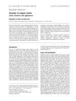
Fig. 1.1. Schematic of a nuclear reactor that produces thermal neutrons. Fuel rods
(a)contain
235
U atoms which when they encounter moderated neutrons undergo
fission producing ∼2.5 high-energy neutrons/
235
U atom. The probability of a fast
(high energy) neutron interacting with a
235
U atom is small. To sustain the chain
reaction, neutrons must be slowed down or thermalized by passing through a mod-
erator. In practice, moderators such as H
2
O, D
2
O, graphite, or beryllium are used,
filling the space in the reactor core around the fuel rods. For reasons of cost, H
2
O
is the most commonly used moderator (b) Thermal neutrons with a peak flux cen-
tered at ∼1.2
˚
A can either be extracted directly from the reactor via a beam tube
(c) or can be furthered slowed down by interaction with another, colder moder-
ator, for example, a vessel of liquid hydrogen (d) These cold neutrons, with their
Maxwellian distribution shifted toward lower energies, can be transported over many
meters to the various spectrometers by
58
Ni-coated optically flat glass surfaces
(e) through a process known as total external reflection