ADVANCES IN ORGAN BIOLOGY THE BIOLOGY OF THE EYE docx
Bạn đang xem bản rút gọn của tài liệu. Xem và tải ngay bản đầy đủ của tài liệu tại đây (13 MB, 408 trang )
ADVANCES IN
ORGAN BIOLOGY
Volume 10
2006
THE BIOLOGY OF THE EYE
ADVANCES IN ORGAN BIOLOGY
E. Edward Bittar, Series Editor
VOLUME 1. Pregnancy and Parturition
Edited by Tamas Zakar, 1996
VOLUME 2. The Synapse: In Development, Health and Disease
Edited by Barry W. Festoff, Daniel Hantai, and Bruce A. Citron, 1997
VOLUME 3. Retinoids: Their Physiological Function and Therapeutic Potential
Edited by G.V. Sherbet
VOLUME 4. Heart Metabolism in Failure
Edited by Ruth Altschuld and Robert A. Haworth, 1998
VOLUME 5. Molecular and Cellular Biology of Bone
Edited by Mone Zaidi, 1998
VOLUME 6. Myocardial Preservation and Cellular Adaptation
Edited by Dipak K. Das, 1998
VOLUME 7. Coronary Angiogenesis
Edited by Karel Rakusan, 1999
VOLUME 8. A Functional View of Smooth Muscle
Edited by Lloyed Barr and Gordon J. Christ, 2000
VOLUME 9. The Renal Circulation
Edited by Warwick P. Anderson, Roger G. Evans, and Kathleen M. Stevenson,
2000
VOLUME 10. The Biology of the Eye
Edited by Jorge Fischbarg, 2006
ADVANCES IN
ORGAN BIOLOGY
THE BIOLOGY OF THE EYE
Edited by: JORGE FISCHBARG
Lazlo Z. Bito Professor of Physiology and
Cellular Biophysics
Columbia University
New York, NY
USA
VOLUME 10
2006
2006
AMSTERDAM • BOSTON • HEIDELBERG • LONDON
NEW YORK • OXFORD • PARIS • SAN DIEGO
SAN FRANCISCO • SINGAPORE • SYDNEY • TOKYO
Academic Press is an imprint of Elsevier
ELSEVIER B.V. ELSEVIER Inc. ELSEVIER Ltd. ELSEVIER Ltd.
Radarweg 29 525 B Street The Boulevard 84 Theobalds Road
P.O. Box 211, 1000 AE Suite 1900, San Diego Langford Lane, Kidlington London WC1X 8RR
Amsterdam, The Netherlands CA 92101-4495, USA Oxford OX5 1GB, UK UK
# 2006 Elsevier B.V. All rights reserved.
This work is protected under copyright by Elsevier B.V., and the following terms and conditions apply to its use:
Photocopying
Single photocopies of single chapters may be made for personal use as allowed by national copyright laws. Permission of the
Publisher and payment of a fee is required for all other photocopying, including multiple or systematic copying, copying for
advertising or promotional purposes, resale, and all forms of document delivery. Special rates are available for educational
institutions that wish to make photocopies for non-profit educational classroom use.
Permissions may be sought directly from Elsevier’s Rights Department in Oxford, UK: phone (+44) 1865 843830, fax (+44) 1865
853333, e‐mail: Requests may also be completed on-line via the Elsevier homepage (http://www.
elsevier.com/locate/permissions).
In the USA, users may clear permissions and makepayments through the Copyright Clearance Center, Inc., 222 Rosewood Drive,
Danvers, MA 01923, USA; phone: (+1) (978) 7508400, fax: (+1) (978) 7504744, and in the UK through the Copyright Licensing
Agency Rapid Clearance Service (CLARCS), 90 Tottenham Court Road, London W1P 0LP, UK; phone: (+44) 20 7631 5555;
fax: (+44) 20 7631 5500. Other countries may have a local reprographic rights agency for payments.
Derivative Works
Tables of contents may be reproduced for internal circulation, but permission of the Publisher is required for external resale or
distribution of such material. Permission of the Publisher is required for all other derivative works, including compilations and
translations.
Electronic Storage or Usage
Permission of the Publisher is required to store or use electronically any material contained in this work, including any chapter or
part of a chapter.
Except as outlined above, no part of this work may be reproduced, stored in a retrieval system or transmitted in any form or by
any means, electronic, mechanical, photocopying, recording or otherwise, without prior written permission of the Publisher.
Address permissions requests to: Elsevier’s Rights Department, at the fax and e-mail addresses noted above.
Notice
No responsibility is assumed by the Publisher for any injury and/or damage to persons or property as a matter of products
liability, negligence or otherwise, or from any use or operation of any methods, products, instructions or ideas contained in the
material herein. Because of rapid advances in the medical sciences, in particular, independent verification of diagnoses and drug
dosages should be made.
First edition 2006
Library of Congress Cataloging in Publication Data
A catalog record is available from the Library of Congress.
British Library Cataloguing in Publication Data
A catalogue record is available from the British Library.
ISBN: 0-444-50925-9
The paper used in this publication meets the requirements of ANSI/NISO Z39.48-1992 (Permanence of Paper).
Printed in The Netherlands.
CONTENTS
LIST OF CONTRIBUTORS vii
PREFACE
Jorge Fischbarg xi
WHY THE EYE IS ROUND
Larry S. Liebovitch 1
TEARS AND THEIR SECRETION
Darlene A. Dartt, Robin R. Hodges and
Driss Zoukhri 21
THE CORNEA: EPITHELIUM AND STROMA
Niels Ehlers and Jesper Hjortdal 83
THE CORNEAL ENDOTHELIUM
Jorge Fischbarg 113
CILIARY BODY AND CILIARY EPITHELIUM
Nicholas A. Delamere 127
THE LENS
Guido A. Zampighi 149
THE VITREOUS
Henrik Lund‐Andersen, J. Sebag, Birgit Sander
and Morten la Cour 181
THE RETINA
Morten la Cour and Berndt Ehinger 195
v
THE RETINAL PIGMENT EPITHELIUM
Morten la Cour and Tongalp Tezel 253
THE CHOROID AND OPTIC NERVE HEAD
Jens Folke Kiilgaard and Peter Koch Jensen 273
INNATE AND ADAPTIVE IMMUNITY OF THE EYE
Mogens Holst Nissen and Carsten Ro
¨
pke 291
DRUG DELIVERY TO THE EYE
Ashim K. Mitra, Banmeet S. Anand and
Sridhar Duvvuri 307
THE SCLERA
Klaus Trier 353
INDEX 375
vi CONTENTS
LIST OF CONTRIBUTORS
Banmeet S. Anand Missouri-Kansas City
Kansas City, Missouri
Darlene A. Dartt Schepens Eye Research Institute
Boston, Massachusetts
Nicholas A. Delamere KY Lions Eye Research Institute
University of Louisville School of
Medicine
Louisville, Kentucky
Sridhar Duvvuri Missouri-Kansas City
Kansas City, Missouri
Bernd Ehinger Department of Ophthalmology
University of Lund
Sweden
Niels Ehlers Department of Ophthalmology
University of Arhus
Arhus, Denmark
Jorge Fischbarg Laszlo Z. Bito Professor of Physiology
and Cellular Biophysics in
Ophthalmology
College of Physicians and Surgeons
Columbia University
New York, New York
Jesper Hjortdal Department of Ophthalmology
University of Arhus
Arhus, Denmark
vii
Robin R. Hodges Schepens Eye Research Institute
Boston, Massachusetts
Peter Koch Jensen Department of Ophthalmology
Rigshospitalet, Copenhagen
Denmark
Jens Folke Kiilgaard Department of Ophthalmology
Rigshospitalet, Copenhagen
Denmark
Morten la Cour Department of Ophthalmology
Herlev University Hospital
Denmark
Larry S. Liebovitch Professor and interim Director
Center for Complex Systems and
Brain Sciences
Florida Atlantic University
Boca Raton, Florida
Henrik Lund‐Andersen Department of Ophthalmology
University of Copenhagen
Herlev Hospital, Denmark
Ashim K. Mitra Division of Pharmaceutical Sciences
School of Pharmacy
University of Missouri-Kansas city
Kansas City, Missouri, USA
Mogens Holst Nissen Institute of Medical Anatomy
The Panum Institute
University of Copenhagen
Copenhagen, Denmark
Carsten Ro
¨
pke Institute of Medical Anatomy
The Panum Institute
University of Copenhagen
Copenhagen, Denmark
Birgit Sander Department of Ophthalmology
University of Copenhagen
Denmark
viii LIST OF CONTRIBUTORS
J. Sebag Professor of Clinical Ophthalmology
Doheny Eye Institute
University of Southern California and
VMR Institute, California, USA
Tongalp Tezel Assistant Professor of Ophthalmology
and Visual Sciences
University of Louisville, School of
Medicine
Kentucky Lions Eye Center
Louisville, Kentucky
USA
Klaus Trier Trier Eye Clinic and Research Lab
Hellerup, Denmark
Guido A. Zampighi Departments of Neurobiology
and Physiology
David Geffen School of Medicine
Los Angeles California
Driss Zoukhri Schepens Eye Research Institute
Boston, Massachusetts
List of Contributors ix
This page intentionally left blank
PREFACE
The invitation to edit this book came from Dr. E. Edward Bittar, an
excellent colleague and friend. Along with the invitation came exceedingly
useful suggestions on the contents and format, for which Dr. Bittar is to be
amply credited, as well as for his mentoring this author and advising on steps
to move the book along during difficult periods.
This preface is the only place in which the Editors seemingly can thank
the authors in public for the time and dedication they have spent for this
volume. Seeing how much knowledge has been distilled into crisp text, one
can feel the dedication and love for their field the contributors have. Partly
as a consequence of the seas of acquaintances the editors navigate in, a large
number of the chapters have been written by Scandinavians. That seems
quite natural these days; that part of the World distinguishes itself in love for
Academia and tradition in ophthalmic sciences. For them as well as the
other authors, in this sampling the pleasure of imparting knowledge con-
tinues to be an important driving force in our world, which should be a sign
of hope.
Editing a book of this sort presents a quandary: given two extremes, one
can try to run a regimented production along narrow lanes, or can allow the
authors latitude for them to write as they see fit. In this case, the second
option carried the day hands down. The original instructions asked the
authors to think of a potential audience of newcomers to the field trying to
discover in relatively simple terms what is known about the eye, which are
xi
the areas receiving most of the attention, and where the excitement of
crossing the border and venturing into the unknown lies in every case. I
think the authors have responded brilliantly. Each one in his own style; some
wrote short accounts, others wrote wonderful comprehensive reviews. The
extension of each chapter in some way gives a measure of the width of the
currents crossing that field, and of the complexities that the author felt
compelled to communicate. The Editors have prudently opted for stepping
aside and letting that be.
The subjects covered are an indication of the growth and diversification of
areas of interest in the eye. Years ago, books in this area took great care in
covering the anatomy, and justifiably so, as the future ocular surgeons
needed to start their careers with the best of directions about the area they
would be operating on. As the basic sciences progressed, books included
growing sections on the functionality of the different organs. We have of
course kep t the anatomical separation of subjects and the functional descrip-
tions. However, we have also chosen to add a chapter on the shape of the
eye, in a way perhaps acknowledging that ophthalmology has begun mod-
ifying the corneal shape, and has begun asking what would it take to give the
eye optimal shape for imag e formation. The chapters on drug delivery and
immunology respond to the same activist approach, one in which as we learn
the basics of the eye we learn as well about ocular characteristics that allow
intervention or explain pathology. In addition, although this book addresses
basic mechanisms, the chapters contain mentions to pathology and disease
wherever these subjects arise naturally.
Speaking for the Editors, we have greatly enjoyed the reading of these
materials. I hope the same intellectual fulfillment will be now felt by the
readership.
New York, February 2005 Jorge Fischbar g
xii PREFACE
WHY THE EYE IS ROUND
Larry S. Liebovitch
Abstract 2
I. Introduction . 2
II. Why Are Things Round? . . 4
A. Inanimate Objects
4
B. Animate Objects
5
III. Why Are Eyes Round? 6
A. Optical Properties .
6
B. Eye Movement
8
C. Hollow
8
D. Phylogeny and/or Ontogeny .
9
E. Conclusions
10
IV. Pressure . 10
A. Surface Tension
10
B. Pressure in the Eye .
11
V. Aqueous Flow . 11
A. Balance of Inflow and Outflow
11
B. Inflow .
12
C. Outflow
12
Advances in Organ Biology
Volume 10, pages 1–19.
© 2006 Elsevier B.V. All rights reserved.
ISBN: 0-444-50925-9
DOI: 10.1016/S1569-2590(05)10001-9
1
VI. The Ciliary Body 13
A. Structure .
13
B. Numbers in Science .
13
C. Reynolds Number .
13
D. Peclet Number
14
E. Concentration Number .
15
F. Fluid Transport . . .
15
G. Ion Transport
16
H. Active or Passive . .
17
VII. Large Scale Aqueous Motions 18
VIII. Control of Intraocular Pressure 18
IX. Summary . 19
ABSTRACT
An impressive characteristic about eyes is their round, spherical structure. This
chapter explores the optical, mechanical, structural, phylogenic, and ontogenic
reasons why eyes are round. This exploration is used as a starting point to
describe how the different features of the eye are related to each other, and how
the roundness is maintained by the inflow and outflow of fluid in the eye.
I. INTRODUCTION
If you look up into the night sky at the constellation of the Big Dipper and
have 20/30 or better visual acuity and adequate night vision, you will see that
the next‐to‐the‐last‐star in the handle of the dipper is actually two stars that
are quite close together. One star is brighter than the other. The brighter star
is called Mizar and the fainter Alcor. It is easy to fall into the trap described
by the ancient Arabic proverb that, ‘‘He sees Alcor, but not the full moon.’’
The lesson here is that the most outstanding fact about eyes is not something
arcane, but the obvious fact that eyes are round (i.e., eyes are spheres).
Therefore, this first chapter will focus on the fact that eyes are round. Why
should eyes be round? What does it tell us about how eyes are constructed
and how they work? Not only is this shape similar in different animals, but
the variation in size of the vertebrate eye, from tree shrew to whale, is much
smaller in proportion than the variation in size of these creatures. It will also
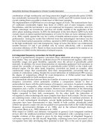
be described how different features of the eye (Figure 1) are related to each
other, and how the roundness is maintained and controlled by the formation,
flow, and removal of fluid in the eye.
2 LARRY S. LIEBOVITCH
Figure 1. The eye.
Why the Eye is Round 3
II. WHY ARE THINGS ROUND?
When I first thought about the roundness of eyes, I realized I did not know
why anything was round. So, I made a list of other round objects to help
organize my thought process. My list consisted of the sun, the earth, the
moon, oranges, frog urinary bladders, basketba lls, and rocks. As you can
see, the list consists of organic animate and inorganic inanimate objects
(Volk, 1985).
A. Inanimate Objects
Before starting with the inanimate objects on the list, the understanding of
the concept of equilibrium is needed. Consider your textbook, unopened, on
a desk. Even though it is static, there are at least two forces at work, making
it that way. It is actually in dynamic equilibrium, subject at every instant, to
opposing forces, which balance it. Gravity is pulling the book down toward
the center of the earth. The desk is pushing it up, preventing it from moving.
All objects that appear static are actually in this balancing act of opposing
forces. If one of the forces were stronger, it would change the object rapidly,
until an opposing force balanced it, and then the object would again be at a
new equilibrium. Objects change so rapidl y when out of equilibrium that we
are not likely to catch sight of them during that time.
What forces are balancing in these inanimate objects? How do those
forces determine the shapes of these objects? In the sun, gravity pulls the
gases of the sun together, pushing all its material toward its center. The
inward pull of gravity raises the temperature, which raises the pressure of
the gas in the sun until the outward pressure of the gas balances the inward
pull of gravity. Both the inward pull of gravity and the outward push of
gas pressure are isotropic. That is, they are equally effective in all directions.
That is why the sun is round. If one of these forces were not isotropic, then
the sun would not be round. Sometimes there are other pressures. If a star is
rapidly rotating, or has a strong magnetic field, then the gas pressure is
weaker along that axis. The gas collapses along that axis, and the star
becomes a flattened disk. The weaker pressure along the axis balances the
weaker gravitational force of the thin mass in the thickness of the disk,
whereas the stronger pressure along the radius of the disk balances the larger
gravitational force of the larger amount of mass in the radial direction.
Thus, round object s exist when forces are isotropic and nonround objects
when forces are not isotropic.
In the earth, the gravitational force pushing inward is balanced by the
outward push of the strength of the rocks, a result of the push of electrons
4 LARRY S. LIEBOVITCH
against each other in adjacent atoms. Both these forces are isotropic, and so
the earth is round. In a basketball, the air pressure pushing outward is
balanced by the tension on the fabric pushing inward. Again, both these
forces are isotropic and so the basketball is round.
B. Animate Objects
In inanimate objects, a round configuration results from a balance of isotro-
pic forces (i.e., forces experienced equally in all directions). But what deter-
mines the shapes of living things? The zoologist and classical scholar,
D’Arcy Thompson attempted to answer this puzzling phenomena in his
book, ‘‘On Growth and Form’’ first publis hed in 1917 (Thompson, 1966).
Although you may not be familiar with his publication, there is a good
chance that you have seen reproductions of his drawings. His exquisite
illustrations of forms of radiolaria, or how the shapes of animals change
from one species to another have been prolifically copied. The seminal point
of Thompson’s book was that genes do not set the blueprint of the shape of
an organism, but they set the rules of how the organism interacts with its
environment. It is then this dynamic interaction between the organism and
its environment that produces the structure.
For example, the final shape of the long bones in the arms and legs is
dependent on forces between osseous cells and the forces of their environ-
ment. Since bone is alive, material is constantly being added and removed
from biochemical reactions by cells within the bone. When a bone is bent,
fluid flows inside the bone. The negative and positive ions in this fluid flow
at different rates generating an electrical voltage. This voltage affects the
cells in the bone, so that their enzymes add more calcium on the electri-
cally negative side of the bend and remove more calcium on the electrically
positive side of the bend. As a result, the bone is rescul pted into a straighter
shape. Bone is very strong at resisting compressive forces pushing inward on
both ends. It is weak at resisting tensile forces pulling outward from both
ends. The resculpting adds material wher e the bone is in compression and
more material is needed. It removes material where the bone is in tension
and excess material is was ted. Thus, the genes, through their complex
programming of cells and their enzymes, have set the rule: add material
where it is needed and remove material where it is not needed. The genes
have set the rule of how the bone interacts with the environment. That rule
and its interaction with the environment then generate the straight shape
of the bone.
Such interactions also sculpt the eye and its surroun ding tissues. In
congenital glaucoma, the increased pressure in the eye stimulates the entire
Why the Eye is Round 5
eye to develop to a larger size than normal. When an eye with retinal
blastoma has to be enucleated at an early age to prevent cancer from
spreading, the bones of that orbit do not grow as large as the other orbit,
because the pressure of the eye is needed to stimulate their normal growth.
For the living things in my list, how much shape is determined solely by
the genes and how much by the rules of interaction with the environment set
by the genes? I have my own guesses about oranges and frog urinary
bladders. What are your guesses? To answer these questions you must ask
yourself, ‘‘What forces are ba lancing to determine the shape?’’ and ‘‘What is
the mechani sm of feedback between the world and the tissue?’’
III. WHY ARE EYES ROUND?
A. Optical Properties
My first guess was that since the most important function of the eye is to
form our image of the world, there must be an optical reason why eyes are
round.
The eye focuses light onto the retina. Most people think that this focusing
is performed by the lens in the eye. However, light is bent most sharply when
it passes through an interface of materials of different refractive indices. In
the eye, the difference in refractive index is much larger at the air–tissue
interface of the cornea (the clear front surface of the eye), than at the fluid–
tissue–fluid interface of the lens. Thus, two‐third of the focusing of light is
done by the cornea and only one‐third by the lens. The lens does the fine‐
tuning of the focusing of the image. The cornea controls the overall quality
of the image. It is problems of the cornea that produce nearsightedness,
farsightedness, or astigmatism that can be corrected by glasses or contact
lenses.
Is the eye round to achieve the best optical image on the retina, where the
light is detected and transformed into electrical signals? There are a number
of different aberrations, ways in which the focus of images on the retina are
not perfect. Important deviations include spherical aberration (where a light
ray in the center of the cornea reaches a focus that is closer to the cornea
than a ray at the periphery of the cornea), and chromatic aberration (where a
ray of blue light reaches a focus that is closer to the cornea rather than a ray
of red light). Another aberration is that the cornea focuses images onto a
spherical surface rather than a flat surface. Moreover, this spherical surface
has a different radius for vertical and horizontal images on the cornea. The
retina of the eye is a spherical surface whose radius is a good compromise
between those two different radii. This looks like a good reason why eyes
might be round, but actually, it is only a very small effect.
6 LARRY S. LIEBOVITCH
In fact, the image of the world on the retina does not need to be in very
good focus across the entire retina. Brown notes that ‘‘the optical character-
istics of the eye are nicely matched to the receptors [photoreceptors] and
neural components (Records, 1979).’’ Only a very small part of the retina,
the fovea, requires light to be accurately focused. This is because the neural
components of the retina that sense light only have high resolution in the
fovea. There are about 100 million photoreceptors, rods, and cones in the
eye that convert light into electrical signals. There are 1 million retinal
ganglion nerve cells that carry the informat ion out of the eye into the brain.
This enormous number of nerve cells is about one‐third of all the afferent
nerve fibers bringing information into the brain. But even with this large
number of nerve cells, there are still 100 photoreceptors for each nerve cell.
Hence, the light from every photoreceptor does not individually reach the
brain. Only in the fovea there is a 1:1 coupling between photoreceptors and
nerve cells. Away from the fovea, the output from many photoreceptors is
processed and blended together into far fewer nerve cells that reach the
brain. Thus, throughout most of the retina the neural pixels (picture ele-
ments) are coarse. In most of the retina, the eye sacrifices spatial resolution
for enhanced sensitivity at low‐light levels, as well as enhanced resolution of
how the light level is changing in time.
The spatial resolution is high only in the fovea, which senses an area that
is about two degrees (2
) across, only four times the diameter of the full
moon. Everything else in your image of the world is fuzzy. The look of the
world, its sharp edges and beautiful colors, is an illusion generated high up in
the neural pathway located in the visual cortex in the back of your head. The
eye is not like a camera. It is more like an electronic information sampling
system. The brain moves the high resolution, clear image fovea to sample
interesting features such as an ornate edge or a flashing light. It samples
phenomena that look interesting. What you see depends primarily on what
you saw before and what you are thinking now. This information is
combined into the fiction of a clear , stable world.
A sharp, clear image is not needed across most of the retina because the
neural elements there that detect light do not have a high‐spatial resolution.
A coarse image is a nice match to the coarse neural elements. Most of the
retina provides a wide angle, low‐resolution detection system to spot poten-
tial predators. The spherical retina may provide a useful detector for such
system. Sharp, clear images are only needed in the fovea, a region 3 mm in
diameter. A spherical shape is not needed to produce a clear image over such
a small target. For example, when light is dim, at the bottom of the ocean or
late at night on the land, animals have developed long cylindrical eyes with
large, fast (high f ratio) lenses that maintain focus and clear images to the
central area of their flattened retinas.
Thus, it does not seem as if roundness is a necessity for optical efficiency.
Why the Eye is Round 7
B. Eye Movement
I used to ask scientists at eye research conferences why they thought eyes
were round. Inevitably, the answer that I received was that eyes were round
because this was the best shape for rapid and accurate eye movements. It is
mechanically easy to rotate a round eye in a round socket to aim it at any
direction. Spheres also have the lowest moment of inertia for their mass and
thus require the least force to move.
Is this the reason why eyes are round? In his classic book on the vertebrate
eye, Walls notes that the ‘‘primitive function of the eye muscles was not to
aim the eye at objects at all [but] designed to give the eyeball the
attributes of a gyroscopically stabilized ship, for the purpose of maintaining
a con stancy of the visual field despite chance buffetings and twistings of an
animal’s body by water currents and so on’’ (Walls, 1963).
Let’s examine the evolutionary sequence (Lythgoe, 1979). Fishes lack the
fovea needed for sharp vision. They do not need to aim their eyes accurately,
so they do not follow objects with their eyes. Amphibians also have limited
eye movement capabilities. Neckless frogs turn their entire bodies in order to
change their direction of gaze. Reptiles show variation in their eye move-
ment. Some, like the Gila monster, have eyes that are fixed in their head.
Others, like the chameleon, can use one eye to look forward and the other to
look backward at the same time. Birds, the descendants of dinosaurs, have
better vision than humans. Some birds have extended high‐ resolution areas
on their retinas that cover a huge field of view. Other birds have more
pigments in their photoreceptors for enhanced color resolution or extra
structures to deliver more oxygen to the retina. Yet, their eyes are fixed
and immobile. It is only mammals that have rapid and accurate movements.
This idea of roundness to facilitate eye motion, which seems obvious to
many scientists, when considered in more detail, seems less convincing. The
evolutionary record is whispering to us that eyes were round before they
moved rapidly or accurately. Thus, it does not seem as if the eye is round
primarily for eye movement reasons.
C. Hollow
Perhaps it is the hollow inside, which is significant. A spherical shell, inflated
with fluid, can provide a clear optical pathway to the retina unobstructed by
bones and ligaments. The spherical shape also provides the shortest, there-
fore the quickest, pathways for oxygen and nutrients to reach the interior
structures of the eye and for wastes to leave them. A convoluted interior
space, with serpentine passageways, would reduce the efficiency of such
diffusion.
8 LARRY S. LIEBOVITCH
But the eye has not taken full advantage of this unobstructed interior
space. Except in the core of the fovea, one layer of blood vessels that nourish
the retina and two layers of synapses of nerve cells, lie in front of the
photoreceptors. Light passes through these cells to reach the photoreceptors.
These obstructions affect the image on the retina. You have probably
observed this blood flow. On a clear day, when you look at a bright blue
sky (but not the sun, which can cause severe and permanent damage) you
can see tiny white specks darting around. This image is called the blue
entoptic phenomenon. The white specks are white blood cells moving in front
of the photoreceptors. The photoreceptors become adapted to the more
numerous red blood cells shadowed against the blue sky, but then detect
and respond to the occasional white blood cell. Experimentally, the speed of
the white dots on a computer screen has been matched with the speed of
these white specks to measure relative retinal microcirculation. To calibrate
the system, a few volunteers wore a neck cuff to reduce the circulation to
head so that the speed of the dots on the computer screen could be related
quantitatively to the blood flow in the retina.
The eye has taken some, but not complete advantage, of this hollow
space. Thus, it does not appear that the eye is round primarily for structural
reasons to create a hollow space.
D. Phylogeny and/or Ontogeny
Walls notes, ‘‘The great German anatomist Froriep once likened the ‘sud-
den’ appearance of the vertebrate eye in evolution to the birth of Atena, fully
grown and fully armed, from the brow of Zeus.’’ There are no intermediate
anatomical adaptations. Animals either have eyes that form images or spots
that detect the amount of light. Perhaps roundness is a consequence of
evolutionary pressures that produced the vertebrate eye. This idea is sup-
ported by the anatomical evidence found in the eyes of the cephalopods,
such as squid and octopus. Their eyes evolved separately from the vertebrate
eye, yet except for some small differences, their anatomy is strikingly similar.
One of the few differences is that the cephalopod eye has nerves, which travel
from the back of the photoreceptors, rather than the front of them, so that
they do not interfere with the light pathway to the photoreceptors. Tripathi
notes, ‘‘The final resemblance between the two types of eye [cephalopod and
vertebrate] makes this one of the most striking cases of convergence in
evolutionary history (Davson and Graham, 1974).’’ Convergence means that
similar adaptive pressures led to similar anatomical structures. Perhaps,
those pressures also dictated the roundness of the eye.
Maybe the answer lies not in phylogeny, the evolutionary history of a
species, but in ontogeny, the developmental history of each new individual.
Why the Eye is Round 9
The structures of the eye need to be axis‐symmetric along the line of rotation
which brings light through the eye into the retina. Perhaps, developmental
processes that form spherical structures are the embryo’s path of least
resistance to form such axis‐symmetric structures.
Although speculation on species‐specific evolution or individual develop-
ment is both interesting as well as attractive, the hard evidence in support of
these ideas is lacking. Thus, it does not seem that the eye is round primarily
for phylogeni c or ontogenic reasons.
E. Conclusions
Neither optical, nor movement, nor structural, nor evolutionary, nor devel-
opmental reasons seem to be the primary reason why the eye is round.
IV. PRESSURE
Although we do not understand why the eye is round, we do understand
how it is round. As explained earlier, the roundness of the eye reflects a
balance of two opposing forces. The outward force exerted by the pressure
of the fluid inside the eye is balanced by the inward tension in the shell of
the eye.
A. Surface Tension
The tension in the outer layers of the eye is called surface tension. If we
were to make a small cut on the eye, the surface tension would be the force
pulling the two sides of the cut away from each other. For a given pressure
inside, the sphere is the shape that has the lowest surface tension. Containers
for gas under pressure of any shape other than spherical require stronger
walls. In the inorganic world, it is harder to manufacture spheres than
cylinders, thus, most gas containers are cylinders. However, the material of
these cylinders must be made twice as strong as would be needed for a sphere
to hold the same pressure of gas.
In a cylinder, the surface tension across a cut in a curved direction is equal
to that for a sphere of the same radius under the same pressure, but the
surface tension for a cut in the long direction has twice the surface tension.
This is the reason that the skin of frankfurters always tears in the long
direction when cooked. The surface tension is twice as great in the lengthwise
direction. Since the frankfurter skin is equally strong in both directions, it
always breaks along the long direction, where the force tearing at it is twice
that of the force tearing at it in the curved direction.
10 LARRY S. LIEBOVITCH
B. Pressure in the Eye
The fluid that flows in the eye is called the aqueous humor. It flows out of the
ciliary body, passes in front of the lens, moves through the pupil, and
circulates in the space behind the cornea. As discussed earlier, the outward
force from the fluid pressure of the aqueous humor inside the eye is isotropic,
felt equally in all directions. The inward force of the surface tension in the
outer shell of eye is also isotropic. The balance between these inward and
outward forces determines the spherical shape of the eye.
Since the force of the fluid pressure inside the eye is isotropic, a pressure
increase in one part of the eye causes a pressure increase everywhere
throughout the eye. In glaucoma, the pressure increase in the aqueous
humor in the front of the eye is transmitted to the back of the eye. Although
the pressure increase is caused by events in the front of the eye, the damage
to vision is due to the effects of this pressure in the back of the eye. The
increased pressure crimps the retinal nerve and blood flow, killing retinal
ganglion cells either by cutting off the transport of essential materials along
the inside of their axons, or the blood supply that nourishes them from the
outside. The loss of vision results from the death of these nerve cells.
The hardness of the eye to touch is not determined by the toughness of the
fabric of the eye, but by the fluid pressure inside the eye. When the pressure
is high, the eye is hard. When the pressure is low, the eye is soft.
However, this is not the whole story. There is an additional factor. I have
always felt that when my bicycle tires are old, no matter how much I pump
them up, they never feel quite as hard as new tires. In the eye too, when the
fabric is compromised, the shape and hardness of the eye change. For
example, the shape of the cornea changes in keratoconus where the collagen
in the cornea is weakened. In pathological myopia, there is a slow mechani-
cal yielding of the fabric, and the eye steadily enlarges in time.
V. AQUEOUS FLOW
A. Balance of Inflow and Outflow
The eye is round because it is inflated by the pressure from the fluid inside. Is
that what is necessary to maintain its shape, that is, to fill it once with
aqueous humor under pressure? Nothing lasts forever. For example, my
bicycle tires lose about 20% of their air every week. In order to maintain
the pressure in the eye, we need to push fluid in and have it leak out in a very
precise system. At first thought, it seems unbelievably wasteful to push fluid
into the eye just to let it leak out again, but it’s actually the most basic
Why the Eye is Round 11
biological trick to expend energy for the sake of control. Balancing the
inflow and outflow of aqueous humor provides a way to maintain and
control the pressure inside the eye.
Soon we will see in detail how the aqueous humor is produced, and how it
leaks out of the eye. The important point to remember here is that there is a
balance of inflow and outflow. If the inflow was greater than the outflow, the
fluid inside the eye would continually increase, and the eye would burst.
If the inflow were less than the outflow, the fluid inside the eye would
continually decrease, and the eye would collapse.
The flow of aqueous humor out of the eye is dr iven by the pressure
inside the eye. The resistance to the flow of aqueous out of the eye deter-
mines the intraocular pressure inside the eye. If it is hard for the aqueous
humor to leave the eye, then more aqueous accumulates in the eye. This
increases the pressure within the eye, which forces more aqueous out. The
pressure continues to increase until the aqueous flow out of the eye equals
the aqueous flow into the eye. The pressure at which this balance occurs is
determined by the resistance to the outflow of aqueous humor leaving
the eye.
Thus, there is always a balance in the amount of aqueous entering and
leaving the eye.
B. Inflow
The aqueous humor is generated by the ciliary body, a wiggly layer of tissue,
two cells thick, along the edge of the ciliary muscle in the inside angle of the
eye, a little back from where the clear cornea merges into the white sclera.
From the ciliary body, the aqueous humor flows into the posterior chamber
behind the lens. Then it passes through the pupil into the anterior chamber
in front of the lens.
C. Outflow
The aqueous humor in the anterior chamber leaves the eye by passing
through a series of structures in the angle of the eye inside of where the
cornea merges with the sclera. On its way out of the eye, the aqueous flows
through a coarse filter and then a fine filter, called the trabecular meshwork.
Then it flows through a layer of cells and into a tube called Schlemm’s
canal that circles the cornea. From the canal it flows through collecting
channels that bring it to the veins. It is not known which of these structures
offers the most resistance to the flow. Some recent evidence suggests that
the cells that line Schlemm’s canal offer the most resistance to the flow, and
thus determine the intraocular pressur e inside the eye.
12 LARRY S. LIEBOVITCH