INFLAMMATORY BOWEL DISEASE – ADVANCES IN PATHOGENESIS AND MANAGEMENT ppt
Bạn đang xem bản rút gọn của tài liệu. Xem và tải ngay bản đầy đủ của tài liệu tại đây (20.4 MB, 344 trang )
INFLAMMATORY BOWEL
DISEASE – ADVANCES IN
PATHOGENESIS AND
MANAGEMENT
Edited by Sami Karoui
Inflammatory Bowel Disease – Advances in Pathogenesis and Management
Edited by Sami Karoui
Published by InTech
Janeza Trdine 9, 51000 Rijeka, Croatia
Copyright © 2011 InTech
All chapters are Open Access distributed under the Creative Commons Attribution 3.0
license, which allows users to download, copy and build upon published articles even for
commercial purposes, as long as the author and publisher are properly credited, which
ensures maximum dissemination and a wider impact of our publications. After this work
has been published by InTech, authors have the right to republish it, in whole or part, in
any publication of which they are the author, and to make other personal use of the
work. Any republication, referencing or personal use of the work must explicitly identify
the original source.
As for readers, this license allows users to download, copy and build upon published
chapters even for commercial purposes, as long as the author and publisher are properly
credited, which ensures maximum dissemination and a wider impact of our publications.
Notice
Statements and opinions expressed in the chapters are these of the individual contributors
and not necessarily those of the editors or publisher. No responsibility is accepted for the
accuracy of information contained in the published chapters. The publisher assumes no
responsibility for any damage or injury to persons or property arising out of the use of any
materials, instructions, methods or ideas contained in the book.
Publishing Process Manager Bojan Rafaj
Technical Editor Teodora Smiljanic
Cover Designer InTech Design Team
First published January, 2012
Printed in Croatia
A free online edition of this book is available at www.intechopen.com
Additional hard copies can be obtained from
Inflammatory Bowel Disease – Advances in Pathogenesis and Management,
Edited by Sami Karoui
p. cm.
ISBN 978-953-307-891-5
free online editions of InTech
Books and Journals can be found at
www.intechopen.com
Contents
Preface IX
Part 1 Pathogenesis of Inflammatory Bowel Disease 1
Chapter 1 The Role of COX-2 Inhibitors on Experimental Colitis 3
Ana Paula R. Paiotti, Ricardo Artigiani-Neto, Daniel A.
Ribeiro,
Sender J.
Miszputen and Marcello Franco
Chapter 2 Intestinal Barrier Dysfunction:
The Primary Driver of IBD? 23
Pieter Hindryckx and Debby Laukens
Chapter 3 Adenosine Receptors: New Targets to Protect Against
Tissue Damage in Inflammatory Bowel Symptoms 41
Sebastian Michael, H W. Rauwald, Haba Abdel-Aziz,
Dieter Weiser, Christa E. Müller, Olaf Kelber and Karen Nieber
Chapter 4 Role of Dipeptidyl Peptidase
IV/CD26 in Inflammatory Bowel Disease 59
Dijana Detel, Lara Batičić Pučar, Ester Pernjak Pugel,
Natalia Kučić, Sunčica Buljević, Brankica Mijandrušić Sinčić,
Mladen Peršić and Jadranka Varljen
Chapter 5 The Roles of Interleukin-17 and
T Helper 17 Cells in Intestinal Barrier Function 89
Elizabeth Trusevych, Leanne Mortimer and Kris Chadee
Chapter 6 Pathogenesis of Inflammatory Bowel Diseases 111
Yutao Yan
Part 2 Advances in Diagnosis of Inflammatory Bowel Disease 135
Chapter 7 The Role of Imaging in
Inflammatory Bowel Disease Evaluation 137
Rahul A. Sheth and Michael S. Gee
VI Contents
Chapter 8 Health-Related Quality of Life in
Inflammatory Bowel Disease 151
Ramiro Veríssimo
Chapter 9 Validation of a Quantitative Determination Method of
Paramino-Salicylic Acid by High-Performance Liquid
Chromatography and Its Application in Rat Plasma 165
Ibrahima Youm, Malika Lahiani-Skiba and Mohamed Skiba
Chapter 10 Approach to the Management of the Pregnant Inflammatory
Bowel Disease Patient: Successful Outcome 177
Flavio M. Habal
Chapter 11 Bone Morphogenetic Proteins and Signaling
Pathway in Inflammatory Bowel Disease 199
Ivana Maric, Tamara Turk Wensveen,
Ivana Smoljan,
Zeljka Crncevic Orlic and Dragica Bobinac
Chapter 12 Genetic Differentiation of Fungi of the
Genus Candida Isolated from Patients with
Inflammatory Bowel Diseases 221
Danuta Trojanowska, Marianna Tokarczyk,
Małgorzata Zwolińska-Wcisło, Paweł Nowak,
Sebastian Różycki and Alicja Budak
Chapter 13 A 9-Year Retrospective Study of Hospitalized
IBD Patients in Shanghai Rui Jin Hospital 233
Tianle Ma, Lulu Sheng, Xiaodi Yang, Shuijin Zhu,
Jie Zhong, Yaozong Yuan and Shihu Jiang
Part 3 Management of Inflammatory Bowel Disease 247
Chapter 14 The Role of Diet, Prebiotic and Probiotic
in the Development and Management of
Inflammatory Bowel Diseases (IBD) 249
A.S. Abdulamir, Muhammad Zukhrufuz Zaman,
R.R. Hafidh and F. Abu Bakar
Chapter 15 The Use of Pomegranate
(Punica granatum L.) Phenolic Compounds
as Potential Natural Prevention Against IBDs 275
Sylvie Hollebeeck, Yvan Larondelle, Yves-Jacques Schneider
and Alexandrine During
Chapter 16 Drug Targeting in IBD Treatment –
Existing and New Approaches 301
Katerina Goracinova, Marija Glavas-Dodov,
Maja Simonoska-Crcarevska and Nikola Geskovski
Preface
Inflammatory bowel diseases (IBD), such as ulcerative colitis and Crohn's disease, are
chronic and relapsing conditions, characterized by abdominal pain, diarrhea, bleeding
and malabsorption. IBD is considered to be a hyper-inflammatory state due to
disturbed interactions between the immune system and the commensal bacterial flora
of the gut. In recent years, many studies have focused on etiopathogeny of IBD, as well
as on advances in diagnosis tools. A better comprehension of mechanisms of the new
drugs used for treatment of IBD has enabled new approach strategies in the
management of Crohn’s disease and ulcerative colitis.
There is accumulating evidence on the importance of microbes in the development
and maintenance of both the intestinal and immune systems. No evidence that
inflammatory bowel disease is caused by a single agent has been found, whereas a
number of microbes have been strongly associated with the presence of disease. The
majority of recent studies support a role for the ability of intestinal pathogens to
promote chronic inflammation in individuals with genetic susceptibility and/or other
environmental factors, which remain to be identified.
The lower gastrointestinal tract houses trillions of microbial cells, representing a large
diversity of species in relatively well-defined phylogenetic ratios that are associated
with maintenance of key aspects of host physiology and immune homeostasis. It is
therefore not surprising that many GI inflammatory diseases, including inflammatory
bowel disease, are associated with substantial changes in the composition of these
microbial assemblages, either as a cause or consequence of host inflammatory
response.
Inflammatory bowel diseases are the consequence of a dysregulated mucosal immune
system. The mucosal immune system consists of two arms, innate and adaptive
immunity, that have been studied separately for a long time. In the last several years,
there has been a huge increase in the discovery of inflammatory bowel disease
susceptibility genes. However, similar advances in identifying and defining
environmental risk factors associated with IBD have lagged behind.
Treatment of inflammatory bowel disease has changed in recent years. Potential future
treatment goals in Crohn’s disease include reduction in bowel damage, prevention of
complications and maintaining long term remission. Combination therapy with
X Preface
infliximab and azathioprine demonstrated superior rates of sustained clinical
remission, when compared to standard therapy. In ulcerative colitis, potential
treatment goals include sustained clinical remission, sustained mucosal healing and
reduction of rates of colorectal dysplasia and cancer. Although tumor necrosis factor
antagonists are effective for the treatment of Crohn's disease and ulcerative colitis, lack
and loss of clinical response is a clinical challenge. Accordingly, the use of therapeutic
drug monitoring has been proposed as a means to optimize treatment. Several
observational studies have demonstrated a relationship between anti-TNF agent
serum drug concentrations and/or antidrug antibody presence, and various
symptomatic and objective clinical endpoints. However, these relationships are not
absolute, and although some algorithms for the use of therapeutic drug monitoring in
clinical practice have been proposed, none have yet been validated in a prospective
clinical trial.
Dr. Sami Karoui
Department of Gastroenterology A. La Rabta Hospital
Tunis
Part 1
Pathogenesis of Inflammatory Bowel Disease
1
The Role of COX-2 Inhibitors
on Experimental Colitis
Ana Paula R. Paiotti
1
, RicardoArtigiani-Neto
1
, Daniel A.
Ribeiro
1,2
,
Sender J.
Miszputen
3
and Marcello Franco
1
Universidade Federal de São Paulo, Escola Paulista de Medicina
1
Departament of Pathology
2
Departament of Biosciences
3
Division of Gastroenterology
Brasil
1. Introduction
Since the introduction of acetylsalicylic acid (aspirin) as the first nonsteroidal
antiinflammatory drug (NSAID) in 1897, NSAIDs have been widely used in the
management of pain and inflammation (Botting, 2010; Vane et al., 1990; Wallace, 1997).
Today, they are classified as traditional nonsteroidal antiinflammatory drugs (tNSAIDs),
characterized by differing degrees of antiinflammatory, analgesic and antipyretic activity.
tNSAIDs are among the most widely used medicines in the world. Unfortunately, they are
associated with dose-dependent gastrointestinal (GI) adverse events ranging from
dyspepsia (10-20%) to symptomatic and complicated ulcers (1-4%) (Scheiman, 2006; Wolfe et
al., 1999). The mechanism of tNSAIDs action is attributed to the cyclooxygenase (COX)
inhibition (Botting, 2010; Vane, 1971). Cyclooxygenase is a key rate-limiting enzyme that
exists in at least two isoforms: COX-1 is observed constitutively expressed in various tissues,
whereas COX-2 does not appear to be expressed except at very low levels in most tissues
and is rapidly upregulated in response to growth factors and cytokines. More recently,
COX-2 has been implicated in several distinct cellular mechanisms, such as angiogenesis,
proliferation and the prevention of apoptosis (Dempke et al., 2001). New antiinflammatory
drugs have been synthesized, such as selective COX-2 inhibitors (anti-COX-2), however,
these drugs may present side effects, such as the ability to modify the epithelial barrier.
Inflammatory bowel disease (IBD) is a common chronic gastrointestinal disorder
characterized by alternating periods of remission and active intestinal inflammation.
The precise etiology of IBD, including Crohn´s disease (CD) and ulcerative colitis (UC),
remains unclear. However, environmental factors, immunological disturbances, genetic
influences and the presence of certain chemical mediators (cytokines) have been established
as putative participants in the pathogenesis of the disease (Barbieri, 2000; Lashner, 1995;
Podolsky, 2002).
In the last few decades, the development of experimental models for studying IBD has
greatly contributed to enhance understanding of the immunological mechanisms involved,
such as changes in the gut epithelial barrier (Colpaert et al, 2001; Shorter et al, 1972). IBD
seems to occur when luminal antigens from the bacterial flora stimulate the immune system
Inflammatory Bowel Disease – Advances in Pathogenesis and Management
4
in the gut barrier towards an exacerbated, genetically defined response. Patients present an
increase in the amount of intestinal bacterial antigen compared to healthy individuals
(Bonen & Cho, 2003).
In particular, some human and animal studies have shown the prime
importance of gut epithelial barrier integrity and changes that lead to deregulation of the
immune system as a result of the loss of intestinal homeostasis (Élson et al., 1995).
A possible association between the use of NSAIDs and the relapse of IBD has been
repeatedly suggested. IBD patients seek relief in NSAIDs for non-IBD-related pains
(arthralgias, arthritides) and these drugs are also prescribed for the symptons of
extraintestinal manifestations of IBD, such as peripheral arthritis, sacroiliitis, ankylosing
spondylitis, and osteoporosis-related fractures. NSAIDs are considered to be the first-line
treatment for the abnormalities just mentioned (i.e, relieve pain and treat inflammation).
It has been reported that CD is associated with gut barrier dysfunction and that some
patients express an instestinal barrier hyperresponsiveness to NSAIDs (Gornet et al., 2002).
Thus, clinicians are concerned that the treatment with NSAIDs could increase the risk of
disease aggravation relapse in controlled patients. A large number of people suffering from
IBD take NSAIDs and COX-2 inhibitors for various reasons, as the efficiency of these drugs
in pain control seems to be unquestioned. In some patients, exacerbation disease happens;
however it is uncertain whether NSAIDs are implicated in IBD relapse or whether COX-2
inhibitors are safer than NSAIDs.
NSAIDs have been implicated in the onset or the exacerbation of IBD in a number of studies
and case reports, whereas in other studies, no relationship has been found between NSAID
treatment and an increase in significant disease flares. On the other hand, COX-2 inhibitors
have a smaller incidence of toxicity to the small bowel or colon, as recent studies indicate
that COX-2 inhibitors are prescribed more often than NSAIDs in patients who are older,
sicker, and have risk factors associated with NSAID gastropathy (Bonner et al., 2000; Bonner
et al., 2004; Kurahara et al, 2001; Vane et al., 1998). Is the concept that the use of NSAIDs is
associated with relapse of IBD is true? For this reason, many studies are conducted with the
use of COX-2 in experimental models. So, the objective of this review is to describe the role
of COX-2 inhibitors on different experimental models of colitis.
2. COX-1/ COX-2 concept, biochemistry and structural comparisons
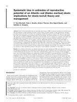
Cyclooxygenase (COX) or prostaglandin H2 synthase (PGHs) is the enzyme that catalyzes
the first two steps in the biosynthesis of the prostaglandins (PGs) from the substrate
arachidonic acid (AA). These are the oxidation of AA to the hydorxyendoperoxide PGH2.
The PGH2 is transformed by a range of enzymes and nonenzymic mechanisms into the
primary prostanoids, PGD
2
, PGE
2
, PGF
2α
, PGI
2
and thromboxane A
2
(TXA
2
) (DeWitt &
Smith , 1988) (Figure 1).
COX activity has long been studied in preprarations from sheep seminal visicles, and this
enzyme was cloned by three separate groups in 1988 (DeWitt & Smith , 1988; Merlie et al,
1988; Yokoyama et al., 1988;). The discovery of a second form of COX in the early 1990s was
the most important event in prostanoid biology in almost 20 years. Induction of this isoform,
COX-2, by several stimuli associated with cell activation and inflammation assured the
relevance of this finding to inflammatory disease in general. A clear sign of the therapeutic
value of this discovery is that in the relatively short time of about five years, several highly
effective anti-inflammatory agents and new therapeutic areas have become subjects for
The Role of COX-2 Inhibitors on Experimental Colitis
5
investigation (Bakhle & Botting, 1996; Botting, 2010; Herschman, 1996; Jouzeau et al., 1997;
Luong et al., 1996).
Fig. 1. The arachidonic acid cascade.
The inducible enzyme COX-2 is very similar in structure and catalytic activity to the
constitutive COX-1. The biosynthetic activity of both isoforms can be inhibited by aspirin
and other NSAIDs (Botting, 2010; Vane, 1971). Both isoforms have a molecular weight of
71 K and are almost identical in length, with just over 600 aminoacids, of which 63% are
in an identical sequence. However, the human COX-2 gene at 8.3 kb is a small immediate
early gene, whereas human COX-1 originates from a much larger 22-kb gene. The gene
products also differ, with the mRNA for the inducible enzyme being approximately 4.5 kb
and that of the constitutive enzyme being 2.8 kb (Bakhle & Botting, 1996; Botting, 2010;
Jouzeau et al., 1997).
The three-dimensional X-ray crystal structure of human or murine COX-2 (Mancini et al,
1994; Picot etal., 1994) can be superimposed on that COX-1 (Lecomte et al., 1994); the
residues that form the substrate binding channel, the catalytic sites, and the residues
immediately adjacent are all identical except for two small variations. In these two positions,
the same substitutions occur: Ile in COX-1 is exchanged for Val in COX-2 at positions
434 and 523 (the residues in COX-2 are given the same number as their equivalent
aminoacids in COX-1).
In spite of this structural identify, there are clear biochemical differences between the
isoforms in substrate and inhibitor selectivity. For example, COX-2 will accept a wider
Inflammatory Bowel Disease – Advances in Pathogenesis and Management
6
range of fatty acids as substrates than will COX-1 (Bakhle & Botting, 1996; Botting, 2010).
Thus, although both enzymes can utilize AA and dihomo-γ-linolenate equally well, COX-2
oxygenates other fatty acid substrates, such as eicosapentaenoic acid, γ-linolenic acid, α-
linolenic acid, and linoleic acid more efficiently than does COX-1. Also, COX-2 acetylated by
aspirin on Ser 530 will still oxidize AA but to 15-HETE, whereas similarly acetylated COX-1
will not oxidize AA at all (Griswold & Adams, 1996; O’Neill et al., 1994; Wong et al., 1997).
In addition (see below), inhibitors will differentiate between COX-2 and COX-1 with over
1000-fold selectivity (Gierse et al., 1996; Luong et al., 1996).
Supporting evidence is strongest from the work on COX-2-selective inhibitors; mutation of
Ile 523 to Val in the COX-1 protein allows COX-2-selective inhibitors to bind and inhibit
PGH
2
formation without altering the K
m
for AA (Guo et al., 1996), and the reverse mutant of
COX-2 in which Val 523 is exchanged for Ile shows inhibitor binding and selectivity profiles
comparable to those of wild-type COX-1 (Bhattacharyya et al., 1996; Mancini et al., 1995).
The structural basis for this has been shown clearly in the crystal analyses of COX-2, which
have used either the human or the murine protein, each bound to a nonselective COX-1 or
COX-2 inhibitor. The smaller size of Val 523 allows the inhibitor access to a side pocket off
the main substrate channel in COX-2-access that is denied sterically by the longer side chain
of Ile in COX-1. Selective inhibitors of COX-2 do not bind to Arg 120, which is used by the
carboxylic acid ot the substrate AA and by the COX-1-selective or-nonselective NSAIDs, all
of which are carboxylic acids (Ren et al., 1995a; Ren et al., 1995b).
Another striking structural difference between the isoforms, but of unknown significance, is
the absence of a sequence of 17 amino acids from the N terminus and the insertion of a
sequence of 18 amino acids at the C terminus of COX-2 i comparison to COX-1. This
accounts for the different numbering for the analogous residues in the two isoforms (e.g. the
acetylatable serine is Ser 530 in COX-1 but Ser 516 in COX-2). The C-terminal insert in COX-
2 does not alter the last four amino acids residues, which in both proteins form the signal for
attachment to the membrane of the endoplasmic reticulum (ER). However, COX-2 is located
on the nuclear membrane as well as on the ER, while COX-1 is found attached only to the
membranes of the ER. The reason for this selective localization may lie in the different
sequence of the C terminus. It is relevant that in the X-ray structural analysis of either
isoform, the three-dimensional structures of the last 18 C-terminal residues in COX-1 and
the last 30 residues in COX-2 were not resolved, implying a marked flexibility in this region
of the proteins even in the crystalline form (Hudson et al., 1993; Mitchell et al., 1993; Morita
et al., 1995; Otto & Smith, 1994; Regier et al., 1993). Although emphasis has been placed here
on the differences between isorforms, the extensive overall structural and biochemical
similarity between COX-1 and COX-2 must be reiterated. Both use the same endogenous
substrate, AA, and form the same product by the same catalytic mechanism. Their major
difference lies in their pathophysiological functions.
2.1 Physiological and pathological functions of COX-1 and COX-2
Chronic inflammation is an excellent example of a disease that represents a malfunction of
normal host defense systems. Thus, rather than classifying PG biosynthesis into
physiological and pathological, it may be better to use the classification applied to the COX
isoforms: either constitutive or induced. COX-1 activity is constitutive, present in nearly all
cell types at a constant level; COX-2 activity is normally absent from cells, and when
induced, the protein levels increase and decrease in a matter of hours after a single stimulus
(Bakhle & Botting, 1996; Botting, 2010; Jouzeau et al., 1997).
The Role of COX-2 Inhibitors on Experimental Colitis
7
The main reason for labeling COX-1 and COX-2 as physiological and pathological,
respectively, is that most of the stimuli known to induce COX-2 are those associated with
inflammation, for example, bacterial lipopolysaccharide (LPS) and cytokines such as
interleukin (IL)-1, IL-2, and tumor necrosis factor alpha (TNF-α). The anti-inflammatory
cytokines, IL-4, IL-10, and IL-13, will decrease induction of COX-2, as will the corticosteroids
(Bakhle & Botting, 1996; Luong et al., 1996). The physiological roles of COX-1 have been
deduced from the deleterious side effects of NSAIDs, which while inhibiting PG
biosynthesis at inflammatory sites, also inhibit constitutive biosynthesis. Thus, COX-1
provides PGs in the stomach and intestine to maintain the integrity of the mucosal
epithelium and its inhibition leads to gastric damage, hemorrhage and ulceration.
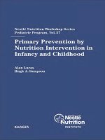
2.2 Mechanisms of NSAID injury to the gastrointestinal mucosa
For evaluation of the validity of new potentially less toxic NSAIDs it is mandatory to clearly
understand the pathogenesis of NSAID induced ulceration (Figure 2). Both aspirin and non-
aspirin NSAIDs inhibit the COX pathway of prostaglandin synthesis (Botting, 2010; Hudson
et al., 1993; Mitchell et al., 1993; Vane, 1971). This represents the basis of anti-inflammatory
action but is also responsible for the development of side effects in the gastrointestinal tract
and kidney as well as inhibition of platelet aggregation. Inhibition of prostaglandin
synthesis can exert injurious actions on the gastric and duodenal mucosa as it abrogates a
number of prostaglandin dependent defence mechanisms. Inhibition of COX leads to a
decrease in mucus and bicarbonate secretion, reduces mucosal blood flow, and causes
vascular injury, leucocyte accumulation, and reduced cell turnover, all factors that
contribute to the genesis of mucosal damage. Within this broad spectrum of events, the
microvascular damage appears to play a central role. Prostaglandins of the E and I series are
potent vasodilators that are continuously produced by the vascular endothelium. Inhibition
of their synthesis by an NSAID leads to vasoconstriction (Gana et al., 1987). Furthermore,
inhibition of prostaglandin formation results in a rapid and significant increase in the
number of neutrophils adhering to the vascular endothelium in both gastric and mesenteric
venules (Asako et al., 1992 a;b; Wallace et al., 1993). Adherence is dependent on expression
of the â2 integrin (CD11/CD18) on neutrophils and intercellular adhesion molecule on the
vascular endothelium (Wallace et al., 1993). Neutrophil adherence in turn causes
microvascular stasis and mucosal injury through ischaemia and release of oxygen derived
free radicals and proteases (Vaananen et al., 1991).
The severity of experimental NSAID gastropathy was markedly reduced in rats rendered
neutropenic by pretreatment with antineutrophil serum or methotrexate (Lee et al., 1992;
Wallace et al., 1990) Recently, Wallace et al (2000) provided evidence for an isoenzyme
specific role of COX in the homeostasis of the gastrointestinal microcirculation. Thus in rats,
the selective COX-1 inhibitor SC-560 decreased gastric mucosal blood flow without affecting
leucocyte adherence to mesenteric venules. In contrast, the selective COX-2 inhibitor
celecoxib markedly increased leucocyte adherence but did not reduce gastric mucosal blood
flow. Only concurrent treatment with the COX-1 and COX-2 inhibitor damaged the gastric
mucosa, suggesting that reduction of mucosal blood flow and increase in leucocyte adhesion
have to occur simultaneously to interfere with mucosal defence. Inhibition of prostaglandin
synthesis thus plays a key role in induction of mucosal injury but does not represent the
only pathway by which NSAIDs can damage the gastrointestinal mucosa. NSAIDs can also
induce local damage at the site of their contact with the gastrointestinal mucosa. Topical
Inflammatory Bowel Disease – Advances in Pathogenesis and Management
8
application of NSAIDs increases gastrointestinal permeability allowing luminal aggressive
factors access to the mucosa. Aspirin and most non-aspirin NSAIDs are weak organic acids.
In the acidic milieu of the stomach, they are converted into more lipid soluble unionised
acids that penetrate into the gastric epithelial cells. There, at neutral pH, they are reionised
and trapped within the cell causing local injury. Having entered gastric mucosal epithelial
cells, NSAIDs uncouple mitochondrial oxidative phosphorylation. This effect is associated
with changes in mitochondrial morphology and a decrease in intracellular ATP and
therefore a reduced ability to regulate normal cellular functions such as maintenance of
intracellular pH. This in turn causes loss of cytoskeletal control over tight junctions and
increased mucosal permeability. The ability of NSAIDs to uncouple oxidative
phosphorylation stems from the extreme lipid solubility and position of a carboxyl group
that acts as a proton translator (Mahmud et al., 1996; Somasundaram et al., 2000). A further
mechanism involved in the topical irritant properties of NSAIDs is their ability to decrease
the hydrophobicity of the mucus gel layer of the gastric mucosa. NSAIDs can convert the
mucus gel from a non-wettable to a wettable state and in experimental animals this effect
has been shown to persist for several weeks or months after discontinuation of NSAID
administration. Gastric mucosal lesions can also occur in a non-acidic milieu, such as
following rectal application. With oral administration, gastric acid however appears to
enhance NSAID induced damage. More extensive and deeper erosions occur at low pH and
an elevation in gastric pH above 4 is necessary to prevent this acid related component.
Prostaglandins do not represent a unique pathway to protect the gastric mucosa. Nitric
oxide (NO) has the potential to counteract potentially noxious effects of inhibition of
Fig. 2. Pathogenesis of NSAID-induced intestinal lesions (Taken from Thiéfin &
Beaugerie, 2005).
The Role of COX-2 Inhibitors on Experimental Colitis
9
prostaglandin synthesis, such as reduced gastric mucosal blood flow and increased
adherence of neutrophils to the vascular endothelium of the gastric microcirculation. NO
has well characterised inhibitory effects on neutrophil activation/adherence demonstrated
in various tissues.
2.3 Chronic inflammatory bowel disease and COX-2
The potential role for prostaglandins in the inflammatory process underlying chronic IBD
has been a focus of controversy. Under the hypothesis that prostaglandins may be
protective, treatment with exogenous prostaglandins was investigated but found to
exacerbate the diarrhea. The possibility that proinflammatory mechanisms might be
involved prompted trials of NSAID therapy. However, studies of various NSAIDs in
patients with ulcerative colitis showed either no improvement or an exacerbation of the
symptoms (Rampton & Sladen, 1981). In keeping with these early findings, some reports
suggested a deleterious effect of NSAIDs on the course of IBD (Evans et al., 1997; Felder et
al., 2000). The magnitude of the risk, however, remains controversial (Bonner et al., 2002;
Nion-Lamurier et al., 2003). The recent review article meets different studies including
original papers, case reports, reviews, controlled trials and databases about exacerbation of
IBD associated with the use of NSAIDs (Kefalakes et al., 2009). The Table 1 showed the
mechanisms of action of NSAIDs and COX-2 inhibitors in patients with IBD.
2.4 Development of the “COXIBs”
The identification of the COX-2 isoenzyme opened the door to development of NSAIDs
which selectivity inhibit COX-2. The main goal of which was to decrease the GI toxicity. The
first generation of selective COX-2 inhibitors came from animal models in which
compounds were sought that were potent anti-inflammatory agents with minimal side
effects on the stomach (Nimesulide, etodolac and meloxicam) (Carvalho et al., 2004). The
discovery of the specificity these products was in reality found after the sale, being due,
mainly on clinical and experimental observations reduced incidence of gastrointestinal side
effects, and subsequently confirmed by in vitro studies. The nimesulide is considered an
aberrant example of NSAIDs, with good power in vivo inflammatory models, but with
weak inhibition in vitro preparations of COX. The nimesulide and display specificity of
action on COX-2, has other effects that further enhance their anti-inflammatory activity, as
inhibition of neutrophil activation and antioxidant properties. Based on in vitro studies
initially suggested that meloxicam selectively inhibited COX-2. However, when tested in
vivo, in humans, its specificity for COX-2 was only about ten times higher than that for COX-
1, with further platelet inhibition (Panara et al., 1999). The molecular modification of these
drugs, especially those of nimesulide, in order to increase its COX-2 selectivity, resulted in
structures without a carboxylic group and the presence of a sulphonamide or sulphone
group, resulting specific inhibitors in the second generation. This group includes celecoxib,
rofecoxib, valdecoxib, parecoxib (pro-drug of valdecoxib), APHS [o-(acetoxyphenyl)hept-2-
ynyl sulfide] and etoricoxib (Fitzgerald & Patrono, 2001; Kulkarni et al., 2000).
Coxib spare COX-1 and firstly inhibit COX-2 function therefore decrease but do not
eliminate NSAIDs associated GI toxicity and are efficacious as tNSAIDs in relieving pain.
Data from large GI outcomes studies have characterised the GI effects of coxib. The
Celecoxib Longterm Arthritis Safety Study (CLASS Study) that compared high dose
Celecoxib (400 mg bid), diclofenac (75 mg bid), and ibuprofen (800 mg 3 times daily)
Inflammatory Bowel Disease – Advances in Pathogenesis and Management
10
showed that symptomatic ulcers were significantly less common among celecoxib users
than tNSAIDs users; however ulcer complication rates were not significantly different
(which was probably due to the confounding factor of concomitant low-dose aspirin use
which was present in 22% of patients) (Silverstein et al., 2000). However, a recent meta-
analysis of available trials of the Cochrane collaboration confirms that celecoxib at any
dosewas associated with statistically less GI events (Moore et al., 2005). Moreover, the
results of another large outcomes study, celecoxib vs naproxen and diclofenac in
osteoarthritis patients (SUCCESS I Study), confirmed the significantly better safety profile of
celecoxib compared with tNSAIDs (Singh et al., 2006). The Vioxx Gastrointestinal Safety of
Rofecoxib trial (VIGOR Study) concluded that rofecoxib users had 50% fewer GI events
compared with naproxen users (Bombardier et al., 2000). Later, in the comparison of
lumiracoxib with naproxen and ibuprofen in the Therapeutic Arthritis Research and
Drug Mechanism of action
Conventional NSAIDs COX-1 and COX-2 → PGE reduction
Surface membrane phospholipid interaction
Effect on mitochondrial energy metabolism
(oxydase phosphorilation inhibition → ATP
deficiency → ↑ mucosal permeability)
Escalation of intestinal inflammatory activity
Enhancement of enterohepatic circulation
Formation of drug enterocyte adducts
COX-independent damage to the small
intestine
Small-bowel enteropathy → blood loss →
hypoalbuminemia
↑ TNF-α, IL-1, NO release
Lower the thromboxane production
COX-1 inhibitors Impairs mucosal microcirculatory blood flow
Lower the thromboxane production
Impairs mucous secretion and acid regulation
Impair renal blood flow and platelet
aggregation
COX-2 inhibitors Imunomodulatory and anti-inflammatory role
on the GI tract (selective COX-2 inhibition →
PGE reduction)
Loss of vasodilation
Increased of vascular permeability
May delay epithelial proliferation
Delay wound healing
↑ Oxygen metabolites (LTB4, TNF)
↑ Leukoc
y
te adherence to the vascular
endothelium
Table 1. Mechanisms of action of NSAIDs and COX-2 inhibitors in patients with IBD (Taken
from Kefalakes et al., 2009).
The Role of COX-2 Inhibitors on Experimental Colitis
11
Gastrointestinal Event Trial (TARGET), showed a 75% decrease in adverse GI events with
the coxib (Schnitzer et al., 2004). It is important to emphasise that although the incidence of
adverse GI events increased in relation to the presence of GI risk factors, the differences
from NSAIDs were maintained in subgroups of patients with and without risk factors
(Skelly et al., 2003).
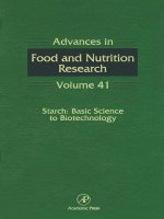
The lumiracoxib is a novel highly selective COX-2 inhibitor. Lumiracoxib differs structurally
from others drugs in the class of selective COX-2 inhibitors (Figure 3) (Brune & Hinz 2004;
Mangold et al., 2004). Differently, the lumiracoxib is a phenyl acetic acid derivative. It has
the highest selectivity (selective for COX-2 compared with COX-1 in the human whole blood
assay with a ratio of 515:1 in healthy subjects and a fairly short plasm half-life (3-6 hours)
compared with other COX-2 selective inhibitors (Esser et al., 2005). In endoscopic studies,
lumiracoxib has been associated with a rate of acute gastric injury and chronic ulcer
formation that does not differ form placebo (Rordorf et al., 2003) and which was
significantly lower than with the NSAID ibuprofen and with celecoxib (Hawkey et al., 2004;
Kivitz et al 2004).
Notwithstanding, it is important to note that 3 of the above commented outcome studies
(CLASS, TARGET and SUCCESS studies) (Schnitzer et al., 2004; Silverstein et al., 2000;
Singh et al., 2006), one endoscopy study (Solomon et al., 2005) and several
epidemiological studies (Lanas et al., 2005) have shown that the concomitant use of low-
dose aspirin and coxib or tNSAIDs increases further the risk of upper GI bleeding in
NSAIDs users and attenuates the GI advantage of a coxib over an tNSAID.A recent meta-
analysis of RCTs has shown that coxib plus low-dose ASA use was associated with a
lower risk of upper GI complications when compared to non-selective NSAID plus low-
dose ASA (Rostom et al., 2009). These gastrointestinal benefits have to be balanced against
the known cardiovascular risks, particularly with long-term use. The VIGOR and
Adenomatous Polyp Prevention on Vioxx Trial Investigators (APPROVe) studies showed
that rofecoxib were associated with increased risk of cardiovascular events after 12 and 36
months of treatment when compared to naproxen (VIGOR) or placebo (APPROVe)
(Bombardier et al., 2000; Bresalier et al., 2005). Other outcome studies have shown also
that celecoxib at doses of 400 mgbid or 200 mgbid (Laine et al., 2004), but not 400 mg once
a day (Arber et al., 2006) is associated with increased risk of cardiovascular events.
Observational studies have shown, however, that celecoxib at 200 mg/day dose was not
associated with increased risk of cardiovascular events (Bombardier et al., 2000;
Silverstein et al., 2000). Recent observational studies have shown that also most NSAIDs
(including nonselective) may be associated with increased cardiovascular risk and this
may be different for the different compounds, dose and length of treatment (Chan et al.,
2006; Lanas et al., 2005; McHippisley-Cox & Coupland, 2005). Of all traditional NSAIDs,
diclofenac have been found to be the one increasing the CV risk the most (Mc Gettigan &
Henry, 2006). In the MEDAL program etoricoxib at the dose of 60–90 mg/day was found
to be not different to diclofenac in the incidence of CV events (Cannon et al., 2006). The
study also showed no differences in the incidence of upper GI complications between
these 2 compounds, although the total number of events (symptomatic ulcers and
complications) was statistically lower in etoricoxib users (Laine et al, 2007). Lastly, both
tNSAIDs and coxib may also increase blood pressure and reduce kidney function.
Following, we describe the effects of these COX-2 inhibitors on differents studies on
experimental colitis models.
Inflammatory Bowel Disease – Advances in Pathogenesis and Management
12
Fig. 3. The chemical structures of some COX-2 inhibitors.
2.5 COX-2 inhibitors on experimental colitis models
The role of selective inhibition of COX-2 for the inflammatory process and the course of
experimental and human colitis is controversially discussed, even though increased levels of
prostaglandins (PGE
2
and PGI
2
) and other eicosanoids were detected in both colitis models
and patients with chronic inflammatory bowel disease, which correlates well with the
disease activity. PGE
2
is produced by mononuclear cells in the lamina propria and is
dependent on COX-2 expresion. It modulates the intestinal immune response, including the
differentiation of T cells and the production and release of proinflammatory cytokines.
During the course of inflammatory bowel disease and experimental colitis, some
prostanoids are released and subsequently modulate the course of the disease.
Animal models are used extensively to study the pathogenesis and pathophysiology of IBD
and to evaluate therapies. The more extensively used models were: acetic acid colitis,
dextran sodium sulphate (DSS) and 2,4,6’-trinitrobenzene sulphonic acid (TNBS). Acetic-
acid-induced colitis in rats resembles human ulcerative colitis in histology, eicosanoid
The Role of COX-2 Inhibitors on Experimental Colitis
13
production and excessive oxygen-derived free radicals release by inflamed mucosa (Millar
et al., 1996). DSS-induced ulcerative colitis is accompanied by erosion and ulceration as well
as inflammatory cell infiltration, characteristics resembling those of human ulcerative colitis
(Okayama et al., 2007). TNBS-induced colitis is accompanied by marked thickening of the
colonic wall, infiltration of polymorphonuclear leukocytes and ulceration, resembling the
human Crohn´s disease (Morris et al., 1989). A number of animal studies have reported the
positive effect of COX-2 inhibition, others exacerbation of colitis (Table 2).
Study Model of colitis Dru
g
Results
Reuter et al. (1996) TNBS diclofenac (10m
g
/k
g
)
naproxen (5mg/kg)
etodolac (10 or 50mg/kg)
nabumetone (25 or 75mg/kg)
L745,337 (1 or 5mg/kg)
unfavorable
Lesch et al. (1999) TNBS NS-398*
SC-58125*
PD-138387*
*dose of 100mg/kg
unfavorable
Karmeli et al. (2000) Acetic-acid or
iodoacetamide
nimesulide (10m
g
/k
g
)
SC-236 (6mg/kg)
favorable
Cuzzocrea et al. (2001) DNBS celecoxib (5m
g
/k
g
)
favorable
Martin et al. (2003) TNBS rofecoxib
favorable
Martin et al. (2005) DSS Rofecoxib (2.5-10m
g
/k
g
)
favorable
Sin
g
h et al. (2003) Acetic-acid;
LTB4-induced IBD)
nimesulide (9 and 18m
g
/k
g
)
favorable
Zhan
g
et al. (2004) TNBS celecoxib (1.25m
g
/k
g
)
unfavorable
El-Medan
y
et al. (2005) Acetic-acid celecoxib (5m
g
/k
g
)
rofecoxib (2.5mg/kg)
favorable
Kruschewski et al. (2006) TNBS NS-398 (10m
g
/k
g
)
favorable
Tsubouchi et al. (2006) DSS rofecoxib
unfavorable
Dudh
g
aonkar et al. (2007) TNBS rofecoxib (10m
g
/k
g
)
favorable
Oka
y
ama et al. (2007) DSS celecoxib (3m
g
/k
g
)
unfavorable
Paiotti et al. (2009) TNBS lumiracoxib (6m
g
/k
g
)
unfavorable
Table 2. COX-2 inhibitors on experimental colitis.