HUMAN PAPILLOMAVIRUS AND RELATED DISEASES – FROM BENCH TO BEDSIDEA CLINICAL PERSPECTIVE potx
Bạn đang xem bản rút gọn của tài liệu. Xem và tải ngay bản đầy đủ của tài liệu tại đây (15.79 MB, 360 trang )
HUMAN PAPILLOMAVIRUS
AND RELATED DISEASES –
FROM BENCH TO BEDSIDE
A CLINICAL PERSPECTIVE
Edited by Davy Vanden Broeck
Human Papillomavirus and Related Diseases – From Bench to Bedside
– A Clinical Perspective
Edited by Davy Vanden Broeck
Published by InTech
Janeza Trdine 9, 51000 Rijeka, Croatia
Copyright © 2011 InTech
All chapters are Open Access distributed under the Creative Commons Attribution 3.0
license, which allows users to download, copy and build upon published articles even for
commercial purposes, as long as the author and publisher are properly credited, which
ensures maximum dissemination and a wider impact of our publications. After this work
has been published by InTech, authors have the right to republish it, in whole or part, in
any publication of which they are the author, and to make other personal use of the
work. Any republication, referencing or personal use of the work must explicitly identify
the original source.
As for readers, this license allows users to download, copy and build upon published
chapters even for commercial purposes, as long as the author and publisher are properly
credited, which ensures maximum dissemination and a wider impact of our publications.
Notice
Statements and opinions expressed in the chapters are these of the individual contributors
and not necessarily those of the editors or publisher. No responsibility is accepted for the
accuracy of information contained in the published chapters. The publisher assumes no
responsibility for any damage or injury to persons or property arising out of the use of any
materials, instructions, methods or ideas contained in the book.
Publishing Process Manager Ivona Lovric
Technical Editor Teodora Smiljanic
Cover Designer InTech Design Team
First published January, 2012
Printed in Croatia
A free online edition of this book is available at www.intechopen.com
Additional hard copies can be obtained from
Human Papillomavirus and Related Diseases – From Bench to Bedside
– A Clinical Perspective, Edited by Davy Vanden Broeck
p. cm.
ISBN 978-953-307-860-1
free online editions of InTech
Books and Journals can be found at
www.intechopen.com
Contents
Preface IX
Part 1 Clinical Aspects of
Human Papillomavirus Related Diseases 1
Chapter 1 Human Papillomavirus: Biology and Pathogenesis 3
José Veríssimo Fernandes and
Thales Allyrio Araújo de Medeiros Fernandes
Chapter 2 Immunohistochemistry in the Diagnosis of
Squamous Intraepithelial Lesions of the Uterine Cervix 41
Evanthia A. Kostopoulou and George Koukoulis
Chapter 3 Screening Methods in Prevention of Cervical Cancer 65
Robert Koiss
Chapter 4 Clinical Manifestations of Genital HPV Infection 83
Edison Natal Fedrizzi
Part 2 Human Papillomavirus Vaccines 99
Chapter 5 Development of New Human Papillomavirus Vaccines 101
Carmen Rodríguez-Cerdeira, Silvia Díez-Moreno,
E. Sánchez and Alfonso Alba
Chapter 6 Current Insight into Anti-HPV Immune Responses
and Lessons for Prophylactic and Therapeutic Vaccines 125
Isabelle Bourgault-Villada and Simon Jacobelli
Chapter 7 Plant Production of Vaccine Against HPV:
A New Perspectives 147
Markéta Šmídková, Marcela Holá,
Jitka Brouzdová and Karel J. Angelis
Chapter 8 Development of Vaccines and Gene Therapy
Against HPV Infection and Cervical Cancer 177
Zoraya De Guglielmo Cróquer
and Armando Rodríguez Bermúdez
VI Contents
Part 3 Human Papillomavirus in Non-Uterine Disease 195
Chapter 9 Epidemiology of HPV in Head and Neck Cancer 197
Márcio Campos Oliveira, Maria da Conceição Andrade
and Fabrício dos Santos Menezes
Chapter 10 Implications of Human Papillomavirus Infections in
the Biology of Head and Neck Cancers 221
Descamps Géraldine, Duray Anaëlle,
Delvenne Philippe
and Saussez Sven
Chapter 11 The Role of Human Papillomavirus
in Head and Neck Cancers 279
Lucinei Roberto Oliveira, Andrielle Castilho-Fernandes,
Alícia Greyce Turatti Pessolato, Régia Caroline Peixoto Lira,
João Paulo Oliveira-Costa, Luciana Souza Chavasco,
Fabiana Alves Miranda, Ivan de Oliveira Pereira,
Edson Garcia Soares
and Alfredo Ribeiro-Silva
Chapter 12 Human Papillomavirus in Donor Semen in Belgium 305
K.W.M. D’Hauwers, W.A.A. Tjalma, U. Punjabi and C.E. Depuydt
Chapter 13 The Impact of Human Papillomavirus
on Cancer Risk in Penile Cancer 319
Angela Adamski da Silva Reis and Aparecido Divino da Cruz
Preface
Cervical cancer is the second most prevalent cancer among women worldwide, mainly
affecting young women. Infection with Human Papilloma Virus (HPV) has been
identified as the causal agent for this condition. The natural history of cervical cancer
is characterized by slow disease progression, generally taking over 10 years, from the
initial infection with HPV, to the diagnosis of cancer. In essence, cervical cancer is a
preventable disease, and treatable if diagnosed in early stage. Historically, the
introduction of the Pap smear has markedly reduced the number of new cases in
countries with an effective prevention program. The burden of disease is highest in
developing countries, with peak incidence in Eastern Africa. Recently, prophylactic
vaccines became available, equally contributing to a better disease prevention.
Unfortunately, the global burden of disease is still very high.
In the first section of this book, clinical aspects of HPV related disease are highlighted.
Innovative clinical diagnostic tools are discussed and Dr Fedrizzi has provided a
highly illustrative contribution on the clinical manifestation of HPV related disease.
The introduction of the HPV prophylactic vaccine has been an important recent
development in the fight against cervical cancer. The second section focuses on HPV
vaccine related issues. Immune responses of the current vaccine are presented by Dr
Bourgault-Villada, and options for the next generation vaccines, or more efficient
production strategies, are discussed. Although HPV is most prominently known from
its role in cervical carcinogenesis, the virus is also involved in other conditions. In the
third section, HPV in non-uterine disease is discussed. Epidemiology and role of HPV
in head-and-neck tumors are addressed. HPV also affects men, and this section covers
the impact of HPV on penile cancers and its prevalence in semen.
This book will be a useful tool for both researchers and clinicians dealing with cervical
cancer, and it will provide them with the latest information in this field.
Dr Davy Vanden Broeck, MSc, PhD
Team Leader HPV/Cervical Cancer Research
International Centre for Reproductive Health
Ghent University
Belgium
X Preface
Acknowledgements
The editor of this book would like to express sincere thanks to all authors for their
high quality contributions. The editor expresses the gratefulness to Ms. Bojana
Zelenika and Ms. Ivona Lovric, process managers, for their continued cooperation.
Part 1
Clinical Aspects of
Human Papillomavirus Related Diseases
1
Human Papillomavirus:
Biology and Pathogenesis
José Veríssimo Fernandes
1
and
Thales Allyrio Araújo de Medeiros Fernandes
2
1
Federal University of Rio Grande do Norte
2
University of Rio Grande do Norte State
Brazil
1. Introduction
The human papillomavirus (HPV) is one of the most common causes of sexually transmitted
disease in both men and women around the world, especially in developing countries,
where the prevalence of asymptomatic infection varies from 2 to 44%, depending on the
population and studied region (Sanjosé et al., 2007). Most HPV infection is transient and
some studies show that the majority of sexually active individuals are exposed to and
acquire infection from this virus at some phase in their lives (Baseman and Koutsky, 2005;
Trottier and Franco, 2006). HPV infection is more prevalent in young adults, at the
beginning of their sexual activity, with a subsequent decline in the prevalence rate with
increasing age, likely as a result of development of an immune response against the virus
and reduction of sexual activity (Castle et al., 2005; Fernandes et al., 2009; Chan et al., 2010).
HPV can infect basal epithelial cells of the skin or inner-lining tissues and are categorized as
cutaneous types or mucosal types. Cutaneous types are epidermotropic and infect the
keratinized surface of the skin, targeting the skin of the hands and feet. Mucosal types infect
the lining of the mouth, throat, respiratory, or anogenital tract epithelium (Burd, 2003).
Some HPVs are associated with warts while others have been well established as the main
risk factor of invasive cervical cancers and their associated pre-cancerous lesions (Clifford et
al., 2005; Zekri et al., 2006; Muñoz et al., 2006). However, only few HPV-infected individuals
progress to invasive cervical cancer (Burd, 2003). Most infected individuals eliminate the
virus without developing recognized clinical manifestation. (Bosch et al., 2008).
Today, more than 150 different HPV types have been cataloged and about 40 can infect the
epithelial lining of the anogenital tract and other mucosal areas of the human body. Based
on their association with cervical cancer and precursor lesions, HPVs can also be classified
as high-risk (HR-HPV) and low-risk (LR-HPV) oncogenic types. LR-HPV types, such as
HPV 6 and 11, can cause common genital warts or benign hyperproliferative lesions with
very limited tendency to malignant progression, while infection with HR-HPV types,
highlighting HPV 16 and 18, is associated with the occurrence of pre-malignant and
malignant cervical lesions (Muñoz et al., 2003; Bosch et al., 2002; Bosch et al., 2008). HR-HPV
types are also associated with many penile, vulvar, anal, and head and neck carcinomas,
and contribute to over 40% of oral cancers (Stanley, 2010).
Human Papillomavirus and Related Diseases
– From Bench to Bedside – A Clinical Perspective
4
Persistent infection with HR-HPV is unequivocally established as a necessary cause of cervival
cancer (Trottier & Franco, 2006). The critical molecules for initiation and progression of this
cancer are the oncoproteins E5, E6, and E7, that act largely by overcoming negative growth
regulation by host cell proteins and by inducing genomic instability, a hallmark of HPV-
associated cancers (Munger et al., 2004; Moody & Laimins, 2010).
Once HPV transmission to the genital tract occurs through sexual contact, the risk factors for
the infection and cervical lesions, including cervical cancer, are the same classic risk factors
for other sexually transmitted diseases. The number of sexual partners is the risk factor
more consistently associated with genital HPV infection and therefore with cervical cancer.
In addition, other indicators of sexual behavior and reproductive activities, heredity,
immune and nutritional status, and smoking can contribute in some way to the
development of cervical cancer (Tarkowski et al., 2004; Muñoz, 2006; Fernandes et al., 2010).
In this chapter we will discuss the biology and pathogenesis of human papillomavirus,
analyzing some specific aspects of their interactions with the infected host and specific host
cell components.
2. Biologic properties of HPV
2.1 Structure of viral particle and regulation of gene expression
The human papillomavirus (HPV) is a relatively small non-enveloped virus that contains a
double-stranded closed circular DNA genome, associated with histone-like proteins and
protected by a capsid formed by two late proteins, L1 and L2. Each capsid is composed of 72
capsomeres, each of which is composed of five monomeric of 55kDa units that join to form a
pentamer corresponding to the major protein capsid, L1. The L1 pentamers are distributed
forming a network of intra- and interpentameric disulfide interactions which serve to
stabilize the capsid (Sapp et al., 1995). In addition to L1, minor capsid proteins with
approximately 75kDa exist within the virion and are called the L2 protein. To assemble the
viral capsid, the pentamers join to copies of L2 that occludes the center of each pentavalent
capsomere. (Jo & Kim 2005; Buck et al., 2008; Conway & Meyers, 2009). Thus, each virion
contains 72 copies of the L1, the major component of the capsid, and a variable number of
copies of L2, a secondary component of the viral capsid, forming a particle with icosahedra
symmetry and approximately 50 to 60 nm in diameter ( Burd, 2003; Longworth & Laimins,
2004; zur Hausen, 2009).
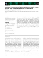
Fig. 1. The structure of HPV. (Adapted from Swiss Institute of Bioinformatics, Viral Zone. -
Available in )
Human Papillomavirus: Biology and Pathogenesis
5
The viral genome of the HPV consists of a single molecule of double-stranded and circular
DNA, containing approximately 8000 base pairs and harboring an average of 8 open reading
frames (ORFs) (Jo & Kim 2005; Zheng & Baker, 2006). In a functional point of view, the HPV
genome is divided into three regions. The first is a noncoding upstream regulatory region
(URR) or long control region (LCR) that has regulatory function of the transcription of the
E6 and E7 viral genes; The second is an early region (E), consisting of six ORFs: E1, E2, E4,
E5, E6, and E7, which encodes no structural proteins involved in viral replication and
oncogenesis. The third is a late (L) region that encodes the L1 and L2 structural proteins. The
LCR region of the anogenital HPVs ranges in size between 800-900 pb, representing about
10% of the genome, and varies substantially in nucleotide composition between individual
HPV types (Fehrmann & Laimins, 2003; Jo & Kim, 2005).
Only one strand of the double-stranded DNA serves as the template for viral gene
expression, coding for a number of polycistronic mRNA transcripts. (Stanley et al., 2007).
The regulation of viral gene expression is complex and controlled by cellular and viral
transcription factors. Most of these regulations occur within the LCR region, which contains
cis-active element transcription regulators. These sequences are bound by a number of
cellular factors as well as the viral E2 product (zur Hausen, 1996). A large number of cellular
transcription factors have been identified and the dysfunction of some of them appears to
play a significant role in papillomavirus-linked carcinogenesis (Thierry et al., 1992; Hamid &
Gaston, 2009).
The transcription start sites of viral promoters differ depending on the virus type, but, in all
types, promoter usage is keratinocyte differentiation-dependent (Smith et al., 2007). The
replication origin and many transcriptional regulatory elements are found in the upstream
LCR region. The virus early promoter, differentiation-dependent late promoter, and two
polyadenylation signals define three general groups of viral genes that are coordinately
regulated during host cell differentiation. The E6 and E7 genes maintain replication
competence. E1 E2, E4, E5, and E8 are involved in virus DNA replication, transcriptional
control, beyond other late functions and L1 and L2, responsible for the assembly of viral
particles (Bodily & Laimins, 2011).
The regulation of expression of the late genes in genital HPVs is not well understood.
However, it has been shown that the second, or later, promoter is initiated in a
differentiation-dependent manner, and thus is activated only when cells are grown in the
host’s stratifying/differentiating tissue. Once activated, the later promoter directs
transcription from a heterogeneous set of start sites and will serve to produce a set of
transcripts that facilitate the translation of L1 and L2 proteins (Smith et al., 2007; Conway &
Meyers, 2009). Activation of the later promoter is accompanied by acceleration of viral DNA
replication and by high levels of viral protein expression. As a result, virus copy-number
amplifies from 50 copies to several thousands of copies per cell. So when a late promoter is
activated, the expression of genes will occur, encoding the structural proteins L1 and L2,
which join to assemble the capsids and to form virions (Stanley et al., 2007).
2.2 Functions of viral proteins
E1 Protein
The E1 protein represents one of the the most conserved proteins among different HPV
types. It has DNA-binding functions and a binding site in the origin of replication localized
Human Papillomavirus and Related Diseases
– From Bench to Bedside – A Clinical Perspective
6
in the LCR region. It assembles into a hexameric complex, supported by the E2 protein, and
the resultant complex has helicase activity and initiates DNA bidirectional unwinding,
constituting a prerequisite for viral DNA replication (Wilson et al., 2002; Frattini & Laimins,
1994). The carboxyl terminal domain of E1 has an ATPase/helicase activity and is necessary
and sufficient for oligomerization. This domain also interacts with the E2 protein and
subunit p70 of DNA polymerase α, but is not sufficient to support replication (Amin et al.,
2000). A segment of approximately 160 amino acid residues upstream of the
ATPase/helicase domain is the DNA-binding domain (Titolo et al., 2003). A stretch of about
50 amino acids within the amino terminus of E1 acts as a localization regulatory region
(LCR) and contains a dominant nuclear export sequence (NES) and a nuclear localization
signal (NSL), which are regulated by phosphorylation (Deng et al., 2004).
E2 protein
The E2 open reading frame of HPV gives rise to multiple gene products by alternative RNA
splicing. The proteins derived from the E2 gene are involved in the control of viral
transcription, DNA replication, and segregation of viral genomes (McPhillips et al., 2006;
Kadaja et al., 2009). These different E2 types represent the major intragenomic regulators
(Bouvard et al., 1994).
The E2 protein can bind to factors on mitotic chromatin and join the virus genome to host
cell chromosomes during mitosis; it contributes to coordinating the HPV DNA replication
with host cell chromosome duplication, allowing the viral genomes to be distributed to the
daughter cell. This constitutes an important requirement for the persistence of virus DNA in
undifferentiated basal cells (McPhillips et al., 2006). Furthermore, the E2 protein interacts
with E1 and stimulates viral DNA replication, favoring the binding of E1 to the origin of
replication ( Seo et al., 1993; Chow et al., 1994).
In lesions containing HPV episomes, the E2 protein directly represses the expression of early
genes as a mechanism to regulate the copy number. In addition, it has been reported that
HPV E2 proteins are able to repress telomerase promoter activity mediated by the HPV E6
protein (Hamid et al., 2009). Integration of the HPV genome in the host cell chromosome
usually disrupts E2 expression, causing a deregulated expression of early viral genes,
including E6 and E7, and this event can favor the transformation of human cells and the
transition into a malignant state (Romanczuk & Howley, 1992)
In addition to the full-length E2 protein, the infected cells can express an E8^E2C transcript,
in which the small E8 domain is fused to the C-terminal domain of E2 (E2C). The full-length
E2 protein forms heterodimers with repressor forms of E2, and these E2 heterodimers serve
as activators of transcription and replication during the viral cycle. The single-chain E2
heterodimer in the HPV 18 genome initiates genome replication, but is not sufficient for
long-term replication of the HPV 18 genome. This is due to the capacity of HPV18 in
encoding the repressor E8/E2, which acts as a negative regulator of HPV18 genome
replication (Kurg et al., 2010). Moreover, it has been shown that inactivation of E2 in the
HPV16 genome increases E6/E7 transcription (Soeda et al., 2006), and that mutation of
E8^E2C in the HPV31 or HPV16 genome increases the genome copy number and the E6/E7
transcription, suggesting that the transcriptional repressing by E8^E2C has an important
role in viral replication (Lace et al., 2008). It was also noted that the E2C domain not only
mediates specific DNA binding but has also an additional role in transcriptional repression
Human Papillomavirus: Biology and Pathogenesis
7
by recruitment of co-repressors, such as the CHD6 protein. This suggests that repression of
the E6/E7 promoter by E2 and E8^E2C involves multiple interactions with host cell proteins
through different protein domains (Fertey et al., 2010).
E4 protein
Despite being considered an early protein, E4 is exclusively located in the differentiated
layers of the infected epithelium (zur Hausen, 1996). Although its expression occurs in
highly differentiated cells that express the capsid genes and synthesize new progeny virions,
and coincides with the onset of vegetative viral DNA replication, E4 is not found in virion
particles. The role of this protein in the virus life cycle has not yet been determined, but E4 is
not required for transformation or episomal persistence of viral DNA, but interacts with the
keratin networks and causes their collapse (Doorbar et al., 1991).
It has been suggested that E4 may have an important role in favoring and supporting the
HPV genome amplification, besides regulating the expression of late genes, controlling the
virus maturation, and facilitating the release of virions (Londgworth & Laimins 2004). E4
also interacts with and disrupts the organization of intermediate filaments. The role of E4 in
providing the release of virus is supported by the association of E4 with the cornified cell
envelope (CCE), a highly resistant structure under the plasmatic membrane of differentiated
keratinocytes in the stratum corneum. Furthermore, E4 may play role in regulating gene
expression and has been shown to induce G2 arrest in a variety of cell types (Londgworth &
Laimins 2004).
E5 protein
The E5 protein is a small hydrophobic peptide, approximately 83 amino acids in size that
localizes primarily to the endoplasmic reticulum. When expressed alone, HPV E5 has weak
oncogenic properties. But in tissue culture assays, HPV E5 can enhance the transforming
activity of E6 and E7, suggesting that it may have a supportive role in tumor progression.
The localization of E5 to the endoplasmic reticulum suggests its activity may be related to
the trafficking of cytoplasmic membrane proteins through this cellular compartment. E5 has
also been reported to alter the activity of the epidermal growth factor receptor (EGFR), in
addition to reducing the surface levels of major histocompatibility complex (MHC) class I
proteins, modulating the MAPK pathway and altering the levels of caveolin 1 (Moody &
Laimins, 2010).
The E5 protein varies in length and primary amino acid sequence among the different
papillomaviruses, but maintains its hydrophobic nature that promotes fusion between cells
(Hu et al., 2009). HPV16 E5 has all the characteristics of fusogenic proteins, including
localization in plasma membrane, high level of hydrophobicity, and the ability for dimmers.
Moreover, HPV16 E5 has been identified to be necessary and sufficient to induce cell-cell
fusion with formation of tetraploid cell and cytokinesis failure (Hu et al., 2009).
The fusogenic activity of the HR-HPV E5 protein contributes to fusion among cells
generating aneuploidy with tetraploid cells and chromosomal instability. These events seem
to precede and favor integration of HPV genomes, which in turn, leads to expression of
viral-cellular fusion transcripts and further enhances expression of the E6-E7 genes,
rendering transformed cells strong growth advantages (Ziegert et al., 2003). Thus, the cell
fusion HR-HPV E5-induced and cell cycle deregulation seems to have an important role in
Human Papillomavirus and Related Diseases
– From Bench to Bedside – A Clinical Perspective
8
the early stages of the transformation process. This suggests that HR-HPV E5-induced cell
fusion can be a critical event in the early stage of the development of HPV-associated
cervical cancer (Gao and Zheng et al., 2010).
As the E5 gene is frequently deleted in cervical cancers, it is believed that the E5 protein may
play a role in the early stages of the process of cellular transformation, but is dispensable for
the maintenance of malignant transformation (zur Hausen, 1996).
E6 protein
The HPV E6 protein is formed by approximately 150 amino acids and contains two zinc-like
fingers joined by an interdomain linker of 36 amino acids, flanked by short amino (N) and
carboxy (C) terminal domains of variable lengths (Howie et al., 2009). The best known
property of the E6 proteins of HR-HPVs is the ability to bind and degrade the tumor-
suppressor protein p53, through the recruitment of the E6-associated protein (E6-AP), a
cellular E3 ligase that does not bind to p53 in the absence of E6. Both E6 proteins from HR-
HPV and LR-HPV bind to p53, but the interaction is stronger in HR-HPV (Lechner et al.,
1994).
The E6 protein can overcome the cell arrest and proapoptotic activities of p53 by targeting
p53 for degradation, inactivating the Mdm2 pathway. E6 can also inhibit the transcriptional
activities of p53 independently of E6-AP (Thomas et al., 2005). Three different mechanisms
have been proposed to explain this p53 inactivation: The first is inhibiting the binding of p53
to its target sequence in the genome; second, E6 may be able to inhibit p53 signaling by
maintaining it in cytoplasm; and third, the mechanism employed by E6 to inhibit p53
activity is the abrogation of the transactivation of p53 responsive genes via interaction with
either the CBP/p300 or hADA3 histone acetyltransferases. The E6 proteins have been shown
to bind to p300, and this interaction inhibits p35 acetylation at p53 dependent sites, leading
to decreased expression from p53. However, unlike p300, E6 interaction with hADA3 results
in hADA3 degradation (Kumar et al., 2002). E6 may also inhibit p53 activation by blocking
the p14/ARF pathway. Thus, E6 is able to modulate transcription of p53-dependent genes
by both degradation of p53 and by interaction with the p300 and hADA3 transactivators
(Shamanin et al., 2008).
The degradation or blocking of the p53 function inhibit apoptotic signaling that would
eliminate the HPV infection cell. There are two major apoptotic pathways that can be
triggered by different stresses: the extrinsic and intrinsic pathways. The E6 protein is able to
disrupt both pathways to facilitate a cytoprotective environment and prevent cell death
(Howie et al., 2009).
In addition, E6 is able to modulate transcription from other cellular signaling pathways as
well as potentiating its ability to act as a diverse modulator of host cell signaling. It has been
shown that E6 interact with three different proteins, such as a novel protein termed E6-
targeted protein 1 (E6TP1) in an E6-AP dependent manner (Wooldridge et al., 2007), beyond
another protein with GAP activity, tuberin, that can also be bound and degraded by E6
(Zeng et al. 2008). Furthermore, HR-HPV E6 has been shown to interact with two proteins
that are part of the innate immune response to viral infection: interferon regulatory factor-3
(IFR-3) and toll-like receptor 9 (TLR9) (Hasan et al., 2007). Exogenous expression of HPV16
E6/E7 has been shown to inhibit TLR9 transcription, leading to a functional loss of TLR9
signaling pathways within the cell (Hasan et al., 2007).
Human Papillomavirus: Biology and Pathogenesis
9
HR-HPV E6 is also able to interact with members of the PDZ family of proteins, promoting
its proteasome-mediated degradation, an activity that seems to be required for induction of
cervical cancer (Shai et al., 2007). HR-HPV E6 PDZ binding can mediate suprabasal cell
proliferation and this is thought to occur by uncoupling the cell proliferation and polarity
control that exist in a differentiated epithelium (Sterlinko et al., 2004). LR-HPV E6 does not
contain the PDZ-binding motif and therefore cannot target these proteins. Degradation of
PDZ proteins results in cellular transformation due to loss of cell-cell contact and loss of cell
polarity (Storrs and Silverstein, 2007). In addition, it has been demonstrated that the
degradation of phosphatase PTPN13 by E6 results in anchorage-independent growth and a
Ras-dependent invasive phenotype (Spanos et al., 2008).
Another function of the HR-HPV E6 protein that is important for immortalization is their
ability to activate the expression of the catalytic subunit of telomerase (hTERT). Thus, the E6
protein is able to promote the maintanance of the telomere, through the action of
telomerase. Interestingly, over-expression of hTERT in conjunction with E7 is sufficient to
immortalize human primary keratinocytes. The HPV E2 proteins are reported to repress
hTERT promoter activity, but the interplay of E6 and E2 during the regulation of this
promoter has not been investigated (Hamid et al., 2009).
E7 protein
The E7 protein has around 100 amino acids in length and contains three conserved regions:
CR1, CR2, and CR3 (Münger and Howley, 2002). It will induce cellular proliferation by
binding to several cellular factors. The best characterized of these interactions is with the RB
tumor suppressor and the related family members p107 and p130. The binding of high-risk
E7 to pRB disrupts the interaction between pRB and E2F, a family of transcription factors,
resulting in the constitutive expression of E2F-responsive genes, such as cyclin A and cyclin
E, and promotes premature S phase entry, DNA synthesis, and the progression of cell cycle
(Zerfass et al., 1995). Thus, in cells overexpressing the HPV E7 protein, this checkpoint
control at G1/S transition is lost and the cells will continue their cell cycle, causing an
uncontrolled cellular proliferation. Moreover, E7 induces the degradation of pRb via the
proteasome-dependent pathway, using a mechanism that involves association with and
reprogramming of the cullin 2 ubiquitin ligase complex (Jo & Kim, 2005; Huh et al., 2007).
HPV E7 can also associate directly with cdk2/cyclin A and cylin E complexes, resulting in
an increased cdk2 activity (Nguyen & Münger, 2008). Another action of E7 that contributes
to cellular immortalization is its interaction with the CDK inhibitors (CKI) p21 and p27,
efficiently neutralizing their inhibitory effects on CDK2 activities, an important factor for G1
to S phase entry and progression (Moody & Laimins, 2010). The ability of E7 to inactivate
these CKIs may contribute to its capacity to abrogate TGF-β mediated growth inhibition.
Moreover, TGF-β also induces a cdk4/cdk6 specific CKI, P15Inkb, and p15Inkb-induced
growth suppression, and these actions may require functional pRB, which is targeted for
degradation by E7 (McLaughlin-Drubin & Münger, 2009). High-risk E7 has further been
shown to increase the levels of the CDC25A phosphatase, which can induce tyrosine
dephosphorylation of CDK2, promoting its activation (Moody & Laimins, 2010).
E7 also affects the expression of S phase genes by directly interacting with E2F factors and
with histone deacetylases (HDAC): E7-E2F6 interaction prevents repression of gene
expression by E2F6, maintaining a S phase environment conductive for viral replication
Human Papillomavirus and Related Diseases
– From Bench to Bedside – A Clinical Perspective
10
(McLaughlin-Drubin et al., 2008), and E7-HDAC binding facilitates HDAC removal at
promoters to activate transcription (Longworth & Laimins, 2004).
Another major apoptotic pathway targeted by HPV proteins is anoikis, a form of apoptosis
that is triggered when normal cells attempt to divide in the absence of a matrix (Tasaki et al.,
2005). E6 and E7 interact with some factors involved with anoikis, such as paxillin, fibulin 1,
and p600 (Huh et al., 2005), promoting the prevention of anoikis.
Furthermore, E6 and E7 interfere with the effects of various growth inhibitory cytokines that
are induced following infection. High-risk HPV proteins repress the transcription of many
IFN-inducible genes (Chang & Laimins, 2000; Kanodia et al., 2007; Tindle, 2002) and block
apoptosis binding to TNF receptor 1, inhibiting the formation of the death-inducing
signaling complex and consequent transduction of apoptotic signals (Filippova et al., 2002).
The exsposure to E7 in a non-inflammatory epithelial environment can also be sufficient to
induce a peripheral tolerance to E7 in the cytotoxic T lymphocytes population (Tindle, 2002).
E6 also interacts with the adaptor protein FAS-associated protein with death domain
(FADD) and caspase 8 to block cell death in response to FAS and TRAIl. Also, E6 can
interfere with induction of the extrinsic and intrinsic (mitochondrial) apoptotic pathways
through interactions with the pro-apoptotic Bcl2 members BAK and BAX, as well as by
upregulation of the inhibitors of apoptosis such as the inhibitor of apoptosis protein 2 (IAP2,
also known as BIRC2) and survivin (also known as BIRC5) (Garnett & Duerksen-Huges,
2006).
L1 protein
The L1 gene corresponds to a sequence of about 1200 base pairs, which encodes a structural
protein highly conserved among different HPV types, the (Xu et al., 2006). The L1 protein is
formed by five monomeric units of 55kDa that join to form a pentameric structure, totaling
72 per each capsid ( Buck et al., 2008). The L1 protein is highly immunogenic and has
conformational epitopes that induce the production of neutralizing type-specific antibodies
against the virus, which prevent the infection (Carter et al., 2003), making it the target of
prophylactic vaccines (Villa et al., 2007; D’Andrilli et al., 2010).
Comparison among L1 sequences of different papillomaviruses suggests a conserved
heparin-binding domain at the C-terminus, and the cleavage of this domain from L1
prevents binding to both heparin and human keratinocytes (Culp et al., 2006; Selinka et al.,
2007). Thus, it is believed that the L1 major capsid protein contains the major determinant
required for initial attachment of the viral particles to cell surface receptors, HSPGs, and
therefore has an important role in infection (Schiller et al., 2010).
L2 protein
L2 is a secondary component of viral capsid and it is present in a variable number of copies
per each capsid, being located on the inner surface in the central cavity below the pentamers
of L1, where they are arranged to form the capsid (Buck et al., 2008). Despite the paucity of
L2 in the virion, this protein has recently been shown to have many more functions than a
simple structural role. L2 contributes to the binding of virion in the cell receptor, favoring its
uptake, transport to the nucleus, and delivery of viral DNA to replication centers. Besides,
E2 helps the packaging of viral DNA into capsids and, due to the presence of a usual
Human Papillomavirus: Biology and Pathogenesis
11
neutralization epitope in L2 proteins of many papillomaviruses, it may be instrumental in
conferring immunity across different types of HPV. L2 also contributes to the interaction of
virion in the cell surface. Two distinct regions in the N-terminal protein of L2 interact with
the cell surface, and this interaction occurs after an initial low-specificity interaction between
L1 and the cell surface. After this, a conformational switch occurs in the capsid, exposing the
L2 epitopes and promoting interactions with a more specific secondary receptor. The
cleavage of the N-terminus of L2 is necessary for the binding of L1 to the secondary
receptor, an indication that L2 has an important role in HPV infection (Schiller et al., 2010) .
Protein Functions
E1 Viral DNA replication
E2 Control of viral transcription, DNA replication, and segregation of viral
genomes.
E4 Favor and support the HPV genome amplification, besides regulating the
expression of late genes, controlling the virus maturation, and facilitating the
release of virions
E5 Enhance the transforming activity of E6 and E7; Promotes fusion between cells,
generating aneuploidy and chromosomal instability; Contribute to immune
response evasion.
E6 Bind and degrade the tumor-suppressor protein p53, inhibiting apoptosis;
Interact with proteins of the innate immune response, contributing to immune
evasion and persistence of virus;Activate the expression of telomerase.
E7 Bind and degrade the tumor-suppressor protein pRB; Increase cdk activity;
Affects the expression of S phase genes by directly interacting with E2F factors
and with histone deacetylases; Induce a peripheral tolerance in cytotoxic T
lymphocytes (CTL) and Downregulate the expression of TLR9, contributing
to immune response evasion
L1 Major capsid protein; contains the major determinant required for attachment
to cell surface receptors. It is highly immunogenic and has conformational
epitopes that induce the production of neutralizing type-specific antibodies
against the virus.
L2 Minor capsid protein; L2 contributes to the binding of virion in the cell
receptor, favoring its uptake, transport to the nucleus, and delivery of viral
DNA to replication centers. Besides, E2 helps the packaging of viral DNA into
capsids.
Table 1. The HPV proteins and functions
3. HPV Infection
The HR-HPVs have the ability to infect several types of epithelial cells, but they can cause
cancer more frequently in the uterine cervix (Timmons et al., 2010). The cervical cancer
arises preferentially in the cervical transformation zone (TZ), located in the boundary
Human Papillomavirus and Related Diseases
– From Bench to Bedside – A Clinical Perspective
12
between the squamous epithelium of ectocervix and the columnar epithelium of endocervix.
Basal cells in the TZ retain the ability to differentiate, a property required for virion
production (Crum & McKeon, 2010). The basal cells in TZ are more susceptible to HPV
infection in that there are fewer overlying layers than in other locations. In addition, the
presence of hormones, such as estrogen and progesterone, that orchestrate cervical changes
during menstruation and childbirth, can help both HPV infection and cancer development
(Timmons et al., 2010; Roberts et al., 2007; Chung et al., 2008).
It has been reported that two types of cells are present in the basal layer of cervix. The first
type comprises the transit amplifying (TA) cells, which are proliferating cells that are able to
undergo terminal differentiation. TA cells divide and differentiate, representing the majority
of cells in the suprabasal layers. The second class of basal cells is the stem cells, which have
unlimited proliferation potential but divide only rarely in order to replenish the TA pool,
serving as reserve cells to enable long-term maintenance of the tissue. Only one daughter
cell of a stem cell division goes on to become a TA cell, while the other remains a stem cell.
It is unclear which cells in the basal layer are the target of HPV infection, and perhaps both
cell classes can be infected. If this is true, infection of stem cells could lead to one long-term
persistent infection, whereas infection of TA cells could lead to short-term infections,
followed by a cure (Jones et al., 2007).
Studies in vitro and in vivo revealed that the L1 major capsid protein contains the major
determinant required to the initial attachment of the viral particles to the cell surface
receptor, the heparan sulfate proteoglycans (HSPGs). Laminin-5 can also contribute to the
binding of viral capsids to the extracellular matrix (ECM) in the epithelial cell lines (Culp et
al., 2006; Selinka et al., 2007).
In vivo, the viral particles bound efficiently to regions of the basement membrane (BM) only
after these regions had been exposed by mechanical or chemical trauma of the epithelium.
The L1 capsid protein binds to HSPGs in segments of the BM exposed after epithelial
trauma. After this, L1 undergoes a conformational change that exposes the N-terminus of
the L2 minor capsid protein, which is cleaved by furin or the closely related protein
convertase (PC) 5 and 6 (Richards et al., 2006). L2 proteolisis exposes a previously occluded
surface of L1 that binds to an undetermined cell surface receptor on keratinocytes that have
migrated over the BM to close the wound. This receptor is still unknown, but in vitro studies
indicate the α6-integrin as a possible candidate (Kines et al., 2009). The cleavage of L2 may
be necessary due to the fact that the surface intact of the epithelia apparently contains
sulfation patterns that do not bind capsids. Binding to the BM may promote the preferential
interaction with basal keratinocytes that are migrating over the exposed BM to close the
wound. Thus, papillomaviruses (PV) are the only viruses that initiate the infectious process
at an extracellular site (Schiller et al., 2010).
The capsids are internalized via the keratinocytes-surface receptor and subsequently surf
toward the cell body. The first phase in infection is the internalization, which usually occurs
2-4 h after cell surface binding (Culp et al., 2004). The pathway involved in internalization
and intracellular trafficking is still unclear, but it seems to occur slowly and asynchronously
over a span of several hours (Schiller et al., 2010). Clatrin-mediated endocytosis has been
pointed out to be like the endocytic pathway for the majority of HPV types. However, some
studies suggest that they can enter through a caveolae-mediated pathway and not via
clatrin-mediated endocytosis (Smith et al., 2007). On the other hand, it has been proposed
Human Papillomavirus: Biology and Pathogenesis
13
that HPV-16 initially enters via clatrin-coated pits but the traffic occurs through caveosomes
to eventually reach the endoplasmic reticulum (Hindmarsh et al., 2007; Laniosz et al., 2008).
Moreover, it has been suggested that the capsids might be internalized via a novel pathway
involving tetraspanin-enriched microdomains (Spoden et al., 2008).
The uncoating is not observed until 8-12 h after cell surface binding, and it seems that L2 has
a critical role in the endosome escape (Kamper et al., 2006). The cytoplasm transport along
microtubules is mediated by protein complex, and L2 has been found to interact with the
microtubule network via the motor protein dynein during infectious entry (Florin et al.,
2006). After the entry of the viral genome into the nucleus, the complexes predominantly
localize in distinct punctate nuclear domains designated as ND10 bodies or promyelotic
leukemia (PML) oncogenic domains (PODs). There is evidence that cell division is required
for establishment and expression of the viral genome in the nucleus (Pyeon et al., 2009).
4. Life cycle of HPV
The HPV life cycle begins with infection of stem cells in the basal layer of the epithelium.
After the entry in the cells, the virus requires the expression of E1 and E2 genes to maintain
a low number of copies of genome. These proteins bind to the viral origin of replication and
recruit cellular DNA polymerases and other proteins necessary for DNA replication (Hamid
et al., 2009). In the suprabasal layer, the expression of genes E1, E2, E5, E6 and E7
contributes to the maintenance of the viral genome and induces cell proliferation ,
increasing the number of HPV-infected cells in the epithelium, resulting in a higher number
of cells that will eventually produce infectious virions (Hamid & Gston, 2009; Lazarczyk et
al., 2009). In the more differentiated cells of this same layer of the epithelium occurs the
activation of differentiation-dependent promoter and maintenance of gene expression E1,
E2, E6 and E7. Furthermore, there will be activation of the expression of E4 gene, whose
product will induce amplification of the viral genome replication, greatly increasing the
number of virus copies per cell, at the same time that occurs the expression of genes L1 and
L2 (Nakahara et al., 2005; Lazarczyk et al., 2009). In the granular layer, the products of late
genes, the major and minor proteins of the viral capsid, L1 and L2 respectively, gather to
assembly of the viral capsids and formations of virions, which reach cornified layer of the
epithelium and are released (Lazarczyk et al., 2009).
For a better understanding, the life cycle of HPV was divided into two parts: a maintenance
phase and differentiation-dependent phase (Bodily & Laimins, 2011).
4.1 Maintenance phase
HPV virions infect cells in the basal epithelial layer that become exposed through
microlesions. The viral capsid binds initially to the basal cell layer and infection occurs
when activated keratinocytes move into the wound, to the upper layers of the epithelium
(Kines et al., 2009). HPV genomes replicate in the nucleus of the basal cell layer, where the
viral replication is considered nonproductive and the virus establishes itself as a low-copy-
number episome by using the host DNA replication machinery (Moody & Laimins, 2010). In
this way, viral proteins are expressed at very low levels in undifferentiated cells, and this
contributes to immune avasion and persistence (Bodily & Laimins, 2011).