HUMAN PAPILLOMAVIRUS AND RELATED DISEASES – FROM BENCH TO BEDSIDE A DIAGNOSTIC AND PREVENTIVE PERSPECTIVE potx
Bạn đang xem bản rút gọn của tài liệu. Xem và tải ngay bản đầy đủ của tài liệu tại đây (10.92 MB, 334 trang )
HUMAN
PAPILLOMAVIRUS AND
RELATED DISEASES –
FROM BENCH TO
BEDSIDE A DIAGNOSTIC
AND PREVENTIVE
PERSPECTIVE
Edited by Davy Vanden Broeck
Human Papillomavirus and Related Diseases – From Bench to Bedside A Diagnostic and
Preventive Perspective
/>Edited by Davy Vanden Broeck
Contributors
Miguel Angel Arrabal-Polo, Miguel Arrabal-Martin, Sergio Merino-Salas, Fernando López-Carmona Pintado, Salvador
Arias-Santiago, Jacinto Orgaz-Molina, Maria Sierra Giron-Prieto, Santiago Melón, María De Oña, Marta-Elena Alvarez-
Argüelles, João Paulo Oliveira-Costa, Giorgia Silveira, Danilo Figueiredo Soave, Juliana Silva Zanetti, Andrielle Castilho-
Fernandes, Lucinei Roberto Oliveira, Alfredo Ribeiro-Silva, Fernando Augusto Soares, Manuel Rodriguez-Iglesias,
Fátima Galán-Sánchez, Claudie Laprise, Helen Trottier, João Oliveira-Costa, Mara Celes, Bruna Riedo Zanetti, Angela
Adamski Da Silva Reis, Daniela De Melo Silva, Aparecido Divino Da Cruz, Cláudio Carlos Da Silva, Ralf Hilfrich, Evanthia
Kostopoulou, Mueller, Penelope Duerksen-Hughes
Published by InTech
Janeza Trdine 9, 51000 Rijeka, Croatia
Copyright © 2013 InTech
All chapters are Open Access distributed under the Creative Commons Attribution 3.0 license, which allows users to
download, copy and build upon published articles even for commercial purposes, as long as the author and publisher
are properly credited, which ensures maximum dissemination and a wider impact of our publications. However, users
who aim to disseminate and distribute copies of this book as a whole must not seek monetary compensation for such
service (excluded InTech representatives and agreed collaborations). After this work has been published by InTech,
authors have the right to republish it, in whole or part, in any publication of which they are the author, and to make
other personal use of the work. Any republication, referencing or personal use of the work must explicitly identify the
original source.
Notice
Statements and opinions expressed in the chapters are these of the individual contributors and not necessarily those
of the editors or publisher. No responsibility is accepted for the accuracy of information contained in the published
chapters. The publisher assumes no responsibility for any damage or injury to persons or property arising out of the
use of any materials, instructions, methods or ideas contained in the book.
Publishing Process Manager Danijela Duric
Technical Editor InTech DTP team
Cover InTech Design team
First published April, 2013
Printed in Croatia
A free online edition of this book is available at www.intechopen.com
Additional hard copies can be obtained from
Human Papillomavirus and Related Diseases – From Bench to Bedside A Diagnostic and Preventive
Perspective, Edited by Davy Vanden Broeck
p. cm.
ISBN 978-953-51-1072-9
free online editions of InTech
Books and Journals can be found at
www.intechopen.com
Contents
Preface VII
Section 1 Diagnostic and Preventive Aspects of HPV-Related
Diseases 1
Chapter 1 Molecular Diagnosis of Human Papillomavirus Infections 3
Santiago Melón, Marta Alvarez-Argüelles and María de Oña
Chapter 2 Molecular Tools for Detection Human Papillomavirus 27
Angela Adamski da Silva Reis, Daniela de Melo e Silva, Cláudio
Carlos da Silva and Aparecido Divino da Cruz
Chapter 3 HPV Diagnosis in Vaccination Era 57
Fátima Galán-Sánchez and Manuel Rodríguez-Iglesias
Chapter 4 HPV L1 Detection as a Prognostic Marker for Management of
HPV High Risk Positive Abnormal Pap Smears 93
Ralf Hilfrich
Chapter 5 Ancillary Techniques in the Histopathologic Diagnosis of
Squamous and Glandular Intraepithelial Lesions of the
Uterine Cervix 117
Evanthia Kostopoulou and George Koukoulis
Chapter 6 Human Papillomavirus Prophylactic Vaccines and Alternative
Strategies for Prevention 149
Lis Ribeiro-Müller, Hanna Seitz and Martin Müller
Section 2 Clinical Aspects of HPV-Infections 185
Chapter 7 Clinical Manifestations of the Human Papillomavirus 187
Miguel Ángel Arrabal-Polo, María Sierra Girón-Prieto, Jacinto
Orgaz-Molina, Sergio Merino-Salas, Fernando Lopez-Carmona
Pintado, Miguel Arrabal-Martin and Salvador Arias-Santiago
Chapter 8 Human Papillomavirus Infection and Penile Cancer: Past,
Present and Future 221
João Paulo Oliveira-Costa, Giórgia Gobbi da Silveira, Danilo
Figueiredo Soave, Andrielle de Castilho Fernandes, Lucinei Roberto
Oliveira, Alfredo Ribeiro-Silva and Fernando Augusto Soares
Chapter 9 The Role of Human Papillomavirus in Pre-Cancerous Lesions
and Oral Cancers 241
Danilo Figueiredo Soave, Mara Rubia Nunes Celes, João Paulo
Oliveira-Costa, Giorgia Gobbi da Silveira, Bruna Riedo Zanetti,
Lucinei Roberto Oliveira and Alfredo Ribeiro-Silva
Chapter 10 Epidemiology of Anogenital Human Papillomavirus
Infections 269
Claudie Laprise and Helen Trottier
Chapter 11 Modern Molecular and Clinical Approaches to Eradicate HPV-
Mediated Cervical Cancer 287
Whitney Evans, Maria Filippova, Ron Swensen and Penelope
Duerksen-Hughes
ContentsVI
Preface
Cervical cancer is the second most prevalent cancer among women worldwide, mainly af‐
fecting young women. Infection with Human Papilloma Virus (HPV) has been identified as
the causal agent for this condition. The natural history of cervical cancer is characterized by
slow disease progression, generally taking over 10 years from the initial infection with HPV
till cancer. In essence, cervical cancer is a preventable disease, and treatable if diagnosed in
early stage. Historically, the introduction of the Pap smear has markedly reduced the num‐
ber of new cases countries with an effective prevention program. The burden of disease is
highest in developing countries, with peak incidence in Eastern Africa. Recently, prophylac‐
tic vaccines became available, equally contributing to a better disease prevention. Unfortu‐
nately, the global burden of disease is still very high
In the first section, diagnostic and preventive aspects of HPV related diseases are highlight‐
ed. The first two chapters describe in detail molecular approaches in HPV detection. With
the introduction of the vaccine, novel challenges arose, Dr. Rodriguez-Iglesias highlights the
need for differential screening strategies in the post-vaccine era. Prognosis towards progres‐
sion of lesions has for many years been an important challenge, and Dr. Hilfrich describes
how the L1 protein can be instrumental in disease prediction. In the following chapter, ancil‐
lary techniques in the histopathologic diagnosis of squamous or glandural lesions are dis‐
cussed. Dr. Mueller contributed a holistic chapter on HPV prevention.
The second section focusses on updated clinical aspects of HPV infections, including general
clinical manifestations of HPV infections, penile cancers, head and neck tumors. Dr. Trottier
describes the epidemiology of anogenital HPV infections, and dr. Duerksen-Hughes provid‐
ed an overview of modern molecular approaches to eradicate HPV induced cervical cancer.
The last chapter provides insights in the analysis of the native virus.
This book will be a useful tool for both researchers and clinicians dealing with cervical can‐
cer, and will provide them with the latest information in this field.
Prof. Dr. Davy Vanden Broeck, MSc, PhD.
Team leader HPV/cervical cancer research
International Centre for Reproductive Health
Ghent University
Belgium
Section 1
Diagnostic and Preventive Aspects of HPV-
Related Diseases
Chapter 1
Molecular Diagnosis of Human Papillomavirus
Infections
Santiago Melón, Marta Alvarez-Argüelles and
María de Oña
Additional information is available at the end of the chapter
/>1. Introduction
Human Papillomavirus (HPV) is arguably the most common sexually transmitted agent
worldwide, either in its clinical (genital warts) or subclinical presentation in men and women.
The main interest in HPV relates to its recognized as a causal and necessary factor for cervical
cancer one of the most common cancers in women (80% of cases in most developing countries,
with an annual incidence of almost half a millon and a mortality rate of approximately 50%)
[1-5], and other types of cancer, such as penis, anal or oral cancer [6].
The overall prevalence of HPV in cervix in women in the general population is 10%. This
prevalence is higher in the less developed world than in more developed regions [7, 8]. A
review of studies has also shown prevalence of HPV in men as usually 20% or greater,
depending on population tested and the type and number of anatomic sities evaluated [9].
HPV infection is most common in sexually active young women 25 years of age or younger
but cervical cancer is common in older woman, suggesting infection at younger age and slow
progression to cancer [10].
The most significant predictor for adquiring HPV infection in men or women appears to be
the life time number of sexual parteners [11,12,13]. For women, the sexual activity of their
partner(s) is also important, with increased risk of adquiring HPV if their partner had, or
currently has, other partners [12].
Not all women infected with high-risk HPV develop cervical cancer, other factors are neces‐
sary: genotype, persistent infection, viral variants, viral load, integration, coinfection, age of
30 years old, inmunosupresión, smoking, condom use, coinfections, long-term use of oral
contraceptives, parity and circumcision. [10, 12, 14-24]
© 2013 Melón et al.; licensee InTech. This is an open access article distributed under the terms of the Creative
Commons Attribution License ( which permits unrestricted use,
distribution, and reproduction in any medium, provided the original work is properly cited.
About 189 HPV genotypes have been sequence and classified according to their biological
niche, oncogenic potential and phylogenetic position [25]. From them, about 40 can infect the
genital tract [26]. HPV types are classified based on their association with cervical cancer and
precursor lesion into low-risk types (LR-HPV), which are found mainly in genital warts, high-
risk types (HR-HPV), which are frequently associated with invasive cervical cancer and
undetermined risk types (table 1) [27, 28, 29].
Risk category HPV types
High-risk 16,18,31,33,35,39,45,51,52,56,58,59,68,73,82
Low-risk 6,11,40,42,43,44,54, 61,70,72, 81, 83, 89
Undetermined risk 26,53,66
Table 1. HPV types classification according their oncogenic potential
Worldwide, HPV-16 is the most common HPV type across the spectrum of HPV related
cervical lesions. In women with ICC (invasive cervical cancer), the most common HPV types
are HPV-16,18,33,45,31 and 58 [30, 31], but among these genotypes, certain variants have linked
to different clinical outcomes. It is now generally accepted that HPV has co-existed with its
human host over a very long period of time and has evolved into multiple evolutionary
lineages [25, 32]. Intratypic variants of HPV16 have been identified from different geographic
locations and are classified according to their host ethnic groups as European (including
prototypes and Asian types), Asian American, African and North American [33]. Through
epidemiological and in-vitro experimental studies, natural variants of HPV16 have shown
substantial differences in pathogenicity, immunogenicity and tumorigenicity. IARC Study [34]
and IARC Meta-analysis [31] are very robust in identifying that HPV-16 and 18 contibute
approximately 70% of all ICC. HPV-16,18 and 45 are the three most relevant types in cervical
adenocarcinoma [30]. The geographical variation in type distribution is of minor significance
variation.
Among men and women, cancers of the ano-genital tract and their precursor lesions have been
strongly linked to infection with sexually transmited human papillomavirus. In men, HPV
infection has been strongly associated with anal cancer and is associated with approximately
85% of the anal squamous cell cancers that accur annually worldwide. Likewise, approxi‐
mately 50% of cancers of penis have been associtated to HPV infection [35]. Genital warts are
a common sexually transmitted condition with an estimated prevalence of 1-2% of young
adults [36]. Although having genital warts is not associated with mortality, represent a
significant public health problem (clinical symptoms and psychosocial problems) and
healthcare costs for society [37-39]. More than 90% of genital warts are related to HPV-6 and
11 (low risk genotypes) in general these types are not associated with malignant lesions,
however 20-50% of these also contained coinfection with oncogenic HPV types [39-41].
On the other hand, between 33-72% of oropharyngeal cancers, and 10% of cancer of the larynx
may be attributed to HPV infection [42-44].
Human Papillomavirus and Related Diseases – From Bench to Bedside A Diagnostic and Preventive Perspective
4
2. Etiopathogenesis of HPV
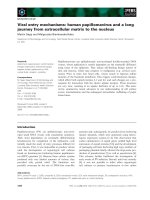
The HPV virion has a double-stranded, circular DNA genome of approximately 7900bp, with
eight overlapping open reading frames, comprising early (E), and late (L) genes and an
untranslated long control region, within an icosahedral capsid. The L1 and L2 genes encode
the mayor and minor capsid proteins. The capsid contains 72 pentamers of L1, and a pproxi‐
mately 12 molecules of L2. The early genes regulate viral replication and some have transfor‐
mation potential. Late genes L6 and L7 code for structural capsid proteins which encapsidate
the viral genome. (Figure 1).
Figure 1. Organization of the HPV genome. Adapted from Doorbar J. [45]
Infection by papillomaviruses requires that virus particles gain access to the epithelial basal
layer and enter the dividing basal cells. Having entered the epithelial tissues, the HPV virus
enters the nucleus of a basal epithelial cell, where early genes E1 and E2 are expressed,
replicating the viral genome and transcribing messenger RNA needed for viral replication; in
addition to its role in replication and genome segregation, E2 can also act as a transcription
factor and can regulate the viral early promoter and control expression of the viral oncogenes
(E6 and E7). At low levels, E2 acts as a transcriptional activator, whereas at high levels E2
represses oncogene expression [45]. As the host cells differentiate, genes E4 and E5 assist in
the production of the viral genome by controlling epidermal growth factor. E6 and E7 are viral
oncogenes which now become important. E6 causes degradation of the tumour suppressor
gene p53, while E7 completes for retinoblastoma protein (pRb), allowing the transcription
factor E2F to drive cell proliferation processes. The p16 protein, encoded by the suppressor
gene CDKN2A (MTS1, INK4A) at chromosome 9p21, is an inhibitor of cyclin dependent
kinases (cdk)which slows cell cycle by inactivating the function of the complex-cdk4 and cdk6-
cyclin D. These complexes regulate the control point of the G1 phase of the cell cycle with
subsequent phosphorylation and inactivation of retinoblastoma (pRb), which E2F released and
which allows cells to enter S phase. It has been demonstrated existence of a correlation between
pRb and p16 reciprocal, which is why there a strong overexpression of p16 both in carcinomas
Molecular Diagnosis of Human Papillomavirus Infections
/>5
as in lesions premalignant cervix. In cervical cancer, pRb is functionally inactivated from the
initial stages of cervical carcinogenesis as a consequence of expression of HPV E7 gene. Genes
E6 and E7 therefore act to remove two principle mechanisms of cell defence, and drive the cell
replication machinery towards production of new virus particles. E6 and E7 are also known
to promote oncogenesis. [45]
On the other hand, integration of HPV-DNA into the host DNA is a well known topic in cervical
cancer. Integration of HPV 16 DNA correlates with dysfunction of HPV E1 or E2 ORF, which
are active during HPV replication. E2 loss of function allows up-regulation of E6 and E7
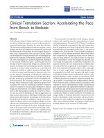
oncoproteins, because E2 is a repressor of E6 and E7. (Figure 2).
Figure 2. The location in squamous epithelium of the main stages of the papillomavirus life cycle. [46]
3. Diagnosis of HPV infections
Despite the promising outcomes, vaccination does not exempt from performing periodic
control visits, because the effects of the vaccine at 15-20 years and the role other genotypes
with oncogenic capacity not included in the vaccine may play are still unknown. Furthermore,
there is still a large population of women which has had no access to it. Then, secondary
prevention by screening and treatment will continue to be crucially important in cervical
cancer prevention programs. Moreover, the fact that infection by HPV provokes long-term
symptomatology demands a close follow-up (screening) of those individuals susceptible to
infection in order to avoid related problems.
Currently, cervical cancer screening is acknowledged as the most effective approach for
cervical cancer control. The primary screening and diagnostic methods have been cytology
Human Papillomavirus and Related Diseases – From Bench to Bedside A Diagnostic and Preventive Perspective
6
and histology, but two limitations of the Pap smear exist: low specificity leading to the need
for repeat screening at relatively short intervals and cervical cancer screening, based on Pap
smear, remains beyond the economic resources of nation in developing world. This econom‐
ic disparity has meant that cervical cancer incidence and mortality rates in the developing
world have remained high, with large reductions in these rates being limited primarily to
the industrialized world. Thus, the reduction of cervical cancer in developing nations remains
an unmet need of high priority. Since the link between HPV and cervical cancer is known
and numerous large scale studies have been done, molecular methods to detect HPV DNA
in clinical specimens (vaginal, urethral, paraurethral, anal or pharyngeal exudates, biop‐
sies, and, especially, endocervical exudates) have been introduced into screening algorithms.
Increased sensitivity has important clinical outcomes because reduce mortality and an
elongation of screening, and implies better compliance with screening and lower cost [47]. An
Italian study showed that HPV-based screening is more effective than cytology in preventing
invasive cervical cancer, by detecting persistent high-grade lesions earlier and providing
longer low-risk period [48].
HPV serves as paradigm for the use of NAATs for its diagnosis and typification due to how
difficult it is to obtain the virus via cell cultures or to develop indirect diagnosis techniques [49].
The first protocols for detect HPV were described about 20 years ago, using L1 consensus
primers PCR systems, particularly MY09/11 and GP5+/6+ [50-52]. These primer systems have
been widely used to study the natural history of HPV and their rule in the development of
genital cancer [53-55]. Nowadays, several kits are commercially available which allow for the
detection of the virus or the detection and typification of the most relevant HPVs: Amplicor
HPV test and Linear array HPV Genotyping test (Roche Diagnostics, Switzerland), Innolipa
HPV Genotyping Extra (Innogenetics, Belgium), Biopat kit (Biotools, Spain) or Clart Papillo‐
mavirus 2 (Genómica, Spain). The latter uses microarray technology to increase the number
of hybridizations in a reduced space. Besides genome amplification, direct hybridization
protocols on the sample (hybrid capture) approved by the FDA for diagnosing HPV in women
(Hybrid Capture II, Digene, USA) is also used. These protocols identify high and low-risk
genotypes without specifying the infecting genotype.
The sensitivity of such methods has left out cytological methods (Papanicolau), which are less
sensitive and specific. This high degree of sensitivity allows to extending the period between
control visits of women to 5 or 6 years [56, 57].
3.1. Signal amplification systems
The Hybrid Capture II system (HCII, Digene, USA) is a non radioactive signal amplifica‐
tion method based on the hybridization of the target HPV-DNA to labeled RNA probes in
solution. The resulting RNA-DNA hybrids are captured onto microtiter wells and are detected
by specific monoclonal antibody and chemiluminiscence substrate, providing a semi-
quantitative measurement of HPV-DNA. Two different probe cocktails are used, one
containing probes for five low-risk gentypes: HPV 6, 11, 42,43 and 44 and the other contain‐
ing probes for 13 high-risk genotypes: HPV 16,18,31,33,35,39,45,51,52,56,58,59 and 68.
Molecular Diagnosis of Human Papillomavirus Infections
/>7
However, HCII has some limitations. It distinguishes between the high-risk and low-risk
groups but does not permit identification of specific HPV genotypes. Hybrid Capture II
(HCII) has been shown to have similar analytic sensitivity to some PCR methods for HPV
DNA detection [58], but present cross-reactivity of the two probe cocktails can reduce the
clinical relevance of a positive result [59, 60].
The Hybrid Capture III (HCIII, Digene, USA) is being evaluated as the next generation of
hybrid capture clinical assays. A primary technical distinction between HCIII and HCII is that
HCIII employs a biotinylated DNA oligonucleotide specific for selected HPV DNA sequences
(HPV16 and HPV18) for the capture of the DNA-RNA complexes on streptavidin-coated wells,
to reduce false positivity [59].
3.2. Target amplification systems (PCR)
Type specific primers designed to amplify exclusively a single HPV genotype can be use but
multiple type-specific PCR reactions must be performed separately to detect the presence of
HPV in a sample. This method is labor-intensive, a little bit expensive and the type –specificity
of each PCR primer set should be validated. Alternatively, consensus or general PCR primers
can be used to amplify a broad-spectrum of HPV types: genome amplification protocols (PCR)
with degenerate primers targeted towards the L1 gene fragment (MY09/MY11) allow for the
detection of a wide range of viral subtypes, which are then identified with specific probes [50,
61]. Other consensual primers (PGMY, GP5+/GP6+ or SF10) used on the same target enhance
diagnostic sensitivity [52, 62, 63]. Thanks to these protocols, the low and high cancer progres‐
sion risk genotypes were identified [25].
Amplification protocols have also experimented great advancements with the application of
real-time PCR, which reduces reaction times (e.g. HPV RealTime test, Abbot, USA; GenoID,
Hungary). In fact, it is now possible to automate the whole process (Cobas® 4800 HPV Test
with 16/18 Genotyping, Roche Diagnostics, Switzerland).
Type-specific PCR primers can be combined with fluorescent probes to real-time detection
[64-66] although multiplexing several type specific primers within one reaction can be technical‐
ly difficult. Broad-spectrum PCR primers have also been used in real-time PCR [67, 68].
The HCII method and consensus PCR assays are currently the most frecuently applied. In last
years, RT-PCR is being introduced in clinical microbiology laboratories.
3.3. Full spectrum genotyping
About 40 different HPV types (involved in human genital infections) have been identified
based on DNA sequence analysis so far, with a subset of these being classified as high risk.
DNA of these types is found in almost all cervical cancers, however, regional variation in the
distribution of certain HPV types should be taken into account in the composition of screening
“cocktails” for high-risk HPV types from different populations [29]. The diversity of virus
types and the incidence of multiple infections have made it necessary to develop reliable
methods to identify the different genotypes, for epidemiological studies as well as for the
Human Papillomavirus and Related Diseases – From Bench to Bedside A Diagnostic and Preventive Perspective
8
patient follow up [69]. Over the last few years, virus genotyping has become an important way
to approach cervical cancer. Then HPV genotype detection could increase specificity in a
routine screening program or in post –treatment follow-up (i.e. test of cure) by differentiating
transient and sequential infection from persistent infection [70-72].
Population-based genotyping characterizations pre- and post-vaccination will be important to
determinate vaccine effectiveness and potential unmasking of niche replacements by non-
vaccines HPV types in cytologically normal women and women with low and high grade
lesions.
Genotyping assays have been developed, like GP5+/6+ reverse line blot, or MY90/11 dot-blot.
Based in these technologies, specific kits have been comercializated: PGMY09/11 linear array
(Linear Array® HPV genotyping test; Roche Molecular Systems, Switzeland) and SPF10 LiPA
25 (Inno-LiPA® HPV test, Innogenetics, Belgium). The assays are based on consensus broad
spectrum PCR which are subsequently differentiated by type-specific oligonucleotide probe
hybrydizacion. These assays have the ability to identify multiple several viruses in cases of
multiple infections. In the last years, others assays for HPV genotyping has been commercial‐
ised and introduced in clinical and research laboratories with full or partial automation
(PapilloCheck HPV-Screening Test, Greiner Bio-One; Clart HPV2, Genomica, Infiniti HPV
Genotyping assay, Autogenomics; Cobas 4800 HPV Test, Roche diagnostics; Real Time High
Risk HPV test, Abbott Molecular) [73]
As already reported and in spite of its limitations, sequencing could be considered the gold
standard for HPV genotyping, due to the possibility of identifying virtually all virus types
without mistaken classifications through cross-reactions among similar types, which can occur
using tests based on hybriditation [74, 75]. Nevertheless, it was disadvantaged at identifying
genotypes in samples with multiple infections, in which viral sequences overlap and it is not
possible to distinguish the various types [74, 76].
In any case, genotyping is a technology that has to be incorporated in the HPV surveillance.
Waiting for massive sequencing, now the most promising field is automated methods, because
simplifies the testing procedure, increases the sample processing capability, minimizes the
human errors, facilitates the quality assurance, reduces the cost and can be developed in
multiples laboratories.
4. Screening and progression prognostic biomarkers technologies
Because molecular testing for HR-HPV DNA may detect infection too early in the process, with
only a small subset of women developing disease that progresses to cancer, there is interest in
defining secondary markers that have potential application in identification of women who
need to be followed more closely because they are at higher risk of developing high-grade
lesions [77]; especially, when the positive predictive value of current screening strategies will
be diminished in a vaccinated population [78]. Then, the impetus for new screenig or progre‐
sion technologies in the developed world is thus predominately driven by the need to increase
Molecular Diagnosis of Human Papillomavirus Infections
/>9
positive predictive value and reduce over-manegement of low-grade and often transient
abnormalities.
In these situations, several surrogate markers are in research.
4.1. HPV viral load
Several studies have suggested that a high HPV-DNA viral load may be a candidate marker
that could help identify women at greater risk of CIN progression [64, 65, 79, 80]. It has been
reproted that average HPV DNA copy number increases significantly with the grade of CIN
mainly for HPV 16, but not for other HR-HPV types [81-83]. Some studies have pointed out
that high viral load in cytological normal epithelium could also be a risk factor for neoplasic
progression but other studies suggested an important limitation to the utility in screening
algorithms for the sustancial overlap oh HPV load values between women without and with
CIN and the common presence of more than one carcinogenic HPV type [64, 84].
Real- time PCR techniques have been developed to quantify HPV in clinical samples. More‐
over, the HCII provides semiquantitative measurement of HPV–DNA, and some studies have
demonstrated that the estimated HCII load correlated well with the precise load generated by
RT-PCR [85-86]. However, real-time PCR assays more accurately measure HPV 16 viral load
by adjusting the signal obtained for HPV 16 DNA with the amount of cellular DNA calculated
for amplification of a human gene, therefore providing a more accurate viral load [64, 65, 87,
88]. However, due to low multiplicity for different HR-HPV types, real-time PCR methods are
not suitable as a high-throughput screening tool.
4.2. HPV mRNA
Although HR-HPV genotypes are associated with any grade of dysplasia, these types can be
detected in a significant proportion of women with normal cytology. It is konwn that HPV E6
and E7 genes are overexpressed throughout the thickness of epithelial cells in high-grade
lesions and cancer. Then, mRNA could be more efficient than cytology for the triage of HPV
DNA-positive women, and provides high speficity for high grade cervical intraepithelial
neoplasia identification [69, 89-93].
Some authors have developed a real time reverse transcriptase amplificatios (RT–PCR) for
HPV detection strategies and suggested that it may be more specific for the detection of
symtomatic infections and quantitative increased coordinately with severity of the lesion
[94, 95].
These assays incorporates NASBA amplification of E6/7 mRNA transcripts prior to type
specific detection via molecular beacons for HPVs 16,18,31,33,and 45. Initial data, on the
pronsotic value and specificity for underlying disease, is promising, but the value of this
method compared with DNA based assays remains to be determined in large-scale prospective
studies [96,97].
Detection of human papillomavirus (HPV) E6/E7 oncogene expression may be more predictive
of cervical cancer risk than test HPV-DNA.Commercial test targeting HPV mRNA has been
Human Papillomavirus and Related Diseases – From Bench to Bedside A Diagnostic and Preventive Perspective
10
developed: NucliSENS-EasyQ® HPV E6/E7 mRNA assay (Biomerieux, USA) and Aptima HPV
test (Gen-Probe, USA) both are a type-specific E6/E7 mRNA test for HR-HPV types performed
in one NASBA reaction NucliSENS-EasyQ® HPV E6/E7 mRNA assay detected HPV
16,18,31,33 and 45 with detection and genotyping and Aptima HPV test detects E6/E7 mRNA
of 14 oncogenic types HPV16,18,31,33,35,39,45,51,52,56,58,59,66, and 68.
4.3. HPV integration (E2/E6-7 ratio)
Most HR-HPV infections are either latent or permissive. Latent infections are not very well
defined, but it is assumed that the viral genome is maintained as an episome in the basal
and parabasal cells of the epithelium without inducing obvious phenotypic alterations in the
host cell.
The transformation process is characterized by the deregulation of viral oncogenes E6 and E7
in cycling cells which ultimately results in chromosomal instability and the accumulation of
mutations. The underlying mechanisms for deregulation are manifold. Integration of the HPV
genome is a characteristic step in cervical carcinogenesis and its appearance correlates with
the progression of precancerous lesions (CIN2/3) to invasive carcinoma [98-100].
However, integration is not mandatory in this process and was shown to be HPV-type
dependent. Vinokurova and colleagues observed that HPV16, 18 and 45 were substantially
more often present in an integrated state compared with HPV types 31 and 33 [101].
The loss of the viral E2 gene is a common consequence of HPV integration. This event may
lead to an elevated expression of the oncogenes E6 and E7 due to the fact that E2 is no longer
able to repress the expression of the viral oncogenes in trans [102, 103 ]. However, in a recent
analysis of biopsy material no correlation between the expression levels of viral oncogene
transcripts and the physical state of the viral genome was found [104.
Several investigators have also focussed on the impact integration may have on the host
genome. Methods for detection of integrated HPV have been described [87, 105. However, they
are affected by similar limitations described for HPV viral load. On the other hand, cervical
epithelial cells for women with CIN may simultaneously countain episomal and integrated
HPV DNA. Recent data suggest that integration frequency in CIN3 and ICC is variable by HPV
genotype, further reducing the desired gains in specificity [101].
4.4. E6-T350G HPV 16 variant
A variety of HPV types have been characterized on the basis of differences greater than 10%
in L1 gene sequence [25]. Isolates of the same type are referred to as “variants” when the
nucleotide sequences of their coding genes differ by less than 2%, or when the non-coding
region (LCR) differs by as much as 5% [106]. HPV 16 is one of the most important HPV
genotypes wich cause serius cervical disorders, but amoung these genotypes, certains variants
have been linked to different clinical outcomes. HPV 16 variants have been grouped into six
distinct phylogenetic branches: E (European), AA (Asian-American), Af1(African 1), Af2
(African 2), NA (North American), As (Asian) with different geographic distributions. Most
Molecular Diagnosis of Human Papillomavirus Infections
/>11
HPV16 variants from European and North American samples were classified as European
prototype (EP) [107]. Several studies have shown that the infection by the European L83V
HPV16 variant, harbouring a nucleotide substitution at position 350 in the E6 gene (E6-T350G),
is a risk factor for advanced cervical disease although some discrepant results have also been
found [21, 104, 108, 109 ].
Detection of HPV variant has been performed mainly by Sanger sequencing, pyrosequencing
or high resolution melting analysis [110, 111]. A new one-step allelic discrimination real time
PCR assay to detect the E6-T350G HPV 16 variant was evaluated in clinical samples, this novel
allelic discrimination assay is a fast sensitive and specific method [24].
4.5. p16 enzyme linked inmunosorbent assay
Protein p16 is a cell cycle regulation protein which accumulates in abnormal epithelial cells
infected with HR-HPVs as a result of a loss of negative regulation by the retinoblastoma protein
induced by E7 expresion [112]. In immunostaining studies, p16 (INK4a) has shown potential
as a marker of high grade cervical intraepithelial neoplasia (CIN) and invasive cervical cancer
[113, 114]. A recent literature report demonstrates different p16 accuracy according to different
anatomical sub-sites. In this complex scenario the p16-IHC test alone or in association to
CDKN2a promoter methylation could be used only as screening methods but need to be
associated with molecular tests in order to detect HPV-DNA and to assess its integration status.
Furthermore, non-dysplastic cells, particulary methaplastic, atrophic and endocervical cells,
may display p16 immunoreactivity, thereby reducing specificity [115].
4.6. Methylation profile
Methylation of CpG islands within gene promoter regions can lead to silencing of gene
expression. Methylation of tumor-relevant genes has been identified in many cancers: p16
methylation is the paradigm for epigenetic inactivation of a tumor suppressor gene, leading
to abrogation of cell cycle control, escape from senescence, and induction of proliferation.
Methylation has been detected already at precancerous stages, suggesting that methylation
markers may have value in cervical cancer screening [116]. Furthermore, methylated DNA is
a stable target and allows for flexibility of assay development.The detection of methylated
genes from cervical specimens is technically feasible and represents a source for detecting
potential biomarkers of relevance to cervical carcinogenesis. In particular, there is the ultimate
hope of finding methylation markers that, among HPV-infected women, would indicate the
presence of CIN2+ and risk of cancer.
A clear role of methylation in carcinogenesis has been demonstrated only for 6 genes (DAPK1,
RASSF1, CDKN2A, RARB, MLH1, and GSTP1 [117].
During the last years, several new platforms have been developed that allow for accurate high-
throughput genome-wide DNA methylation profiling [118]. Markers or marker panels
identified in these approaches could be translated to smaller scaled assays such as Methylight
to be used in cervical cancer screening, but their use is in research.
Human Papillomavirus and Related Diseases – From Bench to Bedside A Diagnostic and Preventive Perspective
12
4.7. Human telomerase RNA component (hTERC)-gain
It has been generally accepted that carcinogenesis involves the progressive accumulation of
genetic abnormalities. Gain at 3q is a common feature of squamous-cell carcinoma (SCC), with
an overlapping area of gain at 3q26 having been reported in SCC at different anatomic sites
[119], including cervix of the uterus [120, 121].
The human telomerase RNA component (hTERC) gene, localized on chromosome 3q26,
encodes the RNA component of human telomerase, and acts as a template for the addition of
the repeat sequence [122]. Genetic studies have shown that amplification of hTERC gene might
be an early event commonly involved in the progression of CIN to cervical cancer [123-127].
Amplification of hTERC gene has been identified in many tumor samples and immortalized
cell lines using techniques such as fluorescence in situ hybridization (FISH) and Southern blot
analysis, suggesting that transcription is upregulated during tumorigenesis [128]. Lan YL et
al. confirm that measuring hTERC gene gain could be a useful biomarker to predict the
progression of CIN-I or –II to CIN-III and cervical cancer [129]. The present limitation to this
assay is the technical complexity and requeriment of highly trained individuals to interpret
the FISH staining, however automated methods for reading TERCH FISH slides are under
development.
4.8. Other proliferation/cell cycle markers
HPV contributes to neoplastic progression predominantly through the action of two viral
oncoproteins (E6 and E7) and is manifested by changes in the expression of host cell cycle
regulatory proteins [130]. Such differentially expressed host proteins and nucleic acids may
have a role as “biomarkers” of dysplastic cells.
To date, a wide array of molecular markers has been evaluated. Three markers that have shown
the greatest potential are the cyclin dependant kinase inhibitor p16
INK4
[131, 132] and the DNA
replication licensing proteins CDC6 (cell division cycle protein 6) and MCM5 (mini chromo‐
some maintenance 5) [133]. Some authors found that three markers showed a linear correlation
between their presence or absence and the grade of dysplasia [132].
5. Summary
In summary, the relevance of HPV infections requires a close monitoring, especially in certain
groups o individuals (e. i. Women older than 30 years old). The accuracy of methos using
NAATs has emerged as election in the control of HPV infection. But the search is ongoing for
safer: more precise markers which may allow for a better control of the infection [134]. These
markers include genome quantification via real-time PCR, viral integration into the human
genome via E2-E1/E6-E7 genes ratio or the search of viral variants by sequencing, pyrose‐
quencing or allelic discrimination techniques [24, 109, 135].
Molecular Diagnosis of Human Papillomavirus Infections
/>13
Addition of new technologies into existing, highly efective screening programs are considered
according to the ability to increase the efficiency of the program (high sensitivity with
reduction in unnecessary follow-up of minor, transient infection) [136].
The table 2 presents a summary of the technologies relative to their intended or perceived
benefit and limitations compared to existing screening and progression prognostic biomarkers
methods [136].
Technology Benefits Limitations
HCII Non radioactive signal amplification method Not identification of specific HPV genotypes
Distinguishes between the high-risk and low-risk
HPV
Cross-reactivity between high-risk and low-risk HPV
Similar analytic sensitivity to some PCR methods
for HPV DNA detection
PCR Non radioactive signal amplification method Contamination
Low cost
Amenable to use with many-samples
HPV
genotyping
Discrimination of HPV-18/18 from other high-risk
types may have greater positive predictive value.
Moderate to high complexity even with
standardized commercial reagents.
May differentiate sequential infection with
different types from persistent infection with the
same type.
Very difficult to establish consensus primer-based
genotyping de novo with adequate quality control
Useful for test of cure. Algorithms may be too complicated to be readily
translated into clinical practice.
Amenable to use with self-sampling. High cost
Compatible with many collection buffers.
Objective output.
HPV mRNA Potential to increase specificity Moderate to high complexity
Objective output. RNA less stable, not compatible with some
common collection buffers
Compatibility with self-sampling unknown
High cost
HPV viral load Potential to increase specificity High complexity
Objective and quantitative output. Not pronostic (except for HPV 16)
Requires type-specific quantitation
High cost
Human Papillomavirus and Related Diseases – From Bench to Bedside A Diagnostic and Preventive Perspective14
Technology Benefits Limitations
HPV
integration
Potential to increase specificity Moderate complexity for DNA methods
Objective output. Very high complexity to detect integrated
transcripts
Integrated DNA may not be transcriptionally active
Requires type-specific assay
Common occurrence of mixed episomal and
integrated HPV in cervical intraepithelial neoplasia
High cost
p16 enzyme
liked
inmunobsorbe
nt assay
Single analyte (p16protein) to detect infection
with any high-risk HPV
Moderate complexity
May increase specificity by detecting active
infection
Compatibility with self-sampling unknown
Subjective output Not compatible with all collection buffers
Cost may be lower than DNA/RNA test Order of sampling may affect performance
Low specifity
Methylacion
profile
As a marker of disease and not infection, may
increase specificity
High complexity
Compatible with urine sampling Sensitivity limited; questionable reproducibility
Objective output. High cost
TERC-gain As a marker of disease and not infection, may
increase specificity
Very high complexity
Subjective output High cost
May be useful as a pronostic marker
Other
proliferation/
cell cycle
markers
As a marker of disease and not infection, may
increase specificity
High complexity
Subjective output Questionable reproducibility
High cost
TERC: telomerase RNA component. Adapted from Gravitt et al [135]
Table 2. Screening and progression prognostic biomarkers technologies.
Molecular Diagnosis of Human Papillomavirus Infections
/>15
Author details
Santiago Melón, Marta Alvarez-Argüelles and María de Oña
Virology Unit (Microbiology Service), Hospital Universitario Central de Asturias (HUCA),
Oviedo, Asturias, Spain
References
[1] Ferlay J, Bray F, Pisani P, Parkin DM. Globocan 2000: cancer incidence, mortality and
prevalence world wide.IARC CancerBase no.5. Lyons, France: IARC press 2001.
[2] Parkin DM, Bray F, Ferlay J, Pisani P. Estimating the world cancer burden: Globocan
2000. Int J Cancer 2001; 94:153-6.
[3] Zur Hausen H. Papillomaviruses and cancer: from basic studies to clinical application.
Nat Rev Cancer 2002;(2):342-50.
[4] Bosch FX, Lorincz A, Muñoz N, Meijer CJ, Shah KV. The casual relation between human
papilomavirus and cervical cancer. J Clin Pathol 2002;55:244-65.
[5] Steenbergen RDM, de WildeJ, Wilting SM, Brink AATP, Snijders PJF, Meier CJLM.
HPV-mediated transformation of the anogenital tract. J Clin Virol 2005; 32(suppl):43-51.
[6] Georgieva S, Iordanov V, Sergieva S. Nature of cervical cancer and other HPV-
associated cancers. J Buon 2009(14): 391-8.
[7] Castellsague X, de San José S, Aguado T, Louie KS, Bruni L, Muñoz et al., editors. HPV
and Cervical cancer in the world. 2007 Report. WHO/ICO Information Centre on HPV
and Cervical cancer (HPV Information Centre). Vaccine 2007; 25(suppl 3).
[8] De San José S, Diaz M, Castellsague X, Clifford G, Bruni L, Muñoz N et el. Worldwide
prevalence and genotype distribution of cervical human papillomavirus DNA in
women with normal cytology: a meta-analysis. Lancet Infect Dis 2007;7(7):453-9.
[9] Dunne EF Nielson CM, Stone KM, Markowitz LE, Giuliano AR.Prevalence of HPV
infection among men: a systemic review of the literature. J Infect Dis 2006; 194:1044-57.
[10] Molano M, van den Brule A, Plummer M, Weiderpass E, Poso H, Arslan A, por el HPV
Study group. Determinants of clearance of human papillomavirus infections in
Colombian womens with normal cytology: a population –based, 5 years follow-up
study. Am J Epidemiol. 2003;158:486-94.
[11] Partridge JM, Koutsky LA. Genital human papillomavirus infection in men. Lancet
Infect Dis 2006; 6:21-31.
[12] Vaccarella S, Franceschi S, Herrero R, Muñoz N, Snijders PJ, Clifford GM, et al. Sexual
behaviour, condom use, and human papillomavirus: pooled analysis of the IARC
Human Papillomavirus and Related Diseases – From Bench to Bedside A Diagnostic and Preventive Perspective
16
human papilomavirus prevalence surveys. Cancer Epidemiol Biomarkers Prevention
2006;15: 326-33.
[13] Wiley D, Masongsong E. Human Papillomavirus: the burden of infection. Obstet
Gynecol Surv.2006:61(suppl)3-14.
[14] Smith JS, Herrero R, Bosetti C, Muñoz N, Bosch FX, Eluf-Neto J, Castellsagué X, Meijer
C, van den Brule A, Franceschi S, Ashley R, por el International Agency for Research
on Cancer (IARC) Multicentric Cervical Cancer Study Group. Herpes simplex virus-2
as a human papillomavirus cofactor in the etiology of invasive cervical cancer. J Nat
Cancer Inst. 2002;94:1604-13.
[15] Castellsagué X, Muñoz N. Cofactors in human papillomavirus carcinogenesis-role of
parity, oral contraceptives, and tobacco smoking. J Natl Cancer Inst Monogr.
2003;31:20-28.
[16] Palefsky JM, Holly EA. Inmunosuppression and co-infection with HIV. J Natl Cancer
Inst Monogr. 2003;31:41-6.
[17] Hopman AH, Kamps MA, Smedts F, Speed EJ, Herrington CS, Ramaekers FC. HPV in
situ hybridization: impact of different protocols on the detection of integrated HPV. Int
J Cancer.2005;115:419-28.
[18] Khan M, Castle P, Lorincz A, Wacholder S, Sherman M, Scott D, et al. The elevated 10-
year risk of cervical precancer and cancer in women with human papillomavirus (HPV)
type 16 or 18 and the possible utility of type-specific HPV testing in clinical practice. J
Natl Cancer Inst. 2005;97:1072-9.
[19] Trimble C, Genkinger J, Burke A, Hoffman S, Helzlisouer K, Diener-West M, et al.
Active and passive cigarette smoking and the risk of cervical neoplasia. Obstet Gynecol.
2005;105:174-81.
[20] Tseng HF, Morgenstern H, Mark T, Peters RK. Risk factors for penile cancer: results of
a population-based case-control study in Los Angeles Country (United States). Cancer
Causes Control 2005;2:298.
[21] Grodzki M, Besson G, Clavel C, Arslan A, Franceschi S, Birembaut P,et al. Increased
risk for cervical disease progression of French women infected with the human
papillomavirus type 16 E6-350G variant. Cancer Epidemiol Blomarkers Prev.
2006;15:820-2.
[22] Peter M, Rosty C, Couturier C, Radvanyi F, Teshima H, Sastre-Garau X. MYC activation
associated with the integration of HPV DNA at the MYC locus in genital tumors.
Oncogene. 2006;25:5985-93.
[23] Winer R, Hughes J, Feng Q, O´Reilly S, Kiviat N, Colmes K, et al. Condom use and the
risk of genital human papillomavirus infection in young woman. N Engl J Med . 2006;
354:2645-54.
Molecular Diagnosis of Human Papillomavirus Infections
/>17