RENAL CELL CARCINOMA pot
Bạn đang xem bản rút gọn của tài liệu. Xem và tải ngay bản đầy đủ của tài liệu tại đây (6.07 MB, 154 trang )
RENAL CELL CARCINOMA
Edited by Hendrik Van Poppel
Renal Cell Carcinoma
Edited by Hendrik Van Poppel
Published by InTech
Janeza Trdine 9, 51000 Rijeka, Croatia
Copyright © 2011 InTech
All chapters are Open Access distributed under the Creative Commons Attribution 3.0
license, which allows users to download, copy and build upon published articles even for
commercial purposes, as long as the author and publisher are properly credited, which
ensures maximum dissemination and a wider impact of our publications. After this work
has been published by InTech, authors have the right to republish it, in whole or part, in
any publication of which they are the author, and to make other personal use of the
work. Any republication, referencing or personal use of the work must explicitly identify
the original source.
As for readers, this license allows users to download, copy and build upon published
chapters even for commercial purposes, as long as the author and publisher are properly
credited, which ensures maximum dissemination and a wider impact of our publications.
Notice
Statements and opinions expressed in the chapters are these of the individual contributors
and not necessarily those of the editors or publisher. No responsibility is accepted for the
accuracy of information contained in the published chapters. The publisher assumes no
responsibility for any damage or injury to persons or property arising out of the use of any
materials, instructions, methods or ideas contained in the book.
Publishing Process Manager Adriana Pecar
Technical Editor Teodora Smiljanic
Cover Designer InTech Design Team
Image Copyright Sebastian Kaulitzki, 2011. Used under license from Shutterstock.com
First published December, 2011
Printed in Croatia
A free online edition of this book is available at www.intechopen.com
Additional hard copies can be obtained from
Renal Cell Carcinoma, Edited by Hendrik Van Poppel
p. cm.
ISBN 978-953-307-844-1
free online editions of InTech
Books and Journals can be found at
www.intechopen.com
Contents
Preface VII
Chapter 1 Radiologic Imaging of Renal Masses 1
Vincent G. Bird and Victoria Y. Bird
Chapter 2 Tubulocystic Carcinoma of
the Kidney, a Rare Distinct Entity 37
Shreenath Bishu, Laurie J. Eisengart and Ximing J. Yang
Chapter 3 Active Surveillance of Renal Cortical Neoplasms 43
Adam C. Mues, Joseph A. Graversen and Jaime Landman
Chapter 4 Partial Nephrectomy for the Treatment of Renal Masses:
Oncologically Sound and Functionally Prudent 51
Eric A. Singer, Gopal N. Gupta and Gennady Bratslavsky
Chapter 5 Image-Guided Percutaneous Ablation of Renal Tumors 67
Majid Maybody, Joseph P. Erinjeri and Stephen B. Solomon
Chapter 6 Radiofrequency Ablation for Renal
Tumor, Past, Present and Future 79
Vilar D. Gallego
Chapter 7 Renal Tumors in Patients with
von Hippel-Lindau Disease: “State of Art Review” 93
Mario Alvarez Maestro, Luis Martinez-Piñeiro
and Emilio Rios Gonzalez
Chapter 8 Exploitation of Aberrant Signalling Pathways as Useful
Targets for Renal Clear Cell Carcinoma Therapy 111
Carol O’Callaghan and Orla Patricia Barry
Preface
There is an obvious increase in diagnosis of renal cell carcinoma due to the
widespread use of imaging, and it now constitutes 4 % of cancer in adults.
Nevertheless, we have been unable to decrease renal cell carcinoma mortality for
uncertain reasons, although a lot of progress was made in diagnosis and imaging,
recognition of different genetic and pathological entities, management of localized
disease (from active surveillance over conventional surgery to minimal invasive
techniques), as well as in research on new drug treatments for advanced stages of the
disease, potentially combined with surgery. The purpose of this book, which tackles a
number of separate interesting topics, is to provide further insight into the disease and
the management of early and advanced renal cell carcinoma.
The volume is divided into different parts; the first part covers the characterization of
renal masses with imaging, and the second part covers pathology, highlighting a rare
distinct entity next to the well-known RCC classification. In the management section,
active surveillance, partial nephrectomy and radiofrequency ablation are presented. A
separate chapter reports on the state of the art management of Von Hippel Lindau
disease, and in the final chapter, conventional and aberrant signaling pathways are
explored as possible targets for therapy.
In the first section on imaging and characterization of renal masses, histopathologic
and clinical nature of different subtypes of renal masses is outlined. The information
on characteristics of these lesions on advanced imaging are synthesized and are of
great value in the management decision making. The authors discuss the different
renal tumor types, their natural history, their imaging characteristics and demonstrate
how imaging can be used in conjunction with other diagnostic tools to optimally
characterize renal masses.
The second chapter describes a rare distinct pathological entity of renal cell carcinoma,
namely the tubulocystic carcinoma, originally described as a subtype of collecting duct
carcinoma but rather low grade. Since tubulocystic carcinoma is not yet included in
the current WHO classification it deserves special attention from uropathologists and
the treating urologists.
Although we are unable to decrease renal cell carcinoma mortality, there is an ever
growing interest in active surveillance of renal cortical neoplasm and its popularity is
VIII Preface
increasing, probably because more and more small tumors are incidentally detected.
Some of them can be benign lesions, but others will definitely be renal cell carcinoma
and therefore knowledge of the natural history of these small renal masses is
important. The concept of active surveillance of patients with a reduced life
expectancy or with a high risk general health status that could make any surgical
procedure hazardous is an interesting one. The place of renal biopsy once such a
conservative approach is contemplated will be discussed.
Nonetheless, surgical removal of renal masses remains the gold standard, and in the
next chapter, the dramatic impact of total nephrectomy on kidney function and on
cardiovascular consequences will be highlighted using a hereditary renal cancer
population managed at the NCI. The authors show that partial nephrectomy is
functionally prudent and can preserve a maximum of renal function while at the same
time being oncologically sound and yielding local control rates comparable to radical
nephrectomy. Recent advances in laparoscopic and robot assisted minimal invasive
surgery will allow earlier convalescence without the incisional complications seen
with open surgical procedures.
The next chapter of the book deals with image guided percutaneous ablation of renal
tumors. Although no randomized clinical trial has shown equivalence of thermal
ablation as compared to surgery, these techniques have recently evolved and their
application is likely to increase in the years to come. In the first paper, the general
concepts of percutaneous image guided ablation are discussed, followed by a
description of different ablation modalities, like radiofrequency ablation and
cryoablation, microwave, laser, irreversible electroporation and high intensity focused
ultrasound. The place of image guiding with ultrasound, computed tomography and
magnetic resonance imaging is presented and different pre-operative and intra-
operative details are discussed. The experience with these ablation modalities seems at
least promising.
One of them, namely radiofrequency ablation, is discussed in detail in the next
chapter, going back to the history of RFA, generators and electrodes, the mechanism of
action and the detailed technical application. Importantly, predictive factors for
success, clinical outcomes and follow up schedule are presented. Finally, the authors
discuss a couple of future prospects that can be applied in combination with
percutaneous ablation that constitutes a new field of investigation.
The following chapter provides a nice overview of renal tumors in general, and more
specifically renal cell carcinoma in patients with Von Hippel Lindau disease. The
historical aspects, genetic disorders and clinical diagnosis are extensively described.
Guidelines for screening and surveillance of patients with Von Hippel Lindau disease
and renal tumors, not only renal cell carcinomas but also renal cysts and benign
tumors, are discussed. Next to excisional nephron sparing surgery, the newer minimal
invasive techniques are also put into perspective.
Preface IX
Finally, in the last chapter, targeted therapy for renal cell carcinoma has been
described extensively, starting with the molecular biology underlying RCC. A number
of registered drugs, and a number of others that are in the pipeline are discussed in an
updated overview. The limitations of the currently available targeted agents are
acknowledged, which is a reason why the authors extend their discussion onto new
possible targets, combination treatments and treatment sequencing that will become
more and more relevant in the near future. Researchers and clinicians will have to
work together and develop a collaborative strategy to optimize the use of available
agents, as well as continue developing clinical trials in order to raise the success rates
of medical treatment of patients with RCC.
We are grateful to all authors and researchers who have contributed to the content of
this work on renal cell carcinoma. Their great enthusiasm made coordinating the book
and collaborating with the InTech staff to produce this book a great pleasure.
Dr. Hendrik Van Poppel
Director of the European School of Urology,
EAU Board Member,
ICUD Treasurer,
Chairman Dept. of Urology, University Hospitals Leuven,
Belgium
1
Radiologic Imaging of Renal Masses
Vincent G. Bird and Victoria Y. Bird
University of Florida, College of Medicine,
Department of Urology and Veteran’s Administration Medical Center, Gainesville, Florida
USA
1. Introduction
Renal cell carcinoma is the third most common urologic malignancy. In the United States in
2010, 58,000 individuals were diagnosed with renal cell carcinoma, of which approximately,
13,000 died. [1] The European Union has experienced a relative increase of 30% for this
malignancy over the past two decades. [2] The countries of central and eastern Europe have
among the highest recorded incidence of kidney cancers worldwide, with the highest rates
observed in the Czech Republic [3] Incidence of this tumor is increasing, the reasons for
which are not entirely understood at the current time. Of concern is that despite advances in
localized surgical treatment, mortality rates have not decreased.
It is the very nature of this disease that renal masses have long been considered “to be renal
cell carcinoma until proven otherwise.“ The foundation for characterization of renal masses
involves imaging. Of critical importance is whether noninvasive imaging can accurately
guide diagnosis so as to properly identify those who need intervention, and those,
depending on their individual clinical circumstance, who can be safely observed. This goal,
which lays at the foundation of research relating to renal masses, has not yet been achieved.
Nonetheless, significant advances in understanding of tumor biology have now clarified
that renal cell carcinoma is composed of a group of heterogeneous malignancies with
distinct genetic abnormalities and natural histories. Advances in imaging technology and
techniques has also aided in differentiating benign renal lesions from their malignant
counterparts. This new collective understanding of renal masses has given new impetus to
more specific characterization of renal masses. This new understanding also allows for a
new perspective on the fundamental nature of renal masses that may in turn lead to more
precise image based characterization.
1.1 Nature of renal masses
Renal masses are now commonly found as incidental lesions on imaging studies done for
other indications. However, review of large series demonstrate that renal masses, notably
clinical stage 1 (less than 7.0 cm), are quite heterogeneous. In consideration of this group of
renal masses, approximately 20% of them will be benign, with only 20-25% of those that are
renal cell carcinoma demonstrating potentially aggressive kidney cancer at time of
diagnosis. [4,5,6,7] These renal masses are most commonly diagnosed on abdominal
imaging, whether in the form of ultrasound, computerized tomography, or magnetic
resonance imaging.
Renal Cell Carcinoma
2
Table 1 summarizes the overall nature of renal masses in the adult population in terms of
histological type. Frequency of these lesions is variable, dependant on criteria used for
assessing and compiling data. As is the case for many organ systems, though a number of
rare uncommon entities are at times found at time of treatment, the focus of the current
discussion is on those entities most commonly found. Surgical series may omit patients not
candidates for surgery or not desiring intervention. These series may also exclude masses
seen on imaging not reaching surgical interventional criteria by the individual investigator.
Imaging series may lack complete histopathologic corroboration of all of the lesions noted if
not all patients with said lesions underwent treatment where tissue would then be available
for histopathologic evaluation. As aforementioned, in many compiled series, notably those
focusing on smaller renal masses and associated nephron-sparing procedures, the incidence
of benign lesions ranges 20-25%, which is not insignificant.
Renal cyst
Simple
Complex (Bosniak II, III, and, IV)
Cystic nephroma
Adenoma
Oncocytoma
Angiomyolipoma
Nephroblastoma (Wilm’s Tumor)
Transitional cell carcinoma
Renal cell carcinoma
Clear cell
Papillary
Granular
Chromophobe
Collecting duct
Sarcomatoid
Renal medullary
Metastases
Other
Sarcomas
Hemangiopericytoma
Leiomyoma
Table 1. Renal Masses
Nonetheless; patients with advanced symptoms still at time times present with higher stage
disease. Larger renal masses (greater than 4 cm) are often malignant forms of renal cell
carcinoma and are less of a diagnostic dilemma [9]. Moreover, larger renal masses,
regardless of nature, have often already compromised a large portion of underlying renal
architecture so as to make any meaningful renal salvage unlikely. In contrast, due to their
heterogeneous nature, characterization of small masses is of great importance in that such
Radiologic Imaging of Renal Masses
3
image related information can better stratify patients for different management options. It is
likely those with relatively small benign tumors can be spared surgical intervention. Those
with tumors of low malignant potential, if having concomitant significant medical co-
morbidity and limited life expectancy may potentially be candidates for active surveillance,
and those that are generally considered good surgical candidates, can undergo definitive
treatment [10]. Though a large number of well accepted possible surgical interventions,
many minimally invasive, now exist for treatment of these masses, these procedures are
associated with a degree of patient risk, both in terms of surgical-perioperative risks, and
longer term risks associated with potential loss of renal function. To date, there remains
question as to overall impact on renal function in procedures where partial nephrectomy
takes place under renal ischemia.
1.2 Imaging of renal masses
A number of reports in the radiologic literature suggest that Hounsfield attenuation value
and enhancement patterns of renal tumors on computerized tomography may be a valuable
non-invasive technique to help distinguish both benign tumors and the different subtypes of
renal cell carcinoma. [11, 12] This is already often the case for angiomyolipomas, which due
to their fat content, are often readily identified and assigned a radiologic diagnosis.
Nonetheless, a small percentage of angiomyolipomas are fat poor, and thus elude diagnosis
by this means.
To date, techniques and analyses of computerized tomographic data have generally not
provided a reliable means of differentiation between oncocytoma and RCC. However, more
recent findings suggest that image based characterization, namely by means of differential
enhancement characteristics seen on computerized tomography, may indeed aid in the
differentiation of benign and malignant renal masses. [13] This is in part due to newer
generation scanners for performance of computerized tomography (CT), which can
accomplish more data acquisition-related tasks in a relatively shorter period of time. It is
also in part due to looking at the image-related characteristics of these lesions in a new or
different perspective.
1.3 Renal tumor characteristics
There are a number of tumor characteristics that may aid in their more specific identification
by imaging techniques. Such features include size, location, gross morphology, fat content,
degree of vascularity (generally relative to the surrounding normal renal parenchyma),
nature of vascularity (i.e. rate that contrast is taken up and eliminated from the tumor),
growth rate, and a large number of antigenic/cell specific characteristics which may be
amenable to specific radiolabelling. A number of studies have been performed with the aim
of identifying clinical parameters that may better suggest the benign or malignant nature of
a renal mass. These studies suggest that in addition to smaller tumor size- age, female
gender, cystic nature, no associated gross hematuria, non-smoking history, and peripheral
nature are all more associated with benign masses. [14, 15, 16] Nonetheless, these clinical
findings are without a high enough degree of certainty for confirming or ruling out
malignant tumor.
This chapter will explore the unique features of different renal masses and the strengths and
weakness of different imaging modalities. As is often the case, different imaging modalities
offer a unique perspective on characteristics of any given renal mass. The developing role of
Renal Cell Carcinoma
4
nuclear medicine for detection of tumor through metabolic characteristics will also be
explored. Indeed it may be the case that a constellation of image-related findings may be
useful for specific non-invasive characterization of a renal mass, and as such may preclude
invasive interventions or further guide management for the patient.
The aims of this discussion are to outline the histopathologic and clinical nature of different
subtypes of renal masses, to synthesize this information with characteristics of these lesions
seen on advanced abdominal imaging, and to shed light on a means for pre-operative, and
possibly non-invasive characterization of these lesions. Such information is of great value to
patients with renal masses in terms of assessment of overall risk and decision-making.
2. Features of renal cysts, angiomyolipoma, oncocytoma, and renal cell
carcinoma
Though there are a relatively large number of different entities that manifest as renal masses
in the adult population, the majority that are common in clinical practice are only of a few
types, namely renal cysts, complex renal cysts, angiomyolipoma, oncocytoma, and the
histopathologic types of renal cell carcinoma. Moreover, little data exists regarding the more
rare entities that manifest as renal masses, and as such, the establishment of general
principles regarding their nature is not possible at the current time. As such the attention of
this discussion is focused on those more common entities.
2.1 Renal cysts
Though the majority of cysts noted on imaging are simple renal cysts, there is a variety of
complex cystic masses, which are to a degree indeterminate in nature, as their probability of
harboring malignancy is at times difficult to predict. As such, this matter merits significant
attention.
Evidence based classification schemes, based on specific image related findings, have been
devised in order to help both physicians and patients understand the relative probability
that any given cystic mass may harbor malignant renal tumor. Bosniak first introduced his
classification of renal cysts in 1986, and has since made refinements in its use. [17] The
Bosniak classification system for renal cysts was first developed based on CT findings.
However, it has been applied to other imaging modalities; namely ultrasonography and
magnetic resonance imaging (MRI). Bosniak did not recommend that ultrasonography be
relied upon for differentiation of surgical from nonsurgical complex cystic renal masses.
However, he stated that MRI is useful for characterizing complex cystic renal masses
because lesion vascularity, manifesting as enhancement, can be evaluated. The Bosniak
classification has been applied to MRI in a reliable manner. [18]
The Bosniak classification system has been widely used by both urologists and radiologists
due to its clinical practicality. The currently used classification scheme is shown in table 2.
[37] Using the lesion’s morphology and enhancement characteristics, each cystic renal lesion
can be categorized into one of five groups (categories I, II, IIF, III, and IV) each with
associated recommendations for patient treatment. [19, 20, 21, 22] Recent modifications
include the use of the IIF category. Follow-up study has shown that many lesions that are
well marginated, contain multiple hairline thin septa, have minimal smooth thickening of
their wall or septa without measurable enhancement, or contain calcifications, which can be
thick, nodular, and irregular, are often benign and as such can be followed. [23] Upon
follow-up imaging, any further changes in findings for the lesion in question may then
Radiologic Imaging of Renal Masses
5
require surgical intervention. Follow-up study has also shown that presence of thick,
nodular, and irregular calcification is not as significant as once thought. Rather, it is the
presence of associated enhancement that increases risk of malignancy in such lesions. [23,
24]) The critical parameter in the Bosniak classification system is the presence of
enhancement, that is to say, presence or absence of tissue vascularity, which generally
manifests on imaging as enhancement. The association of this finding with malignant cystic
lesions had been the chief reason why many felt that standard ultrasonography alone is not
adequate in the evaluation of such lesions. Category III and IV lesions are mainly
characterized by enhancement. Nonetheless, with the advent of contrast enhanced renal
ultrasound, this issue is now under further investigation.
The goal of the Bosniak classification scheme is to identify those cystic masses with a
reasonably high probability of being malignant and thus minimizing the number of benign
renal masses that are removed. Category II lesions are generally benign, and can be
followed with periodic renal imaging. Statistical probability for malignancy is
approximately 50% for category III lesions. Category IV lesions are mostly all malignant
tumors. [25]
CATEGORY DESCRIPTION
I
A beni
g
n simple c
y
st with a hairline thin wall that does not contain septa,
calcifications, or solid components. It measures water density and does not
enhance.
II
A beni
g
n c
y
st that ma
y
contain a few hairline thin septa in which
“perceived” enhancement may be present. Fine calcification or a short
segment of slightly thickened calcification may be present in the wall or
septa. Uniformly high attenuation lesions <3 cm (so-called high-density
cysts) that are well marginated and do not enhance are included in this
g
rou
p
. C
y
sts in this cate
g
or
y
do not re
q
uire further evaluation.
IIF
(F for follow-
up)
Cysts that may contain multiple hairline thin septa or minimal smooth
thickening of their wall or septa. Perceived enhancement of their septa or
wall may be present. Their wall or septa may contain calcification that may
be thick and nodular, but no measurable contrast enhancement is present.
These lesions are generally well marginated. Totally intrarenal
nonenhancing high-attenuation renal lesions >3 cm are also included in this
category. These lesions require follow-up studies to prove benignity.
III
“Indeterminate” cystic masses that have thickened irregular or smooth
walls or septa in which measurable enhancement is present. These are
surgical lesions, although some will prove to be benign (eg, hemorrhagic
cysts, chronic infected cysts, and multiloculated cystic nephroma), some
will be malignant, such as cystic renal cell carcinoma and multiloculated
cystic renal cell carcinoma.
IV
These are clearly malignant cystic masses that can have all the criteria of
category III, but also contain enhancing soft-tissue components adjacent to,
but independent of, the wall or septum. These lesions include cystic
carcinomas and require surgical removal.
Table 2. Bosniak classification for cystic renal masses [19]
Renal Cell Carcinoma
6
Biopsy of a renal mass is often entertained as another means to differentiate surgical from
nonsurgical cystic lesions, however there are a number of studies that point to the lack of
reliability or need for doing so in the majority of cases pertaining specifically to cystic renal
masses.[26, 27, 28, 29, 30] Furthermore, although reportedly rare, biopsy of a neoplastic
lesion can cause needle tumor seeding
and, in cystic masses, potential spillage and
implantation of malignant cells.
14,17
In contrast, biopsy of solid renal masses is quite
common, and more so since active surveillance may be a possible management option.
2.2 Renal angiomyolipoma
Angiomyolipomas (AML) are benign neoplasms that, as their name implies, consist of
varying amounts of mature adipose tissue, smooth muscle, and blood vessels. [31, 32] It is
postulated that angiomyolipomas are derived from the perivascular epithelioid cells.[33]
Growth of this neoplasm may be hormone dependent, which is suggested by both its
predominance in the female and adult population. [33, 34] 20% to 30% of AMLs are found in
patients with tuberous sclerosis syndrome, an autosomal dominant disorder characterized
by mental retardation, epilepsy, and adenoma sebaceum (distinctive skin lesion). Of those
with tuberous sclerosis, 50% develop renal AML. [33, 34, 35] Renal AML is also more often
bilateral and multifocal in this group. In tuberous sclerosis patients renal AML occurs at a
2:1 female: male ratio. [34, 35] In the absence of the tuberous sclerosis complex the lesion
occurs more often in female patients, with most patients presenting later in life, during the
fifth or sixth decade.[33, 34]
Though non-malignant, renal AML may still be of concern in that they can be associated
with serious or fatal hemorrhage. Risk of hemorrhage has been associated with both
pregnancy and tumor size. [33, 36]. More than 50% of renal AML are now found incidentally
due to more prevalent use of abdominal imaging for the evaluation of a wide variety of
nonspecific complaints. [37] On rare occasions these neoplasms may display unusual
behavior. Benign AML involving the renal vein and vena cava as a tumor thrombus has
been reported. [183]. Further, despite their benign nature, extrarenal occurrences have been
reported in renal hilar lymph nodes, retroperitoneum, and liver. Direct extension into the
venous system has been reported. A uniformly benign clinical course in such cases has
argued in favor of multifocal origin rather than metastasis [33, 38, 32 39]. However, very
rare malignant variants of AML have been described.[40, 41] Recent review of this matter
suggests that benign AML and more aggressive variants may all arise from perivascular
epitheloid cells and that they exist as a spectrum of disease, with the majority having a
benign nature. [184, 185]. AML behaving in an irregular manner generally require intense
histopathologic scrutiny to ensure proper diagnosis.
2.3 Renal oncocytoma
Literature review suggests that this benign behaving renal histopathologic type represents
approximately 3% to 11% of all solid renal masses [42, 43, 44, 45] Mean age at presentation
and male-to-female predominance are similar for oncocytoma and RCC, and although
oncocytomas are more likely to be asymptomatic (58% to 83%), most RCCs are now also
diagnosed incidentally. [46, 44, 42, 47] Mean tumor size for oncocytomas has ranged from 4
to 6 cm in most series-again, similar to RCC. However, giant oncocytomas (16-18 cm) have
been reported. [186, 187]. It is important to mindful that even very large lesions may be
benign, and that if possible, organ sparing surgery should be a consideration. [48, 49, 50, 51].
Familial renal oncocytomatosis is relatively uncommon, but also has been noted.
Radiologic Imaging of Renal Masses
7
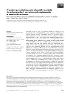
Fig. 1. CT shows 16 x 12 x 15 cm tumor at the caudal pole of the right kidney and 11 x 8.5 12
cm tumor at cranial pole of left kidney. Ponholzer A, Reiter WJ, and Maier U. (2002) Organ
sparing surgery for giant bilateral renal oncocytoma. J. Urol; 2002: 2531-2532.
In gross appearance, renal oncocytomas are light brown or tan, homogeneous, and well
circumscribed tumors. Similar to most renal tumors, they are not truly encapsulated. A
central scar may be seen in approximately one third of cases [54], but prominent necrosis is
generally not seen. [52, 53] Historically, hypervascularity has been thought to be lacking [52,
53], but these lesions often enhance intensely on contrast enhanced imaging. Oncocytomas
cells are packed with numerous large mitochondria, which contributes to their distinctive
staining characteristics. Hemorrhage is found in 20% to 30% of cases [44, 54]. Extension into
the perinephric fat has also been reported in 11% to 20% of cases, [46, 54] however this is
most often not the case in the smaller such lesions that have been resected in recent series. A
transitional histopathologic type has been described in the Birt-Hogg-Dubé syndrome, in
which renal oncocytomas, chromophobe renal cell carcinoma, and distinctive cutaneous
lesions often develop [55]. These transitional neoplasms exhibit features of both oncocytoma
and chromophobe RCC, and some authors have hypothesized that there may be a spectrum
of tumors spanning both of these histopathologic types [55, 56, 57].
It has long since been known that renal oncocytomas often manifest atypia. However, it has
been realized that “atypical”features are part of the renal oncocytoma morphological
spectrum and do not appear to affect tumor behavior adversely. [54] Indeed the dilemma of
oncocytoma relates to these atypia and its apparent relationship with chromophobe renal
cell carcinoma. To date, considerable debate still exists in cases where both of these entities
are present. Morphology is often used as the standard. There are a number of reports
documenting the coexistence of renal cell carcinomas, not limited to chromophobe RCC, in
primary oncocytomas. Though two studies from two referral centers reported a co-existence
of 10%-32% [58, 59], a larger number of studies report this incidence to be 0 to 7.2% [54, 60,
61, 62, 63, 64]. This same issue limits the utility of fine-needle aspiration or biopsy [43, 50,
51]. It is possible that evolving molecular and immunohistochemical techniques may further
aid in clarification of this issue. Historically, most renal oncocytomas cannot be
differentiated from malignant RCC by clinical or radiographic means [65].
Renal Cell Carcinoma
8
2.4 Renal cell carcinoma
Renal cell carcinoma is the most lethal of all urologic cancers, with more than 40% of
patients with this malignancy dying of their cancer. It accounts for 2% to 3% of all adult
malignant neoplasms. [66, 67, 190] There is a male-to-female predominance of 3:2 [66]. Renal
cell carcinoma is primarily a disease of elderly patients, presenting in the sixth and seventh
decades of life [67]. The majority of cases of RCC are believed to be sporadic; the United
States National Cancer Institute estimates that only 4% are familial. A number of familial
conditions associated with renal cell carcinoma are described in the literature. Incidence
rates are 10% to 20% higher in African Americans for unknown reasons [68, 190].
The incidence of RCC has increased since the 1970s by an average of 3% per year. This
appears in part to be due to the more prevalent use of abdominal imaging for the evaluation
of a variety of abdominal, gastrointestinal, and other nonspecific complaints [68]. This trend
has correlated with an increased proportion of incidentally discovered and localized tumors
and with improved 5-year survival rates for patients with this stage of disease [67, 69].
However, other factors must also be at play because Chow and colleagues [68] have
documented a steadily increasing mortality rate from RCC per unit population since the
1980s, and this was observed in all ethnic and both sex groups[191].
Most sporadic RCCs are unilateral and unifocal. However satellite lesions are known to
occur, and are often small and difficult to identify by preoperative imaging, intraoperative
ultrasonography, or visual inspection; they appear to be the main factor contributing to local
recurrence after partial nephrectomy. [70, 71] Bilateral involvement can be synchronous or
asynchronous and is found in 2% to 4% of sporadic RCCs, although it is considerably more
common in patients with von Hippel-Lindau disease or other familial forms of RCC.[ 56, 72,
73]. Multifocal disease, which is found in 10% to 20% of cases, is more common in
association with papillary histology and familial RCC. [74, 75]
All RCCs were traditionally thought to arise primarily from the proximal convoluted
tubules, and this is probably true for most clear cell and papillary variants. However, more
recent data suggest that the other histologic subtypes of RCC, such as chromophobe and
collecting duct RCC, are derived from the more distal components of the nephron. [76, 77,
78] As of recent, urologic researchers have recognized the distinct nature of the various
histopathologic subtypes of RCC through advances in molecular genetics, which has greatly
aided in their proper classification.[72]
All RCCs are, by definition, adenocarcinomas, derived from renal tubular epithelial cells [78,
53, 79]. Most RCCs share ultrastructural features, such as surface microvilli and complex
intracellular junctions, with normal proximal tubular cells, and they are believed to be
derived from this region of the nephron [73]. All of these recent developments indicate that
RCC is not a single malignant neoplasm but rather comprises several different tumor
subtypes, each with a distinct genetic basis and unique clinical features.
Most RCCs are round to ovoid and circumscribed by a pseudocapsule of compressed
parenchyma and fibrous tissue rather than a true histologic capsule. RCCs commonly
consist of yellow, tan, or brown tumor interspersed with fibrotic, necrotic, or hemorrhagic
areas; they are generally not uniform in gross appearance. Cystic degeneration is found in
10% to 25% of RCCs and appears to be associated with a better prognosis compared with
purely solid RCC [88, 89, 90, 91, 190]. Calcification can be stippled or in plaque form, and is
found in 10% to 20% of RCCs. Aggressive local behavior is not uncommon with RCC and
manifests in a number of ways. Invasion and penetration into the collecting system or renal
capsule are found in approximately 20% of cases, although displacement of these structures
Radiologic Imaging of Renal Masses
9
is more commonly seen. Further local spread to involve adjacent organs or the abdominal
wall is often precluded by Gerota's fascia, although some high-grade RCCs may penetrate
this barrier. A notable feature of RCC is its occasional involvement of the venous system,
which is found in 10% of RCCs, more often than in any other tumor type. [ 92, 93]
RCC TYPE PREVALENCE ORIGIN KEY FEATURES
Clear cell 70-80% Proximal tubule Hypervascular, aggressive
behavior common, most likely
to have sarcomatoid features
Papillary 10-15% Proximal tubule Hypovascular, multifocality is
common
Chromophobe 3-5% Cortical portion of
the collecting duct
Prognosis better than
conventional
Some aggressive variants
Rare
Collecting duct
Medullary
Unclassified
< 1%
Collecting duct
Calyceal
epithelium
Infiltrative, generally aggressive
Infiltrative,
g
enerall
y
a
gg
ressive,
patients with sickle cell trait
Generally aggressive
Table 3. Subtypes of renal cell carcinoma [55, 72, 73, 76, 80, 79]
RCC has long been recognized as one of the most vascular of cancers as reflected by the
distinctive neovascular pattern exhibited on renal angiography. Unlike upper tract
transitional cell carcinomas, most RCCs are not grossly infiltrative, with the exception of
collecting duct RCC and some sarcomatoid variants. [73] Tumors smaller than 3 cm were
previously classified as benign adenomas, but some small tumors have been associated with
metastases, and most pathologists agree that with the exception of oncocytoma, there are no
reliable histologic or ultrastructural criteria to differentiate benign from malignant renal cell
epithelial tumors [73].
The vascular nature of RCC plays a special role in imaging of this tumor. Various studies
have tried to differentiate between the clear cell and papillary subtypes of RCC based on the
degree of enhancement on contrast-enhanced CT as well as MRI. As a general point, a
number of these studies have shown excellent accuracy, with higher early enhancement in
clear cell renal cancers. [94] In addition, clear cell RCC has a strong association with necrosis
and retroperitoneal collateral circulation that is best seen on MRI [95]. On the other hand,
papillary RCCs show homogeneous low-level enhancement on both CT and MRI [94, 96].
This will be discussed in detail for each type of imaging modality.
The indications for percutaneous renal biopsy or aspiration in the evaluation of solid renal
masses have traditionally been limited, primarily related to concerns about sampling error,
difficulty interpreting limited tissue given the inherent similarities between the eosinophilic
variants of RCC and oncocytoma, and recognition of the improved diagnostic accuracy of
cross-sectional imaging such as CT or MRI. [82, 83] Eighty-three percent to 90% of solid
renal masses thought to be suspicious for RCC based on careful radiographic evaluation
prove to be RCC on final pathologic analysis [84]. Fine-needle aspiration biopsy (FNAB)
cannot significantly improve on this degree of diagnostic certainty and is unlikely to
influence clinical management in the majority of cases [27, 85]. More recently, FNAB has
been reassessed, and several groups have shown enthusiasm for this approach. Some
Renal Cell Carcinoma
10
studies suggest improved sensitivity and specificity, particularly when FNAB is combined
with molecular analysis for CA-9 expression or other markers for RCC. Molecular analysis
could also assess HMB-45 expression to evaluate for atypical AML, and genetic analysis for
oncocytoma may also be available in the near future. In addition, FNAB could influence
clinical management of small renal masses if markers of clinical aggressiveness could be
established and reliably evaluated on limited pathologic material. The potential
complications of FNAB include bleeding, infection, arteriovenous fistula, needle track
seeding, and pneumothorax. [82] In general, the incidence of complications has been
reduced significantly since the introduction of smaller gauge needles. Tumor location,
operator expertise, and number of biopsy attempts can also influence complication rates.
Perinephric bleeding can be detected by CT scan in 90% of cases, but clinically significant
hemorrhage resulting in gross hematuria is much less common (5% to 7%) and is almost
always self-limited [86, 87]. Only five cases of needle track seeding have been reported with
RCC. Overall, the estimated incidence of needle track seeding with urologic malignant
neoplasms is less than 0.01%; most occur with poorly differentiated transitional cell
carcinoma [82].
3. Imaging modalities
Anatomic imaging modalities historically used for the characterization of renal masses
include intravenous contrast-enhanced plain film radiography, ultrasonography,
computerized tomography, and magnetic resonance imaging. Imaging via nuclear medicine
and hybrid nuclear medicine/computerized tomography techniques has also emerged.
Plain film radiography and computerized tomography require use of ionizing radiation.
Advances in radiation technology and equipment design have resulted in lower does and
overall exposure, but total exposure is additive. Total exposure to ionizing radiation for a
patient is of consideration, as there are carcinogenic risks associated with exposure to large
cumulative doses of radiation. Total radiation exposure is measured in a number of different
manners. Doses may be measured as both skin doses and total effective radiation dose.
Effective absorbed radiation, in Sieverts, can also be measured. Cumulative evidence from a
number of studies suggests that three-phase CT scan exposes patients to approximately two
to three times as much radiation as approximately 12 plain film radiographs (though not
that many be required) taken during the course of intravenous pyelography (IVP). [97, 98,
99] More advanced CT protocols, namely multiphase protocols, have made this issue of
more significant concern. Possibilities for limiting radiation in these studies include
eliminating a phase (which may then limit examination accuracy) or reducing mA during
some of the phases of the study. [100] At the current time, low dose CT protocols are being
investigated, primarily in the setting of urinary lithiasis, with a focus on maintaining
diagnostic accuracy while decreasing exposure. Patients of higher body mass index may not
be ideal for low dose CT [101]. Newer subtraction techniques may also allow for fewer runs
through the scanner
Intravenous contrast-enhanced plain film radiography and computerized tomography both
also involve the use of intravenous iodinated contrast agents, which carry an albeit low, but
real risk. Iodinated contrast media entail risk in three manners: (1) metabolic effect relating
to their hypertonicity, (2) inducement of acute renal dysfunction, and (3) idiosyncratic
contrast material reactions.
Radiologic Imaging of Renal Masses
11
Risks associated with hypertonicity include increased cardiac output and decreased
peripheral vascular resistance due to volume expansion [102], inhibition of the coagulation
cascade by high osmolar contrast agents [103], and renovascular dilation followed by
renovascular constriction, with a result decrease in glomerular filtration rate. [104]
Acute impairment in renal function (increase in serum creatinine of 0.5 to 1.0 mg/dl or 25-
50% decrease in GFR) occurs in 1/1,000-5,000 patients without known risk factors. This
condition is generally nonoliguric, however, when it is accompanied by oliguria, risk of
permanent renal damage exists. [105, 106] Risk factors increasing the risk of acute renal
dysfunction include dehydration, pre-existing renal insufficiency, diabetic nephropathy,
congestive heart failure, hyperuricemia, proteinuria, and multiple administrations of
contrast material in a short time period. [107]. General recommendations to avoid acute
renal dysfunction include ample pre-examination hydration, avoidance of dehydrating
preparations, and a reduction in total contrast used, if feasible.
Patients taking metformin for control of diabetes mellitus are advised to stop this
medication 48 hours prior to contrast administration. When renal function and urine output
are demonstrated to be normal after imaging, metformin therapy may be resumed. The
concern for these patients is that if acute renal dysfunction occurs during metformin
administration, these patients may develop significant lactic acidosis. [108]
Idiosyncratic contrast material reactions may occur in mild, moderate, and severe forms.
Mild reactions include metallic taste, sensation of warmth, sneezing, coughing, and mild
urticaria. Moderate reactions include vomiting, more severe urticaria, headache, edema, and
palpitations. These reactions can be symptomatically treated as needed. However, severe
reactions, which include hypotension, bronchospasm, laryngeal edema, pulmonary edema,
and loss of consciousness, require immediate intervention. Severe reactions occur in less
than 0.1% of patients, and it is estimated that 80% of these may be avoided by use of low
osmolar contrast media. [109, 110]
All contrast-associated risks appear to be lowered by use of low osmolar contrast media
currently available. Administration of prophylactic corticosteroids and use of low osmolar
contrast media have been shown to decrease the risk of contrast reaction, but not eliminate
it. [111] Methylprednisolone 32 mg oral may be given every 12 hours starting 24 hours prior
to examination. This is continued 12-24 hours after the examination to ensure that all
contrast material has been excreted. [112] Diphenhydramine 50 mg oral may also be
administered before contrast administration. All patients offered intravenous iodinated
contrast studies should be counseled and informed of these associated risks.
Many imaging studies relate to differentiation of benign and malignant lesions. Nonetheless
they will be presented in the following where to they relate best to the discussion.
3.1 Contrast-enhanced plain film radiography
The IVP, or intravenous pyelogram (also IVU-intravenous urogram) had long been a
mainstay of urologic diagnostic imaging. This test requires use of intravenous iodinated
contrast. IVP generally requires bowel preparation for optimal imaging results, an absence
of patient contraindications to intravenous iodinated contrast administration, repeated
imaging over thirty to sixty minutes, and at times more prolonged imaging if renal
obstruction is present.
After a scout film is taken, contrast is injected, at which time nephrotomograms are
obtained. These images may reveal abnormalities of the renal parenchyma. However recent
Renal Cell Carcinoma
12
studies comparing IVP to other imaging modalities, namely computerized tomography,
show that sensitivity for detection and characterization of renal masses is limited. [113]
Findings suggestive of a mass seen on IVP generally require other types of imaging for both
corroboration and specific delineation.
IVP may provide valuable information pertaining to the pyelocalyceal system, including the
existence of hydronephrosis, hydroureteronephrosis, existence of urinary stones, and
“filling defects” which include a variety of diagnostic possibilities. However, IVP has
limited sensitivity for renal parenchymal pathologies. Previous studies have documented
the limited sensitivity of small renal masses, particularly when masses are less than 3 cm
[113, 114, 115, 116]. CT urography has gained great popularity in that it provides reliable
assessment of both the renal parenchyma and collecting system. A prospective comparison
of contrast enhanced CT and IVP in initial evaluation of microscopic hematuria
demonstrated that examination with CT had better diagnostic yield for a wide variety of
pathologies. [113]
IVP has fallen out of favor at many institutions for a variety of reasons that include; risk of
contrast toxicity, time consumption involved in performance of test, and limited diagnostic
accuracy in triage setting where diagnoses are often still uncertain. In many institutions it is
now only rarely performed.
3.2 Ultrasonography
Ultrasound requires neither iodinated or gadolinium-based contrast administration, nor
ionizing radiation. Its mechanism of action is based on the transmission of a pulse of high
frequency sound energy into the patient. These sound waves are then reflected, refracted, or
absorbed, depending on type of tissue encountered. The ultrasound transducer also acts as a
receiver, which receives the returning echoes. Collected input is processed by a computer
for the creation of a composite image. [117] Ultrasound is performed in a real time manner
which allows for the technician and physician to modify and review different aspects of the
examination. Newer machines also have significantly improved resolution.
Ultrasonography is used widely throughout medicine. It may be used by anyone with proper
training and experience in almost any clinical scenario; radiology suite, emergency room,
clinic, and under sterile conditions in the operating room. The applications of ultrasonography
are so extensive that they are beyond enumeration here. However, it is of critical importance to
understand that imaging performed with this modality is operator-dependent. This generally
requires that the interpreter of the study be present during the actual real-time study. Image
quality is also closely related to the quality of equipment being used.
Ultrasonography is performed with use of a variety of probes depending on parameters that
include patient body habitus and structures to be imaged. In the case of the kidney, 3.5 to 5
MHz transducers are typically used. Transducers of this range are used to obtain adequate
depth of penetration without substantial loss of resolution. Bony structures and bowel gas
may both interfere with renal imaging, more so on the left than the right.
Ultrasound is commonly employed as an initial imaging exam due to its relative low cost,
relative ease of performance, and lack of need for ionizing radiation. The strength of
ultrasonography is its ability to differentiate solid versus cystic renal structures. In the
hands of an experienced ultrasonographer, it is quite reliable for the identification and
confirmation of simple renal cysts. Historically, there has been concern regarding low
sensitivity for detection of small renal masses. [114. 116] However there is evidence that well
Radiologic Imaging of Renal Masses
13
performed duplex ultrasonography may be highly accurate in the diagnosis and staging of a
large number of renal masses, including those cases where renal vein or caval thrombus are
involved. As active surveillance has become an option for select patients with renal masses,
type of surveillance imaging has become an important issue. A recent study comparing size
of renal masses noted on US, CT, and MRI, comparing imaging results to final pathology,
demonstrated all three modalities accurately predicted pathologic tumor size. This study
included relatively small renal masses as well. [118] However, there may be limitations with
use of ultrasound in respect to the identification of lymphadenopathy. [119]
Renal ultrasonography also has a large role in imaging in patients with azotemia, those with
severe contrast allergy, pregnant patients, neonates, and children. Ultrasonography may at
times be quite useful for assessment of renal masses in the intraoperative setting, both for
open cases and laparoscopic cases. In the open setting it may be useful for the identification
of relatively small masses completely hidden within the renal parenchyma, or in cases
where multiple small tumors, not all of which are immediately amenable to manual
palpation, are suspected. Laparoscopic ultrasound may be useful in a similar fashion, and
may also be used in concert with different ablative devices that are at times used in the
treatment of select renal masses. [120]
3.2.1 Advances in ultrasound technology and technique
Concerns with use of renal ultrasound for detection of renal masses is noted from earlier
data showing that renal masses 3 cm and less are detected only 67-79% of the time. [121].
One must consider that renal tumors may at times have similar echogenicity to normal renal
parenchyma. However, with improvements in ultrasound technology, namely in terms of
resolution and Doppler technology, this may no longer be the case. More recently, contrast-
enhanced ultrasonography (CEUS) has been introduced as another means of further
characterizing renal masses. Ultrasound contrast agents (gas filled microbubbles covered by
a stabilizing shell) are injected as liquids, and then manifest as a gas when in the
bloodstream. This contrast agent, in its gas form, improves the detection of Doppler signals
and helps better reveal both normal and abnormal vascularity. [122] At time of performance
of the study, the contrast agent is injected, and when evident on imaging, the patient holds
their breath while a number of ultrasound frames are obtained for analysis. CEUS entails
less risk than contrast-enhanced computerized tomography, can show even subtle tumor
blood flow, and may serve as a viable alternative for patients unable to undergo contrast
enhanced CT or MRI for any variety of reasons. [123] In study of complex cystic renal
masses CEUS has shown to be comparable in characterization of such masses with use of the
Bosniak system. [124, 125] CEUS may also improve visualization of renal vessels, detection
of vascularity in solid renal masses, and detection of vascularity in septa and walls of
complex renal masses. [126, 127, 128] Use of microbubbles in this manner has been shown to
have a high safety profile, with incidence of adverse reactions lower than that observed for
iodinated and gadolinium based contrast agents.[129]
3.2.2 Ultrasonography and differentiation of renal masses
Angiomyolipomas most often are associated with distinct radiographic findings, which
generally relate to their fat content. The typical but not diagnostic finding of AML on
ultrasonography is a well-circumscribed, highly echogenic lesion, often associated with
shadowing. [37, 25] The finding of shadowing should suggest an AML rather than a small,
Renal Cell Carcinoma
14
echogenic RCC, which rarely shadows [25]. In terms of differentiation and characterization
of solid renal masses using ultrasonography, the presence of shadowing, a hypoechoic rim,
and intratumoral cysts are thought to be important findings that may help distinguish
angiomyolipomas from other solid lesions, namely renal cell carcinoma. [130] Nonetheless,
these findings are not reliable enough to make such a diagnosis with reasonable certainty.
Although conventional ultrasound has not resulted in more specific characterization of renal
masses in terms of histopathologic type/subtype, this will issue will likely be re-evaluated
with introduction of CEUS.
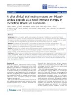
Fig. 2. Renal tumor at the lower pole of the right kidney. Transverse scans. a. A well-defined
expansive, hyperechoic mass is observed at grey-scale ultrasound. b. The mass shows
intense enhancement at CEUS. Siracusano S, Bertolotto M, Ciciliato S, et al (2011) World J
Urol. Epub ahead of print.
3.3 Computerized tomography
Sir Godfrey Newbold Hounsfield, (28 August 1919 – 12 August 2004) and Allan MacLeod
Cormack (February 23, 1924 – May 7, 1998) shared the 1979 Nobel Prize for Physiology/
Medicine for developing the diagnostic technique of X-ray computed tomography (CT).
Beyond doubt, the introduction of the CT scanner is a critical point in the evolution of
medicine.
Since time of inception, CT has taken an increasing role in the realm of imaging. In 2006,
over 60 million CT examinations were performed in the United States. [131] In 2008, over
140 million CT examinations were performed worldwide. [132] Similar to plain film
radiography, images obtained by this modality are created due to the attenuation of photons
by the body tissue being examined. A thin collimated x-ray beam is generated on one side of
the patient. Detectors on the opposite side of the patient then measure the amount of
transmitted radiation. In any given transverse plane being examined these measurements
are repeated as the x-ray beam rotates around the patient. During processing, the
measurements are taken and placed into a matrix of CT values that correspond to
attenuation of a given tissue volume within the patient. The gray scale of each CT pixel is
related to the amount of radiation absorbed at that point. This value is called the attenuation
value, and is commonly denoted in Hounsfield units. Clinically relevant CT Hounsfield unit
values are shown in table 3. [133, 117]
Radiologic Imaging of Renal Masses
15
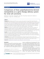
Fig. 3. Sir Godfrey Newbold Hounsfield, (28 August 1919 – 12 August 2004) standing beside
early CT scanner. Source: unknown
Absorber CT numbers (HUs)
Bone +1000
Calculus +400 or greater
Calcification +160 OR GREATER
Acute hemorrhage +50 to 90
Soft Tissue +10 to 50
Water 0
Fat -100
Air -1000
Table 4. Hounsfield Values [133]
Obtained measurements are then taken from a given transverse slice of body tissue and are
mathematically processed by a computer that then reconstructs a cross-sectional image of the
body. Early CT scanners obtained transverse images one slice at a time. However, later
generation spiral (or helical) CT scanners allow patient movement on a gantry with
simultaneous tunnel rotation with continuous x-ray exposure. This arrangement allows for
rapid acquisition of volumetric data from the patient being examined during the time in which
breathing is suspended. The acquisition of volumetric data is of great value in terms of renal
imaging in that it allows for greater accuracy and detail in terms of evaluating renal
parenchymal masses and renal vasculature, particularly in terms of visualization of small renal
masses and supernumerary renal blood vessels. Studies have indeed shown that thin slice
overlapping reconstructions are quite useful for differentiation and accurate depiction for
renal masses as small as 5 mm. [188]) Data acquired in this manner also allows for high quality
CT angiography and three dimensional reconstructions in a variety of manners. [117]