SOFT TISSUE TUMORS pptx
Bạn đang xem bản rút gọn của tài liệu. Xem và tải ngay bản đầy đủ của tài liệu tại đây (16.99 MB, 282 trang )
SOFT TISSUE TUMORS
Edited by Fethi Derbel
Soft Tissue Tumors
Edited by Fethi Derbel
Published by InTech
Janeza Trdine 9, 51000 Rijeka, Croatia
Copyright © 2011 InTech
All chapters are Open Access distributed under the Creative Commons Attribution 3.0
license, which permits to copy, distribute, transmit, and adapt the work in any medium,
so long as the original work is properly cited. After this work has been published by
InTech, authors have the right to republish it, in whole or part, in any publication of
which they are the author, and to make other personal use of the work. Any republication,
referencing or personal use of the work must explicitly identify the original source.
As for readers, this license allows users to download, copy and build upon published
chapters even for commercial purposes, as long as the author and publisher are properly
credited, which ensures maximum dissemination and a wider impact of our publications.
Notice
Statements and opinions expressed in the chapters are these of the individual contributors
and not necessarily those of the editors or publisher. No responsibility is accepted for the
accuracy of information contained in the published chapters. The publisher assumes no
responsibility for any damage or injury to persons or property arising out of the use of any
materials, instructions, methods or ideas contained in the book.
Publishing Process Manager Marija Radja
Technical Editor Teodora Smiljanic
Cover Designer Jan Hyrat
Image Copyright Photosani, 2011. Used under license from Shutterstock.com
First published October, 2011
Printed in Croatia
A free online edition of this book is available at www.intechopen.com
Additional hard copies can be obtained from
Soft Tissue Tumors, Edited by Fethi Derbel
p. cm.
ISBN 978-953-307-862-5
free online editions of InTech
Books and Journals can be found at
www.intechopen.com
Contents
Preface IX
Part 1 Fundamental Aspects of Soft Tissue Tumors 1
Chapter 1 Considerations for Treatment Development
in Rhabdomyosarcoma: In Vitro Assessment
of Novel DNA Binding Drugs 3
Steven J. Wolf, Laurence P.G. Wakelin and Daniel R. Catchpoole
Chapter 2 Telomere Maintenance Mechanisms
in Soft Tissue Sarcomas 31
Matthew J. Plantinga and Dominique Broccoli
Part 2 Diagnosis and Investigations in Soft Tissue Tumors 51
Chapter 3 Classification of Soft Tissue Tumors by
Machine Learning Algorithms 53
Jaber Juntu, Arthur M. De Schepper, Pieter Van Dyck,
Dirk Van Dyck, Jan Gielen, Paul M. Parizel and Jan Sijbers
Chapter 4 Medical Theory on Orthopedics Combining
Molecular Imaging with Clinical Practice 69
Jing jing Peng
Chapter 5 Imaging Findings of Adipocytic Tumors 91
Jun Nishida, Shigeru Ehara and Tadashi Shimamura
Part 3 Types of Soft Tissue Tumors 101
Chapter 6 Pediatric Soft Tissue Tumors 103
Ezequiel Trejo-Scorza, Belinda Beatriz Márquez Álvarez,
Carlos José Trejo-Scorza and Simón Paz-Ivannov
Chapter 7 Head and Neck Soft Tissue Sarcoma 117
Rogelio Gonzalez – Gonzalez, Ronell Bologna – Molina,
Omar Tremillo – Maldonado, Ramon Gil Carreon – Burciaga
and Marcelo Gomez Palacio - Gastelúm
VI Contents
Chapter 8 Dermatofibrosarcoma Protuberans – Special Challenges of
Management in Resources Constrained Countries 143
Titus Osita Chukwuanukwu and Stanley Anyanwu
Chapter 9 Clinical and Molecular Biology of Angiosarcoma 149
N.J. Andersen, R.E. Froman, B.E. Kitchell and N.S. Duesbery
Chapter 10 Gastrointestinal Stromal Tumours: A Contemporary
Review on Pathogenesis, Morphology and Prognosis 175
Muna Sabah
Part 4 Treatment of Soft Tissue Tumors 207
Chapter 11 Treatment of Synovial Sarcoma in Children 209
Shvarova Anna Viktorovna, Rykov Maxim Yurjevich,
Karseladze Appolon Irodionovich
and Ivanova Nadezhda Mikhailovna
Chapter 12 Novel Therapeutic Targets in Soft Tissue Sarcomas 217
Quincy S.C. Chu and Karen E. Mulder
Part 5 Prognosis of Soft Tissue Tumors 253
Chapter 13 Prognostic Factors in Soft Tissue Sarcoma 255
Luiz Eduardo Moreira Teixeira, Jose Carlos Vilela
and Ivana Duval De Araujo
Chapter 14 Metastatis of Soft Tissue Sarcomas 263
Fethi Derbel, Sonia Ziadi, Medi Ben Hadj Hamida,
Jaafar Mazhoud, Mohamed Ben Mabrouk,
Abdallah Mtimet, Sabri Youssef, Ajmi Chaouch,
Ali Ben Ali, Ibtissam Hasni, Mrad Dali Kaouthar,
Jemni Hela, Moncef Mokni and Ridha Ben Hadj Hamida
Preface
Soft tissue tumors include a heterogeneous group of diagnostic entities, most of them
benign in nature and behavior. Malignant entities, soft tissue sarcomas, are rare
tumors that account for 1% of all malignancies. These are predominantly tumors of
adults, but 15% arise in children and adolescents. The wide biological diversity of soft
tissue tumors, combined with their high incidence and potential morbidity and
mortality represent challenges to contemporary researches, both at the level of basic
and clinical science. Determining whether a soft tissue mass is benign or malignant is
vital for appropriate management.
This book is the result of collaboration between several authors, experts in their fields;
they have tried to convey to the reader the complexity of soft tissue tumors and the
diversity in the diagnosis and management of these tumors.
In this book, entitled Soft Tissue Tumors, we have highlighted many of the significant
advances in the diagnosis and treatment of soft tissue tumors; we tried to offer a
comprehensive overview of this wide field, from basic mechanisms underlying soft
tissue tumors to current advances and future directions in the prevention, early
detection and management of these neoplasms.
The presented textbook is subdivided into five sections termed: Fundamental Aspects
of Soft Tissue Tumors, Diagnosis and Investigations,Types and Classification of Soft
Tissue Tumors, Treatment of Soft Tissue Tumors, Prognosis of Soft Tissue Tumors.
Although this book does not cover all aspects related to soft tissue tumors, it is
intended for at least two kinds of readers : residents of intermediate and advanced
courses in medicine; oncologist, pathologist, surgeon, radiologists and all doctors
whatever their specialty.
As editor in chief of this textbook, I would like to acknowledge the efforts made by all
of the contributing authors and the entire editorial team in the publishing of this book
especially Mrs Marija Radja for her very precious collaboration. Their dedication to the
publication of the most contemporary and comprehensive scientific data has this
excellent work as a result. I would like to dedicate this textbook to all my colleagues
surgeons, pathologist, oncologist and radiologist at Sahloul Hospital, the past and
X Preface
actual deans of our university Professor Mohsen Jeddi, Sahloul Essoussi, Abdelkrim
Zbidi, Nejib Mrizak and Ali Mtiraoui for their encouragement, and Professor Moncef
Mokni pathologist in the department of pathology at Farhat Hached Hospital in
Sousse.
I would like to express a special dedication to our first dean and professor of
pathology who had dedicated a lot of his scientific efforts to the soft tissue tumors. His
name is Professor Chedly Bouzakkoura. He passed away last year.
I also dedicate this book especially to Professor Ridha Ben Hadj Hamida, and
Professor Rached Letaief, surgeons at the department of surgery in Sousse. A special
dedication to Professor Gharbi Slaheddine and Hamadi Farhat, the first surgeons at
Sousse University who died a few years ago.
You have taught me, since my first steps as resident, not only to manage different
types of digestive malignancies using the highest quality surgical and medical care
but as well instilled in me a sense of responsibility to improve as a person and as a
clinician.
Fethi Derbel
Professor of General and Digestive Surgery
University Hospital Sahloul
Sousse
Tunisia
Part 1
Fundamental Aspects of
Soft Tissue Tumors
1
Considerations for Treatment Development in
Rhabdomyosarcoma: In Vitro Assessment
of Novel DNA Binding Drugs
Steven J. Wolf
1,3
, Laurence P.G. Wakelin
2
and Daniel R. Catchpoole
1,3
1
The Biospecimens Research Group and Tumour Bank, Children’s Cancer Research Unit,
The Kids Research Institute, The Children’s Hospital at Westmead, Westmead, NSW,
2
The School of Medical Science, The Faculty of Medicine,
The University of New South Wales, Sydney, NSW,
3
Faculty of Medicine, The University of Sydney, NSW,
Australia
1. Introduction
Rhabdomyosarcoma (RMS) is the most common soft tissue sarcoma in children and is
believed to originate from mesenchymal cells that resemble undifferentiated striated muscle
cells (Wexler and Helman, 1997). It is a relatively rare tumour type with approximately 350
patients below the age of 20 diagnosed each year in the USA (Gurney et al, 1999). Incidence
in Australia is also low with only 31 RMS cases out of the total 1,003 childhood cancers
diagnosed between 2001 and 2005 in the state of NSW (Tracey et al 2007). Histological
staining of tumour samples led to the classification of two distinct forms of tumour types:
embryonal (ERMS) and alveolar (ARMS). ERMS is the most common histologically
diagnosed variant of the disease and is associated with an earlier onset, most commonly
around the age of 2 to 5 years (Qualman et al, 1998). Diagnosis of ERMS is made when the
cells fit the criteria of appearing as stroma-rich spindle cells which are not densely packed
and show no alveolar pattern of growth which characterises ARMS. Variant forms of ERMS,
including botryoid and spindle cell types, have been described as being histologically
similar to standard ERMS (Wexler and Helman, 1997).
Treatment of rhabdomyosarcoma employs a multimodal approach that utilizes surgical,
radiological and chemotherapeutic protocols. Unlike in the treatment of adult sarcomas,
surgical removal of the tumour mass in paediatric RMS patients is usually only attempted if
complete resection can be guaranteed without causing cosmetic or developmental damage
to the child. For this reason chemotherapy is the frontline option in the treatment of
paediatric RMS both as a means of local tumour mass control and for the prevention of
residual and micrometastatic disease (Stevens, 2005). Over 70% of patients with non-
metastatic RMS will respond well to chemotherapy and reach a 5 year event free survival
milestone. Patients with metastatic or stage IV ERMS however, and those with ARMS who
generally present at diagnosis with an advanced metastatic form of the cancer, continue to
face a poor prognosis as a result of diminished tumour response to the current
chemotherapy options. Currently, less than 30% of patients with metastatic disease survive
Soft Tissue Tumors
4
without relapse and despite this drastic difference in tumour response, chemotherapy
protocols continue to utilize the same compounds regardless of tumour subtype,
progression or stage (Wexler and Helman, 1997).
Without agents to target specific molecular pathways and proteins of RMS, such as the PAX3-
FKHR chimeric protein, chemotherapy protocols continue to utilize general cytotoxic
compounds that rely on the rapid proliferation of tumour cells for selectivity and optimal
efficacy. Most of these agents bind to DNA and disrupt key molecular processes involved in
DNA transcription and replication. Treatment usually involves the vinca alkaloid vincristine,
the transcription inhibitor actinomycin D and the alkylating prodrug cyclophosphamide
(Breitfeld et al, 2005). Several other general cytotoxic agents, including the topoisomerase
poisons etoposide, doxorubicin, epirubicin, topotecan and irinotecan as well as the alkylating
agents ifosfamide and carboplatin have also been used in alternative treatment protocols and
large scale clinical trials (Table 1). Many agents included in RMS clinical trials and standard
treatment protocols can be broadly classified as general cytotoxic agents, a large proportion,
including etoposide, doxorubicin and topotecan, specifically target and poison the function of
the topoisomerase enzymes, whilst actinomycin D is a transcription inhibitor that has been
successful in the treatment of a wide variety of tumours, including RMS.
2. DNA binding agents underpin RMS therapy – a review of clinical trials
Prior to the 1970s the prognosis for RMS patients was extremely poor regardless of tumour
subtype. The earliest large scale collaboration to be established was the Intergroup
Rhabdomyosarcoma Study Group (IRSG), a joint effort between US and Canadian
researchers. Five trials were carried out by this group between 1972 and 2000 at which point
the group merged into the Children’s Oncology Group (COG) under which more recent
trials have been carried out. Patients enrolled in IRSG or COG clinical trials were grouped
based on various prognostic factors before an appropriate treatment schedule was assigned.
The second collaboration to be established was the European based group ‘International
Society for Paediatric Oncology’ (SIOP) which launched several large cohort trials in 1975,
1984, 1989 and 1995 from which many findings were reported. A selection of key findings
from IRSG, COG and SIOP clinical trials are presented in summarized form in Table 1. With
5 year event-free survival rates (EFS) reaching 70%, patients with gross residual tumour
were believed to have benefited the most in early studies. It was clear however, that patients
with stage III or IV RMS required more intense chemotherapy than those in stage I and II
and it was concluded that despite the successes of the VAC combinational therapies,
introducing additional agents, such as topoisomerase I poisons, would help subdue the
onset of local and distant failures. The prognosis for patients with non-metastatic RMS
continued to improve after the fourth and fifth IRSG studies were completed, yet despite
years of large cohort clinical trials and the subsequent retrospective analysis of data,
response rates in patients with metastatic ERMS and ARMS remained considerably low.
This has been attributed to many factors including combination chemotherapy leading to
additive and overlapping adverse side effects which limit the dosages used as well as
intrinsic or acquired drug resistance mechanisms.
3. Therapeutic advancement in RMS requires new agents
It is clear from this review of the chemotherapeutic treatment options available for RMS that
novel agents are desperately required to improve the prognosis for patients with metastatic
Considerations for Treatment Development in Rhabdomyosarcoma:
In Vitro Assessment of Novel DNA Binding Drugs
5
Study
RMS
Classification
Protocol Tested Results References
IRSG
Study I
Group I VAC + R No benefit from
additional R
Maurer
et al, 1988
Group II VC + R + A No benefit from
additional A
Group III + IV VAC + R + D No benefit from
additional D
IRSG
Study II
Group III Intense repetitive pulse
VAC + radiation
or VDC + radiation
Improvement over IRSG
1:
(SR) increased -50% to
66%
(CRR) increased - 56% to
73%.
Maurer
et al, 1993
Groups I - IV VDC No improvement vs.
VAC. Fatal side effects
Groups I – II VA + C No improvement from
additional C
Groups I – II Repetitive pulse VAC Improvement over IRSG
1
IRSG
Study IV
Groups I – II VAC, with either VAI or
VIE
3yr EFS: 75% VAC,
77% VAI, 77% VIE
Overall EFS of 83%.
Surgery + VAI + VIE
was equally effective as
VAC only
Crist
et al, 2001
IRSG
Study IV
Intermediate
risk ERMS
3yr FFS improved due to doubling of alkylating
agent dosage compared to the treatment protocol
used in IRSG study III. Cyclophosphamide or
ifosfamide had same effect.
Baker
et al, 2000
High Risk /
Stage IV
VAI or VIE ever
y
3 wks /
12 wks +
VAC every 3 wks for 36
wks.
63% OR
(12 weeks)
Sandler
et al, 2001
SIOP
MMT89
Group III,
Stage III
Novel treatment which
combined 6 drugs (IVA)
+ (CbEV) + (IVE).
60% OS (5yr)
versus
42% OS (5yr) MMT84
Stevens
et al, 2005
Independe
nt Phase I
Recurring
solid tumours
varying doses of Cb +
fixed doses of I + E
33% OR
(4% increased)
Marina
et al, 1993
Indepe-
ndent
Phase I/II
Refractor
y
STS
sarcomas
ICbE 32% CR
63% OR
Kung
et al, 1995
CCG
Study I
27 RMS
patients in a
total cohort of
ICbE 78% 1yCR, 33% 2yCR,
66% OR
ERMS ARMS
Van
Winkle
et al, 2005
Soft Tissue Tumors
6
Study
RMS
Classification
Protocol Tested Results References
97 STS
patients
82% 1 yr OS 40% 1 yr
OS
46% 2 yr OS 20% 2 yr
OS
SIOP
MMT89
Untreated
Stage IV RMS
Single Course C, Epi + V 53% Total OR
ERMS ARMS
46% OR 58% OR
Frascella
et al, 1996
IRSG V Stage IV RMS T or T + VAC 46% Total OR
ERMS ARMS
28% OR 65% OR
Pappo
et al, 2001
Independe
nt Trial
Intermediate
risk RMS
VDC + EI at 3 week
intervals over a total 10
cycle course.
91% OS
85% EFS
Arndt
et al, 1998
Table 1. Results from Selected RMS clinical trials involving general cytotoxic compounds.
A=actinomycin D, C=cyclophosphamide, D=Doxorubicin, E=etoposide, Epi=Epirubicin,
I=ifosfamide, R=radiotherapy, V=vincristine, Cb=Carboplatin; CR=Complete Response,
CRR=Complete Response Rate, EFS=Event Free Survival, OR=Overall Response,
OS=Overall Survival, SR=Survival Rate.
or stage IV ERMS and ARMS. To date, the only genetic abnormality consistently associated
with ARMS is the t(2:13)/t(1:13) translocations that produce the oncogenic PAX3/7-FKHR
chimeric proteins. One day these may be targeted by small molecules or genetic based
therapies, however, the immediate future of RMS treatment remains highly dependant on
general cytotoxic agents. Unfortunately, all of the available general cytotoxic agents are
associated with adverse side effects that place severe limitations on the concentrations of
drug that can be administered to children with the disease. To minimize these side effects
each agent is used in low doses both in combination with other general cytotoxic agents and
over an extended period of time. Such treatment protocols rarely guarantee full recovery
and often promote the development of drug resistance mechanisms within the cancer cells
that manifest themselves either during initial rounds of therapy, or more commonly,
following tumour relapse.
Optimization of existing chemotherapy protocols, and the introduction of established
cytotoxic agents into RMS clinical trial, has resulted in improved response rates for ERMS
patients in recent decades. Despite this, ARMS and metastatic ERMS, are still associated
with a poor prognosis (Breitfeld and Meyer, 2005). With such a high dependency on general
cytotoxic agents for the treatment of RMS, novel compounds with improved efficacy and
fewer side effects must be developed. Efforts to improve the outcome in poor prognosis
patient groups focus largely on trials involving new combinations of existing clinically-
active compounds. Some of the most commonly used agents in RMS protocols exploit the
fragility of DNA transcription, and chromosome integrity, by physically interfering with
these processes and structures. For example, actinomycin D inhibits transcription by
intercalating into DNA and impeding the progression of DNA-dependant RNA
polymerases. Etoposide, along with the anthracyclines, camptothecin and its analogues, trap
topoisomerases in their DNA cleavable complexes, resulting in the accumulation of DNA
double strand breaks, fragmented chromosomes, and cell death at mitosis (Pommier Y,
Considerations for Treatment Development in Rhabdomyosarcoma:
In Vitro Assessment of Novel DNA Binding Drugs
7
2006). Given the apparent importance of these biochemical targets in RMS therapy, here, we
have investigated the efficacy of a number of novel DNA binding transcription inhibitors
and topoisomerase poisons in 5 RMS cell lines that represent both ERMS (RD and JR1) and
ARMS (RH30, RH3 and RH4) tumour subtypes. We have also compared their activity with
that of the established transcription inhibitors actinomycin D, chromomycin, and
nogalamycin, and the topoisomerase poisons etoposide, amsacrine, doxorubicin,
mitoxantrone, and topotecan. Each new agent has been designed with altered DNA
association/dissociation kinetics, improved tumour penetration compared to the established
agents and with this in mind, their efficacy and vulnerability to common mechanisms of
resistance are examined.
3.1 Novel DNA binding cytotoxic agents
With a range of novel cytotoxic compounds available to us through colleagues at the
University of New South Wales and the Auckland Cancer Society Research Centre, we
aimed to assess the efficacy of selected agents from various classes in an in vitro RMS cell
line model that best represented both subtypes of the disease. In doing so it was our
intention to identify agents with the potential to expand treatment options for RMS patients
and further improve the efficacy of chemotherapy protocols that utilize general cytotoxic
agents. Each of the novel compounds assessed in this study contain tricyclic carboxamide
moieties that act as DNA intercalating chromophores and have previously been shown to be
cytotoxic in leukaemia and/or solid tumour cell lines (Wakelin et al, 2003; Baguley et al,
1995; Atwell et al, 1984).
One group of novel transcription inhibitors (Figure 1A) contain dual intercalating
chromophores that are joined via their 9-amino groups by linker chains of various structures
and contain N,N-dimethylaminoethyl (DMAE) active side chains. These agents bind to
DNA in a bisintercalating threading fashion inspired by the binding mechanism of
nogalamycin (Wakelin et al, 2003). In this design the carboxamide sidechains spear the DNA
helix and make bonding interactions with guanine bases in the major groove to promote
transcription inhibition by enhancing DNA residence time without increasing binding
affinity. This is a desirable characteristic for activity in solid tumours where tumour
penetration correlates inversely with DNA binding affinity (Wakelin et al, 2003). Differences
in these compounds are found in their linker chains with flexibility, charge and length all
varying. With the linker chains laying in the minor groove of the DNA helix they play a
crucial role in the overall activity of the compound by placing a physical block in the path of
DNA tracking enzymes (Wakelin et al, 2003).
The second class of transcription inhibitors (Figure 1B,C) contain representatives of
phenazine-1-carboxamide dimers bridged via their side chains with alkylamino linkers of
various structures (Spicer et al, 2000). Within this class, the clinical candidate
MLN944/XR5944 bisintercalates with its linker in the DNA major groove making hydrogen
bonding interactions to guanines in a sequence specific manner. This compound possesses a
unique mechanism of action, including the inhibition of transcription factor binding to
DNA, which ultimately leads to the inhibition of transcription (Byers et al, 2005). The
bis(phenazine-1-carboxamides) studied are of two structural types: SN26356
(MLN944/XR5944) and SN26700 are 9-methylphenazines joined via a dicationic -
(CH2)2NH(CH2)NH(CH2)2- linker, and differ in that SN26700 has the amines substituted
with a methyl group (Figure 1B). SN26871 has an N-methylated monocationic -
(CH2)3N(Me)(CH2)3- linker and an 8,9-benzphenazine chromophore (Figure 1C). 
Soft Tissue Tumors
8
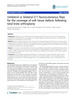
Fig. 1. Molecular structure of (A) novel transcription inhibitors Bis(9-
aminoacridinecarboxamides), C8 DMAE, C3NC3 DMAE and C2pipC2 DMAE, (B-C) novel
transcription inhibitors Bis(phenazine-1-carboxamides), 26356 (MLN944/XR5944), 26700
and 26871 (D) novel topoisomerase poisoning, monointercalating acridine-4-carboxamides,
DACA, 9-amino-DACA, AS-DACA and SN16713.
A third class of novel compounds, also structurally based around the acridine-4-
carboxamide intercalating chromophore, have previously been identified as topoisomerase
poisons (Finlay et al., 1996) and act as monointercalating agents that feature electron-
withdrawing moieties in place of a single active side chain (Figure 1D). N-[2-
(dimethyl)aminoethyl]-acridine-4-carboxamide (DACA), a dual topoisomerase I/II poison
and the parent compound from this class of agents, was unsuccessfully taken into phase II
clinical trial in patients with non-small cell lung carcinoma, advanced ovarian cancer,
recurrent glioblastoma and advanced colorectal cancer (Twelves et al, 2002; Caponigro et al,
2002). 9-amino derivatives of DACA, however, have greater cytotoxic and dose potencies,
and modifications in the 5-position, such as the methyl sulphone group in AS-DACA,
promote solid tumour activity (Atwell et al, 1987). In contrast to DACA, 9-amino-DACA and
AS-DACA appear to be more specific poisons for topoisomerase II (Bridewell et al, 2001)
with AS-DACA, a less lipophillic derivative (Haldane et al,1999) also known to have a wide
spectrum of activity in solid tumours (Atwell et al,1987).
4. Screening novel agents indicates differential response
A panel of 5 RMS cell lines were selected for in vitro assessment of cytotoxicity of novel and
established transcription inhibitors and topoisomerase poisons. RD and JR1 were selected to
represent the ERMS subtype whilst RH30, RH3 and RH4 were selected to represent the
ARMS subtype. We assessed the cytotoxicity of a range of novel and established
transcription inhibitors and topoisomerase poisons against 5 established RMS cell lines.
MTT cell viability assays were used to determine cell survival after a 72 hour exposure to
each compound. Published IC
50
values (Wolf et al, 2009) are plotted as ‘Δ Plots’ which
Considerations for Treatment Development in Rhabdomyosarcoma:
In Vitro Assessment of Novel DNA Binding Drugs
9
graphically represent the differences in efficacy of each drug in each cell line relative to the
median (m) IC
50
of all drugs in all cell lines (Figure 2).
Fig. 2. Δ plots showing variations in drug potency in 5 RMS cell lines. IC
50
s are plotted as a log
10
measure of sensitivity or resistance against the median (m) IC
50
of all agents across all cell
lines (m = 600 nM). This measure of potency, taken as a whole across all RMS cell lines,
serves to highlight the relative differences in drug efficacy. (Wolf, 2009)
Our findings enable classification of these agents into 3 classes; those that are potent in all 5
cell lines; those that show differential responses across the panel; and those that require
higher concentrations to be toxic in all cell lines. The first class includes the naturally
occurring transcription inhibitors actinomycin D, chromomycin and nogalamycin, which are
the most potent amongst the agents studied, the topoisomerase II poisons doxorubicin and
mitoxantrone, and the experimental acridine-4-carboxamide topoisomerase II poison 9-
amino-DACA. Class two includes the bis(phenazine-1-carboxamide) SN 26356, otherwise
known as MLN944/XR5944, identified as a transcription inhibitor and topoisomerase I
poison, the topoisomerase I poison topotecan, and the acridine-4-carboxamide
topoisomerase poison AS-DACA. AS-DACA and topotecan have the same spectrum of
cytotoxic activity, which is complementary to that of SN 26356. Agents such as those
described in group 2 may offer alternative treatment options for RMS tumours unresponsive
to the traditional chemotherapy protocols.
4.1 Cytotoxicity of novel and established transcription inhibitors in RMS cells
The antitumour antibiotics actinomycin D, chromomycin and nogalamycin are amongst the
classical template inhibitors of transcription, each binding to DNA reversibly, but
dissociating slowly so as to present a long-lived block to the passage of RNA polymerases.
Actinomycin D is a monofunctional intercalating agent which places bulky cyclic peptides
in the DNA minor groove, chromomycin is a minor groove binding agent (Yang et al, 1999)
and nogalamycin is a monofunctional threading agent which intercalates with its nogalose
sugar lying in the minor groove and its bicyclic amino sugar spearing the duplex making
hydrogen bonding interactions with guanines in the major groove (Li and Krueger, 1991).
All are known to bind selectively to GC-rich sequences and block RNA polymerase
progression by placing a bulky group in the DNA minor groove. Furthermore, all cause
Soft Tissue Tumors
10
similar profound perturbation to transcription profiles (Zilhif et al, 2006). We have found
that all three agents have indistinguishable activity in the 5 RMS cell lines and that they are
the most potent agents studied, with activity in the nM range (Figure 2). Seemingly, the fine
details of how they interact with DNA to block RNA polymerase do not affect their
cytotoxicity. With actinomycin D routinely used in RMS protocols (Table 1), this observation
suggests that chromomycin and nogalamycin are worthy of consideration for inclusion in
clinical studies.
The development of the bisintercalating bis(9-aminoacridine-4-carboxamide) transcription
template inhibitors was inspired by the threading mechanism of nogalamycin (Wakelin et
al, 2003). Their threading design, in which the carboxamide sidechains spear the DNA helix
to make bonding interactions with guanine bases in the major groove, promotes
transcription inhibition by enhancing DNA residence time without increasing binding
affinity, a desirable characteristic for activity in solid tumours where tumour penetration
correlates inversely with DNA binding affinity. The three examples studied here, C8 DMAE,
C3NC3 DMAE and C2pipC2 DMAE, despite having IC
50
values in human leukaemia CCRF-
CEM cells of 35, 50 and 63 nM respectively (Wakelin et al, 2003), and similar potencies (nM)
in a range of human cancer cell lines (Wakelin unpublished), are found to be about 4 to 40
times less potent in the rhabdomyosarcoma cells, which is some 100 to 1000 times less active
than the naturally occurring transcription inhibitors. RD is the only RMS cell line that could
be considered sensitive and is the only one in which all three threading dimers produced
IC
50
s marginally lower than m (Figure 2). The origins of the intrinsic resistance of the RMS
cell lines to these agents are unclear.
This generalized resistance to the bisacridines also extends to the
bis(phenazinecarboxamide) dimers, with one important exception. These compounds were
designed as bisintercalating topoisomerase I and II poisons (Spicer et al, 2000), but their
actual mechanism of action is complex and appears to involve both transcription inhibition,
along with topoisomerase I poisoning (Byers et al, 2005). The three compounds studied here
are potently cytotoxic in mouse leukemia P388, mouse Lewis lung and Jurkat human
leukemia cells (Gamage et al, 2001). The toxicity of SN26700 and SN26871 however, is
diminished some 35 to 2200 times in the RMS panel, with their IC
50
s clustering around m or
greatly exceeding it (Figure 2). The exceptional response is found with SN26356 which was
used in clinical trial as MLN944/XR5944 (Verborg et al, 2007). Its potent activity in previous
studies is maintained in the RD, RH3 and RH4 cell lines, with an average IC
50
of about 40
nM. The origins of this selectivity are unknown, but our findings point to the importance of
considering SN26356 as a possible clinical trial candidate in RMS.
4.2 Cytotoxicity of novel and established topoisomerase poisons in RMS cells
The trapping of topoisomerases in a cleavable complex with DNA is a well established
mechanism of action of many DNA binding drugs (Li and Liu, 2001). Representative
topoisomerase poisons, both established and novel, were examined in this study, and
produced widely ranging results. For example, amongst the clinically used topoisomerase II
poisons, etoposide and amsacrine were uniformly, poorly active across the RMS cell line
panel, with IC
50
s all greater than m, ranging from 600nM to 22mM (Figure 2). Such a finding
sits oddly with the inclusion of etoposide in clinical RMS protocols (Van Winkle et al, 2005).
In contrast, doxorubicin and mitoxantrone are uniformly active in the RMS cells with
average IC
50
s of about 200nM and 400nM respectively, a finding that supports their
Considerations for Treatment Development in Rhabdomyosarcoma:
In Vitro Assessment of Novel DNA Binding Drugs
11
inclusion in clinical studies. The only clinical topoisomerase I poison studied, topotecan,
produced a differential response with activity of 10nM and 140nM in RH30 and JR1 cells,
but IC
50
s of 1mM to 15mM in the remaining 3 RMS lines. Interestingly, this is the inverse
selectivity of SN26356, which is inactive in RH30 and JR1, and raises the intriguing question
of the potential clinical activity of their use in combination.
The novel topoisomerase poisons evaluated are structurally based on the acridine-4-
carboxamide chromophore, the parent compound of which, DACA (Figure 1), has been
identified as a dual topoisomerase I/II poison (Finlay et al., 1996). Despite its wide solid
tumour activity and its clinical evaluation (Twelves et al, 2002; Caponigro et al, 2002;
Haldane et al, 1993), it shows poor potency in all RMS cell lines with IC
50
s about 2 to 4 mM.
In contrast, its dicationic derivative, 9-amino-DACA, which binds to DNA 6-fold more
tightly than DACA and is only weakly active as a topoisomerase I poison (Finlay et al.,
1996), is 10 times more potent in all RMS cell lines, making its activity comparable to that of
doxorubicin and mitoxantrone (Figure 2). Although the extra charge on the chromophore of
9-amino-DACA enhances cytotoxic potency and antileukaemic activity in mouse tumour
models (Atwell et al, 1987), it diminishes solid tumour activity as a consequence of poor
tumour penetration due to its elevated DNA affinity. Electron withdrawing substituents in
the acridine 5-position lower the chromophore pK, and AS-DACA, bearing a 5-
methylsulphone, has a neutral chromophore at physiological pH, binds DNA with an
affinity between that of DACA and 9-amino-DACA, and is intermediate between these two
agents with respect to topoisomerase selectivity (Finlay et al, 1996). These characteristics
make it generally more cytotoxic than DACA, and endow it with widespread solid tumour
activity (Atwell et al, 1987). In the RMS panel it returns a differential response, strongly
reminiscent of topotecan, with JR1 and RH30 cells being sensitive, but the remaining three
cell lines have IC
50
s above 1mM (Figure 2). Lastly, within the acridinecarboxamide family,
we examined the activity of SN16713, a monofunctional threading agent that superposes the
structures of amsacrine and 9-amino-DACA, selectively poisons topoisomerase II which has
an IC
50
of 120nM in CCRF-CEM cells (Zihlif et al, 2006) and 7nM in Jurkat leukaemia (Finlay
et al, 1996), is poorly active in RMS cells (Figure 2).
Several novel agents displayed comparable or improved efficacy over their established
counterparts in our in vitro drug cytotoxicity study in RMS cell lines. Despite the resistance
of some cell lines to these agents their overall efficacy necessitates further preclinical
development for possible inclusion in RMS clinical trials. Of particular interest were the
novel agents AS-DACA and 9-amino-DACA. 9-amino-DACA showed efficacy across all cell
lines comparable to the established topoisomerase poisons flagging its potential as a
candidate for future RMS clinical trials. By contrast AS-DACA produced a variable cytotoxic
response across the cell line panel. Many factors may be responsible for this observed
variation, in particular the 190x fold difference observed between two archetypal RMS cell
lines, RD and RH30 (Wolf et al, 2011). The remainder of this discussion will explore our
study of AS-DACA cytotoxicity in two RMS cell lines; RD and Rh30, along with AS-DACA-
resistant cell line we derived from RH30, named Res30 (Wolf et al, 2011), as an illustration of
the complexities of developing new treatment strategies for RMS.
5. Causes for differential drug cytotoxicity in RMS cells
Drug “resistance” is a phenomenon that impedes the efficacy of every compound used in
the treatment of cancer at some stage. Mechanisms governing cellular resistance to
Soft Tissue Tumors
12
chemotherapy may be intrinsic, however in most cases they are acquired following repeated
or extended exposure to chemotherapy. Although “acquired” drug resistance is a term that
is used to describe the development of drug resistance within cells that were originally
chemosensitive, it may in fact result from a clonal proliferation of a subpopulation of
intrinsically resistant cells within the original tumour or cell culture. This has been noted to
occur within RMS with resistant, differentiated cells making up the majority of tumour
remaining after chemotherapy treatment (Klunder et al, 2003). The complexity and number
of mechanisms that contribute to drug resistant phenotypes makes identifying and
circumventing the source of the problem a challenge for researchers and clinicians alike.
Some well established mechanisms of resistance include alterations in drug target levels and
function, enhanced drug efflux via membrane bound transport proteins and drug
sequestration/altered intracellular drug distribution. Further, it must be assumed that drug
resistance mechanisms, intrinsic only to certain RMS cell types, act in a manner dependent
on the subtle structural differences which exist between the DNA-binding compounds used.
Given the importance of in vitro studies in pre-clinical drug investigations, it is worthwhile
investigating commonly used RMS cell lines to identify the subtle biological mechanisms
which are intrinsic to them and produce these selective drug resistance phenotypes.
Consequently, in the remaining sections of this review, the impact of different mechanisms
of resistance will be explored, with a specific focus on the differential response of AS-DACA
in RD and RH30 as a paradigm of this complexity.
5.1 ‘Classical’ drug resistance involving transport proteins
One of the most described mechanisms of drug resistance in RMS cell lines, is ATP-Binding
Cassette (ABC) transport protein mediated drug efflux. ABC transport proteins span the
plasma membranes of almost all cells and are responsible for active transport of many
compounds, including a number of agents used in cancer therapy (Klein et al, 1999). In total
49 human genes have been described that encode various ABC transport pumps (Chang,
2007). Whilst each protein is structurally and functionally distinct, all members of the ABC
transport protein family share three conserved sequence motifs within nucleotide binding
domains and are common to many proteins that bind ATP (Leslie et al, 1999). For many
years it was believed that the MDR1 gene, also known as ABCB1, which encodes P-
glycoprotein (P-gp), was the prime contributor to drug efflux (Leslie et al, 1999). Subsequent
studies however led to the identification of several related proteins which have also been
linked to the multidrug resistance phenotype and include multidrug resistance-associated
proteins MRP1 to MRP5 and Breast Cancer Related Protein (BCRP) (Komdeur et al 2003).
5.1.1 Multidrug Resistance-Associated Protein 1 (MRP1)
MRP1 (ABCC1) is a 170kDa protein (190kDa in its glycosylated form), that belongs to the
ABC family of membrane bound transport proteins. MRP1 is comprised of 17
transmembrane segments that are grouped into three transmembrane domains (TMDs), two
cytoplasmic linker regions and two cytoplasmic nucleotide binding domains. This structure
is common to most members of ABCC subfamily. Although the cytoplasmic linker region,
which lies between TMD0 and TMD1, has been shown to be vital for drug transport, loss of
TMD0 does not greatly affect drug transport (Chang, 2007). MRP1 is understood to
transport a greater number of substrates than P-gp, despite being an anion transporter. The
anthracycline antibiotics, vinca alkaloids, folate based antimetabolites, antiandrogens,
Considerations for Treatment Development in Rhabdomyosarcoma:
In Vitro Assessment of Novel DNA Binding Drugs
13
organic anions and heavy metals are just some of the known substrates for MRP1 (Munoz et
al, 2007). This phenomenon has been attributed to the presence of glutathione (GSH) with
several studies indicating that without physiological concentrations of GSH present, MRP1
has no ability to transport unmodified anti-cancer drugs. Hence it is considered that MRP1
may co-transport GSH together with anticancer drugs, or GSH may bind to MRP1 and
enhance the transport of hydrophobic molecules (Chang, 2007).
MRP1 is overexpressed in many tumours including RMS and other soft tissue sarcomas. In
2005 a study that assessed the expression levels of various ABC transport proteins detected
MRP1 in 43% of the surgically resected STS samples examined and found that its expression
correlated to a larger tumour size and age of the patient (>20 years) (Oda et al, 2005).
Similarly, an earlier study reported MRP1 expression in 11 out of 13 paraffin-embedded
primary tumour RMS samples before chemotherapy. In follow up assessments it was found
that a metastasis of a tumour which had previously not expressed the protein did so after
chemotherapy, and showed increased expression in three other primary tumour samples
also following chemotherapy. All other samples however showed equal or decreased levels
of expression following drug exposure (Klunder et al, 2003). In a separate study of 29
paediatric and 16 adult RMS cases it reported that MRP1 was expressed in 56% of cases
however this expression did not contribute to the poorer response to therapy in older RMS
patients (Komdeur et al, 2003).
5.1.2 P-Glycoprotein (P-gp)
P-gp is a 170 kDa protein that predominantly transports cationic or uncharged molecules
and is known to efflux many of the compounds used in RMS therapy including the
anthracycline antibiotics, actinomycin D, etoposide and the vinca alkaloid vincristine
(Larsen et al, 2000). The extent to which P-gp contributes to the poor drug response
associated with metastatic RMS has seen much debate with many studies presenting
conflicting evidence on the matter. In 2003 a study that screened P-gp levels in 13 pairs of
paraffin-embedded RMS samples from patients before and after treatment could not
identify any consistent pattern of change in the expression levels of the protein. Of the 13
samples tested, 4 cases saw a decrease in expression of P-gp, 5 cases showed no change and
only 4 cases showed an increase in expression post treatment (Klunder et al, 2003). Similarly,
in 1996 it was reported that high P-gp expression was not correlated to poor drug response
in RMS patients following therapy (Kuttesch et al, 1996). This study, which used
immunohistochemistry to detect and measure P-gp levels from 71 patients that had been
treated between 1969 and 1991 found no association between the expression levels at
diagnosis and patient outcome following treatment. Instead it was suggested that multidrug
resistance is a consequence of combining agents from several drug classes that subsequently
induce a range of resistance mechanisms within a single population of cells. Another
separate study found that despite a poorer prognosis in older RMS patients, age at diagnosis
has no effect on expression levels of the protein (Komdeur et al, 2003). Whilst these studies
suggested P-gp contributed little to the poor drug response associated with metastatic RMS,
several papers had previously presented a strong relationship between patient prognosis
and P-gp expression level. One such example correlated P-gp levels with relapse in 30
biopsy samples from RMS and STS patients, and found that of the 9 patients with detectable
P-gp levels, all relapsed. Of the 21 patients without detectable P-gp levels, only 1 patient
relapsed (Chan et al, 1990).