BASIC PRINCIPLES OF PERIPHERAL NERVE DISORDERS doc
Bạn đang xem bản rút gọn của tài liệu. Xem và tải ngay bản đầy đủ của tài liệu tại đây (19.19 MB, 288 trang )
BASIC PRINCIPLES
OF PERIPHERAL
NERVE DISORDERS
Edited by Seyed Mansoor Rayegani
Basic Principles of Peripheral Nerve Disorders
Edited by Seyed Mansoor Rayegani
Published by InTech
Janeza Trdine 9, 51000 Rijeka, Croatia
Copyright © 2012 InTech
All chapters are Open Access distributed under the Creative Commons Attribution 3.0
license, which allows users to download, copy and build upon published articles even for
commercial purposes, as long as the author and publisher are properly credited, which
ensures maximum dissemination and a wider impact of our publications. After this work
has been published by InTech, authors have the right to republish it, in whole or part, in
any publication of which they are the author, and to make other personal use of the
work. Any republication, referencing or personal use of the work must explicitly identify
the original source.
As for readers, this license allows users to download, copy and build upon published
chapters even for commercial purposes, as long as the author and publisher are properly
credited, which ensures maximum dissemination and a wider impact of our publications.
Notice
Statements and opinions expressed in the chapters are these of the individual contributors
and not necessarily those of the editors or publisher. No responsibility is accepted for the
accuracy of information contained in the published chapters. The publisher assumes no
responsibility for any damage or injury to persons or property arising out of the use of any
materials, instructions, methods or ideas contained in the book.
Publishing Process Manager Romana Vukelic
Technical Editor Teodora Smiljanic
Cover Designer InTech Design Team
First published March, 2012
Printed in Croatia
A free online edition of this book is available at www.intechopen.com
Additional hard copies can be obtained from
Basic Principles of Peripheral Nerve Disorders, Edited by Seyed Mansoor Rayegani
p. cm.
ISBN 978-953-51-0407-0
Contents
Preface IX
Chapter 1 Pathophysiology of Peripheral Nerve Injury 1
Tomas Madura
Chapter 2 Electrodiagnostic Medicine Consultation
in Peripheral Nerve Disorders 17
S. Mansoor Rayegani and R. Salman Roghani
Chapter 3 Galectin-1 as a Multifunctional Molecule
in the Peripheral Nervous System After Injury 31
Kazunori Sango, Hiroko Yanagisawa,
Kazuhiko Watabe, Hidenori Horie and Toshihiko Kadoya
Chapter 4 Controlled Release Strategy Based
on Biodegradable Microspheres
for Neurodegenerative Disease Therapy 47
Haigang Gu and Zhilian Yue
Chapter 5 Sensory Nerve Regeneration at the CNS-PNS Interface 63
Xiaoqing Tang, Andrew Skuba, Seung-Baek Han,
Hyukmin Kim, Toby Ferguson and Young-Jin Son
Chapter 6 Peripheral Nerve Reconstruction with Autologous Grafts 79
Fabrizio Schonauer, Sergio Marlino,
Stefano Avvedimento and Guido Molea
Chapter 7 Surgical Treatment of Peripheral Nerve Injury 93
Hassan Hamdy Noaman
Chapter 8 Peripheral Nerve Surgery:
Indications, Surgical Strategy and Results 133
Jörg Bahm and Frédéric Schuind
Chapter 9 Neural - Glial Interaction in Neuropathic Pain 147
Homa Manaheji
VI Contents
Chapter 10 An Approach to Identify Nerve Injury-Evoked Changes
that Contribute to the Development or Protect Against
the Development of Sustained Neuropathic Pain 163
Esperanza Recio-Pinto, Monica Norcini and Thomas J.J. Blanck
Chapter 11 Neuropathic Pain Following Nerve Injury 179
Stanislava Jergova
Chapter 12 Contribution of Inflammation
to Chronic Pain Triggered by Nerve Injury 203
S. Echeverry, S.H. Lee, T. Lim and J. Zhang
Chapter 13 Neuropathy Secondary to Chemotherapy:
A Real Issue for Cancer Survivors 215
Esther Uña Cidón
Chapter 14 Basics of Peripheral Nerve Injury Rehabilitation 253
Reza Salman Roghani and Seyed Mansoor Rayegani
Chapter 15 Median and Ulnar Nerves
Traumatic Injuries Rehabilitation 261
Rafael Inácio Barbosa,
Marisa de Cássia Registro Fonseca,
Valéria Meirelles Carril Elui,
Nilton Mazzer and Cláudio Henrique Barbieri
Preface
Peripheral nerve disorders are comprising one of the major clinical topics in
neuromusculoskeletal disorders. Sharp nerve injuries, chronic entrapment syndromes,
and peripheral neuropathic processes can be classified in this common medical topic.
Different aspects of these disorders including anatomy, physiology, pathophysiology,
injury mechanisms, and different diagnostic and management methods need to be
addressed when discussing this topic. The goal of preparing this book was to gather
such pertinent chapters to cover these aspects.
Because different approaches are provided by different disciplines for managing
peripheral nerve disorders, an overview of pertinent topics is needed.
Basic topics such as pathophysiology, regeneration, degeneration, neuropathic pain,
surgical intervention, electrodiagnosis and rehabilitation medicine were covered in
this book.
Multidisciplinary approach to the management of peripheral nerve disorders made
participation of different specialties as a critical and mandatory task. I think this aspect
has accomplished.
The book includes contribution from an international well known group that are
known for their teaching ability and commitments to these topics. I am grateful for
their participation.
S. Mansoor Rayegani, M.D
Professor of Physical Medicine and Rehabilitation,
Shahid Beheshti Medical University, Tehran,
Iran
1
Pathophysiology of Peripheral Nerve Injury
Tomas Madura
Blond McIndoe Laboratories, Plastic Surgery Research, University of Manchester,
Manchester Academic Health Centre, Manchester,
UK
1. Introduction
Peripheral nervous system (PNS) is a complex construction, which serves dual purpose.
Firstly, it disseminates information from the central nervous systems and ensures that this
information is interpreted to the target end - organs. Secondly, it collects information from the
periphery, translates it to nerve signals, processes it and feeds it back to the central nervous
system. The PNS consists of a complex arborisation of peripheral nerves. In order to set a stage
for the information that will be presented further on, I will shortly review the relevant
anatomy first. The peripheral nerves are long extension of neuronal cells, which cells bodies
are located in the spinal chord and dorsal root ganglia (spinal nerves) or in the brain (cranial
nerves). The peripheral nerve consists of nerve fibres and supportive connective tissue. The
connective tissue is organised longitudinally surrounding the nerve fibres and serves a double
function. Firstly, it provides mechanical support for the nerve fibres to withstand stretching
and compression during the body movements. Secondly, it contains blood vessels – vasa
nervorum, which ensure trophic support for the fibres (Gray 1995). The connective tissue is
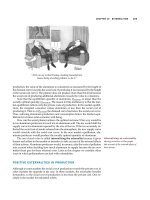
organised in three “layers”. The outermost layer – epineurium – is a thick layer of connective
tissue which ensheaths the nerve and isolates it from the external environment (Fig.1). The
vasa nervorum are continued within this layer and these vessels communicate abundantly
with the network of arterioles and venules found in the connective tissues in the depth of the
nerve. The amount of epineurium differs depending on the individual, thickness of the nerve
and location. There is an evidence that epineurium is thicker around joints (Sunderland 1978).
Deep to epineurium, the axonal fibres are organised in one (unifascicular) or more
(multifascicular) fascicles. The fascicles are enclosed within the second layer of connective
tissue – perineurium (Fig.1). The perineurium is a thick and mechanically strong layer, which
is composed of epithelium-like cells and collagen fibres. The cells are typically organised in
several layers separated by collagen with ample vascular structures running longitudinally
(Thomas and Jones 1967). This stratification gives perineurium a great endurance and ability to
withstand a pressure in excess of 200 mmHg (Selander and Sjöstrand 1978). Deep to
perineurium the endoneurium is found (Fig. 1). It consists of loose collagenous matrix
enveloping the nerve fibres and providing further protection from mechanical forces. The
endoneurium also contains several important cell types. The most abundant one are Schwann
cells, followed by fibroblasts, endothelial-like cells, macrophages and mastocytes (Causey and
Barton 1959). It is important to note that endoneurium contains ample extracellural matrix and
fluid, which is contained at a slightly higher pressure that that surrounding perineurium
(Myers et al. 1978). The reason for that is unknown, although we can speculate that it protects
Basic Principles of Peripheral Nerve Disorders
2
endoneurial space from possible contamination by toxic substances external to the epineural
space.
Fig. 1. Ultrastructure of the peripheral nerve.
(a) Toluidine blue stained transverse section through peripheral nerve of rat.
(b) Detail on thick epineurium enveloping the nerve
(c) Detail on area with peri- and endoneurium.
Pathophysiology of Peripheral Nerve Injury
3
When talking about the injury to the nervous system, it is essential to consider all parts of
this system and also end organs, which are dependent on it. Thus, this review will focus
separately on neural cells, sensory organs and muscle.
1.1 Response of the neural cells
The damage to the neural cells is the most obvious consequence of the injury to the
peripheral nerve. As mentioned above, the nerve is essentially a multi-strand cord-like
structure, which keeps the nerve fibres organised and protected from the external forces.
With the cell bodies being located in the spinal cord and dorsal root ganglia, all the injuries
to the nerves are happening at the level of cellular processes – axons. Perhaps the only
exception to this statement is roots avulsion from spinal cord, for example during brachial
plexus injury. The nerve injury divides neurons into a part, which is proximal and a part,
which is distal to the injury site. These two parts differ significantly from each other, as far
as the reaction to the injury is concerned.
1.1.1 Distal to the injury site (Wallerian degeneration)
More than 160 years have passed since the first report describing the reaction of distal nerve
stump to axotomy. The original work was performed by Augustus Waller and was
presented to the Royal Society of London in 1850. Waller was studying injuries to
glossopharyngeal and hypoglossal nerves in frogs. It is obligatory to quote an excerpt from
his original report here (Waller 1850):
“During the four first days, after section of the hypoglossal nerve, no change is observed in
its structure. On the fifth day the tubes appear more varicose than usual, and the medulla
(term used to describe axons) more irregular. About the tenth day medulla forms
disorganized, fusiform masses at intervals, and where the white substance of SCHWANN
cannot be detected. These alterations, which are most evident in the single tubules, may be
found also in the branches. After twelve or fifteen days many of the single tubules have
ceased to be visible, their granular medulla having been removed by absorption. The
branches contain masses of amorphous medulla.”
This process of disintegration of distal axonal stump after injury is termed Wallerian
degeneration. It is a recognized consequence of a mechanical (but not only) insult to the
nerve. Wallerian degeneration starts almost immediately after axotomy and lasts 3 – 6 weeks
(Geuna et al. 2009). The first sign is disintegration of axons, which starts during first 24 to 48
hours (Stoll et al. 1989). The beginning of this process is characterised by granulation within
axoplasma caused by proteolysis of microtubules and neurofilaments (Lubińska 1982,
Schlaepfer 1977). This is caused by a rapid activation of axoplasmatic proteolyses, which
occurs as a response to intracellular calcium influx (George, Glass, and Griffin 1995,
Schlaepfer and Bunge 1973). An early activation of ubiquitin-proteasome system has been
also shown to play an important role here (Ehlers 2004). Among all the cytoskeletal
structures, the microtubules are thought to disintegrate first (Watts, Hoopfer, and Luo 2003,
Zhai et al. 2003). The loss of microtubular structures then leads to impediment of axonal
transport and further accelerates the degeneration process. The disintegration of
neurofilaments follows shortly and is usually completed within 7 – 10 days. During this
time, the partially disrupted neurofilaments can be detected in the axoplasma only to
Basic Principles of Peripheral Nerve Disorders
4
completely disappear shortly afterwards. One more important point, which needs to be
made, is the direction of the Wallerian degeneration. It seems that the process is
bidirectional. It starts in the zone just below the injury and progresses distally while at the
same time starts at the distal axonal termini (Waxman 1995). Despite the very brisk initiation
of degenerative changes, the distal nerve stump preserves its excitability for a considerable
period of time. When the transacted axons are stimulated distal to the injury zone, it is often
possible to record nerve potentials for up to 10 days. Therefore, it is very important for this
period of refractory excitability to finish, before accurate estimate of the nerve injury extent
can be made by electrophysiological methods.
The processes, which we have discussed so far, were limited to the axon and its inherent
ability to degenerate after injury. To have the full picture of the Wallerian degeneration, we
also need to talk about other cells, which participate and play an integral role in it. In
particular, the role of Schwann cells and macrophages is critical for the Wallerian
degeneration to take place. The Schwann cells are very sensitive to the loss of contact with
axon. In case of dennervation, the Schwann cells change from “supportive” to “reactive”
phenotype. They stop producing myelin (LeBlanc and Poduslo 1990). The continuing
proliferation of Schwann cells leads to formation of Bands of Bungers, which purpose is
thought to be guidance of the regrowing axons (further discussed in the regeneration
subchapter) (Liu, Yang, and Yang 1995). It seems that this phenotypic switch is, at least
partly, a response to neuregulin secretion from the transacted axons (Esper and Loeb 2004).
Activated Schwann cells were found to secret a wide range of immunologically active
substances. In particular, Interleukin (IL) -1B, IL – 6, IL – 10 and Leukaemia Inhibitory
Factor (LIF) were detected abundantly at the injury site in the first few days after injury
(Bolin et al. 1995, Jander et al. 1996, Jander and Stoll 1998, Kurek et al. 1996). These
substances are responsible for attracting immune cells into the distal nerve stump and
orchestrating their function. It was shown, that in the first two days after nerve injury
macrophages and T cells start to infiltrate injury zone, which culminates in infiltration of the
entire distal stump by day 4 (Brück 1997, Perry, Brown, and Gordon 1987). They are
responsible for phagocytosis of the axonal debris and myelin sheaths residua released from
the disintegrating axons and thus finishing the breakdown and elimination of axons.
1.1.2 Proximal to the injury site (proximal end degeneration)
The immediate consequence of axotomy is partial retraction of the proximal stump (Cajal
1928) leaving empty endoneurial tubes lined by Schwann cells. The distance to which the
proximal stump retracts is usually one or two nodes of Ranvier, but that depends on
severity and character of injury. Within the same timeframe the injured axons also seal their
injured axolemma to prevent axoplasma leakage. Shortly after retraction and as early as
hours after axotomy, the proximal stump starts to produce regenerative sprouts (McQuarrie
1985, Meller 1987, Friede and Bischhausen 1980). While these sprouts are forming the cut tip
of the axon swells up, containing endoplasmatic reticulum, mitochondria and microtubules.
This swelling contains products accumulating in the tip of the stump because of disrupted
anterograde axonal transport. One important event happening in the area of the swelling is
reorganisation of microtubular cytoskeleton. In the normal axon the microtubules are
organised longitudinally and all point distally along the axon. After axotomy the
arrangement of microtubules changes and they point against each other (Erez et al. 2007).
Pathophysiology of Peripheral Nerve Injury
5
This swelling is very probably giving the basis for development of axonal end-bulbs, which
occurs within 24 – 48 hours after the injury. The relation between axonal endbulb and axonal
growth cone remains not fully understood (Goldberg, Frank, and Krayanek 1983). A recent
report suggests that depending on the local environment, the injured axons either form
regenerative growth cones or incompetent endbulbs (Kamber, Erez, and Spira 2009). The
successful formation of the growth cone is the ultimate goal of the proximal nerve stump, as
this will be the starting point of the nerve regeneration (see below).
1.1.3 Cell body response
The neurons, which axons were injured and ended up in Wallerian degeneration have lost a
substantial part of their cellular mass. Although we expect them to re-grow their lost parts
and re-establish the functional connection with their end organ, the situation is not always
so favourable. It seems, that the outcome is influenced by location of the lesion in relation to
cell body, type of neuron, physical age and local availability of trophic factors. The most
extreme outcome of nerve axotomy is cellular death of the injured neuron. The proportion of
neuronal cell death in dorsal root ganglia after sciatic nerve lesion in rodents has been
reported to be 10 – 30 % (Ygge 1989, Groves et al. 1997). The number is much lower in
motoneurons, where no significant neuronal death has been observed (Vanden Noven et al.
1993). However, the situation is dramatically different if the nerve (or ventral root) has been
avulsed from the spinal chord. In this case the motoneuronal death can be as high as 80%
(Martin, Kaiser, and A C Price 1999, Koliatsos et al. 1994).
There are several morphological changes in the surviving neurons after axotomy. The most
obvious one is chromatolysis, which is dissolution of the Nissle substance (Cotman 1978,
Kreutzberg 1995). The Nissle substance is a synonym for rough endoplasmatic reticulum
containing mRNA, which has blue and dotty appearance on haematoxylin eosin stain. It is
normally located in the centre of the neuron. The chromatolysis starts within hours of injury
and peaks from 1 – 3 weeks. It usually resolves with reinnervation and the process is more
prolonged and intensified if the distal reinnervation does not occur. The chromatolysis
seamlessly continues either to regeneration or to neuronal death (Martin, Kaiser, and Price
1999). It is not entirely understood what makes the neuron to initiate chromatolysis. It seems
that local synthesis of regulatory proteins on the axonal level and their linking to the dynein
retrograde motor are at the start of the process (Hanz and Fainzilber 2006). Another early
event after axotomy is swelling of the neuronal body and increase of nucleolar size. Later,
the nucleus is displaced under the cell membrane and if the reinnervation does not occur,
the neuron undergoes atrophy. One more important morphological change after neuronal
injury is a reduction of dendritic arborisation. This dendritic retraction leads to a decrease of
the number of synaptic connections of the injured neuron and to a functional isolation of it
(Purves 1975, Brännström, Havton, and Kellerth 1992a). There is an evidence the
motoneurones rebuild their dendritic complex following the reinnervation of target muscle
(Brännström, Havton, and Kellerth 1992b). In contrast, in permanent axotomy this does not
happen (Brännström, Havton, and Kellerth 1992a).
Apart from the morphological changes discussed so far, there is also a great shift on the
functional cellular level. After axotomy, the surviving neurons switch from signal
transmitter “program” to regenerative “program”, or as Fu and Gordon put it from
“signalling mode” to “growing mode” (Fu and T Gordon 1997). The survival of the cell and
Basic Principles of Peripheral Nerve Disorders
6
the mode switch are the first critical steps taken by the neuron towards regeneration. The
switch brings changes to protein expression levels in the way that signalling-associated
proteins become downregulated and growth-associated proteins and structural components
of the cell become upregulated. Gene expression studies have demonstrated changes in
expression patterns of hundreds of genes - the function of many is still yet to be explored
(Kubo et al. 2002, Bosse et al. 2006). There seems to be a similarity between these newly
found expression patterns and protein expression in developing neurons during
embryological development. A group of growth-associated proteins, such as GAP-43 (Skene
et al. 1986), are upregulated during the axonal growth phase up to 100 times and then their
expression drops down upon reinnervation (Karns et al. 1987, Skene et al. 1986). Also, the
expression of cytoskeletal component genes follows the developmental pattern. The
production of neurofilaments gets tuned down (Oblinger and Lasek 1988, Hoffman et al.
1987) whereas the production of tubulins steeply increases (Miller et al. 1989, Hoffman and
Cleveland 1988). Following is the recapitulation of changes in gene expression in the most
important gene categories (Navarro 2009). Upregulated genes include:
Transription factors (c-fos, c-jun, ATF3, NFkB, CREB, STAT)
Neurotrophic factors (NGF, BDNF, GDNF, FGF)
Neurotrophic receptors (Trk, Ret, P75)
Cytokines (TNFa, MCP1)
Growth associated proteins (GAP43)
And the downregulated genes are:
Neurofilaments
Neurotransmitters
Postsynaptic receptors
This is by no means an exhaustive list, but should serve only as a demonstration of the
philosophy behind gene expression alteration following nerve injury.
1.2 Response of the end organs and connective tissues
The multitude of functions that nerves fulfil is only possible because of a fine-tuned crosstalk
between the nerve and its end organs. It is important to note here, that the nerve acts merely as
an interface between the central nervous system and peripheral organs. Thus, for the nerve to
function as intended it must be connected to the end organs. The end organs must not only
function properly, but also have to effectively communicate with the nerve. After the nerve
injury this co-dependent communication circuit gets disrupted. If we look at the nerve
regeneration as a process of re-establishing this communication, we also need to consider the
end organs and their reaction to the nerve injury. This will be in discussed in this subchapter.
1.2.1 Response of muscle
Reaction of the muscle to the dennervation takes place on several levels. The dennervated
muscle changes its structure and its electrophysiological and biochemical properties. It has
not been fully explained why these changes occur. It is probably a mixture of inactivity and
loss of trophic stimuli from the neurons (Midrio 2006). The principal structural change is
atrophy of individual muscle fibres with loss of muscle weight. The weight may decrease to
Pathophysiology of Peripheral Nerve Injury
7
as low as 30% of the muscle original weight (Fu and T Gordon 1995). Under light
microscope the muscle fibres form nuclear knots, which are chains of nuclei with very little
surrounding sarcoplasm. On ultrastructural level we can detect disruption of myofibrils and
disorganisation of sarcomeres. Electrophysiological tests will show decline in Compound
Muscle Action Potential (CMAP), which normally recovers with reinnervation. During
regeneration the muscle motor units can significantly enlarge. This happens due to collateral
sprouting, where one neuron will eventually innervate a higher number of motor plates
then it did originally (Fu and T Gordon 1995). On biochemical level, the dennervated
muscles show decreased uptake of glucose, impaired binding of insulin, decrease of
intramuscular glycogen and also alteration of glycolytic enzymes (Burant et al. 1984,
Donaldson, Evans, and Harrison 1986, DuBois and Max 1983).
1.2.2 Response of sensory organs
The response of the sensory organs is much less studied and understood than that of the
muscle. A successful reinnervation of cutaneous sensory organs depends of a small subset of
Schwann cells found at the terminal ending of neural fibres. The dennervation of the
sensory organs results in the survival of these Schwann cells along with the capsular
structures of sensory organs (Dubový and Aldskogius 1996), which are thought to guide the
axonal regrowth towards their appropriate targets.
2. Axonal regeneration after peripheral nerve injury
As discussed above, the first wave of axonal sprouting occurs as soon as hours after
axotomy (Fawcett and Keynes 1990, Mira 1984). The transected axons produce a great
amount of terminal and collateral sprouts, which are progressing down the endoneurial
tube while being in close contact with the Schwann cells (Nathaniel and Pease 1963, Haftek
and Thomas 1968). This first wave of axonal sprouting is followed by a second wave about
two days later (Cajal 1928, Mira 1984, Cotman 1978). It has been observed that axons may
branch once they reach the distal stump, where one axon may give rise to several branches
(Jenq, Jenq, and Coggeshall 1987, Bray and Aguayo 1974). The early regenerating axons are
growing in the environment, which contains Schwann cells with their basal lamina,
fibroblasts, collagen, immunocompetent cells and axonal debris from degenerating axons.
The Schwann cells and their basal lamina play a crucial and indispensable role in the nerve
regeneration. It was shown that if the Schwann cells are not present in the distal stump, the
regeneration occurs very slowly. This is only thanks to a support of the Schwann cells
migrating from the proximal stump and accompanying the regenerating axons (Gulati 1988,
Hall 1986a). If the migration of the Schwann cells into the distal nerve stump is prohibited
(such as by a cytotoxic agent), the axons fail to regenerate completely (Hall 1986b). As
mentioned above, the Schwann cells react swiftly to the loss of axonal contact by
proliferation and assisting in breaking down the myelin sheaths. While multiplying, they
also migrate and align themselves into longitudinal columns called bands of Bungner
(Waxman 1995, Duce and Keen 1980, Lundborg et al. 1982). The bands of Bungner are
physical guides for regenerating axons. The axons first grow through the injury zone and
then into the bands of Bungner. In order for the regeneration outcome to achieve the pre-
injury state, the axons should ideally grow back into their corresponding columns.
However, the studies on early behaviour of regenerating axons showed that this is not
Basic Principles of Peripheral Nerve Disorders
8
happening. Axons send several regenerative sprouts, which can grow in multitude of
directions and encounter of up to 100 bands of Bungner (Witzel, Rohde, and Brushart 2005).
Some of the axons then grow into them, whereas others may grow freely into the connective
tissue of the nerve, or take an extraneural course. In this setting, the choice of final
regeneration pathway becomes only a matter of chance. This process is termed axonal
misdirection and can significantly hamper the regeneration process. If we consider a
situation where a motor fiber grows into the pathway belonging originally to a sensory
neuron, this will lead into the failure of functional restoration (Molander and Aldskogius
1992, Bodine-Fowler et al. 1997). It seemed, that there was a preferential affinity of
motoneurons to reinnervate motor pathways (Brushart 1993), although a more recent report
did not detect any differences in motor against sensory regrowth (Robinson and Madison
2004). One way to reduce the misdirection, which is fully in our hands, is a meticulous
surgical technique. It is imperative to use an operating microscope to minimise the impact of
a gross misalignment of nerve stumps.
Apart from providing a mechanical guidance for the regenerating axons, the Schwann cells are
also responsible for humoral stimulation of the neuronal outgrowth. The expression of NGF is
stimulated in Schwann cells shortly after nerve injury (Heumann 1987). This happens very
probably as a response to Interleukin-1 secretion by macrophages (Lindholm et al. 1987). Also,
the expression of Neurotrophin 3, 4, 5, 6 as well as Brain – Derived Neurotrophic factor
sharply increase (Funakoshi et al. 1993). The advancement of axons is further facilitated by
growth – promoting molecules, such as laminin and fibronectin (Baron-Van Evercooren et al.
1982, Rauvala et al. 1989). Several studies also demonstrated positive involvement of adhesion
molecules, such as neural cell adhesion molecule (NCAM), neural – glia cell adhesion
molecule (NgCAM), integrins and cadherins (Walsh and Doherty 1996, Seilheimer and
Schachner 1988, Bixby, Lilien, and Reichardt 1988, Hoffman et al. 1986).
In case of myelinated axons, myelination starts as early as eight days after the injury. The
remyelination is thought to recapitulate events from the embryonic development. The
trigger for the start of myelination is axonal radial growth and reaching a certain diameter.
In development it is around 2 µm (Armati 2007). The Schwann cells then rotate around the
axon in their endoneurial tube and form a myelin layer around a length of axon, which will
correspond to an intermodal segment. It is important to note, that there is a constant relation
of 1:1 between a number of cells and internodal segments – i.e. one internodal segment is
always myelinated by only one Schwann cell. The internodal segments tend to be shorter in
regenerated nerves, in comparison to the developing nerves (Vizoso and Young 1948,
Ghabriel and Allt 1977, Minwegen and Friede 1985). This is probably an explanation for
decreased conduction velocity in regenerated nerves (Cragg and Thomas 1964). The
information whether the myelination will occur or not is stored in the axons. The Schwann
cells have an ability to detect that and selectively myelinate appropriate axons (Aguayo et al.
1976, Weinberg and Spencer 1975).
3. Classification of nerve injuries
3.1 Seddon’s classification
Under normal circumstances, the nerves remain connected with their innervation targets
during the whole life of an individual. The most common disturbance to this status quo is a
Pathophysiology of Peripheral Nerve Injury
9
nerve damage by mechanical forces, which results in a loss of ability of the nerve to transfer
stimuli. These forces can act through compression, traction, laceration and direct injection
into the nerve. Moreover, the nerve can get damaged by thermal noxae, electric current,
radiation and metabolic disorders. As a result of the injury the CNS completely, or partially,
looses the ability to communicate with the neural end organs. The extent to which this
happens is greatly variable and depends on the degree of damage to the nerve. The first
classification of the severity of nerve injury was published by Seddon (Seddon 1943) and
was based on his extensive experience with war victims. He classified the nerve injuries to
three degrees, neuropraxia, axonotmesis and neurotmesis and defined the terms as follows:
1. Neurotmesis describes the state of a nerve in which all essential structures have been
sundered. There is not necessarily an obvious anatomical gap in the nerve; indeed, the
epineural sheath may appear to be in continuity, although the rest of the nerve at the
site of damage has been completely replaced by fibrous tissue. But the effect is the same
as if anatomical continuity had been lost. Neurotmesis is therefore of wider
applicability than division.
2. Axonotmesis—here the essential lesion is damage to the nerve fibers of such severity that
complete peripheral degeneration follows; and yet the epineurium and more intimate
supporting structures of the nerve have been so little disturbed that the internal
architecture is fairly well preserved. Recovery is spontaneous, and of good quality,
because the regenerating fibers are guided into their proper paths by their intact
sheaths.
3. Neuropraxia is used to describe those cases in which paralysis occurs in the absence of
peripheral degeneration. It is more accurate than transient block in that the paralysis is
often of considerable duration, though recovery always occurs in a shorter time than
would be required after complete Wallerian degeneration; it is invariably complete.
3.1.1 Neuropraxia
Neuropraxia is a situation where the nerve (or more commonly a segment of it) losses its
ability to propagate action potential while the structural continuity of the axons is fully
preserved. The condition is associated with segmental demyelination of the nerve fibers.
Because the degree of myelination differs depending on the type of nerve fibers, so does the
extent of functional loss and return. The motor fibers are the most susceptible and their
function is lost first and regained last, whereas pain and sympathetic fibers are the opposite
(Sunderland 1978). Typical example of this type of nerve injury is sleeping with the pressure
on the nerve, also called the “Saturday night palsy”. This type of injury usually recovers
within 12 weeks without any intervention.
3.1.2 Axonotmesis
Axonotmesis is an injury resulting in the loss of axonal continuity without any damage to
the connective tissue structures within the nerve. Full Wallerian degeneration and axonal
regrowth occur here and a Tinnel’s sign accompanies the regeneration. The recovery of
function is usually very good, although not as good as in neuropraxia. Surgical intervention
is normally not necessary.
Basic Principles of Peripheral Nerve Disorders
10
3.1.3 Neurotmesis
Damage to the neural connective tissue structures, including endoneurium, perineurium and /
or epineurium is termed neurotmesis. Again, Wallerian degeneration and axonal regrowth
occur and Tinnel’s sign is possible to elicit over the injured nerve. The regeneration process
here is hampered by axonal misdirection, loss of nerve/blood barrier and intraneural scarring.
Injuries interrupting peri- and epineurium require surgical intervention. The outcome is
generally worse than in axonotmesis. This, however, also depends on the relative location
from the innervation target and in general it is difficult to predict.
3.2 Sunderland’s classification
Early work of Sunderland brought about a much deeper understanding of the nerve
ultrastructure (Sunderland, 1947, Sunderland and Bradley 1949). This offered an explanation
for a wide variety of clinical findings and outcomes in the neurotmesis category. Natural
following of this line of thought was extension of the Seddon’s classification, which was
formalised by Sutherland (Sunderland 1978). In the new classification the types I and II
correspond to neuropraxia and axonotmesis respectively. Type III is an injury involving axons
and endoneurium while perineurial and epineurial structures are intact. Sunderland’s type IV
injury is associated with division of axon, endoneurial and perineurial structures. This is a
more significant injury, which often leads to intraneural scarring and requires surgical
intervention to ensure the best possible outcome. Finally, type V of Sunderland’s classification
is a total division of the nerve trunk where all the neuronal and connective tissue structures are
interrupted. It is important to note that in real clinical situation nerve injury is often a
combination of more than one type of injury. This mixed pattern injury has been classed as a
type VI, which was added to the original classification at a later date (Mackinnon 1988).
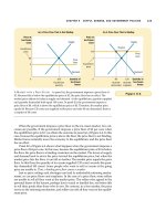
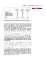
3.3 Correlations among the grade of injury, clinical and electrophysiological findings
and potential for functional recovery
The correlations are found in the following Table 1:
Seddon Neuropraxia Axonotmesis Neurotmesis Neurotmesis Neurotmesis
Sunderland T
y
pe I T
y
pe II T
y
pe III T
y
pe IV T
y
pe V
Pathological
findings
Anatomical
continuity
preserved
Selective
demyelination of
the in
j
ur
y
zone
Axonal continuity
disrupted
(together with
myelin sheath)
Axonal and
endoneurium
continuity
disrupted
Axonal,
endoneurium
and
perineurium
continuity
disrupted
Complete
division of the
nerve
Wallerian
de
g
eneration
No Yes Yes Yes Yes
Motor
paral
y
sis
Complete Complete Complete Complete Complete
Sensory
paral
y
sis
Often partially
spared
Complete Complete Complete Complete
Autonomic
paral
y
sis
Much of the
function spared
Complete Complete Complete Complete
Muscle
atroph
y
Very little Progressive with
time
Progressive
with time
Progressive
with time
Progressive
with time
Pathophysiology of Peripheral Nerve Injury
11
Seddon Neuropraxia Axonotmesis Neurotmesis Neurotmesis Neurotmesis
Sunderland T
y
pe I T
y
pe II T
y
pe III T
y
pe IV T
y
pe V
Tinnel’s si
g
n Absent Present Present Present Present
Electrophysiol
ogical findings
Normal
conduction
proximal and
distal to injury
site
No conduction
through injury
site
No fibrilation
waves
No conduction
distal to injury site
Fibrilation waves
present
No conduction
distal to injury
site
Fibrilation
waves present
No conduction
distal to injury
site
Fibrilation
waves present
No conduction
distal to injury
site
Fibrilation
waves present
Spontaneous
recover
y
Complete Complete Variable None None
Surgery
needed?
No No Varies Yes Yes
Rate of
recovery
Days (up to 3
months)
Slow – 1 mm per
day
Slow – 1 mm
per day
Only after
surgical repair
- 1 mm per day
Only after
surgical repair
- 1 mm per
da
y
Table 1. Classifications of nerve injuries and their correlation with clinical, pathological and
electrophysiological findings.
4. References
Aguayo, A J, J Epps, L Charron, and G M Bray. 1976. “Multipotentiality of Schwann cells in
cross-anastomosed and grafted myelinated and unmyelinated nerves: quantitative
microscopy and radioautography.” Brain Research 104 (1) (March 5): 1-20.
Armati, Patricia. 2007. The biology of Schwann cells : development, differentiation and
immunomodulation. Cambridge, UK: Cambridge University Press.
Baron-Van Evercooren, A, H K Kleinman, S Ohno, P Marangos, J P Schwartz, and M E
Dubois-Dalcq. 1982. “Nerve growth factor, laminin, and fibronectin promote
neurite growth in human fetal sensory ganglia cultures.” Journal of Neuroscience
Research 8 (2-3): 179-193.
Bixby, J L, J Lilien, and L F Reichardt. 1988. “Identification of the major proteins that
promote neuronal process outgrowth on Schwann cells in vitro.” The Journal of Cell
Biology 107 (1) (July): 353-361.
Bodine-Fowler, S C, R S Meyer, A Moskovitz, R Abrams, and M J Botte. 1997. “Inaccurate
projection of rat soleus motoneurons: a comparison of nerve repair techniques.”
Muscle & Nerve 20 (1) (January): 29-37.
Bolin, L M, A N Verity, J E Silver, E M Shooter, and J S Abrams. 1995. “Interleukin-6
production by Schwann cells and induction in sciatic nerve injury.” Journal of
Neurochemistry 64 (2) (February): 850-858.
Bosse, Frank, Kerstin Hasenpusch-Theil, Patrick Küry, and Hans Werner Müller. 2006.
“Gene expression profiling reveals that peripheral nerve regeneration is a
consequence of both novel injury-dependent and reactivated developmental
processes.” Journal of Neurochemistry 96 (5) (March): 1441-1457.
Brännström, T, L Havton, and J O Kellerth. 1992a. “Changes in size and dendritic
arborization patterns of adult cat spinal alpha-motoneurons following permanent
Basic Principles of Peripheral Nerve Disorders
12
axotomy.” The Journal of Comparative Neurology 318 (4) (April 22): 439-451. 1992b.
“Restorative effects of reinnervation on the size and dendritic arborization patterns
of axotomized cat spinal alpha-motoneurons.” The Journal of Comparative Neurology
318 (4) (April 22): 452-461.
Bray, G M, and A J Aguayo. 1974. “Regeneration of peripheral unmyelinated nerves. Fate of
the axonal sprouts which develop after injury.” Journal of Anatomy 117 (Pt 3) (July):
517-529.
Brück, W. 1997. “The role of macrophages in Wallerian degeneration.” Brain Pathology 7 (2)
(April): 741-752.
Brushart, T M. 1993. “Motor axons preferentially reinnervate motor pathways.” The Journal of
Neuroscience: The Official Journal of the Society for Neuroscience 13 (6) (June): 2730-2738.
Burant, C F, S K Lemmon, M K Treutelaar, and M G Buse. 1984. “Insulin resistance of
denervated rat muscle: a model for impaired receptor-function coupling.” The
American Journal of Physiology 247 (5 Pt 1) (November): E657-666.
Cajal, S. 1928. Degeneration and regeneration of the nervous system. London: Oxford University
Press.
Causey, G, and A A Barton. 1959. “The cellular content of the endoneurium of peripheral
nerve.” Brain: A Journal of Neurology 82 (December): 594-598.
Cotman, Carl. 1978. Neuronal plasticity. New York: Raven Press.
Cragg, B G, and P K Thomas. 1964. “The conduction velocity of regenerated peripheral
nerve fibers.” The Journal of Physiology 171 (May): 164-175.
Donaldson, D, O B Evans, and R W Harrison. 1986. “Insulin binding in denervated muscle.”
Muscle & Nerve 9 (3) (April): 211-215.
DuBois, D C, and S R Max. 1983. “Effect of denervation and reinnervation on oxidation of [6-
14C]glucose by rat skeletal muscle homogenates.” Journal of Neurochemistry 40 (3)
(March): 727-733.
Dubový, P, and H Aldskogius. 1996. “Degeneration and regeneration of cutaneous sensory
nerve formations.” Microscopy Research and Technique 34 (4) (July 1): 362-375.
Duce, I R, and P Keen. 1980. “The formation of axonal sprouts in organ culture and their
relationship to sprouting in vivo.” International Review of Cytology 66: 211-256.
Ehlers, Michael D. 2004. “Deconstructing the axon: Wallerian degeneration and the
ubiquitin-proteasome system.” Trends in Neurosciences 27 (1) (January): 3-6.
Erez, Hadas, Guy Malkinson, Masha Prager-Khoutorsky, Chris I De Zeeuw, Casper C
Hoogenraad, and Micha E Spira. 2007. “Formation of microtubule-based traps
controls the sorting and concentration of vesicles to restricted sites of regenerating
neurons after axotomy.” The Journal of Cell Biology 176 (4) (February 12): 497-507.
Esper, Raymond M, and Jeffrey A Loeb. 2004. “Rapid axoglial signaling mediated by
neuregulin and neurotrophic factors.” The Journal of Neuroscience 24 (27) (July 7):
6218-6227.
Fawcett, J W, and R J Keynes. 1990. “Peripheral nerve regeneration.” Annual Review of
Neuroscience 13: 43-60.
Friede, R L, and R Bischhausen. 1980. “The fine structure of stumps of transected nerve fibers
in subserial sections.” Journal of the Neurological Sciences 44 (2-3) (January): 181-203.
Fu, S Y, and T Gordon. 1995. “Contributing factors to poor functional recovery after delayed
nerve repair: prolonged denervation.”
The Journal of Neuroscience: The Official Journal
of the Society for Neuroscience 15 (5 Pt 2) (May): 3886-3895. 1997. “The cellular and
molecular basis of peripheral nerve regeneration.” Molecular Neurobiology 14 (1-2)
(April): 67-116.
Pathophysiology of Peripheral Nerve Injury
13
Funakoshi, H, J Frisén, G Barbany, T Timmusk, O Zachrisson, V M Verge, and H Persson.
1993. “Differential expression of mRNAs for neurotrophins and their receptors after
axotomy of the sciatic nerve.” The Journal of Cell Biology 123 (2) (October): 455-465.
George, E B, J D Glass, and J W Griffin. 1995. “Axotomy-induced axonal degeneration is
mediated by calcium influx through ion-specific channels.” The Journal of
Neuroscience: The Official Journal of the Society for Neuroscience 15 (10) (October): 6445-
6452.
Geuna, Stefano, Stefania Raimondo, Giulia Ronchi, Federica Di Scipio, Pierluigi Tos,
Krzysztof Czaja, and Michele Fornaro. 2009. “Chapter 3: Histology of the
peripheral nerve and changes occurring during nerve regeneration.” International
Review of Neurobiology 87: 27-46.
Ghabriel, M N, and G Allt. 1977. “Regeneration of the node of Ranvier: a light and electron
microscope study.” Acta Neuropathologica 37 (2) (February 28): 153-163.
Goldberg, S, B Frank, and S Krayanek. 1983. “Axon end-bulb swellings and rapid retrograde
degeneration after retinal lesions in young animals.” Experimental Neurology 79 (3)
(March): 753-762.
Gray, Henry. 1995. Gray’s anatomy : the anatomical basis of medicine and surgery. 38th ed. New
York: Churchill Livingstone.
Groves, M J, T Christopherson, B Giometto, and F Scaravilli. 1997. “Axotomy-induced
apoptosis in adult rat primary sensory neurons.” Journal of Neurocytology 26 (9)
(September): 615-624.
Gulati, A K. 1988. “Evaluation of acellular and cellular nerve grafts in repair of rat
peripheral nerve.” Journal of Neurosurgery 68 (1) (January): 117-123.
Haftek, J, and P K Thomas. 1968. “Electron-microscope observations on the effects of
localized crush injuries on the connective tissues of peripheral nerve.” Journal of
Anatomy 103 (Pt 2) (September): 233-243.
Hall, S M. 1986a. “Regeneration in cellular and acellular autografts in the peripheral nervous
system.” Neuropathology and Applied Neurobiology 12 (1) (February): 27-46. 1986b.
“The effect of inhibiting Schwann cell mitosis on the re-innervation of acellular
autografts in the peripheral nervous system of the mouse.” Neuropathology and
Applied Neurobiology 12 (4) (August): 401-414.
Hanz, Shlomit, and Mike Fainzilber. 2006. “Retrograde signaling in injured nerve the axon
reaction revisited.” Journal of Neurochemistry 99 (1) (October): 13-19.
Heumann, R. 1987. “Regulation of the synthesis of nerve growth factor.” The Journal of
Experimental Biology 132 (September): 133-150.
Hoffman, P N, and D W Cleveland. 1988. “Neurofilament and tubulin expression
recapitulates the developmental program during axonal regeneration: induction of
a specific beta-tubulin isotype.” Proceedings of the National Academy of Sciences of the
United States of America 85 (12) (June): 4530-4533.
Hoffman, P N, D W Cleveland, J W Griffin, P W Landes, N J Cowan, and D L Price. 1987.
“Neurofilament gene expression: a major determinant of axonal caliber.”
Proceedings of the National Academy of Sciences of the United States of America 84 (10)
(May): 3472-3476.
Hoffman, S, D R Friedlander, C M Chuong, M Grumet, and G M Edelman. 1986.
“Differential contributions of Ng-CAM and N-CAM to cell adhesion in different
neural regions.”
The Journal of Cell Biology 103 (1) (July): 145-158.
Jander, S, and G Stoll. 1998. “Differential induction of interleukin-12, interleukin-18, and
interleukin-1beta converting enzyme mRNA in experimental autoimmune
Basic Principles of Peripheral Nerve Disorders
14
encephalomyelitis of the Lewis rat.” Journal of Neuroimmunology 91 (1-2) (November
2): 93-99.
Jander, S, J Pohl, C Gillen, and G Stoll. 1996. “Differential expression of interleukin-10 mRNA
in Wallerian degeneration and immune-mediated inflammation of the rat peripheral
nervous system.” Journal of Neuroscience Research 43 (2) (January 15): 254-259.
Jenq, C B, L L Jenq, and R E Coggeshall. 1987. “Numerical patterns of axon regeneration that
follow sciatic nerve crush in the neonatal rat.” Experimental Neurology 95 (2)
(February): 492-499.
Kamber, Dotan, Hadas Erez, and Micha E Spira. 2009. “Local calcium-dependent
mechanisms determine whether a cut axonal end assembles a retarded endbulb or
competent growth cone.” Experimental Neurology 219 (1) (September): 112-125.
Karns, L R, S C Ng, J A Freeman, and M C Fishman. 1987. “Cloning of complementary DNA
for GAP-43, a neuronal growth-related protein.” Science 236 (4801) (May 1): 597-600.
Koliatsos, V E, W L Price, C A Pardo, and D L Price. 1994. “Ventral root avulsion: an
experimental model of death of adult motor neurons.” The Journal of Comparative
Neurology 342 (1) (April 1): 35-44.
Kreutzberg, G. W. 1995. Reaction of the neuronal cell body to axonal damage. S. G. Waxman, J. D.
Kocsis, and P. K. Stys. The Axon: Structure, Function and Pathophysiology. New
York: Oxford University Press.
Kubo, Tateki, Toshihide Yamashita, Atsushi Yamaguchi, Ko Hosokawa, and Masaya
Tohyama. 2002. “Analysis of genes induced in peripheral nerve after axotomy
using cDNA microarrays.” Journal of Neurochemistry 82 (5) (September): 1129-1136.
Kurek, J B, L Austin, S S Cheema, P F Bartlett, and M Murphy. 1996. “Up-regulation of
leukaemia inhibitory factor and interleukin-6 in transected sciatic nerve and muscle
following denervation.” Neuromuscular Disorders 6 (2) (March): 105-114.
LeBlanc, A C, and J F Poduslo. 1990. “Axonal modulation of myelin gene expression in the
peripheral nerve.” Journal of Neuroscience Research 26 (3) (July): 317-326.
Lindholm, D, R Heumann, M Meyer, and H Thoenen. 1987. “Interleukin-1 regulates
synthesis of nerve growth factor in non-neuronal cells of rat sciatic nerve.” Nature
330 (6149) (December 17): 658-659.
Liu, H M, L H Yang, and Y J Yang. 1995. “Schwann cell properties: 3. C-fos expression, bFGF
production, phagocytosis and proliferation during Wallerian degeneration.” Journal
of Neuropathology and Experimental Neurology 54 (4) (July): 487-496.
Lubińska, L. 1982. “Patterns of Wallerian degeneration of myelinated fibres in short and
long peripheral stumps and in isolated segments of rat phrenic nerve.
Interpretation of the role of axoplasmic flow of the trophic factor.” Brain Research
233 (2) (February 11): 227-240.
Lundborg, G, L B Dahlin, N Danielsen, H A Hansson, A Johannesson, F M Longo, and S
Varon. 1982. “Nerve regeneration across an extended gap: a neurobiological view
of nerve repair and the possible involvement of neuronotrophic factors.” The
Journal of Hand Surgery 7 (6) (November): 580-587.
Mackinnon, Susan. 1988. Surgery of the peripheral nerve. New York; Stuttgart; New York:
Thieme Medical Publishers; G. Thieme Verlag.
Martin, L J, A Kaiser, and A C Price. 1999. “Motor neuron degeneration after sciatic nerve
avulsion in adult rat evolves with oxidative stress and is apoptosis.” Journal of
Neurobiology 40 (2) (August): 185-201.
McQuarrie, I G. 1985. “Effect of conditioning lesion on axonal sprout formation at nodes of
Ranvier.” The Journal of Comparative Neurology 231 (2) (January 8): 239-249.
Pathophysiology of Peripheral Nerve Injury
15
Meller, K. 1987. “Early structural changes in the axoplasmic cytoskeleton after axotomy
studied by cryofixation.” Cell and Tissue Research 250 (3) (December): 663-672.
Midrio, Menotti. 2006. “The denervated muscle: facts and hypotheses. A historical review.”
European Journal of Applied Physiology 98 (1) (September): 1-21.
Miller, F D, W Tetzlaff, M A Bisby, J W Fawcett, and R J Milner. 1989. “Rapid induction of
the major embryonic alpha-tubulin mRNA, T alpha 1, during nerve regeneration in
adult rats.” The Journal of Neuroscience: The Official Journal of the Society for
Neuroscience 9 (4) (April): 1452-1463.
Minwegen, P, and R L Friede. 1985. “A correlative study of internode proportions and
sensitivity to procaine in regenerated frog sciatic nerves.” Experimental Neurology 87
(1) (January): 147-164.
Mira, J C. 1984. “Effects of repeated denervation on muscle reinnervation.” Clinics in Plastic
Surgery 11 (1) (January): 31-38.
Molander, C, and H Aldskogius. 1992. “Directional specificity of regenerating primary
sensory neurons after peripheral nerve crush or transection and epineurial suture A
sequential double-labeling study in the rat.” Restorative Neurology and Neuroscience 4
(5) (January 1): 339-344.
Myers, R R, H C Powell, M L Costello, P W Lampert, and B W Zweifach. 1978. “Endoneurial
fluid pressure: direct measurement with micropipettes.” Brain Research 148 (2) (June
16): 510-515.
Nathaniel, E J, and D C Pease. 1963. “Regenerative changes in rat dorsal roots following
Wallerian degeneration.” Journal of Ultrastructure Research 52 (December): 533-549.
Navarro, Xavier. 2009. “Chapter 27: Neural plasticity after nerve injury and regeneration.”
International Review of Neurobiology 87: 483-505.
Oblinger, M M, and R J Lasek. 1988. “Axotomy-induced alterations in the synthesis and
transport of neurofilaments and microtubules in dorsal root ganglion cells.” The
Journal of Neuroscience 8 (5) (May): 1747-1758.
Perry, V H, M C Brown, and S Gordon. 1987. “The macrophage response to central and
peripheral nerve injury. A possible role for macrophages in regeneration.” The
Journal of Experimental Medicine 165 (4) (April 1): 1218-1223.
Purves, D. 1975. “Functional and structural changes in mammalian sympathetic neurones
following interruption of their axons.” The Journal of Physiology 252 (2) (November):
429-463.
Rauvala, H, R Pihlaskari, J Laitinen, and J Merenmies. 1989. “Extracellular adhesive
molecules in neurite growth.” Bioscience Reports 9 (1) (February): 1-12.
Robinson, Grant A, and Roger D Madison. 2004. “Motor neurons can preferentially
reinnervate cutaneous pathways.” Experimental Neurology 190 (2) (December): 407-
413.
Schlaepfer, W W. 1977. “Structural alterations of peripheral nerve induced by the calcium
ionophore A23187.” Brain Research 136 (1) (November 4): 1-9.
Schlaepfer, W W, and R P Bunge. 1973. “Effects of calcium ion concentration on the
degeneration of amputated axons in tissue culture.” The Journal of Cell Biology 59 (2
Pt 1) (November): 456-470.
Seddon, H. J. 1943. “Three types of nerve injury.” Brain
66 (4) (December 1): 237 -288.
Seilheimer, B, and M Schachner. 1988. “Studies of adhesion molecules mediating
interactions between cells of peripheral nervous system indicate a major role for L1
in mediating sensory neuron growth on Schwann cells in culture.” The Journal of
Cell Biology 107 (1) (July): 341-351.