CURRENT BASIC AND PATHOLOGICAL APPROACHES TO THE FUNCTION OF MUSCLE CELLS AND TISSUES – FROM MOLECULES TO HUMANS pdf
Bạn đang xem bản rút gọn của tài liệu. Xem và tải ngay bản đầy đủ của tài liệu tại đây (18.99 MB, 400 trang )
CURRENT BASIC AND
PATHOLOGICAL
APPROACHES TO THE
FUNCTION OF MUSCLE
CELLS AND TISSUES – FROM
MOLECULES TO HUMANS
Edited by Haruo Sugi
Current Basic and Pathological Approaches to
the Function of Muscle Cells and Tissues – From Molecules to Humans
Edited by Haruo Sugi
Contributors
Haruo Sugi, Hiroki Minoda, Takuya Miyakawa, Suguru Tanokura, Shigeru Chaen, Takakazu
Kobayashi, Larissa Lipskaia, Isabelle Limon, Regis Bobe
,
Roger Hajjar, Ricardo Espinosa-Tanguma,
Paola Algara-Suárez, Rebeca Mejía-Elizondo, Víctor Saavedra-Alanís, Paul Fransen, Cor E. Van
Hove, Johanna van Langen, Hidde Bult, Maoxian Deng, Lixia Deng, Yarong Xue, Marcelo J.
Alfonzo, Fabiola Placeres-Uray, Walid Hassan-Soto, Adolfo Borges, Ramona González de Alfonzo,
Itala Lippo de Becemberg, Saima Salim, Sharique A. Ali, Shintaro Nakano, Toshihiro Muramatsu,
Shigeyuki Nishimura, Takaaki Senbonmatsu, Valérie Metzinger-Le Meuth, Eléonore M'Baya-
Moutoula, Fatiha Taibi, Ziad Massy, Laurent Metzinger, Ho-Chang Kuo, Wei-Chiao Chang, Hafidh
I. Al-Sadi, J.M. Ramírez, J.J. Salazar, R. de Hoz, B. Rojas, B.I. Gallego, A.I. Ramírez, A. Triviño, Carla
Máximo Prado, Edna Aparecida Leick, Fernanda Degobbi Tenório Quirino dos Santos Lopes,
Milton A. Martins, Iolanda de Fátima Lopes Calvo Tibério, Sho Shinohara, Satoko Shinohara,
Takanori Kihara, Jun Miyake, Angel Vodenicharov, Canan G. Nebigil, Shiro Mizuno, Hirohisa Toga
and Takeshi Ishizaki
Published by InTech
Janeza Trdine 9, 51000 Rijeka, Croatia
Copyright © 2012 InTech
All chapters are Open Access distributed under the Creative Commons Attribution 3.0 license,
which allows users to download, copy and build upon published articles even for commercial
purposes, as long as the author and publisher are properly credited, which ensures maximum
dissemination and a wider impact of our publications. After this work has been published by
InTech, authors have the right to republish it, in whole or part, in any publication of which they
are the author, and to make other personal use of the work. Any republication, referencing or
personal use of the work must explicitly identify the original source.
Notice
Statements and opinions expressed in the chapters are these of the individual contributors and
not necessarily those of the editors or publisher. No responsibility is accepted for the accuracy
of information contained in the published chapters. The publisher assumes no responsibility for
any damage or injury to persons or property arising out of the use of any materials,
instructions, methods or ideas contained in the book.
Publishing Process Manager Sandra Bakic
Typesetting InTech Prepress, Novi Sad
Cover InTech Design Team
First published July, 2012
Printed in Croatia
A free online edition of this book is available at www.intechopen.com
Additional hard copies can be obtained from
Current Basic and Pathological Approaches to the Function of Muscle Cells and Tissues –
From Molecules to Humans, Edited by Haruo Sugi
p. cm.
ISBN 978-953-51-0679-1
Contents
Preface IX
Section 1 Contractile and Regulatory Mechanisms of
Contraction in Skeletal, Cardiac and Smooth Muscle Cells 1
Chapter 1 The Gas Environmental Chamber as a Powerful
Tool to Study Structural Changes of Living Muscle
Thick Filaments Coupled with ATP Hydrolysis 3
Haruo Sugi, Hiroki Minoda, Takuya Miyakawa,
Suguru Tanokura, Shigeru Chaen and Takakazu Kobayashi
Chapter 2 Calcium Cycling in Synthetic and
Contractile Phasic or Tonic Vascular Smooth Muscle Cells 27
Larissa Lipskaia, Isabelle Limon, Regis Bobe
and Roger Hajjar
Chapter 3 The Role of Sodium-Calcium Exchanger in
the Calcium Homeostasis of Airway Smooth Muscle 45
Ricardo Espinosa-Tanguma, Paola Algara-Suárez,
Rebeca Mejía-Elizondo and
Víctor Saavedra-Alanís
Chapter 4 Contraction by Ca
2+
Influx via
the L-Type Ca
2+
Channel Voltage Window in
Mouse Aortic Segments is Modulated by Nitric Oxide 69
Paul Fransen, Cor E. Van Hove, Johanna van Langen and Hidde Bult
Chapter 5 MAP Kinase-Mediated and MLCK-Independent
Phosphorylation of MLC20 in Smooth Muscle Cells 93
Maoxian Deng, Lixia Deng and Yarong Xue
Chapter 6 Two Guanylylcyclases Regulate
the Muscarinic Activation of Airway Smooth Muscle 113
Marcelo J. Alfonzo, Fabiola Placeres-Uray,
Walid Hassan-Soto, Adolfo Borges,
Ramona González de Alfonzo and Itala Lippo de Becemberg
Chapter 7 Melanophores: Smooth Muscle Cells in Disguise 133
Saima Salim and Sharique A. Ali
VI Contents
Section 2 Pathological Aspects of
Cardiac and Smooth Muscle Cells 159
Chapter 8 Cardiomyocyte and Heart Failure 161
Shintaro Nakano, Toshihiro Muramatsu,
Shigeyuki Nishimura and Takaaki Senbonmatsu
Chapter 9 Implication of MicroRNAs in the Pathophysiology of
Cardiac and Vascular Smooth Muscle Cells 183
Valérie Metzinger-Le Meuth, Eléonore M'Baya-Moutoula,
Fatiha Taibi, Ziad Massy and Laurent Metzinger
Chapter 10 Cardiovascular Lesions of Kawasaki Disease:
From Genetic Study to Clinical Management 207
Ho-Chang Kuo and Wei-Chiao Chang
Chapter 11 Vascular Smooth Muscle Cells and
the Comparative Pathology of Atherosclerosis 233
Hafidh I. Al-Sadi
Chapter 12 Choroidal Vessel Wall: Hypercholesterolaemia-Induced
Dysfunction and Potential Role of Statins 255
J.M. Ramírez, J.J. Salazar, R. de Hoz,
B. Rojas, B.I. Gallego, A.I. Ramírez and A. Triviño
Section 3 Factors Influencing Structure and
Function of Smooth Muscle Cells and Tissues 299
Chapter 13 Different Modulators of Airways and
Distal Lung Parenchyma Contractile
Responses in the Physiopathology of Asthma 301
Carla Máximo Prado, Edna Aparecida Leick,
Fernanda Degobbi Tenório Quirino dos Santos Lopes,
Milton A. Martins and Iolanda de Fátima Lopes Calvo Tibério
Chapter 14 Regulation of Differentiated Phenotypes
of Vascular Smooth Muscle Cells 331
Sho Shinohara, Satoko Shinohara, Takanori Kihara and Jun Miyake
Chapter 15 Structure and Function of Smooth Muscle
with Special Reference to Mast Cells 345
Angel Vodenicharov
Chapter 16 Role of Prokineticin in Epicardial
Progenitor Cell Differentiation to Regenerate Heart 363
Canan G. Nebigil
Chapter 17 Hypoxic Pulmonary Vascular
Smooth Muscle Cell Proliferation 379
Shiro Mizuno, Hirohisa Toga and Takeshi Ishizaki
Preface
This volume consists of 17 short review articles, originally submitted to the Editor under
the theme of “Muscle Cell”. Muscles are classified into three types, skeletal, cardiac and
smooth muscles, according to their structure and function. In vertebrate animals
including humans, skeletal muscle produces body movement, cardiac muscle is
responsible for the heart function as a pump, and smooth muscle is distributed among
various visceral organs and blood vessels to keep the animals alive. In all kinds of muscle,
mechanical activity results from relative sliding of actin and myosin filaments coupled
with ATP hydrolysis, though the mechanism of the myofilament sliding still remains to
be a matter for debate and speculation. On the other hand, the mechanical activity of
muscle is controlled by changes in the intracellular concentration of free Ca
2+
ions. In
skeletal muscle, contraction is initiated by the release of Ca
2+
ions from the intracellular
membranous structure, sarcoplasmic reticulum, while in cardiac muscle contraction is
mainly coupled with influx of Ca
2+
ions from the extracellular space. In smooth muscles,
the origin of Ca
2+
ions activating contraction, i.e. activator Ca
2+
, is variable and is not yet
fully understood, reflecting the complex structure of smooth muscle tissues. Fifty years
ago, smooth muscles were sometimes called “headache muscle” because of extreme
technical difficulties in studying their function. As the readers will become aware,
considerable progress has now been achieved on the research field of smooth muscle cells
and tissues, and smooth muscles are no longer “headache muscle”.
For the sake of convenience for general readers, the book is divided into three parts
according to the subjects of articles. Part I includes articles dealing with basic aspects of
function of skeletal and smooth muscle cells, and also melanocytes which have many
properties common to those of smooth muscles. Part II contains articles dealing with
pathological aspects of cardiac and smooth muscle cell functions, while Part III consists
of articles concerning factors influencing structure and function of cardiac and smooth
muscle cells and tissues.
The Editor believes that these articles are extremely stimulating and informative for
the readers who are interested not only in the basic mechanisms of muscle cell
function, but also in the pathological and clinical aspects of muscle cells and tissues.
Dr. Haruo Sugi
Emeritus Professor, Teikyo University,
Japan
Section 1
Contractile and Regulatory Mechanisms of
Contraction in Skeletal, Cardiac and
Smooth Muscle Cells
Chapter 1
© 2012 Sugi et al., licensee InTech. This is an open access chapter distributed under the terms of the
Creative Commons Attribution License ( which permits
unrestricted use, distribution, and reproduction in any medium, provided the original work is properly cited.
The Gas Environmental Chamber
as a Powerful Tool to Study
Structural Changes of Living Muscle
Thick Filaments Coupled with ATP Hydrolysis
Haruo Sugi, Hiroki Minoda, Takuya Miyakawa,
Suguru Tanokura, Shigeru Chaen and Takakazu Kobayashi
Additional information is available at the end of the chapter
1. Introduction
The gas environmental chamber (or the hydration chamber) has been developed to observe
chemical reactions in water solutions under high magnifications with an electron micro-
scope (for an extensive review, see Buttler & Hale, 1981). The gas environmental chamber
(EC) has been widely used for in situ observation of inorganic substances in the field of
materials science. Fig.1 shows two different types of the EC. One is film-sealed EC, which is
insulated from high vacuum of electron microscope with sealing film at is upper and lower
windows to pass electron beam (Fig.1A). Water vapor (water gas) is constantly circulated
through the EC to keep the specimen in hydrated state. The other is aperture-limited EC,
which has apertures to pass electron beam without any sealing film. Water gas is constantly
injected into the EC, and sucked out of the EC to keep the specimen in hydrated state
(Fig.1B).
In the research field of medical and biological sciences, it was a dream of investigators to
observe living microorganisms moving under an electron microscope with high magnifica-
tions. In order to realize this dream, a number of attempts have hitherto been made to ob-
serve living microorganisms by means of the EC attached to an electron microscope. Such
attempts have been, however, found to be unsuccessful because the function of living mi-
croorganisms are readily impaired by electron beam irradiation. On the other hand, the
function of biological macromolecules, such as proteins and lipids, are expected to be much
more resistant against electron beam irradiation. The experiments to be described in this
chapter were started to ascertain whether the EC was useful in studying dynamic structural
Current Basic and Pathological Approaches to
the Function of Muscle Cells and Tissues – From Molecules to Humans
4
changes of biological macromolecules related to their function. After many considerations,
we decided to study molecular mechanism of muscle contraction using the EC, which was
designed and constructed to be suitable for physiological experiments to investigate dynam-
ic structural changes of hydrated muscle myosin filaments coupled with ATP hydrolysis.
Figure 1. Two types of the EC. (A) Film-sealed EC. (B) Aperture-limited EC. (Fukushima, 1988)
As explained in detail in the following sections, the greatest mystery concerning the
mechanism of muscle contraction is how the myosin heads extending from myosin
filaments convert chemical energy derived from ATP hydrolysis into mechanical work
producing force and motion in muscle. Despite extensive studies, the movement of the
myosin heads still remains as a matter of debate and speculation. The reason for the present
situation in the field of muscle research arises from the fact that the myosin head movement
has been determined only indirectly. The most straightforward way to record the myosin
head movement is to observe the myosin head movement in hydrated myosin filaments,
which retain their physiological function. In the early 1980’s, we had an opportunity to meet
Professor Fukami in Nihon University, who succeeded in preparing the carbon sealing film
for the film-sealed EC at that time and was looking for coworkers to study physiological
function of biological tissues.
The Gas Environmental Chamber as a Powerful Tool to
Study Structural Changes of Living Muscle Thick Filaments Coupled with ATP Hydrolysis
5
We started to work with Fukami’s group using the EC, manufactured by the Japan Electron
Optics Laboratory (JEOL, Ltd, Co., together with the carbon sealing film developed in
Fukami’s laboratory. After the period of trials and errors, encompassed over ten years, we
succeeded in recording the ATP-induced myosin head movement in hydrated myosin
filaments with a number of unexpected findings, which are described in this chapter.
2. The gas environmental chamber (EC)
Fig.2 is a schematic diagram of the film-sealed gas environmental chamber (EC). The EC
consists of a metal compartment (diameter, 3.5mm; depth, 0.8mm) with upper and lower
window frames (copper grids) to pass electron beam. Each window frame has nine
apertures, each having a diameter of 0.1mm. The specimen is placed on the surface of lower
sealing film, and covered by a thin layer of experimental solution by constantly circulating
water vapor through the EC. To obtain clear specimen images, the internal pressure of the
EC is made 60―80 Torr. The flow rate of water vapor is adjusted to 0.1―0.2l/min, so that
thin layer of experimental solution covering the specimen is in equilibrium with the vapor
pressure in the EC (Fukushima et al.,1985; Fukami et al.,1991). The EC was attached to a
200kV transmission electron microscope (JEM 2000EX, JEOL). (Sugi et al.,1997).
Figure 2. Diagram of the film-sealed EC. The upper and lower windows (copper grids with nine aper-
tures) are covered with carbon sealing films held on copper grids. The EC contains an ATP-containing
electrode to apply ATP to the specimen iontophoretically. The image of the specimen is recorded with
the imaging plate (IP) (Sugi et al. , 1997).
Current Basic and Pathological Approaches to
the Function of Muscle Cells and Tissues – From Molecules to Humans
6
3. Carbon sealing film
The most important element of the film-sealed EC is the carbon sealing film developed in
Fukami’s laboratory. In principle, both spatial resolution and contrast of electron
micrographs taken by the EC increases with decreasing thickness of the sealing film.
Preliminary experiments made in Fukami’s laboratory indicated that, to obtain a spatial
resolution < 1 nm, thickness of the sealing film should be 15―20nm. Meanwhile, resistivity
of a sealing film against pressure difference decreases sharply with increasing its area; the
thickness of a sealing film covering a circular aperture of 50μm diameter should be ~100nm
to bear a practical pressure difference.
Figure 3. Photomicrographs of plastic microgrides with holes of small diameters (A), with holes of
nonuniform diameters (B), and with holes of fairly uniform diameters (5―8nm)(C). (Fikushima, 1988).
As it is practically difficult to a hole < 50μm into metal wall of the EC, Fukami & Adachi
(1965) plastic microgrids made from high-molecular organic compound (cellulose
acetobutylate). Examples of microgrids are shown in Fig. 3. Microgrids with small (A) or
nonuniform holes (B) were unsuitable, while microgrids with fairly uniform holes of 5―
8nm diameters (C) were suitable for electron microscopic observation of the specimen.
Fig. 4 illustrates steps to prepare carbon sealing film by covering the microgrid with a thin
layer of carbon film (thickness, ~20nm). First, plastic microgrids prepared on a glass slide is
put onto water surface (a), where the microgrids ( having trapezoidal cross-section) are
floating with longer side dounwards (b). The position of the microgrids are inverted by
The Gas Environmental Chamber as a Powerful Tool to
Study Structural Changes of Living Muscle Thick Filaments Coupled with ATP Hydrolysis
7
means of triacetylcellurose (TAC) membrane, and again put oto water surface (c,d). The
inverted microgrids are then placed on a mica surface, and exposed to evaporated carbon
gas so that the grids are coated with thin carbon layer (e,f). The carbon sealing film prepared
on a mica surface are cut into rectangular pieces of appropriate size, and put onto water
surface (g,h,i). Finally, pieces of the carbon insulating film is placed onto the copper grid, in
such a way that each piece of the insulating film covers nine apertures of copper grid (k).
Figure 4. Diagram showing steps to prepare carbon insulating film supported by copper microgrids
(Fukushima,1988). For explanation, see text
The carbon insulating film prepared by the above method well resisted against pressure
difference up to 1 atm (Fukushima, 1981).
Current Basic and Pathological Approaches to
the Function of Muscle Cells and Tissues – From Molecules to Humans
8
4. Determination of the critical electron dose to impair function of
contractile proteins
Although biological specimens mounted in the EC can be kept in living, hydrated state ,their
function is gradually impaired by electron beam irradiation, thus giving a serious limitation
in the use of the EC for physiological experiments. Therefore, the critical incident electron
dose to impair physiological function of contractile proteins in muscle was determined in by
Suda et al. (1992). They observed muscle myofibrils, consisting of hexagonal array of actin
and myosin filaments, in the EC (magnification, 2500X), and activated them with ATP.
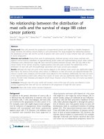
Figure 5. Relation between the total incident electron dose and the survival rate of muscle myofibrils,
expressed as percentage of myofibrils contracted in response to ATP in the microscopic field (Suda et
al.,1992). Note that contraction of myofibrils in response to ATP disappears when the electron dose
exceeds 5 x 10
-4
C/cm
2
.
The results are summarized in Fig.5. When the total incident electron dose was < 5 x 10
-
4
C/cm
2
, all the myofibrils in the electron microscopic field contracted in response to ATP. If,
however, the total incident electron dose was further increased, the ATP-induced myofibril
contraction disappeared in a nearly all-or-none manner, though the myofibrils showed no
appreciable changes in appearance.
The critical electron dose to impair physiological function of contractile proteins was con-
firmed by us with respect to both the ATP-induced myosin head movement and the ATPase
The Gas Environmental Chamber as a Powerful Tool to
Study Structural Changes of Living Muscle Thick Filaments Coupled with ATP Hydrolysis
9
activity of hydrated myosin filaments mounted in the EC. Based on these results, electron
microscopic observation and recording of the specimen was made with a total incident elec-
tron dose < 10
-4
C/cm
2
, being well below the critical dose to impair function of contractile
proteins. In order to fulfill this condition, the specimen in the EC had to be observed with
extremely weak electron beam intensities (at the fluorescent screen) < 5 x 10
-13
A/cm
2
. There-
fore, observation and focusing of the specimen required enormous skill and patience. The
electron beam intensity through the specimen under a magnification of 10,000x was 5 x 10
-
13
x (10,000)
2
= 5 x 10
-5
A/cm
2
. Immediately after the focusing of the specimen, electron beam
was stopped until the time of recording.
5. Background of experiments with the EC
Before describing our experimental results, it seems necessary to give a brief overview of the
experimental work to investigate mechanism of muscle contraction. In the middle1950s,
H.E. Huxley & Hanson (1954) made a monumental discovery that a skeletal muscle consists
of hexagonal lattice of actin and myosin filaments, and that muscle contraction results from
relative sliding between actin and myosin filaments (Fig. 6).
Figure 6. Electron micrographs of longitudinal thin section of rabbi psoas muscle myofibrils (H.E.
Huxley, 1957).
Considerable progress has been made with respect to the structure and function of actin and
myosin filaments after the discovery of sliding filament mechanism in muscle contraction.
As shown in Fig.7A, a myosin molecule is divided into two parts; (1) a long rod called light
meromyosin (LMM) and (2) the rest of myosin molecule consisting of a short rod (S2) and
two heads (S1) is called heavy meromyosin (HMM). In myosin filaments (or thick fila-
ments), LMM aggregates to form filament backbone, which is polarized in opposite direc-
tions on either side of the central part.
While the S1 heads extend laterally from the filament backbone with an axial interval of
14.3nm (Fig.7B). The central part of myosin filament is called the bare region (or bare zone),
where the projection of myosin head is absent.
Current Basic and Pathological Approaches to
the Function of Muscle Cells and Tissues – From Molecules to Humans
10
Figure 7. Ultrastructure of myosin (thick) and actin (thin) filaments and their arrangement within a
sarcomere. (A) Diagram of a myosin molecule. (B) Arrangement of myosin molecules to form a myosin
filament. (C) Arrangement of actin monomers (G-actin) in an actin filament. (D) Longitudinal arrange-
ment of actin and myosin filaments within a sarcomere. Note that the half sarcomere is the structural
and functional unit of muscle (Sugi, 1992).
On the other hand, actin filaments consist primarily of two helical strands of globular actin
monomers (G-actin) , which are wound around each other with a pitch of 35.5nm. The axial
separation of actin monomers in actin filaments is 5.46nm (Fig.7C). In vertebrate skeletal
muscle, actin filaments contain tropomyosin and troponin.
As shown in Fig.7D, actin filaments extend from the Z-line to penetrate in between myosin
filaments, which are located centrally in each sarcomere. Within a sarcomere, the region
containing only actin filaments is called the I-band, whereas the region containing myosin
filaments and part of actin filaments is called the A-band. It has been confirmed by a num-
ber of experimental methods (H.E. Huxley & Hanson,1954; Page & Huxley,1963; Wray &
Holmes,1981) that the filament lengths remain constant irrespective of whether a muscle
shortens or being stretched. Therefore, the central problem in understanding the molecular
The Gas Environmental Chamber as a Powerful Tool to
Study Structural Changes of Living Muscle Thick Filaments Coupled with ATP Hydrolysis
11
mechanism of muscle contraction is: what makes actin and myosin filaments slide past each
other? Since both actin binding site and ATPase activity are localized in the S1 heads of
myosin molecule, it is generally believed that the S1 heads, extending from myosin filament
backbone towards actin filaments, play a key role in converting chemical energy of ATP
hydrolysis into mechanical work producing force and motion in muscle.
Figure 8. Diagrams showing hypothetical attachment-detachment cycle between the myosin S1 head
extending from myosin filament and the sites on actin filament. The myosin head first attaches to actin
filament (top diagram), changes its configuration to move actin filament to the right (middle diagram),
and then detach from actin filament (bottom diagram). Axial spacing of the myosin heads on myosin
filament differs from that of the sites on actin filament, so that the attachment-detachment cycle takes
place asynchronously (H.E. Huxley,1969).
Fig.8 illustrates hypothetical attachment-detachment cycle between the S1 heads and the
corresponding sites on actin filaments. Extensive studies have been made to prove confor-
mational changes (or movement) of the myosin heads coupled with ATP during muscle
contraction. Although experimental methods used include muscle mechanics, time-resolved
X-ray diffraction, chemical probes attached to myosin heads, electron microscopy of quick
frozen muscle fibers, and nucleotide-dependent changes of myosin head crystals, no clear
conclusion has been obtained (Cooke,1986; Hibbard & Trentham,1986, Geeves & Holmes,
1999, A.F. Huxley,1998).
Thus, the myosin head movement coupled with ATP hydrolysis in muscle still remains to be
a matter for debate and speculation. The difficulties in this research field seem to arise from
the fact that numerous myosin heads undergo conformational changes asynchronously, so
that experimental data are statistical to obscure behavior of individual myosin heads. Since
the most straightforward way to study conformational changes in individual myosin heads
electron microscopically, we attempted to record ATP-induced movement of individual
Current Basic and Pathological Approaches to
the Function of Muscle Cells and Tissues – From Molecules to Humans
12
myosin head in using the EC, enabling us to keep myofilaments in hydrated, living state.
As described later, the EC has been proved to be extremely powerful tool in visualizing the
behavior of individual myosin heads under the electron microscope with high magnifica-
tions.
6. Experimental methods
In order to achieve the purpose to record movements of myosin head in hydrated myosin
filaments, the following problems in experimental technique had to be solved: (1) how to
record images of the specimen with extremely weak electron beam intensities, (2) how to
position-mark myosin heads without specimen staining used for conventional electron
microscopy; and (3) how to apply ATP to the specimen without changing its position in the
electron microscopic field. We solved these problems in the following ways.
6.1. Recording of specimen image
Based on the critical electron dose to impair function of contractile proteins (Fig.5), experi-
ments were performed under electron microscopic magnification of 10,000x, and the speci-
men images were recorded on an imaging plate (IP) system (PIX system, JEOL). The IP is
10.2 x 7.7cm in size, and has a sensitivity ~60times that of X-ray film. The exposure time was
0.18s with an electron beam intensity of 1―2 x 10
-12
A/cm
2
. The number of pixels in the IP is
~12,000,000 to give a special resolution mdose, recording of the specimen image can only be
repeated at most 4times. The IP system was developed by Fuji Photofilm Co., and is now
used worldwide not noly for transmission electron microscope, but for other purposes like
time-resolved X-ray diffraction.
6.2. Preparation of synthetic bipolar myosin filaments and position marking of
myosin heads
We decided to use synthetic thick filaments, consisting of myosin-myosin rode mixture,
prepared from rabbi psoas muscle. Myosin was prepared by the method of Perry (1955),
while myosin rod was obtained by chymotryptic digestion of myosin by the method of
Margossian & Lowey (1982). Myosin and myosin rod were mixed at a molar ratio of 1:1, and
were slowly polymerized by dialysis against a solution of low ionic strength (KCl
concentration, 120mM) to bipolar myosin filaments (1.5―3μm in length, and 50―200nm in
diameter at the center) suitable for our experiments. As shown in Fig. 9, the synthetic
filaments are spindle-shaped, and their polarity is reversed across their central region, as
judged from the direction of extension of rod part of HMM (myosin S2) from the filaments.
Though the myosin S1 heads are lost from the filaments, probably due to fixation and
staining procedures, this indicates that the synthetic filaments are bipolar in structure, being
similar to native myosin filaments in muscle.
To position-mark individual myosin heads in the hydrated myosin filaments without stain-
ing procedures, colloidal gold particles (diameter, 20nm; coated with protein A; EY labora-
The Gas Environmental Chamber as a Powerful Tool to
Study Structural Changes of Living Muscle Thick Filaments Coupled with ATP Hydrolysis
13
tories) were attached to the myosin heads, using a site directed antibody (IgG) to the junc-
tional peptide between 50- and 20-kDa segments of myosin heavy chain (Sutoh et al.,1989).
The antibody attaches to only one of the two myosin heads near its distal end facing actin
filaments. Technical details to position-mark individual myosin heads have been described
elsewhere (Sugi et al., 1997). It was essential to position-mark myosin heads sparsely, so that
each gold particle was reasonably separated from neighboring particles.
Figure 9. Conventional electron micrograph of synthetic bipolar myosin filaments. Note that the direc-
tion of extension of rod part of HMM (myosin subfragment 2) from the filaments is reversed across their
central region
6.3. Application of ATP to the specimen
To apply ATP to the specimen without causing its displacement, we used conventional glass
capillary microelectrodes containing 100mM ATP (see Fig.2). By passing current pulses
through the electrode, negatively charged ATP ions are moved out of the electrode. The
iontophoretically released ATP ions from the electrode reach to the specimen by diffusion in
the experimental solution covering the specimen. Normally, a rectangular current pulse
(intensity, 10nA; duration, 1s) from an electronic stimulator was applied to the electrode
through a current clamp circuit (Oiwa et al.,1993). Total amount of ATP released from the
microelectrode was estimated to be ~10―14mol (Oiwa et al.,1991). The time required for the
released ATP to reach the specimen by diffusion was estimated to be <30s by video record-
ing
ATP-induced shortening of myofibrils in the EC under a light microscope. Hexokinase
(50units/ml) and D-glucose (2mM) were added to the experimental solution to eliminate
contamination of ATP (Oiwa et al.,1991). In some experiments, ADP was also applied to the
specimen with similar method.
Current Basic and Pathological Approaches to
the Function of Muscle Cells and Tissues – From Molecules to Humans
14
6.4. Data analysis
Under an electron microscopic magnification of 10,000x, the pixel size on the IP is 2.5 x
2.5nm. In our experimental condition, the number of electrons reaching each pixel is esti-
mated to be at most 7―8. Each IP record of the specimen was divided into a number of
subframes, and each subframe was observed on the monitor screen of electron microscope.
Due to electron statistics, the shape of gold particle images was variable. Particles with near-
ly circular shape were selected to be used for analysis, after an appropriate binning proce-
dure, i.e. the procedure to determine each particle configuration consisting of particles with
electron counts above a certain level. Particle shapes were not markedly altered by the level
of binning.
Then, the center of mass position of each selected gold particle was determined with an
image processor (Nexus Qube System, Nexsus) in the early experiments, and with an ordi-
nary personal computer in the late experiments. The center of mass position was obtained as
the coordinates (two significant figures) within a single pixel where the center of mass posi-
tion was located, and the coordinates, representing the position of the particle, were also
taken to represent the position of the myosin head. The position of the myosin head, deter-
mined by the above method, was compared between the two IP records. The absolute coor-
dinates common to the two IP records were obtained from the position of natural markers,
i.e. bright spots on the carbon sealing film. When the center of mass position was different
between the two IP records, the distance (D) between the two center of mass positions (with
the coordinates X1 and Y
1 and X2 and Y2, respectively) was calculated as D = √(X1―X2)
2
+(Y1-Y2)
2
, and this value was taken as the amplitude of myosin head movement.
7. Experimental results and their interpretation
Prior to the experiments to be described in the following sections,we first made experiments
with the EC using myosin-paramyosin hybrid filaments, in which rabbit skeletal muscle
myosin was bound around the surface of long and thick paramyosin filaments obtained
from molluscan somatic smooth muscle, because this hybrid filaments were very easy to
handle experimentally. Although we established our experimental methods already de-
scribed in the preceding sections during the course of experiments, and succeeded in record-
ing the ATP-induced myosin head movement (Sugi et al.,1997), we do not mention the re-
sults obtained on this hybrid filaments because (1) the space available for this chapter is
limited, and (2) the results obtained from the unusual material may not attract attention of
general readers.
7.1. Stability of myosin head position in the absence of ATP
Fig.10 shows examples of spindle-shaped bipolar myosin filaments with a number of gold
particles bound to individual myosin heads. The particle image consisted of 20―50 dark
pixels with a wide range of gradation, reflecting electron statistics. We first examined
The Gas Environmental Chamber as a Powerful Tool to
Study Structural Changes of Living Muscle Thick Filaments Coupled with ATP Hydrolysis
15
whether the particle position, representing the myosin head position, was stable or changed
with time in the absence of ATP, by comparing the center of mass position of the same parti-
cle between the two IP records of the same filament, taken at an interval of 5―10min, and
then the two IP records were superimposed to detect differences in particle position.
Figure 10. (a and b) Examples of IP records of single bipolar myosin filaments with a number of gold
particles attached to individual myosin heads. (c) Enlarged view of myosin filament shown in (a) (Sugi
et al.,2008).
An example of superimposed tracings of the two IP records is presented in Fig. 11a, in
which open and filled circles of 20nm diameter are drawn around the center of mass posi-
tion of particles in the first and the second records, respectively. It was found that filled
circles in the second record are almost completely covered by open circles in the first record.
This indicates that (1) the filament stick firmly to the surface of carbon sealing film, and that
(2) the position of individual myosin heads on the filament remain almost unchanged with
time. Fig.11b is a histogram showing distribution of the distance between the center of mass
positions of particles in the first and the second records. Among 120 particles on three dif-
ferent pairs of IP records, 93 particles exhibited no significant changes in position (D <
2.5nm), while the rest 27 particles showed only small position changes (2.5nm < D > 5nm).
The stability in position of both the filament and the myosin heads in the absence of ATP pro-
vided an extremely favorable condition for recording the myosin head movement in response
to applied ATP. Although individual myosin heads are believed to continue thermal fluctua-
tion, their mean position, time-averaged over the exposure time of IP recording (0.18s), re-
mains almost unchanged with time. Since the same stability of myosin heads has also been