Pulmonary/Respiratory Therapy Secrets 3E docx
Bạn đang xem bản rút gọn của tài liệu. Xem và tải ngay bản đầy đủ của tài liệu tại đây (5.03 MB, 555 trang )
Parsons & Heffner: Pulmonary/Respiratory Therapy Secrets 3E
PDF version By m_natee
Content
I. BEDSIDE EVALUATION
1 TAKING THE PULMONARY HISTORY
2 PHYSICAL EXAMINATION
3 SMOKING CESSATION
4 PULMONARY REHABILITATION
5 PULMONARY DISABILITY EVALUATION
6 PREOPERATIVE ASSESSMENT OF THE
PULMONARY PATIENT
7 POSTOPERATIVE PULMONARY CARE
II. DIAGNOSTIC IMAGING
8 CHEST RADIOGRAPHS
9 COMPUTED TOMOGRAPHY SCANS AND ULTRASOUND
10 PULMONARY ANGIOGRAPHY AND MAGNETIC RESONANCE IMAGING OF THE CHEST
III. LABORATORY EVALUATION
11 ARTERIAL BLOOD GASES
12 PULSE OXIMETRY
13 PULMONARY FUNCTION TESTING
14 CLINICAL EXERCISE TESTING
IV. PROCEDURES
15 THORACENTESIS AND PERCUTANEOUS PLEURAL BIOPSY
16 BRONCHOSCOPY
17 INTERVENTIONAL PULMONOLOGY
18 CHEST TUBES
19 FLOW-DIRECTED PULMONARY ARTERY CATHETERS
20 MEDIASTINOSCOPY
21 THORACOSCOPY
V. AIRWAY DISEASE
22 ASTHMA
23 CHRONIC OBSTRUCTIVE LUNG DISEASE
24 CYSTIC FIBROSIS
VI. INFECTIOUS DISEASE
25 COMMUNITY-ACQUIRED PNEUMONIA
26 NOSOCOMIAL PNEUMONIA
27 ASPIRATION SYNDROMES
28 FUNGAL PNEUMONIA
29 PARASITIC INFECTIONS
30 VIRAL PNEUMONIA
31 PNEUMONIA PREVENTION
32 EMPYEMA AND LUNG ABSCESS
33 TUBERCULOSIS
34 ATYPICAL MYCOBACTERIA
VII. PULMONARY COMPLICATIONS OF AIDS
35 INFECTIOUS PULMONARY COMPLICATIONS OF HIV INFECTION
36 NONINFECTIOUS PULMONARY COMPLICATIONS OF HIV INFECTION
VIII. PULMONARY VASCULAR DISEASES
37 THROMBOEMBOLIC DISEASE
38 NONTHROMBOTIC PULMONARY EMBOLI
39 PULMONARY HYPERTENSION
IX. INTERSTITIAL LUNG DISEASES
40 GENERAL APPROACHES TO INTERSTITIAL LUNG DISEASE
41 SARCOIDOSIS
42 IDIOPATHIC PULMONARY FIBROSIS
43 COLLAGEN VASCULAR DISEASE
44 BRONCHIOLITIS, BRONCHIOLITIS OBLITERANS, AND SMALL AIRWAY DISEASE
X. VASCULITIS AND IMMUNOLOGIC DISEASES
45 SMALL VESSEL VASCULITIS: WEGENER'S GRANULOMATOSIS, MICROSCOPIC
POLYANGIITIS, AND CHURG-STRAUSS SYNDROME
46 DIFFUSE ALVEOLAR HEMORRHAGE
XI. VENTILATORY DISORDERS
47 SLEEP APNEA SYNDROMES
48 ALVEOLAR HYPOVENTILATION
XII. OCCUPATIONAL AND ENVIRONMENTAL LUNG DISEASES
49 SILICOSIS, COAL WORKERS' PNEUMOCONIOSIS, AND CHRONIC BERYLLIUM
DISEASE
50 ASBESTOS-RELATED LUNG DISEASE
51 HYPERSENSITIVITY PNEUMONITIS AND OTHER DISORDERS CAUSED BY ORGANIC
AGENTS
52 OCCUPATIONAL ASTHMA
53 DRUG-INDUCED LUNG DISEASE
54 RADIATION INJURY TO THE LUNG
55 INHALATIONAL INJURIES
XIII. LUNG NEOPLASMS
56 SOLITARY PULMONARY NODULES
57 LUNG CANCER
58 MALIGNANT PLEURAL EFFUSIONS
59 SYSTEMIC COMPLICATIONS OF LUNG CANCER
60 BENIGN NEOPLASMS OF THE LUNG
61 PULMONARY METASTATIC DISEASE
XIV. RESPIRATORY FAILURE
62 ACUTE RESPIRATORY FAILURE
63 ACUTE RESPIRATORY DISTRESS SYNDROME
64 AIRWAY MANAGEMENT
65 TRACHEOSTOMY
66 NONINVASIVE VENTILATION
67 TRADITIONAL INVASIVE VENTILATION
68 ALTERNATIVE INVASIVE VENTILATORY STRATEGIES
69 WEANING
70 CHRONIC VENTILATORY SUPPORT
XV. END-STAGE LUNG DISEASE
71 OXYGEN THERAPY
72 LUNG TRANSPLANTATION
XVI. PLEURAL DISORDERS
73 PLEURAL EFFUSIONS
74 PNEUMOTHORAX
75 MESOTHELIOMA
75 MESOTHELIOMA
XVIII. SPECIAL CONSIDERATIONS
76 PULMONARY MANIFESTATIONS OF SYSTEMIC DISEASE
I. BEDSIDE EVALUATION
1 TAKING THE PULMONARY HISTORY
Karen A. Fa
g
an MD
1. What is dyspnea and what causes it?
Dyspnea is the subjective sensation of uncomfortable or difficult breathing. Most patients report dyspnea as
"shortness of breath." Patients report dyspnea when their breathing is excessive for the activity that they are
doing. The sensation of dyspnea is produced by stimulation of both central and peripheral receptors that
monitor respiratory muscle activity, hypoxia, hypercapnia, acid-base status, airway irritation, and changes in
the pressure volume characteristics of the lung (i.e., j receptors in lung fibrosis or emphysema).
There are many systems and conditions that contribute to dyspnea, including cardiopulmonary, hematologic,
psychosocial, and environmental (e.g., high altitude) factors; body habitus (i.e., obesity); fever; and level of
exercise. Any situation that increases the work of breathing (i.e., airway obstruction or decreased lung
compliance) also contributes to the sensation of dyspnea.
2. Give the features of dyspnea that are important to distinguish in the pulmonary
and respiratory history
Onset: Acute dyspnea is readily recognized by both patient and physician. Subacute/chronic and
progressive dyspnea, however, may be more difficult to characterize. Exercise tolerance or limitation over
time may be the most useful way to establish the duration of symptoms in these
situations. The patient's report
of changes in exercise capacity over time (from months to years) may identify the onset of symptoms.
Dyspnea at rest is a late finding in respiratory disease.
Positional complaints: Platypnea, shortness of breath experienced upon assuming the upright position, is
most commonly seen in patients with hepatic disease and intrapulmonary shunts.
Orthopnea, dyspnea occurring in the supine position, is most commonly a symptom of cardiac dysfunction.
Paroxysmal nocturnal dyspnea is also a feature of many cardiac diseases. Occasionally, patients with upper
airway lesions may present with complaints of dyspnea or cough while recumbent.
Precipitants: Reliable precipitating factors leading to dyspnea include environmental or occupational
exposures, exposure to animals, and exposure to inhalational agents (industrial or recreational).
Karnani NG, Reisfield GM, Wilson GR: Evaluation of chronic dyspnea. Am Fam Physician 71(8):1529-1537,
2005.
3. What questions should be asked about a patient's smoking history?
page
11
page
12
Smoking-related lung disease is common; thus, a complete, reliable smoking history, including the following
information, is important in the initial assessment of any patient, especially a patient with pulmonary disease:
l
Age at which smoking began
l Type of tobacco used
l
Breaks in smoking history
l Amount of smoking (i.e., pack-years, or packs per day multiplied by the number of years smoked)
A physician caring for a smoking patient should assess previous attempts at smoking cessation and should
determine ways to improve the patient's success. Information should be sought about the presence of other
smokers in the patient's environment, the use of support groups, the use of pharmacologic treatments (i.e.,
nicotine replacement), and prior input from medical personnel.
4. Which features of the family history are important when assessing a patient with
respiratory complaints?
Page 1 of 5Printed from STUDENT CONSULT: Pulmonary/Respiratory Therapy Secrets 3E
12/12/2006 />There may be a hereditary component in several diseases. All patients should be asked about any respiratory
diseases or symptoms in first-degree relatives (i.e., those immediately related to the patient). Early age at
onset of emphysema may suggest a deficiency in alpha
1
antitrypsin. Cough with purulent sputum production
and recurrent infections may suggest a familial form of bronchiectasis (e.g., cystic fibrosis or Williams-
Campbell syndrome). Some patients with pulmonary fibrosis may also have
familial forms. Approximately 20%
of patients with idiopathic pulmonary arterial hypertension have an affected family member.
5. What information should be obtained from a patient who complains of cough?
Coughing is a common complaint of patients. Although cough can be a nonspecific symptom of many
diseases, a good history should begin to limit the differential diagnosis. The history includes descriptions of the
onset, quality, duration, associated expectoration, presence of other respiratory symptoms, and changes in
voice. Cough may be caused by inflammatory, chemical, mechanical, or psychosocial mechanisms.
Sputum production is a key feature of cough. Healthy adults generally do not expectorate any sputum during
the course of the day; thus, sputum production may be considered abnormal. The consistency and color of the
sputum may help identify the source because purulent sputum usually correlates with infectious causes. The
presence and quantity of blood are also important. Fetid-smelling, purulent sputum may indicate the presence
of an anaerobic infection or a lung abscess. Large quantities of sputum (bronchorrhea) can be seen in some
malignancies, bronchiectases, and inflammatory airway diseases. Thick, tenacious sputum associated with
mucous plugs can be seen in patients with cystic fibrosis and asthma (especially allergic bronchopulmonary
aspergillosis). Rarely, patients report expectoration of a chalky or stone-like object, a broncholith, which can
be associated with tuberculosis and some fungal infections.
The time of day during which the cough is worst may help identify a cause. Sinusitis or sinus drainage may
cause a nocturnal or morning cough. Similarly, gastroesophageal reflux may cause symptoms that are worse
at night or when the patient is supine. Upper airway obstruction has the same pattern. Cough after exercise
may indicate reactive airway disease. Nocturnal coughing may indicate the presence of cardiac disease,
especially when associated with paroxysmal nocturnal dyspnea. Cough that occurs during eating may indicate
the presence of a tracheoesophageal fistula.
KEY POINTS: ESSENTIALS FOR EVALUATING COMPLAINTS OF DYSPNEA AND
COUGH
1. Onset (i.e., acute, chronic, or progressive)
2. Precipitants of symptoms (e.g., environmental exposures or allergens)
3. Positional component (e.g., lying down, sitting up, or eating)
4. Sputum production (including color, consistency, and presence of blood)
page
12
page 13
A careful list of past and present medication use is important in evaluating a cough. Chronic dry coughing is
seen with the use of angiotensin-converting enzyme inhibitors in as many as 20% of patients treated with
these antihypertensive agents. Fortunately, the coughing resolves with cessation of the drug. However,
chronic dry coughing with dyspnea may also be a feature of the pulmonary fibrosing diseases; thus, the
medication history may be important in distinguishing the diagnosis.
Aspiration of foreign bodies may also produce both acute and chronic coughing; this possibility should be
considered in children with cough and in adults with a history of impaired consciousness. Hoarseness may be
associated with laryngeal sources of cough. An often-overlooked cause of chronic cough is hair or wax in the
external auditory canal causing stimulation of the vagus nerve.
Holmes RL, Fadden CT: Evaluation of the patient with chronic cough. Am Fam Physician 69(9):2159-2166,
2004.
6. Which features of an asthmatic patient's history suggest severe disease that may
require more aggressive treatment?
If a patient answers yes to any of the following questions, he or she is at increased risk of developing
respiratory failure as a result of an asthma exacerbation:
Page 2 of 5Printed from STUDENT CONSULT: Pulmonary/Respiratory Therapy Secrets 3E
12/12/2006 />l Have you required mechanical ventilation for an exacerbation in the past?
l
Have you needed to be seen in the emergency department (ED) or to be hospitalized for asthma in the
past year?
l
Have you been treated with oral corticosteroids for asthma in the past?
l Have you had an increase in the use of rescue medications (i.e., inhalers) in the past week?
l
Do you frequently wake at night due to your asthma symptoms?
7. Define hemoptysis. How are the cause and severity assessed?
Hemoptysis is the expectoration of blood with coughing. It is a manifestation of a number of different
processes. It is a frightening, occasionally life-threatening complaint that brings patients to medical attention
promptly. Most important in the patient interview is assessment of the quantity and quality of blood and the
presence of any associated symptoms. Massive hemoptysis is usually easily assessed. It is generally greater
than 600 cc in a 24-hour period and can be quite dramatic. More commonly, patients complain of lesser
quantities such as streaks, specks, or clots. It may be difficult to estimate the amount of blood based on such
reports. Use of collection containers may be the best way to establish the amount of blood produced. Other
associated symptoms, such as fullness in one side of the chest or a tickle in the airway, can occasionally
localize the side from which the bleeding is originating.
There are numerous causes of hemoptysis. The presence of associated symptoms may help form a
differential diagnosis of the cause. Sputum production, especially when purulent, may point to an infectious
cause of the hemoptysis. Weight loss and chronic cough in a patient with hemoptysis who smokes may be an
indication of malignancy. Tuberculosis may present with similar symptoms in a patient exposed to the
mycobacterium. Hemoptysis in a patient with heart disease and dyspnea while recumbent may be caused by
pulmonary edema. The presence of chest pain and acute dyspnea may suggest pulmonary embolism.
Corder R: Hemoptysis. Emerg Med Clin North Am, 1(2):421-435, 2003.
8. Can the causes of chest pain be reliably differentiated from one another?
No. Chest pain arises from several sites in the thorax and surrounding organs. Although there are features
that suggest a particular cause of chest pain, it can be frustrating to accurately establish and treat the cause of
the chest pain. History alone can rarely identify the cause of chest pain, but attention to the quality, onset,
duration, related symptoms, and precipitating and alleviating factors may help the observant historian more
carefully evaluate this serious complaint. Chest pain is usually described as pleuritic or nonpleuritic.
page 13
page 14
Pleuritic chest pain, or pain arising from the parietal surface of the pleura, usually can be distinguished easily
from other chest pain syndromes by history. It is usually sharp and relates to respiratory muscle movements
such as inspiration or coughs. It is frequently sudden in onset and may be episodic. Causes of pleuritic chest
pain include pneumonia, pleural effusion, pulmonary infarction, chest wall muscle inflammation, rib fractures,
pneumothorax, and inflammation of the pleura in systemic diseases such as systemic lupus erythematosus
and rheumatoid arthritis.
Nonpleuritic chest pain can be more difficult to characterize than pleuritic chest pain because both
pulmonary
and cardiac disease may present in similar ways. Classic anginal chest pain with pressure-like pain, radiation
to the arm and jaw with associated shortness of breath, nausea, and diaphoresis may be difficult to distinguish
from similar symptoms seen in pulmonary hypertension. Careful attention to medical history of other conditions
and risk factors for coronary artery disease may distinguish the cause of this type of pain. Other important
causes of nonpleuritic chest pain include musculoskeletal, gastroesophageal, pericardial, and aortic disorders.
Subdiaphragmatic processes can also present with referred pain to the chest through irritation of the
diaphragm and its surfaces.
9. What information should be obtained about potential environmental exposures and
occupational history?
Page 3 of 5Printed from STUDENT CONSULT: Pulmonary/Respiratory Therapy Secrets 3E
12/12/2006 />Two distinct environments may be important in evaluating a pulmonary patient: the home and the workplace.
Before a detailed history of either of these locations is undertaken, it is important to have a clear
understanding from the patient of the primary symptoms and whether they relate to a particular location or
activity.
A detailed history of potential exposures in the home encompasses the construction, the site, the furnishings,
the heating and cooling systems, any damage to the home (e.g., water damage), the presence of carpeting,
the type of linens used, and any pets. This information is of particular interest in patients with hypersensitivity
syndromes and asthma or other allergic syndromes. The presence of pets and other animals, currently or
previously, may contribute to allergic and asthmatic symptoms. Pet birds are
frequently overlooked in reporting
animals in the home, so it is important to ask about these specifically.
A detailed occupational history includes all past and current jobs, specific responsibilities at each location, and
information regarding chemicals and other hazardous materials at the workplace. It is especially important to
ascertain whether respiratory protection was worn and, if so, what type. Documented exposures should be
thoroughly reviewed. If necessary, the patient or physician may request job descriptions and material safety
data sheets from the work site. This is especially important in patients with concern for particulate-induced
lung disease or for workers with exacerbations of their respiratory symptoms in the work environment.
10. What is the most important information to obtain when a patient is being
evaluated for an abnormal chest radiograph?
KEY POINTS: ESSENTIALS FOR EVALUATING AN ABNORMAL CHEST
RADIOGRAPH
1. Obtain previous chest radiographs for comparison.
2. Evaluate for associated symptoms such as cough, weight loss, chest pain, or
fever.
3. Obtain a smoking and occupational history (e.g., exposures that may increase
the possibility of cancer, fibrosis).
page
14
page 15
The most important questions to be addressed when a patient has been referred for evaluation of an abnormal
chest radiograph are the following:
l Does the patient have any previous chest radiographs?
l
Are they available for comparison with the current films?
l Can they be obtained?
Direct comparison of prior radiographs may establish a lesion as benign or may suggest that further evaluation
is necessary.
11. A patient's wife complains that he snores and stops breathing at night and that he
falls asleep at embarrassing times during the day. What else do you want to know
about the patient?
l Does he stop snoring for brief intervals in the night? If so, how does he resume snoring?
l
Does he ever have quick, jerky limb movements while asleep?
l Does he complain of not sleeping well or of feeling very sleepy during the day?
l
Does he frequently take naps?
l Does he have headaches in the morning?
l
Has he experienced sexual dysfunction?
These questions may help characterize several sleep disorders, especially
obstructive sleep apnea, which can
affect as many as 20% of adults in the United States. Although the patient can frequently provide adequate
information to the interviewer, it is always important to obtain additional data from family and sleep partners
because the patient may have frequent awakenings that do not fully arouse him but that significantly disturb
his sleep.
Page 4 of 5Printed from STUDENT CONSULT: Pulmonary/Respiratory Therapy Secrets 3E
12/12/2006 />2
PHYSICAL EXAMINATION
Samer Saleh MD
Om P.
Sharma MD, FRCP
1. Describe the general principles underlying a successful physical examination
The physical examination of the chest should be pursued in an orderly manner through inspection, palpation,
percussion, and auscultation. The physical examination is not a routine exercise, but rather a systemic
intellectual activity that should be pursued logically and diligently.
2. Which clinical signs best indicate respiratory distress?
Rapid respiratory rate and the use of accessory muscles of respiration denote the presence of respiratory
discomfort. The rate of normal quiet respiration varies from 12-18 breaths per minute. The diaphragm and the
intercostal muscles perform respiration. Accessory muscles of respiration include the scalene muscles and the
pectorals. During their use, the nostrils flair, the alae nasi contract, and the sternomastoids elevate the
clavicles and the sternum. Large changes in intrathoracic pressure during inspiration and expiration produce
retraction of the intercostal muscles during inspiration, particularly if tracheal obstruction exists. Patients with
advanced emphysema breathe through pursed lips, a maneuver that helps to increase expiratory flow time.
3. What is the significance of paradoxical respiration?
Normal respiration is of two types, thoracic and abdominal. Thoracic respiration, performed by the upper part
of the chest, is seen in normal women, anxious subjects, patients with ascites, and patients
with diaphragmatic
paralysis. In men and young children, respiration is abdominal. During normal respiration, the diaphragm
moves down in inspiration (seen as outward movement of the abdominal viscera) and upward in expiration. In
paradoxical respiration, the diaphragm moves down in expiration and is sucked in during inspiration. This
finding represents diaphragmatic fatigue or paralysis and indicates impending respiratory arrest. In ventilated
patients, it reflects ventilator-patient dysynchrony and requires either adjustment of the
ventilator or sedation of
the patient.
4. How can inspection be useful in a patient with a chest disease?
The patient with a barrel-shaped chest whose supraclavicular spaces are retracted on inspiration clearly has
emphysema. Retraction of the lower lateral chest wall during inspiration in the same patient is a characteristic
called Hoover's sign. A tripod sign is present in patients with respiratory distress when they lean forward on
both upper extremities to help stabilize the clavicle for the action of the accessory respiratory muscles. The
presence of dilated veins on the chest wall is pathognomonic of superior vena cava syndrome. Impaired
movement of part or all of the hemithorax may result from pleural effusion, pneumothorax, pleural tumor, or
fibrosis. Gynecomastia in a man with cigarette stains on the fingers is a telltale sign of lung cancer.
Sharma O: Symptoms and signs in pulmonary medicine. Dis Mon 41:577-640, 1995.
5. Define subcutaneous emphysema
Subcutaneous emphysema is the presence of air in the subcutaneous tissues. It may be caused by the
following: air leaking from within the pleura, for example, from a pneumothorax; mediastinal air, for example,
from a ruptured esophagus; or gas-forming organisms. Subcutaneous emphysema also may be caused
iatrogenically from insertion of chest tubes and central lines.
page 16
page 17
6. What is Tietze's syndrome?
Careful palpation of the chest sometimes reveals
costochondral tenderness, often with swelling, which may be
the source of unexplained pain in the chest. The condition, also called costochondritis, may be caused by
stress or trauma to rib structures at one or more costochondral junctions.
Gilliland B: Relapsing polychondritis and other arthritides. In Braunwald E, Fauci AS, Kasper DL, et al (eds):
Harrison's Principles of Internal Medicine, 15th ed. New York, McGraw-Hill, 2001, p 2013.
Page 1 of 5Printed from STUDENT CONSULT: Pulmonary/Respiratory Therapy Secrets 3E
12/12/2006 />7. How is consolidation distinguished from pleural effusion on pulmonary
examination?
A combination of percussion and auscultatory findings distinguishes consolidation from effusion (Table 2-1).
Table 2-1. PHYSICAL FINDINGS IN PULMONARY CONSOLIDATION AND PLEURAL
Condition Inspection Palpation PercussionAuscultation
ConsolidationRespiratory rate
increased;
movements
decreased on
affected side
No mediastinal shift;
tactile (vocal) fremitus
increased
Dull Bronchial breathing;
bronchophony;
whispering pectoriloquy;
fine crepitations
Pleural
effusion
Movements
diminished
If large, mediastinum
shifted to opposite
side; tactile (i.e., vocal)
fremitus absent
Flat or stony
dull
Breath sounds absent;
sometimes bronchial
and egophonic above
level of fluid
8. Describe egobronchophony
Egobronchophony or egophony is a nasal character imparted to the spoken word because of the presence of
overtones. It is easily recognized: when a patient says "E," it sounds like "A." Egobronchophony is best heard
over an effusion. It represents the area of consolidated or collapsed lung above the effusion.
9. What are rales, crackles, or crepitations?
Crepitations sound like bursting air bubbles and indicate that secretions are present. Table 2-2 summarizes
the differences between fine and coarse crackles.
10. How is airway obstruction identified?
The presence of wheezes or rhonchi is suggestive of airway obstruction. Both are produced by the rapid flow
of air through narrowed bronchi. The walls and secretions of the bronchi vibrate between the closed and
barely open positions, similar to the way a reed vibrates in a musical instrument. Wheezes tend to be of a
higher pitch and a greater intensity than rhonchi, which have a snoring or moaning quality (Table 2-3).
page 17
page 18
Table 2-2. DIFFERENCES BETWEEN FINE AND COARSE CRACKLES
Features Fine Crackles Coarse Crackles
Sound Explosive interrupted sounds (<250
msec); higher in pitch, simulated by
rubbing a lock of hair between the fingers
Explosive interrupted sounds (<250
msec); lower in pitch, simulated by
bubbling liquid
Cause Sudden opening up of previously
collapsed alveoli and small airways
Sudden opening up of previously
collapsed bronchi and large airways;
air bubbling through secretions
Phase of
respiratory
Cycle
End inspiration Early inspiration or, often, expiration
Effect of coughDoes not clear May clear
Settings Pulmonary fibrosis, pneumonia, and heart
failure
Acute bronchitis, severe pulmonary
edema, and chronic bronchitis
Table 2-3. DIFFERENCES BETWEEN WHEEZES AND RHONCHI
Features Wheezes Rhonchi
Page 2 of 5Printed from STUDENT CONSULT: Pulmonary/Respiratory Therapy Secrets 3E
12/12/2006 />Sound Continuous (>250 msec),
high-pitched musical sound;
usually polyphonic
Continuous (>250 msec), low-pitched moaning
sound; frequently monophonic
Cause Vibration of small airways at
point of closure
Vibration of larger airways at point of closure
Phase of
respiratory
cycle
Almost always inspiratory;
occasionally expiratory
Almost always inspiratory; occasionally expiratory
Effect of
cough
May change with cough Clears, at least temporarily
Diseases Asthma or extrinsic
compression of airway by
foreign body, tumor, or
secretion
Acute bronchitis; chronic obstructive pulmonary
disease; extrinsic compression of airway; or
obstruction of the airway by foreign body,
tumor, or
secretions
11. Which findings in a patient with bronchospasm are most ominous?
A silent chest in a tired and lethargic patient with airway obstruction signifies exhaustion and impending
respiratory arrest. Previously heard wheezes disappear because airflow velocity is decreased in obstructed
airways and no sounds are produced. Such a situation requires prompt intubation and mechanical ventilation.
12. How is the severity of bronchospasm assessed?
Although respiratory rate and pulsus paradoxus are useful indicators, they are neither sensitive nor specific
enough for assessing the severity of airway obstruction. The only way to
reliably measure airway obstruction is
by measuring flow rate either by spirometry or by peak-flow meters.
page 18
page 19
13. Describe clubbing and name the five most common pulmonary causes of
clubbing
Clubbing is a bilateral, symmetric fingernail deformity, originally described by Hippocrates. When associated
with
periostitis and arthritis, this syndrome is called hypertrophic pulmonary osteoarthropathy. Pulmonary
causes of clubbing include bronchiectasis, lung abscesses, pulmonary malignancy, cystic fibrosis, and
idiopathic pulmonary fibrosis. Clubbing is not a feature of chronic bronchitis, emphysema, or bronchial asthma.
KEY POINTS: COMMON PULMONARY CAUSES OF CLUBBING
1. Lung cancer
2. Bronchiectasis
3. Lung abscesses
4. Cystic fibrosis
5. Idiopathic pulmonary fibrosis
14. What is the significance of hypertrophic pulmonary osteoarthropathy (HOA)?
Finger clubbing and HOA are different manifestations of the same disease process. HOA includes clubbing,
periosteal inflammation, and synovial effusions. The most frequent cause of HOA is lung cancer. Removal of
the cancer may result in disappearance of HOA.
Martinez-Lavin M: Hypertrophic osteoarthropathy. In Klippel JH, Dieppe PA (eds): Rheumatology. London,
Mosby, 1998, p 8.
15. What are the usual clinical signs in emphysema?
Patients with emphysema present with a relatively quiet chest that is often barrel-shaped and is diffusely
hyperresonant. Breath sounds are vesicular but significantly reduced in intensity. Adventitious sounds are
unusual unless there is concomitant bronchitis or asthma. The expiratory phase of respiration is usually
Page 3 of 5Printed from STUDENT CONSULT: Pulmonary/Respiratory Therapy Secrets 3E
12/12/2006 />prolonged.
Badgett RG, Tanaka DJ, Hung DK, et al: Can moderate chronic obstructive pulmonary disease be diagnosed
by historical and physical findings alone? Am J Med 94:188-196, 1993.
16. Which eye disorders are seen in patients with pulmonary diseases?
Episcleritis and uveitis may be seen in patients with systemic lupus erythematosus and rheumatoid arthritis.
Patients with ankylosing spondylitis and up to 25% of patients with sarcoidosis have uveitis. Optic nerve
involvement with gradual progressive visual loss is also seen in sarcoidosis. Bilateral episodic anterior uveitis
is a feature of Behçet's syndrome. It is often associated with retinal vasculitis.
Keratoconjunctivitis sicca is a feature of Sjögren's syndrome. Choroid tubercles may be seen in patients with
tuberculosis. Wegener's granulomatoses may produce lid edema, nasolacrimal duct obstruction, proptosis,
and conjunctival chemosis.
James DG, Graham E: Oculo-pulmonary syndromes. Semin Respir Med 9:380-384, 1988.
17. Describe the skin findings associated with pulmonary disease
See Table 2-4.
Sharma O: Selected pulmonary cutaneous syndromes. Semin Respir Med 9:239-246, 1988.
page 19
page 20
Table 2-4. SKIN FINDINGS IN PULMONARY DISEASE
Skin Finding Description Pulmonary Disease
Cyanosis Bluish discoloration of extremities, usually
the tips of fingers or lips
Any disease causing hypoxemia,
including cardiac disease; seen
with peripheral vascular disease.
Lupus pernio Chronic bluish granulomatous infiltration of
the nose, cheeks, ears, and, sometimes, the
lips and chin
Sarcoidosis
Erythema
nodosum
Painful red nodules occurring mainly on the
shins
Primary tuberculosis,
coccidioidomycosis, sarcoidosis,
brucellosis, Behçet's disease
Lupus vulgaris Reddish-brown, flat plaques with yellowish-
brown nodules on the head, face, neck,
arms, and legs (in descending order of
frequency); ulceration and scarring are
characteristic
Tuberculosis
Splinter
hemorrhages
Psittacosis pneumonia
Horder's spots Faint pink spots on the trunk Psittacosis pneumonia
Vesicles Clear lesions 2-5 mm in diameter Varicella pneumonia
Cutaneous
ulcers
Tularemia pneumonia
Stevens-
Johnson
syndrome
Mycoplasma pneumonia
KEY POINTS: FINDINGS ON FUNDUS EXAMINATION OF THE PULMONARY
PATIENT
1. Choroid tubercles in tuberculosis
Page 4 of 5Printed from STUDENT CONSULT: Pulmonary/Respiratory Therapy Secrets 3E
12/12/2006 />
2. Papilledema in sarcoidosis
3. Retinal vasculitis in collagen vascular disease and Behçet's disease
4. Papilledema and retinal hemorrhages in hypercapneic respiratory failure
18. What are the pulmonary changes in hepatic cirrhosis?
page 20
page 21
Pleural effusions, namely hepatic hydrothorax, can occur in up to 10% of patients with liver cirrhosis. They are
more common on the right side than on the left side and can occur in the absence of ascites. Spontaneous
bacterial empyema can complicate these effusions. Hepatopulmonary syndrome is characterized by
hypoxemia, platypnea (worsening dyspnea in the upright position), and orthodeoxia (worsening hypoxemia in
the upright position). It results from right-to-left intrapulmonary shunts. Portopulmonary hypertension is
pulmonary arterial hypertension in patients with portal venous hypertension.
deCampos J, Filho L, Werebe E, et al: Hepatic hydrothorax. Semin Resp Critical Care Med 22:665-673, 2001.
19. What is the BODE index?
The BODE index is a grading system that consists of four variables. The
B
stands for body mass index. The
O
stands for the degree of airflow obstruction, measured by the forced expiratory volume in 1 second (FEV
1
)
after a dose of albuterol. The
D
stands for dyspnea, which is measured by the modified medical research
council (MMRC) dyspnea scale. The
E
stands for exercise capacity, measured by the 6-minute walk test. The
BODE index has a score of 0-10; the higher the score, the higher the mortality. It is better than the FEV
1
at
predicting the risk of death from any cause and from respiratory causes among patients with COPD.
Celli BR, Cote CG, Marin JM, et al: The body-mass index, air flow obstruction, dyspnea and exercise capacity
index in chronic obstructive pulmonary disease. N Eng J Med 350:1005-1012, 2004.
20. What are the neurologic signs of worsening hypercapnia in a patient with COPD?
Hypoxia and acute on top of chronic hypercapnia cause many manifestations in patients with decompensated
chronic respiratory failure. Headaches, drowsiness, confusion, and coma (in late stages) can occur. Muscle
twitching, tremors, and asterixis are some of the motor signs. Papilledema can occur in up to 10% of patients
with respiratory insufficiency and reflects the raised
intracranial pressure from hypercapnia-induced cerebral
vasodilation. Flame-shaped retinal hemorrhages and distended retinal veins can also be seen on examination
of the fundus.
Jozefowicz RF: Neurologic manifestations of pulmonary disease. Neurol Clin 7:605-617, 1989.
Printed from STUDENT CONSULT: Pulmonary/Respiratory Therapy Secrets 3E (on 12 December 2006)
©
2006 Elsevier
Page 5 of 5Printed from STUDENT CONSULT: Pulmonary/Respiratory Therapy Secrets 3E
12/12/2006 />3
SMOKING CESSATION
Steven J. Kolpak MD
Thomas
D. MacKenzie MD, MSPH
1. Describe the prevalence of cigarette smoking in the United States in this century
Cigarette smoking became the most popular form of tobacco consumption in the 1920s. Per-capita cigarette
consumption rose sharply during World War II and eventually peaked in the late
1960s at over 4000 cigarettes
per capita per year. The prevalence of cigarette smoking (i.e., the percentage of the adult population who
smoke regularly) peaked at 41% and declined annually until 1990 when the prevalence reached 25%. Since
then, the decline has been much less rapid, reaching 23% in 2002. The good news is that the prevalence of
smoking among high school students has fallen to a new 12-year low after rising steadily in the 1990s.
Currently, 22% of high school students have smoked on at least 1 of the last 30 days, down from a high of
36% in 1997.
Centers for Disease Control and Prevention (CDC): Cigarette Smoking Among Adults-United States, 2002.
Morb Mortal Wkly Rep 53(20):427-431, 2004.
Centers for Disease Control and Prevention (CDC): Cigarette Smoking Among High School Students-United
States, 1991-2003. Morb Mortal Wkly Rep 53:499-501, 2004.
2. What two questions best assess a patient's level of nicotine dependence?
1. How soon after awakening do you smoke your first cigarette? (Less than 30 minutes after awakening
indicates more severe dependence.)
2. How many cigarettes do you smoke per day? (More than 25 indicates severe dependence.)
3. How do you quantify a person's smoking history?
Multiply the average number of packs smoked per day
by the number of years of smoking to get the number of
pack-years of smoking. For example, a 55-year-old woman who began smoking at age 15 and thinks she
smoked an average of 1.5 packs (30 cigarettes) per day has a 60 pack-year smoking history (1.5 packs/day ×
40 years).
4. Is smoking cessation counseling effective?
With intervention, smoking abstinence rates can be significantly increased. There is a strong dose-response
relationship between the intensity (i.e., time spent) of counseling and its effectiveness. Brief advice (<3
minutes) increases quit rates by 30%, low-intensity counseling (3-10 minutes) increases rates by 60%, and
high-intensity counseling (>10 minutes) increases rates by over 100%. Likewise, the number of sessions
included in the intervention shows a positive association with cessation rates.
5. Describe strategies used to promote smoking cessation in a clinical setting
Programs that use several modes of repeated counseling and intervention are the most effective for initial and
long-term cessation. Interventions include clinician (physician and nonphysician) individualized counseling,
telephone counseling, and group counseling. Self-help materials such as pamphlets, cassettes, and videos
work best as an adjunct to clinician advice. The use of
carbon monoxide testing and pulmonary function
testing to give feedback on parameters related to smoking can double quit rates in primary care settings.
page 22
page 23
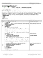
6. List the five A's of smoking cessation counseling
The U.S. Public Health Service lists five steps in the provision of office-based interventions (Fig. 3-1).
1. Ask about smoking at every opportunity: Tobacco exposure should be assessed at every office visit as
the fifth vital sign. It raises the awareness of smokers, nonsmokers, and office staff to the importance of
cessation.
2. Advise all smokers to stop: Physician advice is a powerful and inexpensive tool for smoking cessation,
Page 1 of 6Printed from STUDENT CONSULT: Pulmonary/Respiratory Therapy Secrets 3E
12/12/2006 />especially when given in a "teachable moment" such as an office visit for bronchitis or a tobacco-
related
hospitalization.
3. Assess the patient's willingness to quit: To tailor counseling to the individual patient, determine his or
her readiness to quit and interest in doing so. If the patient is not interested in quitting, provide a
motivational intervention (see question 7). Patients
interested in quitting should be offered assistance or
referred for intensive treatment.
4. Assist patients in the cessation effort: Any health care provider can assist the patient in setting a quit
date, which should be scheduled as soon after the initial counseling session as possible. Offer
recommended pharmacotherapies (first-line therapies, such as nicotine replacement systems and
sustained-release bupropion [bupropion SR], or second-line therapies, such as clonidine and
nortriptyline) to all patients unless these are specifically contraindicated.
5. Arrange follow-up: A follow-up visit or telephone call should occur shortly after the quit date, preferably
within the first week. A second follow-up is recommended within the first month, with further contact as
needed.
7. List the five R's of motivational interventions
The following components of clinical interventions are designed to enhance motivation to quit smoking in
patients who are not ready to make an attempt at quitting.
1. Relevance: Information should be provided that is relevant to the patient's sociodemographic
characteristics, disease status, health concerns, and social situation.
2. Risks: Acute, long-term, and environmental risks should be discussed with the patient.
3. Rewards: The clinician should highlight potential rewards of stopping that seem relevant to the patient.
4. Roadblocks: Barriers to quitting should be elicited. Discuss characteristics of the different treatments
that could eliminate these barriers.
5. Repetition: The motivational intervention should be repeated every time an unmotivated smoker visits
the clinic.
8. Name the typical nicotine withdrawal symptoms
l
Craving for nicotine
l Anger
l
Restlessness
l
Irritability
l Anxiety
l
Increased appetite
l Frustration
l
Difficulty concentrating
9. In the absence of treatment, how long can the symptoms of nicotine withdrawal be
expected to last?
Nicotine withdrawal symptoms begin quickly, as soon as several hours after the last cigarette. They generally
peak within the first few days and are usually minimal by 30 days. Some smokers, however, complain of
tobacco cravings for months or even years after quitting.
10. What happens to pulmonary function tests with smoking? Upon cessation?
page 23
page 24
Figure 3-1 The "Five A's" algorithm.
Page 2 of 6Printed from STUDENT CONSULT: Pulmonary/Respiratory Therapy Secrets 3E
12/12/2006 />The forced expiratory volume in 1 second (FEV
1
) has been used as the primary measure of pulmonary
function in several studies. Among all persons over the age of 45 years, the FEV
1
declines at a rate of
approximately 20 mL/yr as a natural consequence of aging. In the Lung Health Study, patients with chronic
obstructive pulmonary disease (COPD) who continued to smoke showed a steeper rate of decline in FEV
1
of
about 62 mL/yr. Patients who were able to quit successfully reduced their rate of decline to that of
nonsmokers.
Scanlon PD, Connett JE, Waller LA, et al: Smoking cessation and lung function in mild-to-moderate chronic
obstructive pulmonary disease-The Lung Health Study. Am J Respir Crit Care Med 161:381-390, 2000.
11. What are some short-term health benefits of smoking cessation?
page 24
page 25
1. The excess risk of premature coronary heart disease falls by one-half within 1 year of abstinence.
2. Some of the toxic effects of cigarette smoking that may lead to cardiac events, such as increased
platelet activation, elevated carbon monoxide levels, and coronary artery spasm, are immediately
reversible with cessation.
3. Pregnant women who stop during the first 3-4 months of
pregnancy eliminate their excess risk of having
a low-birth-weight baby.
12. What are some long-term benefits of smoking cessation?
Data from 50 years of follow-up on male British physicians suggest that
1. Men who stop smoking before age 50 cut their age-specific mortality rate in half and extend their life by
6 years compared to continuing smokers.
2. Men who quit smoking by age 30 have a similar life expectancy to those who never smoked, which is
10 years longer than that of continuing smokers.
Doll R, Peto R, Boreham J, Sutherland I: Mortality in relation to smoking: 50 years' observations on male
British doctors. BMJ 328:1519, 2004.
KEY POINTS: FIVE CANCERS CAUSALLY RELATED TO CIGARETTE SMOKING
1. Lung (all types)
2. Oral cavity (lip, tongue, mouth floor, and pharynx)
3. Laryngeal
4. Esophageal
5. Pancreatic
13. How much weight do people gain after they quit smoking?
Several studies on the effects of smoking on weight have shown that ex-smokers gain more weight over time
than nonsmokers or active smokers. The typical weight gain associated with smoking cessation ranges from
2.5-4.5 kg (5-10 lb). Women tend to gain slightly more weight than men. Genetic predisposition, younger age,
and reduced physical activity may increase the risk for weight gain.
Rigotti N: Treatment of tobacco use and dependence. N Engl J Med 346: 506-512, 2002.
14. Who should receive nicotine replacement therapy (NRT) when trying to quit
smoking?
The U.S. Public Health Service recommends that all persons who are ready to make a quit attempt, in the
absence of contraindications, should be offered NRT when trying to quit smoking.
Fiore MC, Bailey WC, Cohen SJ, et al: Treating Tobacco Use and Dependence: A Clinical Practice Guideline.
Rockville, MD, U.S. Department of Health and Human Services, 2000, pp 71-75.
Page 3 of 6Printed from STUDENT CONSULT: Pulmonary/Respiratory Therapy Secrets 3E
12/12/2006 />15. How do different types of nicotine replacement therapy work?
Nicotine gum contains nicotine bound in a gum base, which allows the nicotine to be released slowly. Once
released, the nicotine is absorbed through the buccal mucosa.
The nicotine inhalation system consists of a mouthpiece and a cartridge that contains 10 mg of nicotine.
Nicotine is released when air is inhaled through the assembled device. Each puff delivers 13 μg of
nicotine, so
80 puffs are needed to obtain the amount of nicotine found in a typical cigarette.
The nicotine nasal spray is an aqueous solution of nicotine. One spray is delivered to each nostril, and the
nicotine is rapidly absorbed through the mucous membranes. Because of its rapid absorption, the nasal spray
has been found to have more dependence potential than other forms of nicotine replacement.
page 25
page 26
The nicotine transdermal patch is composed of an adhesive base with a thin film of nicotine. The nicotine is
absorbed across the skin, giving relatively steady serum nicotine concentrations.
Nicotine lozenges, the most recently approved replacement formulation, contain nicotine in a hard base. The
slowly dissolving base allows nicotine to be absorbed through the mucous membranes of the mouth.
16. How should smokers use nicotine gum and lozenges?
Nicotine gum and lozenges are both available over the counter in 2- and 4-mg strengths. Patients should be
instructed to chew the gum on a fixed schedule, at least one piece every 1-2
hours, and to continue for at least
1-3 months. The patient should chew it slowly until a tingling sensation is felt and then should hold it between
the cheek and teeth for maximal buccal absorption. Heavy smokers (i.e., those who used more than 25
cigarettes per day) should use the 4-mg dose of gum.
For the lozenges, users should be instructed to use 1-2 lozenges every hour for 6 weeks and then to taper the
dose over the next 6 weeks. Patients should not chew or swallow the lozenge or eat or drink anything while
using it because these activities will decrease buccal absorption of nicotine.
17. How do you decide which form of NRT to use?
All forms of NRT have similar efficacy, resulting in quit rates nearly double those of placebo. Choice of NRT is
largely dictated by patient preference. Patches appear to be the easiest to use and are generally preferred by
patients. Smokers who struggle with the habitual nature of smoking behavior may prefer an inhaler, lozenges,
or gum.
18. What are the contraindications to NRT?
Although package inserts recommend caution in using nicotine products in patients with cardiovascular
disease, studies of patch use show no association between NRT and acute cardiovascular events, even in
patients who smoke intermittently while using the patch. The nicotine nasal spray should not be used in
persons with severe reactive airway disease. Pregnant and breast-feeding smokers should be urged to quit
first without any pharmacologic therapy. NRT should be offered only if the potential benefits of the increased
chance of abstinence afforded by these products outweigh their risks.
19. What is the role of bupropion SR (Zyban) in smoking cessation?
Bupropion SR is a dopamine and norepinephrine reuptake inhibitor. It is the only antidepressant approved in
the United States for smoking cessation. The Cochrane Review reports
that initial quit rates with bupropion SR
are double those of placebo. Bupropion may be more effective in heavier smokers and may also assist in
preventing relapse in successful quitters.
Hays JT, Hurt RD, Rigotti NA, et al: Sustained-release bupropion for pharmacologic relapse prevention after
smoking cessation: A randomized, controlled trial. Ann Intern Med 135(6):423-433, 2001.
Page 4 of 6Printed from STUDENT CONSULT: Pulmonary/Respiratory Therapy Secrets 3E
12/12/2006 />Hughes J, Stead L, Lancaster T: Antidepressants for smoking cessation. Cochrane Database Syst Rev (4):
CD000031, 2004.
20. How is bupropion SR prescribed?
Patients should be instructed to begin bupropion therapy 1 week before their target smoking quit date. The
recommended dosage is 150 mg per day for 3 days and then 150 mg twice daily for the duration of treatment.
There is, however, evidence that success rates are just as high in patients using just 150 mg per day and that
adverse effects may be more common with the higher dose. While smokers are encouraged to continue
bupropion for 7-12 weeks after their quit date, use for several months may improve long-term quit rates.
21. What are the contraindications to bupropion SR therapy?
page 26
page 27
KEY POINTS: FDA-APPROVED PHARMACOLOGIC THERAPIES FOR SMOKING
CESSATION
1. Bupropion SR (Zyban)
2. Nicotine gum
3. Nicotine inhalers
4. Nicotine nasal sprays
5. Nicotine patches
6. Nicotine lozenges
Bupropion SR should not be prescribed to patients who have a seizure disorder, who have a current or former
diagnosis of bulimia or anorexia nervosa, or who have used a monoamine oxidase (MAO) inhibitor within the
previous 2 weeks. Bupropion SR is a Food and Drug Administration (FDA) Class B drug in pregnancy. As with
use of nicotine replacement therapy, bupropion SR should be used only after a pregnant woman has failed to
quit without pharmacotherapy and the benefits of an increased chance of smoking cessation outweigh the
risks of using it.
22. Can combination therapies be used effectively?
1. Nicotine patch with nicotine gum or nasal spray: A meta-analysis of three studies found that
combination nicotine therapy is almost twice as effective as monotherapy. While the patient is receiving
a relatively constant amount of nicotine through the patch, he or she can adjust the dose on an acute
basis using a second agent. Combination therapy is recommended only when monotherapy has failed.
2. Nicotine patch and bupropion SR: One randomized, controlled trial comparing the nicotine patch
alone, bupropion SR alone, and a combination of bupropion SR and the patch found that the
combination is safe and significantly increases quit rates compared to the patch alone but not
compared to bupropion SR alone.
23. Are there other effective pharmacologic therapies for smoking cessation?
Two other drugs, nortriptyline and clonidine, are considered second-line therapies for tobacco dependence.
Neither is FDA approved for this indication. Patients failing first-line treatments may be candidates for either
drug.
1. Nortriptyline: Dosing ranges from 25-150 mg per day, with treatment periods from 12 weeks to 1 year.
Side effects of nortriptyline, such as dry mouth and sedation, often limit its usefulness.
2. Clonidine: An alpha
2
receptor agonist, it has been shown to nearly double the rate of successful
quitting. It is initially started at 0.1 mg twice daily, and therapy generally lasts from 1-3 months. Side
effects similar to nortriptyline can be problematic.
24. Which societal interventions have been instituted to help curb smoking?
1. Increased tobacco taxes
2. Mass-media tobacco education and counteradvertising campaigns
Page 5 of 6Printed from STUDENT CONSULT: Pulmonary/Respiratory Therapy Secrets 3E
12/12/2006 />
3. Business and workplace indoor smoking bans
4. Restricted youth access to tobacco
5. Phone "quitlines" and internet-based counseling resources for patients and healthcare providers
Printed from STUDENT CONSULT: Pulmonary/Respiratory Therapy Secrets 3E (on 12 December 2006)
©
2006 Elsevier
Page 6 of 6Printed from STUDENT CONSULT: Pulmonary/Respiratory Therapy Secrets 3E
12/12/2006 />4
PULMONARY REHABILITATION
Bonnie F. Fahy RN, MN
1. What is the definition of pulmonary rehabilitation?
Pulmonary rehabilitation was defined in 1999 by the American Thoracic Society (ATS) as "a multidisciplinary
program of care for patients with chronic respiratory impairment that is individually tailored and designed to
optimize physical and social performance and autonomy."
American Thoracic Society: ATS statement: Pulmonary rehabilitation-1999. Am J Respir Crit Care Med
159:1666-1682, 1999.
2. Who is a candidate for pulmonary rehabilitation?
Pulmonary rehabilitation should be considered for every patient with chronic lung disease, both obstructive
and restrictive, who, despite optimal medical management, has dyspnea or other respiratory symptoms,
reduced exercise tolerance, any restriction in activities because of lung disease, or impaired health status.
Global Initiative for Chronic Obstructive Lung Disease (GOLD) 2005 Guidelines for the care of the patient with
chronic obstructive pulmonary disease (COPD) recommends that all patients with GOLD Stage II (moderate),
GOLD Stage III (severe), or GOLD Stage IV (very severe) lung disease have pulmonary rehabilitation added
to their therapeutic regime.
3. Who is not a candidate for pulmonary rehabilitation?
Patients who are unable to participate (e.g., because of severe arthritis or a psychiatric disorder) or who have
an unstable concomitant condition (e.g., unstable angina) that may place them at risk are usually not
candidates for pulmonary rehabilitation.
4. Patients with pulmonary hypertension (PH) were once thought not to be
candidates for pulmonary rehabilitation. Has this thinking changed?
Yes. Since the inclusion of patients with PH in pulmonary rehabilitation programs as part of pretransplant care
has been shown to result in improved physical conditioning, patients with PH are now receiving pulmonary
rehabilitation at specialty centers with staff who have experience caring for PH patients.
American Association of Cardiovascular and Pulmonary Rehabilitation: AACVPR Guidelines for Pulmonary
Rehabilitation Programs, 3
rd
ed. Champaign, IL, Human Kinetics, 2004, pp 77-79.
5. What are the goals of pulmonary rehabilitation?
The goals of pulmonary rehabilitation are:
l Relief of symptoms
l Improvement in exercise tolerance
l
Improvement in health status
l Prevention of disease progression by avoidance of complications and exacerbations
l
Reduction in mortality
Pulmonary rehabilitation does not change lung function except for a minimal change that can be attributed to
instruction in the effective use of bronchodilators.
page 28
page
29
6. Why is it important to refer a patient to a comprehensive pulmonary rehabilitation
program rather than sending the patient to a program that provides only exercise
training?
Page 1 of 4Printed from STUDENT CONSULT: Pulmonary/Respiratory Therapy Secrets 3E
12/12/2006 />If the patient is going to invest time and energy in rehabilitation, he or she will appreciate receiving the most
comprehensive services available. Additionally, some insurance providers reimburse for pulmonary
rehabilitation only once in a lifetime, and it is unfortunate when a patient has utilized this one-time benefit on a
suboptimal program.
7. How will I know if I am referring my patient to a comprehensive pulmonary
rehabilitation program?
Question the program coordinator regarding program content or locate a program that has been nationally
certified by the American Association of Cardiovascular and Pulmonary Rehabilitation (AACVPR). An
AACVPR-certified program has been evaluated and found to have the essential standards of care for
pulmonary rehabilitation. A listing of AACVPR-certified programs is available at
8. What is meant by self-management education?
Traditional patient education, which simply provides the patient with information related to the condition and
its
therapy, is being enhanced in pulmonary rehabilitation programs by emphasis being given to self-
management
education. Self-management education teaches specific skills to manage chronic disease and to guide health
behavior modification. These skills increase self-efficacy with the goal of improving clinical outcomes including
adherence to therapies. An example of a self-management skill is early identification of an exacerbation.
KEY POINTS: ESSENTIAL COMPONENTS OF COMPREHENSIVE PULMONARY
REHABILITATION
1. Exercise training
2. Self-management education
3. Psychosocial and behavioral intervention
4. Nutritional therapy
5. Outcome assessment
6. Promotion of long-term adherence
9. What specific topics should be taught during the education sessions?
page 29
page 30
The curriculum presented must be individualized to the patient's needs, with the needs being identified during
the initial assessment. Educational topics include:
l Breathing strategies
l Normal lung function and pathophysiology of lung disease
l
Proper use of medications, including oxygen
l Bronchial hygiene techniques
l
Benefits of exercise and maintaining physical activities
l Energy conservation and work-simplification techniques
l
Eating right
l Irritant avoidance, including smoking cessation
l
Prevention and early treatment of respiratory exacerbations
l Indications for calling the health care provider
l
Leisure, travel, and sexuality
l Coping with chronic lung disease and end-of-life planning
l Anxiety and panic control, including relaxation techniques and stress management
10. Which breathing strategies should be taught in pulmonary rehabilitation?
Traditionally, the breathing strategies that are taught in pulmonary rehabilitation are pursed lip breathing and
diaphragmatic breathing. Pursed lip breathing is known to help prevent airway collapse and to reduce
respiratory rate and dyspnea while improving tidal volume and oxygen saturation. Results from the use of
diaphragmatic breathing have not been as convincing; no data from controlled studies support this breathing
Page 2 of 4Printed from STUDENT CONSULT: Pulmonary/Respiratory Therapy Secrets 3E
12/12/2006 />strategy. Despite these findings, patients find that diaphragmatic breathing, in combination with pursed lip
breathing, allows them to remain in control of their breathing instead of having their breathing control them.
11. How do you exercise a dyspneic, frightened patient?
Start slowly and offer much reassurance. The patient's baseline exercise ability is assessed by a simple
exercise tolerance test (e.g., a walk distance test). From these data, an individualized exercise prescription is
devised, emphasizing endurance rather than speed or strength. Although treadmills are the most common
exercise mode in pulmonary rehabilitation, treadmills may be intimidating to the severely limited patient.
Pushing a rollator (a walker with four large wheels) will build confidence, and transference to the treadmill will
follow. Upper extremity exercises are equally important and follow the same tenet of first building endurance
and then building strength.
12. What are the benefits of adding strength training to endurance training in
pulmonary rehabilitation?
Strength training has greater potential for increasing muscle mass and strength than endurance training.
Strength training may result in less dyspnea, thereby making this type of exercise easier to tolerate than
aerobic training in some patients. For improvements in muscle strength and endurance, a combination of
strength and endurance exercise is optimal.
Ortega F, Toral J, Cejudo P, et al: Comparison of effects of strength and endurance training in patients with
chronic obstructive pulmonary disease. Am J Respir Crit Care Med 166:669-674, 2002.
13. Is a group setting preferred over one-to-one instruction in pulmonary
rehabilitation?
KEY POINTS: SIGNIFICANT AND CLINICALLY MEANINGFUL IMPROVEMENTS
FROM PULMONARY
REHABILITATION
1. Decrease in dyspnea
2. Increase in exercise ability and functional capacity
3. Improved health status
4. Decreased healthcare utilization
page 30
page 31
Yes. The major advantage of a group setting is that it brings patients with similar problems together. "Misery
loves company" rings true. Patients who exhibit what is thought to be situational depression may improve
markedly when they find they are not the only ones with dyspnea or the need for supplemental oxygen. An
occasional patient requires referral for in-depth psychological counseling, which should be available in
rehabilitation programs. Many rehabilitation programs have ongoing maintenance exercise that serves as a
support group. Space should be available for family members to meet while patients are exercising so that
they, too, have a support system.
14. What professional disciplines are included in the pulmonary rehabilitation team?
The core pulmonary rehabilitation team includes the referring physician, the program medical director, and the
program coordinator, who will be an experienced healthcare provider such as a registered nurse, physical
therapist, or respiratory therapist. Other team members may include:
l Exercise physiologists
l
Dietitians
l Occupational therapists
l
Social workers
l Psychologists
15. Describe the cost and duration of a pulmonary rehabilitation program
Page 3 of 4Printed from STUDENT CONSULT: Pulmonary/Respiratory Therapy Secrets 3E
12/12/2006 />
The cost and duration of pulmonary rehabilitation programs vary regionally because of different policies
established by Medicare fiscal intermediaries and other insurance providers. There is no national coverage
policy for pulmonary rehabilitation, resulting in each fiscal intermediary establishing a local coverage
determination (LCD) for pulmonary rehabilitation. Generally, the cost of a program is far less than one hospital
admission. GOLD guidelines state that the optimal length of a program has not been determined, but that the
longer the program continues, the more effective the results.
Printed from STUDENT CONSULT: Pulmonary/Respiratory Therapy Secrets 3E (on 12 December 2006)
©
2006 Elsevier
Page 4 of 4Printed from STUDENT CONSULT: Pulmonary/Respiratory Therapy Secrets 3E
12/12/2006 />5
PULMONARY DISABILITY EVALUATION
Oyebode A. Taiwo MD, MPH
Carrie
A. Redlich MD, MPH
Akshay
Sood MD, MPH
1. Explain the difference between impairment and disability
Impairment means a loss of physical or physiologic function. Disability refers to the impact of the impairment
on the person's life. Impairment may occur without disability. For example, a patient with moderate
emphysema working on a word processor may have measurable impairment and little resultant disability.
Conversely, disability may occur without impairment. For example, an autobody painter with isocyanate
asthma may be disabled from work but may have no measurable impairment if removed from work when the
disease process is at an early stage. Two people with the same impairment may have different resultant
disabilities.
KEY POINTS: DIFFERENCES BETWEEN DISABILITY AND IMPAIRMENT
EVALUATION
1. Impairment means loss of physical or physiologic function.
2. Disability refers to the impact of the impairment on the person's life.
3. Impairment assessment is a medical evaluation performed by physicians.
4. Disability determination is made by administrators based on the information
provided by physicians and on criteria for eligibility.
2. What guides exist for the evaluation of pulmonary impairment and disability?
There are several guides with varying standards that often result in different ratings of pulmonary impairment
and disability. Social Security, the Department of Veterans' Affairs, and the State Workers' Compensation
Boards use different criteria. Other commonly used documents are the American Medical Association (AMA)
Guides to the Evaluation of Permanent Impairment
and the American Thoracic Society (ATS) official
statements. Physicians are encouraged to read through the latest editions of these documents before
evaluating patients under different compensation systems. Many, but not all, states use the AMA
Guides
for
determining workers' compensation. Some pulmonary diseases such as coal workers' pneumoconiosis have
their own specific criteria for impairment and disability (see question 17).
American Medical Association: Guides to the Evaluation of Permanent Impairment, 5th ed. Chicago, American
Medical Association, 2001, pp 87-115.
American Thoracic Society: Evaluation of impairment/disability secondary to respiratory disorders. Am Rev
Respir Dis 133(6):1205-1209, 1986.
Social Security Administration: Disability Evaluation under Social Security. Washington, DC, U.S. Department
of Health and Human Services, 2005, pp 24-35. Available at
Electronic Code of Federal Regulations (e-CFR) Title 38: Pensions, Bonuses, and Veterans' Relief, Volume 1
(Part 4). Schedule for Rating Disabilities: The Respiratory System 4:96-97. Available at
.
page 32
page
33
3. Explain the differences between permanent and temporary disability and between
partial and total disability
A disability may be characterized as either permanent or temporary. A permanent disability is not expected to
improve with time and treatment. On the other hand, a temporary disability is likely to be short-term and the
patient can be expected to improve to a higher level of function. In addition, a disability may be characterized
as partial or total. Total disability usually implies that an individual is unable to do any work, whereas partial
disability allows for some level of functioning in the workplace.
4. Describe the role of a physician in respiratory impairment and disability
Page 1 of 6Printed from STUDENT CONSULT: Pulmonary/Respiratory Therapy Secrets 3E
12/12/2006 />assessment
Physicians determine the presence and severity of a respiratory impairment using objective criteria. Disability
determination is made by administrators (often nonclinicians) based on the information provided by physicians
on impairment.
5. What is the Americans with Disabilities Act (ADA)?
The ADA of 1990 is the federal law that protects individuals who have a disability as defined in the ADA but
are qualified to perform the essential functions of a job, with or without reasonable accommodations. A
disability is defined by the ADA as a physical or mental impairment that
substantially
limits one or more of the
major life activities of such an individual. The ADA prohibits pre-employment medical examinations. An
employer is also prohibited from making inquiries of a job applicant as to whether he or she is an individual
with a disability or as to the nature or severity of
any such disability.
Department of Justice: Americans with Disability Act, Public Law, 1990, pp 101-336. Available at
Matson CC, Holleman WL, Nosek M, Wilkinson W: Impact of the Americans with Disabilities Act on family
physicians. J Fam Pract 36(2):201-206, 1993.
6. What is the role of a physician under the ADA?
After a conditional offer of employment, an employer may require an employment entrance examination. The
purpose of this examination is to determine if an individual can perform the essential functions of a job in a
safe manner. These examinations must be given to all individuals offered the job conditionally, regardless of
health status. The clinician performs the employment entrance examination keeping all the medical
information obtained confidential and advises the employer and nonmedical personnel on issues regarding
restrictions on the work or duties of the employee and necessary accommodations.
Matson CC, Holleman WL, Nosek M, Wilkinson W: Impact of the Americans with Disabilities Act on family
physicians. J Fam Pract 36(2):201-206, 1993.
7. What is accommodation?
Accommodation is the process of making workplace adjustments to permit a person with impairment to
continue to work. Even when impairment cannot be eliminated by medical treatment following an illness, a
physician may serve as a member of an interdisciplinary team to significantly diminish the consequent
disability. For example, modification of chemical exposures may be possible or a worker may be transferred to
another department that does not deal with a specific chemical agent or process.
8. What is workers' compensation?
page 33
page 34
Workers' compensation is a no-fault system of insurance in which insurers or employers pay benefits to an
employee with an injury or illness caused by a workplace exposure or accident. In return, the worker gives up
his or her rights to sue the employer for the injury. Workers' compensation laws vary from state to state.
Physicians play an important role in workers' compensation. They are obligated to diagnose and treat work-
related illness and to assist the patient with the documentation to file claim for benefits. Some states require
that all work-related diseases be reported to the State Department of Health.
9. How does one determine whether a respiratory disease is work related?
A detailed occupational exposure history is required to establish the diagnosis of an occupational lung
disease. This includes all employments. Exposures to specific respiratory agents in the workplace, such as
dusts, fumes, vapors, and allergens, should be noted, clarifying the timing, duration, and dose of exposure
when
possible, as well as the use of a personal respiratory protective device. Temporal associations between
exposures and symptoms should be carefully documented, including onset and changes in relationship to
work. Presence of similar complaints or diseases in coworkers is
also helpful in establishing an occupational
Page 2 of 6Printed from STUDENT CONSULT: Pulmonary/Respiratory Therapy Secrets 3E
12/12/2006 />etiology.
10. Define a preexisting respiratory condition
This refers to any impairment or disease that existed prior to the onset of another disease. For example, an
individual may have a history of preexisting childhood asthma which was made worse by workplace exposure
to respiratory irritants. A preexisting condition can be
exacerbated
if the individual temporarily became worse
and then returned to his or her baseline, or the preexisting condition can be
aggravated
if the individual
became worse but never returned to baseline (i.e., the individual now has persistence of symptoms and
requires more asthma medications). In practice, both new-onset respiratory disorders and preexisting
respiratory disorders made worse by workplace exposures are considered work-related lung diseases; thus,
both are compensable.
11. How certain do I need to be that an impairment is due to an occupational
exposure when dealing with workers' compensation cases?
The level of certainty required in determining causation for workers' compensation is different from the usual
standard of 95% certainty used in medical research. The commonly accepted standard of certainty that an
illness was caused by an occupational exposure is one of "more probable than not," or a level of certainty
greater than 50%.
12. What is the role of pulmonary function tests in evaluating lung impairment?
The most commonly used test of pulmonary function is spirometry. In obstructive airway disease, the
spirometry should be done after the administration of inhaled bronchodilators. In Crapo et al, test results are
expressed as percentage of predicted normal values, which are based on the individual's height, age, and
gender, and need to be race-corrected for African Americans. In addition, the lower limit of normal is
calculated as lying at the 5th percentile of the reference population. The AMA classification of respiratory
impairment is outlined below (Table 5-1). At least one of these measures of ventilatory function should be
abnormal to the degree described in that class if the impairment is to be rated in that class.
American Medical Association: Guides to the Evaluation of Permanent Impairment, 5th ed. Chicago, American
Medical Association, 2001, pp 87-115.
Crapo RO, Morris AH: Standardized single breath normal values for carbon monoxide diffusing capacity. Am
Rev Respir Dis 123(2):185-189, 1981.
Crapo RO, Morris AH, Gardner RM: Reference spirometric values using techniques and equipment that meet
ATS recommendations. Am Rev Respir Dis 123(6):659-664, 1981.
13. Should arterial blood gas levels be measured routinely in evaluating lung
impairment?
page 34
page 35
Table 5-1. AMERICAN MEDICAL ASSOCIATION CLASSIFICATION OF RESPIRATORY
IMPAIRMENT
Test Parameter Class 1: Class 2: Class 3: Class 4:
(As Percentage of
Predicted Normal
Value)
0-9% Impairment
of the Whole
Person
10-25%
Impairment of the
Whole Person
26-50%
Impairment of the
Whole Person
51-100%
Impairment of the
Whole Person
FVC
≥
Lower limit of
normal
≥
60 and <lower
limit of normal
51-59%
≤
50%
FEV
1
≥
Lower limit of
normal
≥
60 and <lower
limit of normal
41-59%
≤
40%
D
L
CO
≥
Lower limit of
normal
≥
60 and <lower
limit of normal
41-59%
≤
40%
Page 3 of 6Printed from STUDENT CONSULT: Pulmonary/Respiratory Therapy Secrets 3E
12/12/2006 />