APPLICATIONS OF MONTE CARLO METHOD IN SCIENCAPPLICATIONS OF MONTE CARLO METHOD IN SCIENCE AND ENGINEERING_1E AND ENGINEERING_1 doc
Bạn đang xem bản rút gọn của tài liệu. Xem và tải ngay bản đầy đủ của tài liệu tại đây (27.81 MB, 552 trang )
APPLICATIONS OF
MONTE CARLO METHOD IN
SCIENCE AND ENGINEERING
Edited by Shlomo Mark and Shaul Mordechai
Applications of Monte Carlo Method in Science and Engineering
Edited by Shlomo Mark and Shaul Mordechai
Published by InTech
Janeza Trdine 9, 51000 Rijeka, Croatia
Copyright © 2011 InTech
All chapters are Open Access articles distributed under the Creative Commons
Non Commercial Share Alike Attribution 3.0 license, which permits to copy,
distribute, transmit, and adapt the work in any medium, so long as the original
work is properly cited. After this work has been published by InTech, authors
have the right to republish it, in whole or part, in any publication of which they
are the author, and to make other personal use of the work. Any republication,
referencing or personal use of the work must explicitly identify the original source.
Statements and opinions expressed in the chapters are these of the individual contributors
and not necessarily those of the editors or publisher. No responsibility is accepted
for the accuracy of information contained in the published articles. The publisher
assumes no responsibility for any damage or injury to persons or property arising out
of the use of any materials, instructions, methods or ideas contained in the book.
Publishing Process Manager Ana Nikolic
Technical Editor Teodora Smiljanic
Cover Designer Martina Sirotic
Image Copyright Qiwen, 2010. Used under license from Shutterstock.com
First published February, 2011
Printed in India
A free online edition of this book is available at www.intechopen.com
Additional hard copies can be obtained from
Applications of Monte Carlo Method in Science and Engineering,
Edited by Shlomo Mark and Shaul Mordechai
p. cm.
ISBN 978-953-307-691-1
free online editions of InTech
Books and Journals can be found at
www.intechopen.com
Chapter 1
Chapter 2
Chapter 3
Chapter 4
Chapter 5
Chapter 6
Chapter 7
Chapter 8
Preface XI
Monte Carlo Simulations in NDT 1
Frank Sukowski and Norman Uhlmann
Application of Monte Carlo Simulation
in Optical Tweezers 21
Yu-Xuan Ren, Jian-Guang Wu and Yin-Mei Li
Enabling Grids for GATE Monte-Carlo Radiation
Therapy Simulations with the GATE-Lab 35
Sorina Camarasu-Pop, Tristan Glatard,
Hugues Benoit-Cattin and David Sarrut
Monte Carlo Simulation for Ion Implantation Profiles,
Amorphous Layer Thickness Formed by the Ion
Implantation, and Database Based on Pearson Function 51
Kunihiro Suzuki
Application of Monte Carlo Simulation
in Industrial Microbiological Exposure Assessment 83
Javier Collado, Antonio Falcó, Dolores Rodrigo,
Fernando Sampedro, M. Consuelo Pina and Antonio Martínez
Monte Carlo Simulation of Radiative Transfer
in Atmospheric Environments for Problems Arising
from Remote Sensing Measurements 95
Margherita Premuda
Monte Carlo Simulation of Pile-up Effect
in Gamma Spectroscopy 125
Ali Asghar Mowlavi, Mario de Denaro and Maria Rosa Fornasier
Monte Carlo Simulations of Microchannel
Plate–Based, Time-Gated X-ray Imagers 141
Craig A. Kruschwitz and Ming Wu
Contents
Contents
VI
Many-particle Monte Carlo Approach
to Electron Transport 167
G. Albareda, F. L. Traversa, A. Benali and X. Oriols
Monte-Carlo Simulation
in Electron Microscopy and Spectroscopy 195
Vladimír Starý
Monte Carlo Simulation of SEM and SAM Images 231
Y.G. Li, S.F. Mao and Z.J. Ding
Monte Carlo Simulation of Insulating Gas
Avalanche Development 297
Dengming Xiao
Monte Carlo Simulation of Electron Dynamics in Doped
Semiconductors Driven by Electric Fields: Harmonic
Generation, Hot-Carrier Noise and Spin Relaxation 331
Dominique Persano Adorno
A Pearson Effective Potential for Monte-Carlo Simulation
of Quantum Confinement Effects in nMOSFETs 359
Marie-Anne Jaud, Sylvain Barraud, Philippe Dollfus,
Jérôme Saint-Martin, Arnaud Bournel and Hervé Jaouen
Monte Carlo Device Simulations 385
Dragica Vasileska, Katerina Raleva and Stephen M. Goodnick
Wang-Landau Algorithm and its Implementation
for the Determination of Joint Density
of States in Continuous Spin Models 431
Soumen Kumar Roy, Kisor Mukhopadhyay,
Nababrata Ghoshal and Shyamal Bhar
Characterizing Molecular Rotations
using Monte Carlo Simulations 451
Bart Verberck
Finite-time Scaling and its Applications
to Continuous Phase Transitions 469
Fan Zhong
Using Monte Carlo Method to Study
Magnetic Properties of Frozen Ferrofluid 495
Tran Nguyen Lan and Tran Hoang Hai
Monte Carlo Studies of Magnetic Nanoparticles 513
K. Trohidou and M. Vasilakaki
Chapter 9
Chapter 10
Chapter 11
Chapter 12
Chapter 13
Chapter 14
Chapter 15
Chapter 16
Chapter 17
Chapter 18
Chapter 19
Chapter 20
Contents
VII
Monte Carlo Simulation for Magnetic
Domain Structure and Hysteresis Properties 539
Katsuhiko Yamaguchi, Kenji Suzuki and Osamu Nittono
Monte Carlo Simulations of Grain Growth
in Polycrystalline Materials Using Potts Model 563
Miroslav Morháč and Eva Morháčová
Monte Carlo Simulations of Grain Growth in Metals 581
Sven K. Esche
Monte Carlo Simulations on Defects
in Hard-Sphere Crystals Under Gravity 611
Atsushi Mori
Atomistic Monte Carlo Simulations in Steelmaking:
High Temperature Carburization
and Decarburization of Molten Steel 629
R. Khanna, R. Mahjoub and V. Sahajwalla
GCMC Simulations of Gas Adsorption
in Carbon Pore Structures 653
Maria Konstantakou, Anastasios Gotzias, Michael Kainourgiakis,
Athanasios K. Stubos and Theodore A. Steriotis
Effect of the Repulsive Interactions on the Nucleation
and Island Growth: Kinetic Monte Carlo Simulations 677
Hu Juanmei and Wu Fengmin
Monte Carlo Methodology for Grand Canonical
Simulations of Vacancies at Crystalline Defects 687
Döme Tanguy
Frequency-Dependent Monte Carlo Simulations
of Phonon Transport in Nanostructures 707
Qing Hao and Gang Chen
Performance Analysis of Adaptive GPS Signal Detection
in Urban Interference Environment
using the Monte Carlo Approach 735
V. Behar, Ch. Kabakchiev, I. Garvanov and H. Rohling
Practical Monte Carlo Based Reliability Analysis
and Design Methods for Geotechnical Problems 757
Jianye Ching
A Monte Carlo Framework to Simulate Multicomponent
Droplet Growth by Stochastic Coalescence 781
Lester Alfonso, Graciela Raga and Darrel Baumgardner
Chapter 21
Chapter 22
Chapter 23
Chapter 24
Chapter 25
Chapter 26
Chapter 27
Chapter 28
Chapter 29
Chapter 30
Chapter 31
Chapter 32
Contents
VIII
Monte Carlo Simulation
of Room Temperature Ballistic Nanodevices 803
Ignacio Íñiguez-de-la-Torre, Tomás González,
Helena Rodilla, Beatriz G. Vasallo and Javier Mateos
Estimation of Optical Properties
in Postharvest and Processing Technology 829
László Baranyai
MATLAB Programming of Polymerization
Processes using Monte Carlo Techniques 841
Mamdouh A. Al-Harthi
Monte Carlo Simulations in Solar Radio Astronomy 857
G. Thejappa and R. J. MacDowall
Using Monte Carlo Simulation
for Prediction of Tool Life 881
Sayyad Zahid Qamar, Anwar Khalil Sheikh,
Tasneem Pervez and Abul Fazal M. Arif
Loss of Load Expectation Assessment in Electricity Markets
using Monte Carlo Simulation and Neuro-Fuzzy Systems 901
H. Haroonabadi
Automating First- and Second-order Monte Carlo
Simulations for Markov Models in TreeAge Pro 917
Benjamin P. Geisler
Monte Carlo Simulations
of Adsorbed Molecules on Ionic Surfaces 931
Abdulwahab Khalil Sallabi
Chapter 33
Chapter 34
Chapter 35
Chapter 36
Chapter 37
Chapter 38
Chapter 39
Chapter 40
Pref ac e
Monte Carlo simulation, the iterative computational method used to examine and in-
vestigate the behavior of physical and mathematical systems utilizing stochastic tech-
niques. It is a widely used method and a successful statistical tool in studying a broad
array of problems, areas and cases in which it is infeasible or impossible to compute
exact results utilizing deterministic algorithms.
The Monte Carlo method has proven to be a very useful statistical sampling computa-
tional technique in a aining approximate numerical solutions to system and quantita-
tive problems which are complex, nonlinear, involve uncertain parameters, and that
are otherwise too complicated to solve analytically. In such areas of problem solving,
when compared to other methods of analysis, Monte Carlo approaches are known to
be the most accurate. Historically, however, because of the relatively large amount of
computational time required, these techniques were considered fairly burdensome.
Nowadays, as a result of the ever-increasing computing power, as well as the increas-
ing availability of distributed resources, these computations can be substantially
accelerated.
In today’s world, with the wide prevalence of novel programming languages and tools,
the rapid growth of computing power and the availability of ever more advanced and
powerful hardware, the need for increasingly complex and powerful computational so-
lutions such as Monte Carlo simulation and applications is growing exponentially. The
utilization of Monte Carlo methods, simulations and applications, is found in widely
disparate fi elds and areas of application such as nuclear physics, reliability, networks,
fi nance and business, engineering, economics, risk analysis, project management, the
study of heat transfer, molecular dynamic analysis, environmental sciences, chemistry,
telecommunications, engineering, games and so forth.
In this book, Applications of Monte Carlo Method in Science and Engineering, we
further expose the broad range of applications of Monte Carlo simulation in the
fi elds of Quantum Physics, Statistical Physics, Reliability, Medical Physics, Polycrys-
talline Materials, Ising Model, Chemistry, Agriculture, Food Processing, X-ray Im-
aging, Electron Dynamics in Doped Semiconductors, Metallurgy, Remote Sensing
XII
Preface
and much more diverse topics. The book chapters included in this volume clearly
refl ect the current scientifi c importance of Monte Carlo techniques in various fi elds
of research.
Shlomo Mark
Negev Monte Carlo Research Center and Department of So ware Engineering,
SCE - Sami Shamoon College of Engineering, Bialik/Basel Sts. Beer Sheva 84100,
Israel
Shaul Mordechai
Department of Physics and the Cancer Research Center,
Ben-Gurion University (BGU), Beer-Sheva, 84105,
Israel
1. Introduction
X-ray techniques are commonly used in the fields of non-destructive testing (NDT)
of industrial parts, material characterization, security and examination of various other
specimens. The most used techniques for obtaining images are radioscopy for 2D and
computed tomography (CT) for 3D imaging. Apart from these two imaging techniques,
where X-ray radiation penetrates matter, other methods like refraction or fluorescence analysis
can also be used to obtain information about objects and materials. The vast diversity of
possible specimen and examination tasks makes the development of universal X-ray devices
impossible. It rather is necessary to develop and optimize X-ray machines for a specific task or
at least for a limited range of tasks. The most important parameters that can be derived from
object geometry and material composition are the X-ray energy or spectrum, the dimensions,
the examination geometries and the size of the detector. The task itself demands a certain
image quality which depends also on the X-ray spectrum, the examination geometry and
furthermore on the size of the X-ray source’s focal spot and the resolution of the detector.
Monte-Carlo (MC) simulations are a powerful tool to optimize an X-ray machine and its key
components. The most important components are the radiation source, e.g. an X-ray tube and
the detector. MC particle physics simulation codes like EGS (Nelson et al., 1985) or GEANT
(Agostinelli et al., 2003) can describe all interactions of particles with matter in an X-ray
environment very well. Almost all effects can be derived from these particle physics processes.
The MC codes are event based. Every single primary particle is generated and tracked along
with all secondary particles until the energy of all particles drops below a certain threshold.
The primaries are generated one after another, since no interactions between particles take
place.
When simulating X-ray sources, in most cases X-ray tubes, the primary particles are electrons.
The electron beam is parameterized by the electrons’ kinetic energy and the intensity profile
along the cross-section of the beam. When hitting the target, X-rays are generated by
interaction of electrons with the medium. The relevant magnitudes for imaging are the X-ray
energy spectrum and the effective optical focal spot size (Morneburg, 1995).
The most used imaging systems in the field of NDT are flat panel detectors. There are two
basic types of detectors: Direct converting semiconductor detectors and indirect converting
scintillation detectors. The type of particle interactions in the respective sensor layer
determines the detection efficiency and effective spatial resolution. Interaction of X-rays in
direct converting detectors produces electron-hole-pairs in the semiconductor materials. The
free charge carriers drift to electrodes, where the current can be measured. MC simulations can
Monte Carlo Simulations in NDT
Frank Sukowski and Norman Uhlmann
Fraunhofer Institute for Integrated Circuits IIS, Development Center X-ray Technology
(EZRT)
Germany
1
describe the X-ray absorption and scattering as well as the electron drift which leads to image
blurring. Measuring X-rays with scintillation detectors works differently. X-rays interact in
the scintillation layer and produce visible photons, which are detected in a CCD or CMOS
chip. In addition to X-ray scattering and electron drift the diffusion of the visible photons
in the scintillation layer contributes greatly to image blurring (Beutel et al., 2000). In any
case, a thicker sensor layer improves the detection efficiency on the one hand, which leads
to shorter measurement times, but decreases the spatial resolution on the other hand. Finding
the optimal trade-off between efficiency and resolution by designing detector properties is an
excellent task for MC simulations.
Another application field of MC simulations are feasibility studies for special examination
tasks in order to evaluate physical limits of different imaging methods. These studies are not
limited to radioscopic methods, but include other ways to obtain information about specimens
like refractive, diffractive and backscatter imaging as well as fluorescence analysis and many
more.
In this chapter MC applications aimed at the optimization of X-ray setups for specific tasks
and feasibility studies are introduced.
The used Monte-Carlo code is called ROSI (ROentgen SImulation), which was developed by
J. Giersch and A. Weidemann at the University of Erlangen (Giersch et al., 2003). Is is an
object oriented programm code and the simulation runs can be parallelized in a computer
network for largely increasing the performance. It is based on the particle physics codes EGS4
for general electromagnetic particle interactions and LSCAT for low energy processes.
2. Simulation of X-ray sources
2.1 X-ray source characteristics in NDT imaging
In common X-ray tubes, radiation is produced by accellerating electrons via a potential
difference between the cathode (the electron emitter) and the anode (the X-ray target). When
the electrons hit the target, they are decelerated hard by collisions with electrons of the
target material or in the coulomb fields of atomic cores. X-ray radiation is produced in two
different processes. Since electrons are charged, acceleration or in this case deceleration can
cause emission of photons. The energy of these photons corresponds to the electrons’ energy
loss during the deceleration process, so the maximum possible energy corresponds to the
acceleration voltage (E
max
= e ·U). This process is called bremsstrahlung. The other process
is called characteristic or fluorescence radiation and takes place when electrons ionize the
target material by hitting bound electrons. The excited atoms change into their ground state
very quickly by electronic transition from a high to the lower vacant energy level. During this
process a photon is emitted, whose energy corresponds to the difference in these energy levels
(Morneburg, 1995).
2.1.1 Energy spectrum
In the field of X-ray imaging the kind of application forces all neccessary source properties.
When penetration techniques like radiography or computed tomography are used, the X-ray
radiation energy is one of the most important parameters. The radiation must partially
penetrate the object to obtain the highest possible contrast between high and low absorbing
parts of the specimen. With X-ray tubes as sources, the energy spectrum can be shaped by
adjusting the tube voltage and using various prefilters. Figure 1 shows spectra between 30
and 450 kV with several prefilters.
2
Applications of Monte Carlo Method in Science and Engineering
Fig. 1. X-ray spectra, normalized to a maximum of 1
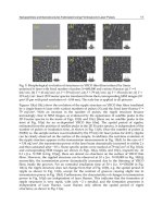
The influence of the image quality can clearly be seen in 2. A Siemensstern with 8 mm thick
iron and copper sections is radiographed. (a) The energy of the X-rays is not sufficient to
penetrate any material, the area behind the object is completely dark. (b) The area behind
the object is still very dark compared to the uncovered area, although a faint contrast
between copper (darker) and iron (lighter) can be seen. Many low energy photons enhance
the brightness in the uncovered area, while they are completely absorbed in the object. (c)
The low-energy photons are filtered out by the prefilter and don’t contribute to either the
uncovered or covered image parts. The difference between these areas is reduced, while the
contrast is enhanced. This spectrum would be a good choice for separating the iron and copper
sections. (d) The vast majority of the photons penetrate the object regardless of the material.
The complete object appears brighter, but the contrast between iron and copper is reduced
again.
2.1.2 Focal spot size
The focal spot size U
F
of the X-ray source is also a very important magnitude and has a
large influence on the spatial resolution of the image, especially when working with high
magnifications M . The magnification is given by the fraction of the focus-detector-distance
FDD and the focus-object-distance FO D. As illustrated in 3, the geometrical unsharpness U
g
of the image is given by
U
g
= U
F
(
1 − M
)
=
d
1 −
FDD
FOD
.(1)
2.1.3 Intensity
With many applications, the measurement time is crucial and should be as short as possible.
The image noise on one hand results from electronic noise in detector systems, but the main
3
Monte Carlo Simulations in NDT
(a) 30 kV without prefilter (b) 160 kV with 4 mm
aluminium prefilter
(c) 160 kV with 4 mm copper
prefilter
(d) 450 kV with 4 mm copper
prefilter
Fig. 2. Images of a Siemensstern. The sections are iron and copper with thickness of 8 mm
each
part originates from poisson noise due to limited quantum statistics. Poisson noise is 1/
N
p
,
where N
p
is the number of events per pixel in one image. For obtaining low-noise images in
a short time, the source intensity must be maximized. The number of emitted photons from
an X-ray source first depends on the tube voltage U. The intensity is roughly proportional to
the squared voltage. Since the voltage shapes the energy spectrum, it is not always desirable
to change it for a given application. The second way to increase the intensity is to increase
the tube current I, which is proportional to the intensity. The electrical power P applied to the
X-ray target is P
= U · I. Unfortunately only about 1% of the electrical power is converted to
X-rays. The vast majority of the electrical power heats up the target, which forces a limitation
in the appliable current. Monte-Carlo simulations can help a great deal to optimize target
material composition and geometry to increase the load capacity of targets or increase the
X-ray conversion efficiency.
2.2 High resolution imaging
As mentioned in the above section, a small focal spot is crucial to achieve a good spatial
resolution when working with high magnifications. High resolution in X-ray imaging means
resolution of object details below 1 micron. For those applications, microfocus X-ray tubes
with transmission targets are commonly used where the target is also the exitation window of
4
Applications of Monte Carlo Method in Science and Engineering
Fig. 3. Geometrical unsharpness due to X-ray source dimension
the tube. The transmission target has a great advantage since the specimen can be placed very
close to the focal spot in order to achive high magnifications. The electron beam in the X-ray
tube is focused onto the target by electronic lenses. The diamater of the beam on the target
surface reaches from 200 nm to several μm and mostly determines the X-ray focal spot size.
But the diffusion of the electrons in the target, which depends largely on the target materials
and layer composition can further increase the focal spot size as shown in 4. To design a target
for smallest possible focal spots, Monte-Carlo simulations of electronic diffusion and X-ray
production processes were performed.
Fig. 4. Geometrical setup of a transmission target
In the simulation a parallel electron beam with electron kinetic energy between 30 and 120 keV
was modeled with a gaussian intensity cross-section in both dimensions. The FWHM value
of the gaussian distribution was 200 nm. The first layer material of the transmission target
5
Monte Carlo Simulations in NDT
is tungsten. Since the X-ray productivity rises with the atomic number proportional to Z
2
,
tungsten with Z
= 74 is a good choice. It has even more advantages, a very high melting point
at over 3000
◦
C, a fair thermal conductivity, mechanical and chemical stability. The X-rays are
produced mainly in the tungsten layer, which is also called the X-ray production layer. In
the simulations, the thickness of this layer was varied from 0.05 to 7 microns (depending on
electron energy). From their point of origin the photons have to pass the remaining target
material to reach the side opposite the electron beam. Therefore the substrate material must
fulfill serveral requirements. The atomic number must be quite low, so the X-rays can pass that
layer without being absorbed, even at low energies. Furthermore, the substrate must have
a good thermal conductivity and a high melting point so that the heat that is generated in
the tungsten layer can be conducted to the air side of the target, where it can be cooled by
fans for example. A performance number can be approximated by the product of thermal
conductivity λ and maximum allowable temperatur T
max
. A further task of the substrate is to
form a mechanical closure of the vacuum vessel against the air pressure. Since the target must
be thin for X-ray transmissibility, the material must be quite stable. Common materials for this
task are beryllium, aluminium, diamond or other carbon configurations. The simulations were
done for a 300 micron thick beryllium substrate, which forms a quite stable vaccum closure.As
simulation results the diameter of the effective focal spot U
F
, i.e. the area where photons are
produced and the X-ray production efficiency were obtained. The total X-ray intensity φ and
the brillance b, which is defined as the intensity divided by the source area are also important
magnitudes for some applications.
b
=
φ
A
F
=
4φ
πU
2
F
(2)
Determining the focal spot size U
F
from simulation data is shown in 5. The two-dimensional
energy distribution of generated X-rays on the target was calculated with ROSI (a). The focal
spot profile was taken from a line profile averaged over the whole target width in one direction
(b). This profile was integrated after normalizing the total X-ray power to a value of 1. The
focal spot is defined as the area where the integral value is between 0.1 and 0.9 (c).
(a) X-ray energy
distribution of all
generation locations
(b) One-dimensional focal spot
profile averaged over whole width
(c) Integral over normalized profile
Fig. 5. Determination of focal spot sizes
In figure 6 the effective focal spot size U
F
(a), the X-ray intensity φ (b) and the brillance b (c) is
shown for several tungsten layer thicknesses and the tube voltages of 30, 70 and 120 kV. The
intensities are calculated per target current.
For each voltage, all curves follow a similar course. The focal spot size can never be smaller
than the diameter of the electron beam, so it is nearly 200 microns in diameter with very thin
6
Applications of Monte Carlo Method in Science and Engineering
tungsten layers, since only a few electrons interact with that layer and are barely scattered
to distant parts of the tungsten. Due to the small interaction probability, the X-ray intensity
is also very low. With thicker tungsten layers, the interaction probability and therefore the
production rate of photons rises rapidly. Since the average scattering angles are quite small,
especially at higher voltages, the electron beam barely broadens in that layer, keeping the focal
spot size almost constant. The brillance rises to a maximum until the tungsten becomes thick
enough so that electrons can be scattered multiply, reaching distant parts of that layer, where
they also produce X-rays. The result is an increase of the focal spot size. The total number of
photons produced and reaching the opposite side of the target still rises until the tungsten
becomes so thick, that the photons are reabsorbed by the tungsten. The focal spot size gets
into saturation and the intensity is again reduced by higher target self-absorption.
(a) Focal spot size (b) X-ray intensity (c) Source brillance
Fig. 6. Optimization of target configuration with nano focus sources
Of course the simulations can also be done with other substrate materials and thicknesses
to find optimal parameters for a specific application. The Monte Carlo simulation can also
calculate the heat deposition in the target volume. The data can then be taken into a heat
transfer simulation tool to calculate the heat load capacity of the whole target.
2.3 High energy imaging
Imaging of very large and dense objects such as freight containers, whole cars (especially
engines) or parts from shipbuilding requires very high energetic radiation in the MeV range
to penetrate these objects. X-ray tubes on the market are available up to voltages of 450 kV,
which is by far not enough. To produce high energy X-rays linear accelerators (LINACs) are
commonly used. The principle in generating X-rays is the same, but the method of accelerating
the electrons differs from X-ray tubes. The electrons are emitted by a gun and accelerated by
bundles in a waveguide through several copper cavities. A high voltage microwave signal is
applied, which accelerates the electron bundles over several cavities up to kinetic energies of
some MeVs.
When electrons hit the target at these energies, X-ray radiation is almost solely produced in
the direction of the impacting electrons, so X-ray targets work exclusively as transmission
targets. The relativistic Lamor formula describes the angular distribution of bremsstrahlung
generation (Jackson, 2006):
dP
dΩ
=
e
2
˙
v
2
4πc
3
sin
2
θ
(
1 − β cos θ
)
5
(3)
At very high energies and small angles, β
= v /c ≈ 1, the denominator decreases with a
power of five and the whole term gets very large. Using high energy X-rays for imaging means
that the radiation field is limited or at least decreases rapidly in intensity at the borders. To
7
Monte Carlo Simulations in NDT
choose appropriate radiation geometries for different object sizes, the radiation field has to be
calculated and taken into account.
We modeled a commonly X-ray target made of 800 μm copper and 450 μmtungsten.The
electron beam was modeled as a parallel and monoenergetic beam. The intensity cross-section
was gaussian in shape with a FWHM value of 1 mm. We calculated the angular X-ray intensity
distribution for energies from 1 to 18 MeV (see 7).
Fig. 7. Simulation setup for X-ray generation with a LINAC target
The results are shown in 8. The theoretically calculated distribution looks quite different to
the simulation results. The Lamor formula assumes all electrons travelling in the forward
direction (θ
= 0
◦
) when generating bremsstrahlung. In reality the electrons can be scattered
by collisions with other electrons and atomic cores while changing their direction before
generating bremsstrahlung. The forward peak is blurred to higher angles. The absolute
intensity increase with electron kinetic energy is described very well and corresponds to the
theory.
2.4 Efficiency optimization
Some applications get along without high resolution or high energy sources. Sometimes a
short measurement time is most essential. Inspection systems within an industrial production
line have to measure prefabricated parts within a production cycle. When inspecting parts
with computed tomography for reconstructing the whole 3-dimensional volume, this task is
quite demanding, since the parts must be radiographed from several hundred points of view
in a short time. The most important component to achieve this is a highly intense radiation
source, that works normally with moderate voltages between 80 to 225 kV. Most X-ray tubes
have fixed targets, where the electron beam hits the same spot on the target the whole time.
The electron beam current is therefore limited due to heating up this focal spot. For medical
X-ray imaging, there are tubes with rotating targets since 1933. The electron beam hits the
target not in a single spot, but in a circular path. The load with rotating targets can be enhanced
by a factor of approximately ten compared to fixed targets. The reasons why rotating targets
are not common in industrial X-ray imaging are locally unstable and quite big focal spots of
8
Applications of Monte Carlo Method in Science and Engineering
(a) Calculated with Lamor formula (b) Simulated with ROSI
Fig. 8. Analytically calculated and simulated angle distrubutions for generated X-rays in a
LINAC target at high energies
about 800 microns or more and their very high price. They only are used where measurement
time is crucial.
With Monte-Carlo simulations some work was done to improve the allowed target load by
modifying both the electron beam geometry and target composition with rotating anodes
(Sukowski, 2007). This work was done with a medical X-ray tube, but since industrial X-ray
tubes are derived from medical tubes, the results can be conveyed to industral tubes without
difficulty. Under variation of the tungsten layer thickness, the emitted X-ray intensity and
energy deposition in the target was simulated. The 3-dimensional energy distribution can be
transferred to finite element simulation programs to calculate the temperature distribution in
steady state while taking cooling effects into account. With the simulation results, optimizing
the electron beam and target geometries is possible.
3. Simulation of X-ray detectors
3.1 Types of detectors commonly used in NDT
In almost all X-ray imaging applications, line or area pixel sensors are used. An X-ray
image is virtually the spatial distribution of the X-ray radiation intensity hitting the sensor
area. When X-rays interact with the sensor material, energy is transferred to the sensor and
converted into an electrical signal. The signals are amplified and digitized pixel by pixel to a
numeric value. The spatial pixel value distribution can be visualized by a color or more often
used grey brightness scale. In a positive X-ray image, bright areas correspond to high X-ray
intensity, where almost no material is between the X-ray source and the detector, while dark
areas are usually covered by thick or heavy parts of the specimen (see 2). In the simulation
studies we focused on characterizing flat-panel pixel detectors with squared or rectangular
surfaces, which are the most used detectors. Basically there are two types of flat-panel detector
technologies that differ in the way of conversion from X-ray energy deposition to an electrical
signal (Beutel et al., 2000).
3.1.1 Indirect converting detectors
Most flat-panel detectors convert the X-ray energy deposition in an indirect way into an
electrical signal. The X-ray detection mechanism is based on a scintillator. X-rays interacting
with a scintillator ionize the atoms, causing emission of fluorescence light due to exited-state
9
Monte Carlo Simulations in NDT
deactivation. The energy level differences of some elements in a typical scintillator are in the
range of some electronvolts. The fluorescence light emitted from the scintillator is therefore
visual light that can be detected by a photo diode array which is arranged just behind the
scintillator layer (Beutel et al., 2000).
3.1.2 Direct converting detectors
Unlike scintillator based detectors, direct converting detectors usually consist of a
semiconductor material as sensor layer. The semiconductor is assembled between two
electrodes. One electrode is continuous over the whole sensor area, while the other electrode
on the opposite side consists of many small solder beads which resemble the detector pixels.
Between the two electrodes a voltage is applied so that the semiconductor is completely
depleted of charge carriers. When X-rays interact with the semiconductor, they transfer energy
to bound valence electrons, generating free electron-hole-pairs,which drift to nearby electrode
beads due to the electrical field within the semiconductor. At the electrodes a current can be
measured, which is proportional to the energy deposited by the X-rays (Beutel et al., 2000).
3.1.3 Detector properties
Regardless of application, a perfect detector should fulfill two essential characteristics. First,
every X-ray photon hitting the detector surface should create a signal. Since X-rays can pass
matter, what makes them usefull after all, they also can pass the detector without being
detected. The fraction of detected photons N
d
to photons hitting the detector N
0
is not
exceeding 1 and is called the detection efficiency η
det
.
η
det
=
N
d
N
0
≤ 1(4)
Especially at high photon energies, the efficiency can be quite low, so the measurement time
must be increased for obtaining low-noise images. The efficiency depends on the choice of
the sensor material, but mainly on the thickness of the sensor layer. Since X-ray intensity
decreases exponentially with the path length in material, increasing the sensor thickness can
significantly improve the detection efficiency.
The second important characteristic for spatial resolving detection systems is the ability to
determine the location where an X-ray photon hits the detector surface. In the best case,
X-rays are not only detected efficiently, they rather should be detected exactly where the
initial interaction took place. Unfortunately, this is usually not the case. When X-rays are
absorbed by a material, their kinetic energy is transferred to one or more electrons. These
electrons propagate through the medium while transferring parts of their kinetic energy to
other electrons until stopped. The path length of electrons in matter can reach some tens
of microns. Therefore the signal is blurred over a certain volume. Another effect can cause
a longer range, but less intense signal blurring. X-rays are not always absorbed by matter,
they can also be scattered, transferring only a part of their energy at the location of their
initial interaction, what is called Compton scattering. The scattered photon with the remaining
energy can be absorbed in a detector volume quite far away (up to some centimeters) from
their first point of interaction, causing two or even more signal spots. These two effects occur
in both detector types and can be calculated very well by ROSI. They depend on the layer
composition (materials and thicknesses) of the detector. In scintillator based detectors there is
one more effect that dominates the signal blurring. When X-rays are converted to visual light
in the scinitllation layer, this light is emitted isotropically to all directions. To be detected, it
10
Applications of Monte Carlo Method in Science and Engineering
has to reach the photo diode layer, where it can be spread over some pixels. This blurring
scales highly with the distance from the point of light generation to the photo diode layer.
Therefore thick scintillators, where light can be produced quite far away from the photo
diode layer often yield a poor spatial resolution. The principle is shown in 9. Signals are
clearer distinguishable with thin scintillators, but the efficiency is reduced. Every application
demands a different trade-off between efficiency and spatial resolution. The generation,
absorption and propagation of visible light in media and on material borders can be described
by DETECT2000 (G. McDonald et al., 2000), also a Monte-Carlo simulation code.
Fig. 9. Signal blurring in a scintillator based detector due to spread of visual photons
For evaluating the relation between the detector properties and their layer composition, one
direct converting and one indirect converting detector with 100 μm pixel pitch each were
modelled with layer compositions shown in 1.
Detector type Direct converting (DIC) Indirect converting (IDC)
Layer composition:
Front cover 100 μmAl 1mmAl
Gap
1mm 1mm
Front electrode
5 μmAl
Sensor
750 μm CdTe 140 μm Gd
2
O
2
S
semiconductor scintillator
Rear electrodes
50 μmsolder
Fiberoptic plate
3mmAl
2
O
3
Electronics 1.5 mm Si 1.5 mm Si
Gap
10 mm
Rear shielding
2 mm steel 400 μmCu
Table 1. Detector layer compositions
11
Monte Carlo Simulations in NDT
