Chapter 013. Chest Discomfort (Part 6) pptx
Bạn đang xem bản rút gọn của tài liệu. Xem và tải ngay bản đầy đủ của tài liệu tại đây (39.9 KB, 5 trang )
Chapter 013. Chest Discomfort
(Part 6)
Acute Chest Discomfort
In patients with acute chest discomfort, the clinician must first assess the
patient's respiratory and hemodynamic status. If either is compromised, initial
management should focus on stabilizing the patient before the diagnostic
evaluation is pursued. If, however, the patient does not require emergent
interventions, then a focused history, physical examination, and laboratory
evaluation should be performed to assess the patient's risk of life-threatening
conditions.
Clinicians who are seeing patients in the office setting should not assume
that they do not have acute ischemic heart disease, even if the prevalence may be
lower. Malpractice litigation related to myocardial infarctions that were missed
during office evaluations is becoming increasingly common, and ECGs were not
performed in many such cases. The prevalence of high-risk patients seen in office
settings may be increasing due to congestion in emergency departments.
In either setting, the history should include questions about the quality and
location of the chest discomfort (Table 13-2). The patient should also be asked
about the nature of onset of the pain and its duration. Myocardial ischemia is
usually associated with a gradual intensification of symptoms over a period of
minutes. Pain that is fleeting or that lasts hours without being associated with
electrocardiographic changes is not likely to be ischemic in origin. Although the
presence of risk factors for coronary artery disease may heighten concern for this
diagnosis, the absence of such risk factors does not lower the risk for myocardial
ischemia enough to be used to justify a decision to discharge a patient.
Wide radiation of chest pain increases probability that pain is due to
myocardial infarction. Radiation of chest pain to the left arm is common with
acute ischemic heart disease, but radiation to the right arm is also highly consistent
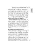
with this diagnosis. Figure 13-1 shows estimates derived from several studies of
the impact of various clinical features from the history on the probability that a
patient has an acute myocardial infarction.
Figure 13-1
Impact of chest pain characteristics on odds of acute myocardial infarction
(AMI). (Figure prepared from data in Swap and Nagurney.)
Right shoulder pain is also common with acute cholecystitis, but this
syndrome is usually accompanied by pain that is located in the abdomen rather
than chest. Chest pain that radiates between the scapulae raises the question of
aortic dissection.
The physical examination should include evaluation of blood pressure in
both arms and of pulses in both legs. Poor perfusion of a limb may be due to an
aortic dissection that has compromised flow to an artery branching from the aorta.
Chest auscultation may reveal diminished breath sounds; a pleural rub; or
evidence of pneumothorax, pulmonary embolism, pneumonia, or pleurisy. Tension
pneumothorax may lead to a shift in the trachea from the midline, away from the
side of the pneumothorax. The cardiac examination should seek pericardial rubs,
systolic and diastolic murmurs, and third or fourth heart sounds. Pressure on the
chest wall may reproduce symptoms in patients with musculoskeletal causes of
chest pain; it is important that the clinician ask the patient if the chest pain
syndrome is being completely reproduced before drawing too much reassurance
that more serious underlying conditions are not present.
An ECG is an essential test for adults with chest discomfort that is not due
to an obvious traumatic cause. In such patients, the presence of
electrocardiographic changes consistent with ischemia or infarction (Chap. 221) is
associated with high risks of acute myocardial infarction or unstable angina (Table
13-4); such patients should be admitted to a unit with electrocardiographic
monitoring and the capacity to respond to a cardiac arrest. The absence of such
changes does not exclude acute ischemic heart disease, but the risk of life-
threatening complications is low for patients with normal electrocardiograms or
only nonspecific ST-T-wave changes. If these patients are not considered
appropriate for immediate discharge, they are often candidates for early or
immediate exercise testing.
Prevalence Finding
Myocardial Unstable
Infarction, % Angina, %
ST elevation ( 1 mm) or Q waves
on ECG not known to be old
79 12
Ischemia or strain on ECG not
known to be old (ST depression 1 mm or
ischemic T waves)
20 41
None of the
preceding ECG
changes but a prior history of angina or
myocardial infarction (history of heart
attack or nitroglycerin use)
4 51
None of the preceding ECG
changes and no prior history of angina or
myocardial infarction (history of heart
attack or nitroglycerin use)
2 14
Note: ECG, electrocardiogram.