Chapter 023. Weakness and Paralysis (Part 4) pps
Bạn đang xem bản rút gọn của tài liệu. Xem và tải ngay bản đầy đủ của tài liệu tại đây (260.18 KB, 6 trang )
Chapter 023. Weakness and Paralysis
(Part 4)
Hemiparesis
Hemiparesis results from an upper motor neuron lesion above the
midcervical spinal cord; most such lesions are above the foramen magnum. The
presence of other neurologic deficits helps to localize the lesion. Thus, language
disorders, cortical sensory disturbances, cognitive abnormalities, disorders of
visual-spatial integration, apraxia, or seizures point to a cortical lesion.
Homonymous visual field defects reflect either a cortical or a subcortical
hemispheric lesion. A "pure motor" hemiparesis of the face, arm, or leg is often
due to a small, discrete lesion in the posterior limb of the internal capsule, cerebral
peduncle, or upper pons. Some brainstem lesions produce "crossed paralyses,"
consisting of ipsilateral cranial nerve signs and contralateral hemiparesis (Chap.
364). The absence of cranial nerve signs or facial weakness suggests that a
hemiparesis is due to a lesion in the high cervical spinal cord, especially if
associated with ipsilateral loss of proprioception and contralateral loss of pain and
temperature sense (the Brown-Séquard syndrome).
Acute or episodic hemiparesis usually results from ischemic or
hemorrhagic stroke, but may also relate to hemorrhage occurring into brain tumors
or as a result of trauma; other causes include a focal structural lesion or
inflammatory process as in multiple sclerosis, abscess, or sarcoidosis. Evaluation
begins immediately with a CT scan of the brain (Fig. 23-3) and laboratory studies.
If the CT is normal and an ischemic stroke is unlikely, MRI of the brain or
cervical spine is performed.
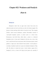
Figure 23-3
An algorithm for the initial workup of a patient with weakness. EMG,
electromyography; LMN, lower motor neuron; NCS, nerve conduction studies;
UMN, upper motor neuronSubacute hemiparesis that evolves over days or weeks
has an extensive differential diagnosis. A common cause is subdural hematoma,
especially in elderly or anticoagulated patients, even when there is no history of
trauma. Infectious possibilities include cerebral abscess, fungal granuloma or
meningitis, and parasitic infection. Weakness from primary and metastatic
neoplasms may evolve over days to weeks. AIDS may present with subacute
hemiparesis due to toxoplasmosis or primary CNS lymphoma. Noninfectious
inflammatory processes, such as multiple sclerosis or, less commonly, sarcoidosis,
merit consideration. If the brain MRI is normal and there are no cortical and
hemispheric signs, MRI of the cervical spine should be undertaken.
Chronic hemiparesis that evolves over months is usually due to a neoplasm
or vascular malformation, a chronic subdural hematoma, or a degenerative disease.
If an MRI of the brain is normal, the possibility of a foramen magnum or high
cervical spinal cord lesion should be considered.
Paraparesis
An intraspinal lesion at or below the upper thoracic spinal cord level is
most commonly responsible, but a paraparesis may also result from lesions at
other locations that disturb upper motor neurons (especially parasagittal
intracranial lesions) and lower motor neurons [anterior horn cell disorders, cauda
equina syndromes due to involvement of nerve roots derived from the lower spinal
cord (Chap. 372), and peripheral neuropathies].
Acute paraparesis may not be recognized as due to spinal cord disease at an
early stage if the legs are flaccid and areflexic. Usually, however, there is sensory
loss in the legs with an upper level on the trunk; a dissociated sensory loss
suggestive of a central cord syndrome; or exaggerated stretch reflexes in the legs
with normal reflexes in the arms. It is important to image the spinal cord (Fig. 23-
3). Compressive lesions (particularly epidural tumor, abscess, or hematoma, but
also a prolapsed intervertebral disk and vertebral involvement by malignancy or
infection), spinal cord infarction (proprioception is usually spared), an
arteriovenous fistula or other vascular anomaly, and transverse myelitis, are
among the possible causes (Chap. 372).
Diseases of the cerebral hemispheres that produce acute paraparesis include
anterior cerebral artery ischemia (shoulder shrug is also affected), superior sagittal
sinus or cortical venous thrombosis, and acute hydrocephalus. If upper motor
neuron signs are associated with drowsiness, confusion, seizures, or other
hemispheric signs, MRI of the brain should be undertaken.
Paraparesis may result from a cauda equina syndrome, for example,
following trauma to the low back, a midline disk herniation, or an intraspinal
tumor; although sphincters are affected, hip flexion is often spared, as is sensation
over the anterolateral thighs. Rarely, paraparesis is caused by a rapidly evolving
anterior horn cell disease (such as poliovirus or West Nile virus infection),
peripheral neuropathy (such as Guillain-Barré syndrome; Chap. 380) or myopathy
(Chap. 382). In such cases, electrophysiologic studies are diagnostically helpful
and refocus the subsequent evaluation.
Subacute or chronic paraparesis with spasticity is caused by upper motor
neuron disease. When there is associated lower-limb sensory loss and sphincter
involvement, a chronic spinal cord disorder is likely (Chap. 372). If an MRI of the
spinal cord is normal, MRI of the brain may be indicated. If hemispheric signs are
present, a parasagittal meningioma or chronic hydrocephalus is likely and MRI of
the brain is the initial test. In the rare situation in which a longstanding paraparesis
has a lower motor neuron or myopathic etiology, the localization is usually
suspected on clinical grounds by the absence of spasticity and confirmed by EMG
and nerve conduction tests.