Chapter 032. Oral Manifestations of Disease (Part 3) potx
Bạn đang xem bản rút gọn của tài liệu. Xem và tải ngay bản đầy đủ của tài liệu tại đây (46.15 KB, 18 trang )
Chapter 032. Oral Manifestations
of Disease
(Part 3)
Diseases of the Oral Mucosa
Infection
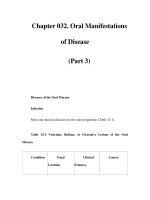
Most oral mucosal diseases involve microorganisms (Table 32-1).
Table 32-
1 Vesicular, Bullous, or Ulcerative Lesions of the Oral
Mucosa
Condition Usual
Location
Clinical
Features
Course
Viral Diseases
Primary
acute herpetic
gingivostomatitis
[herpes simplex
virus (HSV) type 1,
rarely type 2]
Lip and oral
mucosa (buccal,
gingival, lingual
mucosa)
Labial
vesicles that
rupture and crust,
and intraoral
vesicles tha
t
quickly ulcerate;
extremely painful;
acute gingivitis,
fever, malaise, foul
odor, and cervical
lymphadenopathy;
occurs primarily in
infants, children,
and young adults
Heals
spontaneously in 10–
14 days. Unless
secondarily infected,
lesions lasting >3
wee
ks are not due to
primary HSV
infection.
Recurrent
herpes labialis
Mucocutane
ous junction of lip,
perioral skin
Eruption of
groups of vesicles
that may coalesce,
then rupture and
Lasts about 1
week, but condition
may be prolonged if
secondarily infected.
crust; painful to
pressure or spicy
foods
If severe, topical or
oral antiviral may
reduce healing time.
Recurrent
intraoral herpes
simplex
Palate and
gingiva
Small
vesicles on
keratinized
epithelium that
rupture and
coalesce; painful
Heals
spontaneously i
n
about 1 week. If
severe, topical or oral
antiviral may reduce
healing time.
Chickenpox
(varicella-zoster
virus)
Gingiva and
oral mucosa
Skin lesions
may be
accompanied by
small vesicles on
oral mucosa that
rupture to form
shallow ulcers;
may coalesce to
form large bullous
lesions that
Lesions heal
spontaneously within
2 weeks.
ulcerate; mucosa
may have
generalized
erythema
Herpes
zoster (reactivation
of varicella-
zoster
virus)
Cheek,
tongue, gingiva, or
palate
Unilateral
vesicular eruptions
and ulcerati
on in
linear pattern
following sensory
distribution of
trigeminal nerve or
one of its branches
Gradual
healing without
scarring unless
secondarily infected;
postherpetic
neuralgia is common.
Oral acyclovir,
famcyclovir, or
valacyclovir reduce
healing time
and
postherpetic
neuralgia
Infectious
mononucleosis
(Epstein-Barr
Oral mucosa
Fatigue,
sore throat,
malaise, fever, and
Oral lesions
disappear during
convalescence; no
virus) cervical
lymphadenopathy;
numerous small
ulcers usually
appear several days
before
lymphadenopathy;
gingival bleeding
and multip
le
petechiae at
junction of hard
and soft palates
treatment though
glucocorticoids
indicated if tonsillar
swelling
compromises airway
Herpangina
(coxsackievirus A;
also possibly
coxsackie B and
echovirus)
Or
al mucosa,
pharynx, tongue
Sudden
onset of fever, sore
throat, and
oropharyngeal
vesicles, usually in
children under 4
years, during
summer months;
Incubation
period 2–
9 days;
fever for 1–
4 days;
recovery uneventful
diffuse pharyngeal
congestion and
vesicles (1–
2 mm),
grayish-white
surrounded by red
areola; vesicles
enlarg
e and
ulcerate
Hand, foot,
and mouth disease
(coxsackievirus
A16 most
common)
Oral mucosa,
pharynx, palms, and
soles
Fever,
malaise, headache
with oropharyngeal
vesicles that
become painf
ul,
shallow ulcers;
highly infectious;
usually affects
children under age
10
Incubation
period 2–
18 days;
lesions heal
spontaneously in 2–
4
weeks
Primary Gingiva, Acute
Followed by
HIV infection palate, and pharynx
gingivitis and
oropharyngeal
ulceration,
associated
with
febrile illness
resembling
mononucleosis and
including
lymphadenopathy
HIV seroconversion,
asymptomatic HIV
infection, and usually
ultimately by HIV
disease
Bacterial or Fungal Diseases
Acute
necrotizing
ulcerative
gingivitis ("trench
mo
uth," Vincent's
infection)
Gingiva Painful,
bleeding gingiva
characterized by
necrosis and
ulceration of
gingival papillae
and margins plus
lymphadenopathy
and foul odor
Debridement
and diluted (1:3)
peroxide lavage
provide relief within
24 h; antibiotics
in
acutely ill patients;
relapse may occur
Prenatal
(congenital)
syphilis
Palate, jaws,
tongue, and teeth
Gummatous
involvement of
palate, jaws, and
facial bones;
Hutchinson's
incisors, mulberry
molars, glossitis,
mucous patches,
and fissures on
corner of mouth
Tooth
deformities in
permanent dentition
irreversible
Primary
syphilis (chancre)
Lesion
appears where
organism enters
body; may occur on
lips, tongue, or
tonsillar area
Small
papule developing
rapidly into a large,
painless ulcer with
indurated bor
der;
unilateral
lymphadenopathy;
chancre and lymph
nodes containing
spirochetes;
Healing of
chancre in 1–
2
months, followed by
secondary syphilis in
6–8 weeks
serologic tests
positive by third to
fourth weeks
Secondary
syphilis
Oral mucosa
frequently invo
lved
with mucous
patches, primarily
on palate, also at
commissures of
mouth
Maculopapu
lar lesions of oral
mucosa, 5–
10 mm
in diameter with
central ulceration
covered by grayish
membrane;
eruptions occurring
on various mucosal
surfaces and skin
accompanied
by
fever, malaise, and
sore throat
Lesions may
persist from several
weeks to a year
Tertiary
syphilis
Palate and
tongue
Gummatous
infiltration of
palate or tongue
Gumma may
destroy palate,
causing complete
followed by
ulceration and
fibrosis; atrophy of
tongue papillae
produces
characteristic bal
d
tongue and
glossitis
perforation
Gonorrhea
Lesions may
occur in mouth at
site of inoculation
or secondarily by
hematogenous
spread from a
primary focus
elsewhere
Most
pharyngeal
infection is
asymptomatic; may
p
roduce burning or
itching sensation;
oropharynx and
tonsils may be
ulcerated and
erythematous;
saliva viscous and
fetid
More difficult
to eradicate than
urogenital infection,
though pharyngitis
usually resolves with
appropriate
antimicrobial
treatment
Tub
erculosis
Tongue,
tonsillar area, soft
palate
A painless,
solitary, 1–
5 cm,
irregular ulcer
covered with a
persistent exudate;
ulcer has a firm
undermined border
Autoinnoculat
ion from pulmonary
infection usual;
lesions resolve with
appropriate
antimicrobial therapy
Cervicofacia
l actinomycosis
Swellings in
region of face,
neck, and floor of
mouth
Infection
may be associated
with an extraction,
jaw fracture, or
eruption of molar
tooth; in acute
form resembles an
acute pyogenic
abscess, but
contains yellow
"sulfur granules"
(gram-positive
mycelia and their
Typically
swelling is hard and
grows painlessly;
multiple abscesses
with draining tracks
develop; penicillin
first choice; surgery
usually necessary
hyphae)
Histoplasmo
sis
Any area of
the mouth,
particula
rly tongue,
gingiva, or palate
Nodular,
verrucous, or
granulomatous
lesions; ulcers are
indurated and
painful; usual
source
hematogenous or
pulmonary, but
may be primary
Systemic
antifungal therapy
necessary to treat
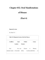
Candidiasis
(Table 32-3)
Dermatologic Diseases
Mucous
membrane
pemphigoid
Typically
produces marked
gingival erythema
Painful,
grayish-white
collapsed vesicles
Protracted
course with
remissions and
and ulceration;
other areas of oral
cavity, esophagus,
and vagina may be
affected
or bullae of full-
thickness
epithelium with
peripheral
erythematous zone;
gingival lesions
desquamate,
leaving ulcerated
area
exacerbations;
involvement of
different sites occurs
slowly;
glucocorticoids may
temporarily reduce
symptoms but do not
control the disease
Erythema
multiforme minor
and major
(Stevens-Johnson
syndrome)
Primarily the
oral mucosa and the
skin of hands and
feet
Intraoral
ruptured bullae
surrounded by an
inflammatory area;
lips may show
hemorrhagic
crusts; the "iris," or
"target," lesion on
the
skin is
pathognomonic;
patient may have
Onset very
rapid; usually
idiopathic, but may
be associated with
trigger such as drug
reaction; condition
may last 3–
6 weeks;
mortality with EM
major 5–
15% if
untreated
severe signs of
toxicity
Pemphigus
vulgaris
Oral mucosa
and skin; sites of
mechanical trauma
(soft/hard palate,
frenulum, lips,
buccal mucosa)
Usually
(>70%) presents
with oral lesions;
fragile, ruptured
bullae and
ulcerated oral
areas; mostly in
older adults
With repeated
occurrence of bullae,
toxi
city may lead to
cachexia, infection,
and death within 2
years; often
controllable with oral
glucocorticoids
Lichen
planus
Oral mucosa
and skin
White striae
in mouth; purplish
nodules on skin at
sites of friction;
occasionally causes
oral mucosal ulcers
a
nd erosive
gingivitis
White striae
alone usually
asymptomatic;
erosive lesions often
difficult to treat, but
may respond to
glucocorticoids
Other Conditions
Recurrent
aphthous ulcers
Usually on
nonkeratinized oral
mucosa (buccal and
labial mucosa, floor
of mouth, soft
palate, lateral and
ventral tongue)
Single or
clusters of painful
ulcers with
surrounding
erythematous
border; lesions may
be 1–
2 mm in
diameter in crops
(herpetiform), 1–
5
mm (minor), or 5–
15 mm (major)
Lesions heal
in 1–
2 weeks but
may re
cur monthly or
several times a year;
protective barrier
with orabase and
topical steroids give
symptomatic relief;
systemic
glucocorticoids may
be needed in severe
cases
Behçet's
syndrome
Oral mucosa,
eyes, genitalia, gut,
and CNS
Multiple
aphthous ulcers
in
mouth;
inflammatory
ocular changes,
ulcerative lesions
Oral lesions
often first
manifestation; persist
several weeks and
heal without scarring
on genitalia;
inflammatory
bowel disease and
CNS disease
Traumatic
ulcers
Anywhere
on oral mucosa;
dentures
frequently
responsible for
ulcers in vestibule
Localized,
discrete ulcerated
lesions with red
border; produced
by accidental biting
of mucosa,
penetration by a
foreign object, or
chronic irritation
by a denture
Lesions
usually heal in 7–
10
days when irrita
nt is
removed, unless
secondarily infected
Squamous
cell carcinoma
Any area in
the mouth, most
commonly on lower
lip, tongue, and
floor of mouth
Ulcer with
elevated, indurated
border; failure to
heal, pain not
prominent; lesions
Invades and
destroys underlying
tissues; frequently
metastasizes to
regional lymph nodes
tend to arise in
areas of
erythro/leukoplakia
or in smooth
atrophic tongue
Acute
myeloid leukemia
(usually
monocytic)
Gingiva Gingival
swelling and
superficial
ulceration followed
by hyperpla
sia of
gingiva with
extensive necrosis
and hemorrhage;
deep ulcers may
occur elsewhere on
the mucosa
complicated by
secondary infection
Usually
responds to systemic
treatment of
leukemia;
occasionally requires
local radiation
therapy
Lymphoma Gingiva, Elevated,
Fatal if
ton
gue, palate and
tonsillar area
ulcerated area that
may proliferate
rapidly, giving the
appearance of
traumatic
inflammation
untreated; may
indicate underlying
HIV infection
Chemical or
thermal burns
Any area in
mouth
White
slough due to
contact with
corrosive agents
(e.g., aspirin, hot
cheese) applied
locally; removal of
slough leaves raw,
painful surface
Lesion heals
in several weeks if
not secondarily
infected
Note: CNS, central nervous system.