The Insects - Outline of Entomology 3th Edition - Chapter 2 pot
Bạn đang xem bản rút gọn của tài liệu. Xem và tải ngay bản đầy đủ của tài liệu tại đây (2.51 MB, 28 trang )
TIC02 5/20/04 4:48 PM Page 21
Chapter 2
E XTERNAL ANATOMY
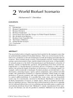
“Feet” of leaf beetle (left) and bush fly (right). (From scanning electron micrographs by C.A.M. Reid & A.C. Stewart.)
TIC02 5/20/04 4:48 PM Page 22
22
External anatomy
Insects are segmented invertebrates that possess the
articulated external skeleton (exoskeleton) characteristic of all arthropods. Groups are differentiated by
various modifications of the exoskeleton and the
appendages – for example, the Hexapoda to which the
Insecta belong (section 7.2) is characterized by having
six-legged adults. Many anatomical features of the
appendages, especially of the mouthparts, legs, wings,
and abdominal apex, are important in recognizing the
higher groups within the hexapods, including insect
orders, families, and genera. Differences between
species frequently are indicated by less obvious anatomical differences. Furthermore, the biomechanical
analysis of morphology (e.g. studying how insects fly or
feed) depends on a thorough knowledge of structural
features. Clearly, an understanding of external anatomy
is necessary to interpret and appreciate the functions
of the various insect designs and to allow identification
of insects and their hexapod relatives. In this chapter
we describe and discuss the cuticle, body segmentation,
and the structure of the head, thorax, and abdomen
and their appendages.
First some basic classification and terminology needs
to be explained. Adult insects normally have wings
(most of the Pterygota), the structure of which may
diagnose orders, but there is a group of primitively
wingless insects (the “apterygotes”) (see section 7.4.1
and Box 9.3 for defining features). Within the Insecta,
three major patterns of development can be recognized
(section 6.2). Apterygotes (and non-insect hexapods)
develop to adulthood with little change in body form
(ametaboly), except for sexual maturation through
development of gonads and genitalia. All other insects
either have a gradual change in body form (hemimetaboly) with external wing buds getting larger at each
molt, or an abrupt change from a wingless immature
insect to winged adult stage via a pupal stage (holometaboly). Immature stages of hemimetabolous insects
are generally called nymphs, whereas those of holometabolous insects are referred to as larvae.
Anatomical structures of different taxa are homologous if they share an evolutionary origin, i.e. if the
genetic basis is inherited from an ancestor common to
them both. For instance, the wings of all insects are
believed to be homologous; this means that wings (but
not necessarily flight; see section 8.4) originated once.
Homology of structures generally is inferred by comparison of similarity in ontogeny (development from
egg to adult), composition (size and detailed appearance), and position (on the same segment and same
relative location on that segment). The homology of
insect wings is demonstrated by similarities in venation
and articulation – the wings of all insects can be derived
from the same basic pattern or groundplan (as explained
in section 2.4.2). Sometimes association with other
structures of known homologies is helpful in establishing the homology of a structure of uncertain origin.
Another sort of homology, called serial homology,
refers to corresponding structures on different segments of an individual insect. Thus, the appendages of
each body segment are serially homologous, although
in living insects those on the head (antennae and
mouthparts) are very different in appearance from
those on the thorax (walking legs) and abdomen (genitalia and cerci). The way in which molecular developmental studies are confirming these serial homologies
is described in Box 6.1.
2.1 THE CUTICLE
The cuticle is a key contributor to the success of the
Insecta. This inert layer provides the strong exoskeleton of body and limbs, the apodemes (internal supports and muscle attachments), and wings, and acts as
a barrier between living tissues and the environment.
Internally, cuticle lines the tracheal tubes (section 3.5),
some gland ducts and the foregut and midgut of the
digestive tract. Cuticle may range from rigid and
armor-like, as in most adult beetles, to thin and flexible,
as in many larvae. Restriction of water loss is a critical
function of cuticle vital to the success of insects on
land.
The cuticle is thin but its structure is complex and
still the subject of some controversy. A single layer
of cells, the epidermis, lies beneath and secretes the
cuticle, which consists of a thicker procuticle overlaid
with thin epicuticle (Fig. 2.1). The epidermis and cuticle together form an integument – the outer covering
of the living tissues of an insect.
The epicuticle ranges from 3 µm down to 0.1 µm in
thickness, and usually consists of three layers: an inner
epicuticle, an outer epicuticle, and a superficial
layer. The superficial layer (probably a glycoprotein) in
many insects is covered by a lipid or wax layer, sometimes called a free-wax layer, with a variably discrete
cement layer external to this. The chemistry of the
epicuticle and its outer layers is vital in preventing
dehydration, a function derived from water-repelling
(hydrophobic) lipids, especially hydrocarbons. These
TIC02 5/20/04 4:48 PM Page 23
The cuticle
23
Fig. 2.1 The general structure of insect
cuticle; the enlargement above shows
details of the epicuticle. (After Hepburn
1985; Hadley 1986; Binnington 1993.)
compounds include free and protein-bound lipids, and
the outermost waxy coatings give a bloom to the external surface of some insects. Other cuticular patterns,
such as light reflectivity, are produced by various kinds
of epicuticular surface microsculpturing, such as close-
packed, regular or irregular tubercles, ridges, or tiny
hairs. Lipid composition can vary and waxiness can
increase seasonally or under dry conditions. Besides
being water retentive, surface waxes may deter predation, provide patterns for mimicry or camouflage, repel
TIC02 5/20/04 4:48 PM Page 24
24
External anatomy
Fig. 2.2 Structure of part of a chitin chain, showing two
linked units of N-acetyl-d-glucosamine. (After Cohen 1991.)
excess rainwater, reflect solar and ultraviolet radiation,
or give species-specific olfactory cues.
The epicuticle is inextensible and unsupportive.
Instead, support is given by the underlying chitinous
cuticle known as procuticle when it is first secreted.
This differentiates into a thicker endocuticle covered
by a thinner exocuticle, due to sclerotization of the
latter. The procuticle is from 10 µm to 0.5 mm thick
and consists primarily of chitin complexed with protein. This contrasts with the overlying epicuticle which
lacks chitin.
Chitin is found as a supporting element in fungal cell
walls and arthropod exoskeletons, and is especially
important in insect extracellular structures. It is an
unbranched polymer of high molecular weight – an
amino-sugar polysaccharide predominantly composed
of β-(1–4)-linked units of N-acetyl-d-glucosamine
(Fig. 2.2).
Chitin molecules are grouped into bundles and
assembled into flexible microfibrils that are embedded
in, and intimately linked to, a protein matrix, giving
great tensile strength. The commonest arrangement of
chitin microfibrils is in a sheet, in which the microfibrils
are in parallel. In the exocuticle, each successive sheet
lies in the same plane but may be orientated at a slight
angle relative to the previous sheet, such that a thickness of many sheets produces a helicoid arrangement,
which in sectioned cuticle appears as alternating light
and dark bands (lamellae). Thus the parabolic patterns
and lamellar arrangement, visible so clearly in sectioned cuticle, represent an optical artifact resulting
from microfibrillar orientation (Fig. 2.3). In the endocuticle, alternate stacked or helicoid arrangements
of microfibrillar sheets may occur, often giving rise to
Fig. 2.3 The ultrastructure of cuticle (from a transmission
electron micrograph). (a) The arrangement of chitin
microfibrils in a helicoidal array produces characteristic
(though artifactual) parabolic patterns. (b) Diagram of how
the rotation of microfibrils produces a lamellar effect owing to
microfibrils being either aligned or non-aligned to the plane of
sectioning. (After Filshie 1982.)
thicker lamellae than in the exocuticle. Different
arrangements may be laid down during darkness compared with daylight, allowing precise age determination in many adult insects.
Much of the strength of cuticle comes from extensive
hydrogen bonding of adjacent chitin chains. Additional
stiffening comes from sclerotization, an irreversible
process that darkens the exocuticle and results in the
proteins becoming water-insoluble. Sclerotization may
result from linkages of adjacent protein chains by
phenolic bridges (quinone tanning), or from controlled
dehydration of the chains, or both. Only exocuticle
becomes sclerotized. The deposition of pigment in the
cuticle, including deposition of melanin, may be associated with quinones, but is additional to sclerotization
and not necessarily associated with it.
In contrast to the solid cuticle typical of sclerites and
mouthparts such as mandibles, softer, plastic, highly
flexible or truly elastic cuticles occur in insects in varying locations and proportions. Where elastic or springlike movement occurs, such as in wing ligaments or for
the jump of a flea, resilin – a “rubber-like” protein – is
present. The coiled polypeptide chains of this protein
function as a mechanical spring under tension or compression, or in bending.
TIC02 5/20/04 4:49 PM Page 25
The cuticle
Fig. 2.4 A specialized worker, or replete, of the honeypot
ant, Camponotus inflatus (Hymenoptera: Formicidae), which
holds honey in its distensible abdomen and acts as a food store
for the colony. The arthrodial membrane between tergal
plates is depicted to the right in its unfolded and folded
conditions. (After Hadley 1986; Devitt 1989.)
In soft-bodied larvae and in the membranes between
segments, the cuticle must be tough, but also flexible
and capable of extension. This “soft” cuticle, sometimes
termed arthrodial membrane, is evident in gravid
females, for example in the ovipositing migratory
locust, Locusta migratoria (Orthoptera: Acrididae), in
which intersegmental membranes may be expanded
up to 20-fold for oviposition. Similarly, the gross
abdominal dilation of gravid queen bees, termites, and
ants is possible through expansion of the unsclerotized
cuticle. In these insects, the overlying unstretchable
epicuticle expands by unfolding from an originally
highly folded state, and some new epicuticle is formed.
An extreme example of the distensibility of arthrodial
membrane is seen in honeypot ants (Fig. 2.4; see also
section 12.2.3). In Rhodnius nymphs (Hemiptera:
Reduviidae), changes in molecular structure of the
cuticle allow actual stretching of the abdominal membrane to occur in response to intake of a large fluid
volume during feeding.
Cuticular structural components, waxes, cements,
pheromones (Chapter 4), and defensive and other compounds are products of the epidermis, which is a nearcontinuous, single-celled layer beneath the cuticle.
25
Many of these compounds are secreted to the outside
of the insect epicuticle. Numerous fine pore canals
traverse the procuticle and then branch into numerous
finer wax canals (containing wax filaments) within
the epicuticle (enlargement in Fig. 2.1); this system
transports lipids (waxes) from the epidermis to the
epicuticular surface. The wax canals may also have a
structural role within the epicuticle. Dermal glands,
part of the epidermis, produce cement and/or wax,
which is transported via larger ducts to the cuticular
surface. Wax-secreting glands are particularly well
developed in mealybugs and other scale insects
(Fig. 2.5). The epidermis is closely associated with
molting – the events and processes leading up to and
including ecdysis (eclosion), i.e. the shedding of the old
cuticle (section 6.3).
Insects are well endowed with cuticular extensions,
varying from fine and hair-like to robust and spine-like.
Four basic types of protuberance (Fig. 2.6), all with
sclerotized cuticle, can be recognized on morphological, functional, and developmental grounds:
1 spines are multicellular with undifferentiated
epidermal cells;
2 setae, also called hairs, macrotrichia, or trichoid
sensilla, are multicellular with specialized cells;
3 acanthae are unicellular in origin;
4 microtrichia are subcellular, with several to many
extensions per cell.
Setae sense much of the insect’s tactile environment.
Large setae may be called bristles or chaetae, with the
most modified being scales, the flattened setae found
on butterflies and moths (Lepidoptera) and sporadically
elsewhere. Three separate cells form each seta, one for
hair formation (trichogen cell), one for socket formation (tormogen cell), and one sensory cell (Fig. 4.1).
There is no such cellular differentiation in multicellular spines, unicellular acanthae, and subcellular microtrichia. The functions of these types of protuberances
are diverse and sometimes debatable, but their sensory
function appears limited. The production of pattern,
including color, may be significant for some of the microscopic projections. Spines are immovable, but if they
are articulated, then they are called spurs. Both spines
and spurs may bear unicellular or subcellular processes.
2.1.1 Color production
The diverse colors of insects are produced by the interaction of light with cuticle and/or underlying cells or
TIC02 5/20/04 4:49 PM Page 26
26
External anatomy
TIC02 5/20/04 4:49 PM Page 27
The cuticle
27
Fig. 2.6 The four basic types of cuticular
protuberances: (a) a multicellular spine;
(b) a seta, or trichoid sensillum; (c)
acanthae; and (d) microtrichia. (After
Richards & Richards 1979.)
fluid by two different mechanisms. Physical (structural)
colors result from light scattering, interference, and
diffraction, whereas pigmentary colors are due to the
absorption of visible light by a range of chemicals. Often
both mechanisms occur together to produce a color
different from either alone.
All physical colors derive from the cuticle and its
protuberances. Interference colors, such as iridescence and ultraviolet, are produced by refraction from
varyingly spaced, close reflective layers produced by
microfibrillar orientation within the exocuticle, or, in
some beetles, the epicuticle, and by diffraction from
regularly textured surfaces such as on many scales.
Colors produced by light scattering depend on the size
of surface irregularities relative to the wavelength of
Fig. 2.5 (opposite) The cuticular pores and ducts on
the venter of an adult female of the citrus mealybug,
Planococcus citri (Hemiptera: Pseudococcidae). Enlargements
depict the ultrastructure of the wax glands and the various
wax secretions (arrowed) associated with three types of
cuticular structure: (a) a trilocular pore; (b) a tubular duct;
and (c) a multilocular pore. Curled filaments of wax from the
trilocular pores form a protective body-covering and prevent
contamination with their own sugary excreta, or honeydew;
long, hollow, and shorter curled filaments from the tubular
ducts and multilocular pores, respectively, form the ovisac.
(After Foldi 1983; Cox 1987.)
light. Thus, whites are produced by structures larger
than the wavelength of light, such that all light is
reflected, whereas blues are produced by irregularities
that reflect only short wavelengths.
Insect pigments are produced in three ways:
1 by the insect’s own metabolism;
2 by sequestering from a plant source;
3 rarely, by microbial endosymbionts.
Pigments may be located in the cuticle, epidermis,
hemolymph, or fat body. Cuticular darkening is the
most ubiquitous insect color. This may be due to
either sclerotization (unrelated to pigmentation) or the
exocuticular deposition of melanins, a heterogeneous
group of polymers that may give a black, brown,
yellow, or red color. Carotenoids, ommochromes,
papiliochromes, and pteridines (pterins) mostly produce yellows to reds, flavonoids give yellow, and tetrapyrroles (including breakdown products of porphyrins
such as chlorophyll and hemoglobin) create reds,
blues, and greens. Quinone pigments occur in scale
insects as red and yellow anthraquinones (e.g. carmine
from cochineal insects), and in aphids as yellow to red
to dark blue–green aphins.
Colors have an array of functions in addition to the
obvious roles of color patterns in sexual and defensive
display. For example, the ommochromes are the main
visual pigments of insect eyes, whereas black melanin,
an effective screen for possibly harmful light rays, can
TIC02 5/20/04 4:49 PM Page 28
28
External anatomy
convert light energy into heat, and may act as a sink for
free radicals that could otherwise damage cells. The red
hemoglobins which are widespread respiratory pigments in vertebrates occur in a few insects, notably in
some midge larvae and a few aquatic bugs, in which
they have a similar respiratory function.
2.2 SEGMENTATION AND TAGMOSIS
Metameric segmentation, so distinctive in annelids,
is visible only in some unsclerotized larvae (Fig. 2.7a).
The segmentation seen in the sclerotized adult or
nymphal insect is not directly homologous with that
of larval insects, as sclerotization extends beyond each
primary segment (Fig. 2.7b,c). Each apparent segment
represents an area of sclerotization that commences in
front of the fold that demarcates the primary segment
and extends almost to the rear of that segment, leaving
an unsclerotized area of the primary segment, the conjunctival or intersegmental membrane. This secondary segmentation means that the muscles, which
are always inserted on the folds, are attached to solid
rather than to soft cuticle. The apparent segments
of adult insects, such as on the abdomen, are secondary
in origin, but we refer to them simply as segments
throughout this text.
In adult and nymphal insects, and hexapods in general, one of the most striking external features is
the amalgamation of segments into functional units.
This process of tagmosis has given rise to the familiar
tagmata (regions) of head, thorax, and abdomen.
In this process the 20 original segments have been divided into an embryologically detectable six-segmented
head, three-segmented thorax, and 11-segmented
abdomen (plus primitively the telson), although varying degrees of fusion mean that the full complement is
never visible.
Before discussing the external morphology in more
detail, some indication of orientation is required. The
bilaterally symmetrical body may be described according to three axes:
1 longitudinal, or anterior to posterior, also termed
cephalic (head) to caudal (tail);
2 dorsoventral, or dorsal (upper) to ventral (lower);
3 transverse, or lateral (outer) through the longitudinal axis to the opposite lateral (Fig. 2.8).
For appendages, such as legs or wings, proximal or
basal refers to near the body, whereas distal or apical
means distant from the body. In addition, structures
Fig. 2.7 Types of body segmentation. (a) Primary
segmentation, as seen in soft-bodied larvae of some insects.
(b) Simple secondary segmentation. (c) More derived
secondary segmentation. (d) Longitudinal section of dorsum
of the thorax of winged insects, in which the acrotergites of
the second and third segments have enlarged to become the
postnota. (After Snodgrass 1935.)
are mesal, or medial, if they are nearer to the midline
(median line), or lateral if closer to the body margin,
relative to other structures.
Four principal regions of the body surface can be
recognized: the dorsum or upper surface; the venter
or lower surface; and the two lateral pleura (singular:
TIC02 5/20/04 4:49 PM Page 29
Segmentation and tagmosis
29
Fig. 2.8 The major body axes and the relationship of parts of the appendages to the body, shown for a sepsid fly.
(After McAlpine 1987.)
pleuron), separating the dorsum from the venter and
bearing limb bases, if these are present. Sclerotization
that takes place in defined areas gives rise to plates
called sclerites. The major segmental sclerites are the
tergum (the dorsal plate; plural: terga), the sternum
(the ventral plate; plural: sterna), and the pleuron (the
side plate). If a sclerite is a subdivision of the tergum,
sternum, or pleuron, the diminutive terms tergite,
sternite, and pleurite may be applied.
The abdominal pleura are often at least partly mem-
branous, but on the thorax they are sclerotized and
usually linked to the tergum and sternum of each segment. This fusion forms a box, which contains the leg
muscle insertions and, in winged insects, the flight
muscles. With the exception of some larvae, the head
sclerites are fused into a rigid capsule. In larvae (but
not nymphs) the thorax and abdomen may remain
membranous and tagmosis may be less apparent (such
as in most wasp larvae and fly maggots) and the terga,
sterna, and pleura are rarely distinct.
TIC02 5/20/04 4:49 PM Page 30
30
External anatomy
Fig. 2.9 Lateral view of the head of a generalized pterygote insect. (After Snodgrass 1935.)
2.3 THE HEAD
The rigid cranial capsule has two openings, one posteriorly through the occipital foramen to the prothorax,
the other to the mouthparts. Typically the mouthparts
are directed ventrally (hypognathous), although sometimes anteriorly (prognathous) as in many beetles,
or posteriorly (opisthognathous) as in, for example,
aphids, cicadas, and leafhoppers. Several regions can be
recognized on the head (Fig. 2.9): the posterior horseshoe-shaped posterior cranium (dorsally the occiput)
contacts the vertex dorsally and the genae (singular:
gena) laterally; the vertex abuts the frons anteriorly
and more anteriorly lies the clypeus, both of which may
be fused into a frontoclypeus. In adult and nymphal
insects, paired compound eyes lie more or less dorsolaterally between the vertex and genae, with a pair
of sensory antennae placed more medially. In many
insects, three light-sensitive “simple” eyes, or ocelli,
are situated on the anterior vertex, typically arranged
in a triangle, and many larvae have stemmatal eyes.
The head regions are often somewhat weakly
delimited, with some indications of their extent coming
from sutures (external grooves or lines on the head).
Three sorts may be recognized:
1 remnants of original segmentation, generally
restricted to the postoccipital suture;
2 ecdysial lines of weakness where the head capsule
of the immature insect splits at molting (section 6.3),
including an often prominent inverted “Y”, or epi-
TIC02 5/20/04 4:49 PM Page 31
The head
cranial suture, on the vertex (Fig. 2.10); the frons is
delimited by the arms (also called frontal sutures) of
this “Y”;
3 grooves that reflect the underlying internal skeletal
ridges, such as the frontoclypeal or epistomal suture,
which often delimits the frons from the more anterior
clypeus.
The head endoskeleton consists of several invaginated
ridges and arms (apophyses, or elongate apodemes),
the most important of which are the two pairs of tentorial arms, one pair being posterior, the other anterior,
sometimes with an additional dorsal component. Some
of these arms may be absent or, in pterygotes, fused
to form the tentorium, an endoskeletal strut. Pits are
discernible on the surface of the cranium at the points
where the tentorial arms invaginate. These pits and the
sutures may provide prominent landmarks on the head
but usually they bear little or no association with the
segments.
The segmental origin of the head is most clearly
demonstrated by the mouthparts (section 2.3.1). From
anterior to posterior, there are six fused head segments:
1 labral;
2 antennal, with each antenna equivalent to an entire
leg;
3 postantennal, fused with the antennal segment;
4 mandibular;
5 maxillary;
6 labial.
The neck is mainly derived from the first part of the
thorax and is not a segment.
2.3.1 Mouthparts
The mouthparts are formed from appendages of all
head segments except the second. In omnivorous
insects, such as cockroaches, crickets, and earwigs,
the mouthparts are of a biting and chewing type
(mandibulate) and resemble the probable basic design
of ancestral pterygote insects more closely than the
mouthparts of the majority of modern insects. Extreme
modifications of basic mouthpart structure, correlated
with feeding specializations, occur in most Lepidoptera,
Diptera, Hymenoptera, Hemiptera, and a number of the
smaller orders. Here we first discuss basic mandibulate
mouthparts, as exemplified by the European earwig,
Forficula auricularia (Dermaptera: Forficulidae) (Fig.
2.10), and then describe some of the more common
modifications associated with more specialized diets.
31
There are five basic components of the mouthparts:
1 labrum, or “upper lip”, with a ventral surface called
the epipharynx;
2 hypopharynx, a tongue-like structure;
3 mandibles, or jaws;
4 maxillae (singular: maxilla);
5 labium, or “lower lip” (Fig. 2.10).
The labrum forms the roof of the preoral cavity
and mouth (Fig. 3.14) and covers the base of the
mandibles; it may be formed from fusion of parts of a
pair of ancestral appendages. Projecting forwards from
the back of the preoral cavity is the hypopharynx,
a lobe of probable composite origin; in apterygotes,
earwigs, and nymphal mayflies the hypopharynx bears
a pair of lateral lobes, the superlinguae (singular:
superlingua) (Fig. 2.10). It divides the cavity into a
dorsal food pouch, or cibarium, and a ventral salivarium into which the salivary duct opens (Fig. 3.14). The
mandibles, maxillae, and labium are the paired appendages of segments 4–6 and are highly variable in
structure among insect orders; their serial homology
with walking legs is more apparent than for the labrum
and hypopharynx.
The mandibles cut and crush food and may be used
for defense; generally they have an apical cutting edge
and the more basal molar area grinds the food. They
can be extremely hard (approximately 3 on Moh’s scale
of mineral hardness, or an indentation hardness
of about 30 kg mm−2) and thus many termites and
beetles have no physical difficulty in boring through
foils made from such common metals as copper, lead,
tin, and zinc. Behind the mandibles lie the maxillae,
each consisting of a basal part composed of the proximal cardo and the more distal stipes and, attached to
the stipes, two lobes – the mesal lacinia and the lateral
galea – and a lateral, segmented maxillary palp,
or palpus (plural: palps or palpi). Functionally, the
maxillae assist the mandibles in processing food; the
pointed and sclerotized lacinae hold and macerate
the food, whereas the galeae and palps bear sensory
setae (mechanoreceptors) and chemoreceptors which
sample items before ingestion. The appendages of the
sixth segment of the head are fused with the sternum
to form the labium, which is believed to be homologous
to the second maxillae of Crustacea. In prognathous
insects, such as the earwig, the labium attaches to the
ventral surface of the head via a ventromedial sclerotized plate called the gula (Fig. 2.10). There are two
main parts to the labium: the proximal postmentum,
closely connected to the posteroventral surface of the
TIC02 5/20/04 4:49 PM Page 32
TIC02 5/20/04 4:49 PM Page 33
The head
head and sometimes subdivided into a submentum and
mentum; and the free distal prementum, typically
bearing a pair of labial palps lateral to two pairs
of lobes, the mesal glossae (singular: glossa) and
the more lateral paraglossae (singular: paraglossa).
The glossae and paraglossae, including sometimes the
distal part of the prementum to which they attach, are
known collectively as the ligula; the lobes may be
variously fused or reduced as in Forficula (Fig. 2.10), in
which the glossae are absent. The prementum with its
lobes forms the floor of the preoral cavity (functionally
a “lower lip”), whereas the labial palps have a sensory
function, similar to that of the maxillary palps.
During insect evolution, an array of different mouthpart types have been derived from the basic design
described above. Often feeding structures are characteristic of all members of a genus, family, or order
of insects, so that knowledge of mouthparts is useful for
both taxonomic classification and identification, and
for ecological generalization (see section 10.6). Mouthpart structure is categorized generally according to
feeding method, but mandibles and other components
may function in defensive combat or even male–male
sexual contests, as in the enlarged mandibles on certain male beetles (Lucanidae). Insect mouthparts have
diversified in different orders, with feeding methods
that include lapping, suctorial feeding, biting, or piercing combined with sucking, and filter feeding, in addition to the basic chewing mode.
The mouthparts of bees are of a chewing and lapping
type. Lapping is a mode of feeding in which liquid or
semi-liquid food adhering to a protrusible organ, or
“tongue”, is transferred from substrate to mouth. In the
honey bee, Apis mellifera (Hymenoptera: Apidae), the
elongate and fused labial glossae form a hairy tongue,
which is surrounded by the maxillary galeae and the
labial palps to form a tubular proboscis containing a
food canal (Fig. 2.11). In feeding, the tongue is dipped
into the nectar or honey, which adheres to the hairs,
and then is retracted so that adhering liquid is carried
into the space between the galeae and labial palps. This
back-and-forth glossal movement occurs repeatedly.
Movement of liquid to the mouth apparently results
from the action of the cibarial pump, facilitated by each
Fig. 2.10 (opposite) Frontal view of the head and dissected
mouthparts of an adult of the European earwig, Forficula
auricularia (Dermaptera: Forficulidae). Note that the head is
prognathous and thus a gular plate, or gula, occurs in the
ventral neck region.
33
Fig. 2.11 Frontal view of the head of a worker honey bee,
Apis mellifera (Hymenoptera: Apidae), with transverse section
of proboscis showing how the “tongue” (fused labial glossae)
is enclosed within the sucking tube formed from the maxillary
galae and labial palps. (Inset after Wigglesworth 1964.)
retraction of the tongue pushing liquid up the food
canal. The maxillary laciniae and palps are rudimentary
and the paraglossae embrace the base of the tongue,
directing saliva from the dorsal salivary orifice around
into a ventral channel from whence it is transported
to the flabellum, a small lobe at the glossal tip; saliva
may dissolve solid or semi-solid sugar. The sclerotized,
spoon-shaped mandibles lie at the base of the proboscis
and have a variety of functions, including the manipulation of wax and plant resins for nest construction,
the feeding of larvae and the queen, grooming, fighting,
and the removal of nest debris including dead bees.
Most adult Lepidoptera and some adult flies obtain
their food solely by sucking up liquids using suctorial
(haustellate) mouthparts that form a proboscis or rostrum (Box 15.5). Pumping of the liquid food is achieved
by muscles of the cibarium and/or pharynx. The proboscis of moths and butterflies, formed from the greatly
elongated maxillary galeae, is extended (Fig. 2.12a) by
increases in hemolymph (“blood”) pressure. It is loosely
coiled by the inherent elasticity of the cuticle, but tight
coiling requires contraction of intrinsic muscles
TIC02 5/20/04 4:49 PM Page 34
34
External anatomy
Fig. 2.12 Mouthparts of the cabbage white or cabbage butterfly, Pieris rapae (Lepidoptera: Pieridae). (a) Positions of the
proboscis showing, from left to right, at rest, with proximal region uncoiling, with distal region uncoiling, and fully extended
with tip in two of many possible different positions due to flexing at “knee bend”. (b) Lateral view of proboscis musculature.
(c) Transverse section of the proboscis in the proximal region. (After Eastham & Eassa 1955.)
(Fig. 2.12b). A cross-section of the proboscis (Fig. 2.12c)
shows how the food canal, which opens basally into the
cibarial pump, is formed by apposition and interlocking
of the two galeae. The proboscis of some male hawkmoths (Sphingidae), such as that of Xanthopan morgani,
can attain great length (Fig. 11.8).
A few moths and many flies combine sucking with
piercing or biting. For example, moths that pierce fruit
and exceptionally suck blood (species of Noctuidae)
have spines and hooks at the tip of their proboscis
which are rasped against the skins of either ungulate
mammals or fruit. For at least some moths, penetration
is effected by the alternate protraction and retraction
of the two galeae that slide along each other. Bloodfeeding flies have a variety of skin-penetration and
feeding mechanisms. In the “lower” flies such as
mosquitoes and black flies, and the Tabanidae (horse
flies, Brachycera), the labium of the adult fly forms a
non-piercing sheath for the other mouthparts, which
together contribute to the piercing structure. In contrast, the biting calyptrate dipterans (Brachycera:
Calyptratae, e.g. stable flies and tsetse flies) lack
mandibles and maxillae and the principal piercing
organ is the highly modified labium. Mouthparts of
adult Diptera are described in Box 15.5.
Other mouthpart modifications for piercing and
sucking are seen in the true bugs (Hemiptera), thrips
(Thysanoptera), fleas (Siphonaptera), and sucking lice
(Phthiraptera: Anoplura). In each order different
mouthpart components form needle-like stylets capable of piercing the plant or animal tissues upon which
the insect feeds. Bugs have extremely long, thin paired
mandibular and maxillary stylets, which fit together to
form a flexible stylet-bundle containing a food canal
and a salivary canal (Box 11.8). Thrips have three
stylets – paired maxillary stylets (laciniae) plus the
left mandibular one (Fig. 2.13). Sucking lice have three
stylets – the hypopharyngeal (dorsal), the salivary
(median), and the labial (ventral) – lying in a ventral
sac of the head and opening at a small eversible proboscis armed with internal teeth that grip the host
during blood-feeding (Fig. 2.14). Fleas possess a single
stylet derived from the epipharynx, and the laciniae
of the maxillae form two long cutting blades that are
TIC02 5/20/04 4:49 PM Page 35
The head
35
Fig. 2.14 Head and mouthparts of a sucking louse,
Pediculus (Phthiraptera: Anoplura: Pediculidae). (a)
Longitudinal section of head (nervous system omitted). (b)
Transverse section through eversible proboscis. The plane of
the transverse section is indicated by the dashed line in (a).
(After Snodgrass 1935.)
Fig. 2.13 Head and mouthparts of a thrips, Thrips australis
(Thysanoptera: Thripidae). (a) Dorsal view of head showing
mouthparts through prothorax. (b) Transverse section
through proboscis. The plane of the transverse section is
indicated by the dashed line in (a). (After Matsuda 1965;
CSIRO 1970.)
ensheathed by the labial palps (Fig. 2.15). The
Hemiptera and the Thysanoptera are sister groups and
belong to the same assemblage as the Phthiraptera
(Fig. 7.2), but the lice at least had a psocopteroid-like
ancestor, presumably with mouthparts of a more
generalized, mandibulate type. The Siphonaptera are
distant relatives of the other three taxa; thus similarities in mouthpart structure among these orders result
largely from parallel or, in the case of fleas, convergent
evolution.
Slightly different piercing mouthparts are found in
antlions and the predatory larvae of other lacewings
(Neuroptera). The stylet-like mandible and maxilla
on each side of the head fit together to form a sucking
tube (Fig. 13.2c), and in some families (Chrysopidae,
Myrmeleontidae, and Osmylidae) there is also a narrow
poison channel. Generally, labial palps are present,
maxillary palps are absent, and the labrum is reduced.
Prey is seized by the pointed mandibles and maxillae,
which are inserted into the victim; its body contents are
digested extra-orally and sucked up by pumping of the
cibarium.
A unique modification of the labium for prey capture
occurs in nymphal damselflies and dragonflies (Odonata
These predators catch other aquatic organisms by
TIC02 5/20/04 4:49 PM Page 36
36
External anatomy
shoots the labium rapidly forwards. Labial retraction
then brings the captured prey to the other mouthparts
for maceration.
Filter feeding in aquatic insects has been studied best
in larval mosquitoes (Diptera: Culicidae), black flies
(Diptera: Simuliidae), and net-spinning caddisflies
(Trichoptera: many Hydropsychoidea and Philopotamoidea), which obtain their food by filtering particles
(including bacteria, microscopic algae, and detritus)
from the water in which they live. The mouthparts of
the dipteran larvae have an array of setal “brushes”
and/or “fans”, which generate feeding currents or trap
particulate matter and then move it to the mouth. In
contrast, the caddisflies spin silk nets that filter particulate matter from flowing water and then use their
mouthpart brushes to remove particles from the nets.
Thus insect mouthparts are modified for filter feeding
chiefly by the elaboration of setae. In mosquito larvae
the lateral palatal brushes on the labrum generate the
feeding currents (Fig. 2.16); they beat actively, causing
particle-rich surface water to flow towards the mouthparts, where setae on the mandibles and maxillae help
to move particles into the pharynx, where food boluses
form at intervals.
In some adult insects, such as mayflies (Ephemeroptera), some Diptera (warble flies), a few moths
(Lepidoptera), and male scale insects (Hemiptera:
Coccoidea), mouthparts are greatly reduced and nonfunctional. Atrophied mouthparts correlate with short
adult lifespan.
Fig. 2.15 Head and mouthparts of a human flea, Pulex
irritans (Siphonaptera: Pulicidae): (a) lateral view of head;
(b) transverse section through mouthparts. The plane of
the transverse section is indicated by the dashed line in (a).
(After Snodgrass 1946; Herms & James 1961.)
extending their folded labium (or “mask”) rapidly and
seizing mobile prey using prehensile apical hooks on
modified labial palps (Fig. 13.4). The labium is hinged
between the prementum and postmentum and, when
folded, covers most of the underside of the head. Labial
extension involves the sudden release of energy, produced by increases in blood pressure brought about by
the contraction of thoracic and abdominal muscles,
and stored elastically in a cuticular click mechanism at
the prementum–postmentum joint. As the click mechanism is disengaged, the elevated hydraulic pressure
2.3.2 Cephalic sensory structures
The most obvious sensory structures of insects are on
the head. Most adults and many nymphs have compound eyes dorsolaterally on head segment 4 and three
ocelli on the vertex of the head. The median, or anterior, ocellus lies on segment 1 and is formed from a
fused pair; the two lateral ocelli are on segment 3. The
only visual structures of larval insects are stemmata,
or simple eyes, positioned laterally on the head, either
singly or in clusters. The structure and functioning of
these three types of visual organs are described in detail
in section 4.4.
Antennae are mobile, segmented, paired appendages.
Primitively, they appear to be eight-segmented in
nymphs and adults, but often there are numerous subdivisions, sometimes called antennomeres. The entire
TIC02 5/20/04 4:49 PM Page 37
The head
37
Fig. 2.16 The mouthparts and feeding currents of a mosquito larva of Anopheles quadrimaculatus (Diptera: Culicidae). (a) The
larva floating just below the water surface, with head rotated through 180° relative to its body (which is dorsum-up so that the
spiracular plate near the abdominal apex is in direct contact with the air). (b) Viewed from above showing the venter of the head
and the feeding current generated by setal brushes on the labrum (direction of water movement and paths taken by surface
particles are indicated by arrows and dotted lines, respectively). (c) Lateral view showing the particle-rich water being drawn into
the preoral cavity between the mandibles and maxillae and its downward expulsion as the outward current. ((b,c) After Merritt
et al. 1992.)
TIC02 5/20/04 4:49 PM Page 38
38
External anatomy
Fig. 2.17 Some types of insect antennae: (a) filiform – linear and slender; (b) moniliform – like a string of beads; (c) clavate or
capitate – distinctly clubbed; (d) serrate – saw-like; (e) pectinate – comb-like; (f ) flabellate – fan-shaped; (g) geniculate – elbowed;
(h) plumose – bearing whorls of setae; and (i) aristate – with enlarged third segment bearing a bristle.
antenna typically has three main divisions (Fig. 2.17a):
the first segment, or scape, generally is larger than
the other segments and is the basal stalk; the second
segment, or pedicel, nearly always contains a sensory
organ known as Johnston’s organ, which responds
to movement of the distal part of the antenna relative
to the pedicel; the remainder of the antenna, called the
flagellum, is often filamentous and multisegmented
(with many flagellomeres), but may be reduced or
variously modified (Fig. 2.17b–i). The antennae are
reduced or almost absent in some larval insects.
Numerous sensory organs, or sensilla (singular:
sensillum), in the form of hairs, pegs, pits, or cones,
occur on antennae and function as chemoreceptors,
mechanoreceptors, thermoreceptors, and hygroreceptors (Chapter 4). Antennae of male insects may be more
elaborate than those of the corresponding females,
increasing the surface area available for detecting
female sex pheromones (section 4.3.2).
The mouthparts, other than the mandibles, are well
endowed with chemoreceptors and tactile setae. These
sensilla are described in detail in Chapter 4.
2.4 THE THORAX
The thorax is composed of three segments: the first
or prothorax, the second or mesothorax, and the
third or metathorax. Primitively, and in apterygotes
(bristletails and silverfish) and immature insects, these
segments are similar in size and structural complexity.
In most winged insects the mesothorax and metathorax are enlarged relative to the prothorax and form a
pterothorax, bearing the wings and associated musculature. Wings occur only on the second and third
segments in extant insects although some fossils have
prothoracic winglets (Fig. 8.2) and homeotic mutants
may develop prothoracic wings or wing buds. Almost
all nymphal and adult insects have three pairs of
thoracic legs – one pair per segment. Typically the legs
are used for walking, although various other functions
and associated modifications occur (section 2.4.1).
Openings (spiracles) of the gas-exchange, or tracheal,
system (section 3.5) are present laterally on the second
and third thoracic segments at most with one pair
per segment. However, a secondary condition in some
TIC02 5/20/04 4:49 PM Page 39
The thorax
39
Fig. 2.18 Diagrammatic lateral view of a wing-bearing thoracic segment, showing the typical sclerites and their subdivisions.
(After Snodgrass 1935.)
insects is for the mesothoracic spiracles to open on the
prothorax.
The tergal plates of the thorax are simple structures
in apterygotes and in many immature insects, but are
variously modified in winged adults. Thoracic terga are
called nota (singular: notum), to distinguish them
from the abdominal terga. The pronotum of the prothorax may be simple in structure and small in comparison with the other nota, but in beetles, mantids, many
bugs, and some Orthoptera the pronotum is expanded
and in cockroaches it forms a shield that covers part of
the head and mesothorax. The pterothoracic nota each
have two main divisions – the anterior wing-bearing
alinotum and the posterior phragma-bearing postnotum (Fig. 2.18). Phragmata (singular: phragma) are
plate-like apodemes that extend inwards below the
antecostal sutures, marking the primary intersegmental folds between segments; phragmata provide
TIC02 5/20/04 4:49 PM Page 40
40
External anatomy
Fig. 2.19 The hind leg of a cockroach, Periplaneta americana (Blattodea: Blattidae), with enlargement of ventral surface of
pretarsus and last tarsomere. (After Cornwell 1968; enlargement after Snodgrass 1935.)
attachment for the longitudinal flight muscles (Fig.
2.7d). Each alinotum (sometimes confusingly referred
to as a “notum”) may be traversed by sutures that mark
the position of internal strengthening ridges and commonly divide the plate into three areas – the anterior
prescutum, the scutum, and the smaller posterior
scutellum.
The lateral pleural sclerites are believed to be derived
from the subcoxal segment of the ancestral insect
leg (Fig. 8.4a). These sclerites may be separate, as in
silverfish, or fused into an almost continuous sclerotic
area, as in most winged insects. In the pterothorax, the
pleuron is divided into two main areas – the anterior
episternum and the posterior epimeron – by an
internal pleural ridge, which is visible externally as
the pleural suture (Fig. 2.18); the ridge runs from
the pleural coxal process (which articulates with the
coxa) to the pleural wing process (which articulates
with the wing), providing reinforcement for these articulation points. The epipleurites are small sclerites
beneath the wing and consist of the basalaria anterior
to the pleural wing process and the posterior subalaria, but often reduced to just one basalare and one
subalare, which are attachment points for some direct
flight muscles. The trochantin is the small sclerite
anterior to the coxa.
The degree of ventral sclerotization on the thorax
varies greatly in different insects. Sternal plates, if pre-
sent, are typically two per segment: the eusternum
and the following intersegmental sclerite or intersternite (Fig. 2.7c), commonly called the spinasternum
(Fig. 2.18) because it usually has an internal apodeme
called the spina (except for the metasternum which
never has a spinasternum). The eusterna of the prothorax and mesothorax may fuse with the spinasterna
of their segment. Each eusternum may be simple or
divided into separate sclerites – typically the presternum, basisternum, and sternellum. The eusternum
may be fused laterally with one of the pleural sclerites
and is then called the laterosternite. Fusion of the
sternal and pleural plates may form precoxal and
postcoxal bridges (Fig. 2.18).
2.4.1 Legs
In most adult and nymphal insects, segmented fore,
mid, and hind legs occur on the prothorax, mesothorax, and metathorax, respectively. Typically, each leg
has six segments (Fig. 2.19) and these are, from proximal to distal: coxa, trochanter, femur, tibia, tarsus,
and pretarsus (or more correctly post-tarsus) with
claws. Additional segments – the prefemur, patella, and
basitarsus (Fig. 8.4a) – are recognized in some fossil
insects and other arthropods, such as arachnids, and
one or more of these segments are evident in some
TIC02 5/20/04 4:49 PM Page 41
The thorax
Ephemeroptera and Odonata. Primitively, two further
segments lie proximal to the coxa and in extant insects
one of these, the epicoxa, is associated with the wing
articulation, or tergum, and the other, the subcoxa,
with the pleuron (Fig. 8.4a).
The tarsus is subdivided into five or fewer components, giving the impression of segmentation; but,
because there is only one tarsal muscle, tarsomere is
a more appropriate term for each “pseudosegment”.
The first tarsomere sometimes is called the basitarsus,
but should not be confused with the segment called
the basitarsus in certain fossil insects. The underside of
the tarsomeres may have ventral pads, pulvilli, also
called euplantulae, which assist in adhesion to surfaces. Terminally on the leg, the small pretarsus
(enlargement in Fig. 2.19) bears a pair of lateral claws
(also called ungues) and usually a median lobe, the
arolium. In Diptera there may be a central spine-like
or pad-like empodium (plural: empodia) which is
not the same as the arolium, and a pair of lateral pulvilli
(as shown for the bush fly, Musca vetustissima, depicted
on the right side of the vignette of this chapter). These
structures allow flies to walk on walls and ceilings.
The pretarsus of Hemiptera may bear a variety of structures, some of which appear to be pulvilli, whereas
others have been called empodia or arolia, but the
homologies are uncertain. In some beetles, such as
Coccinellidae, Chrysomelidae, and Curculionidae, the
ventral surface of some tarsomeres is clothed with
adhesive setae that facilitate climbing. The left side of
the vignette for this chapter shows the underside of the
tarsus of the leaf beetle Rhyparida (Chrysomelidae).
Generally the femur and tibia are the longest leg
segments but variations in the lengths and robustness
of each segment relate to their functions. For example,
walking (gressorial) and running (cursorial) insects
usually have well-developed femora and tibiae on
all legs, whereas jumping (saltatorial) insects such as
grasshoppers have disproportionately developed hind
femora and tibiae. In aquatic beetles (Coleoptera) and
bugs (Hemiptera), the tibiae and/or tarsi of one or
more pairs of legs usually are modified for swimming
(natatorial) with fringes of long, slender hairs. Many
ground-dwelling insects, such as mole crickets (Orthoptera: Gryllotalpidae), nymphal cicadas (Hemiptera:
Cicadidae), and scarab beetles (Scarabaeidae), have
the tibiae of the fore legs enlarged and modified for
digging (fossorial) (Fig. 9.2), whereas the fore legs
of some predatory insects, such as mantispid lacewings
(Neuroptera) and mantids (Mantodea), are specialized
41
for seizing prey (raptorial) (Fig. 13.3). The tibia and
basal tarsomere of each hind leg of honey bees are modified for the collection and carriage of pollen (Fig. 12.4).
These “typical” thoracic legs are a distinctive feature
of insects, whereas abdominal legs are confined to the
immature stages of holometabolous insects. There
have been conflicting views on whether (i) the legs on
the immature thorax of the Holometabola are developmentally identical (serially homologous) to those of the
abdomen, and/or (ii) the thoracic legs of the holometabolous immature stages are homologous with those
of the adult. Detailed study of musculature and innervation shows similarity of development of thoracic legs
throughout all stages of insects with ametaboly (without metamorphosis, as in silverfish) and hemimetaboly
(partial metamorphosis and no pupal stage) and in
adult Holometabola, having identical innervation
through the lateral nerves. Moreover, the oldest known
larva (from the Upper Carboniferous) has thoracic and
abdominal legs/leglets each with a pair of claws, as in
the legs of nymphs and adults. Although larval legs
appear similar to those of adults and nymphs, the term
prolegs is used for the larval leg. Prolegs on the
abdomen, especially on caterpillars, usually are lobelike and each bears an apical circle or band of small
sclerotized hooks, or crochets. The thoracic prolegs
may possess the same number of segments as the adult
leg, but the number is more often reduced, apparently
through fusion. In other cases, the thoracic prolegs, like
those of the abdomen, are unsegmented outgrowths of
the body wall, often bearing apical hooks.
2.4.2 Wings
Wings are developed fully only in the adult, or exceptionally in the subimago, the penultimate stage of
Ephemeroptera. Typically, functional wings are flaplike cuticular projections supported by tubular, sclerotized veins. The major veins are longitudinal, running
from the wing base towards the tip, and are more
concentrated at the anterior margin. Additional supporting cross-veins are transverse struts, which join
the longitudinal veins to give a more complex structure. The major veins usually contain tracheae, blood
vessels, and nerve fibers, with the intervening membranous areas comprising the closely appressed dorsal
and ventral cuticular surfaces. Generally, the major
veins are alternately “convex” and “concave” in relation to the surface plane of the wing, especially near the
TIC02 5/20/04 4:49 PM Page 42
42
External anatomy
Fig. 2.20 Nomenclature for the main areas, folds, and margins of a generalized insect wing.
wing attachment; this configuration is described by
plus (+) and minus (–) signs. Most veins lie in an anterior area of the wing called the remigium (Fig. 2.20),
which, powered by the thoracic flight muscles, is
responsible for most of the movements of flight. The
area of wing posterior to the remigium sometimes
is called the clavus; but more often two areas are
recognized: an anterior anal area (or vannus) and a
posterior jugal area. Wing areas are delimited and
subdivided by fold-lines, along which the wing can
be folded; and flexion-lines, at which the wing flexes
during flight. The fundamental distinction between
these two types of lines is often blurred, as fold-lines
may permit some flexion and vice versa. The claval
furrow (a flexion-line) and the jugal fold (or fold-line)
are nearly constant in position in different insect
groups, but the median flexion-line and the anal
(or vannal) fold (or fold-line) form variable and unsatisfactory area boundaries. Wing folding may be very
complicated; transverse folding occurs in the hind
wings of Coleoptera and Dermaptera, and in some
insects the enlarged anal area may be folded like a fan.
The fore and hind wings of insects in many orders are
coupled together, which improves the aerodynamic
efficiency of flight. The commonest coupling mechanism (seen clearly in Hymenoptera and some Trichoptera)
is a row of small hooks, or hamuli, along the anterior
margin of the hind wing that engages a fold along the
posterior margin of the fore wing (hamulate coupling).
In some other insects (e.g. Mecoptera, Lepidoptera,
and some Trichoptera), a jugal lobe of the fore wing
overlaps the anterior hind wing ( jugate coupling),
or the margins of the fore and hind wing overlap
broadly (amplexiform coupling), or one or more hindwing bristles (the frenulum) hook under a retaining
structure (the retinaculum) on the fore wing (frenate
coupling). The mechanics of flight are described in
section 3.1.4 and the evolution of wings is covered in
section 8.4.
All winged insects share the same basic wing venation comprising eight veins, named from anterior to
posterior of the wing as: precosta (PC), costa (C),
subcosta (Sc), radius (R), media (M), cubitus (Cu),
anal (A), and jugal (J). Primitively, each vein has
an anterior convex (+) sector (a branch with all of its
subdivisions) and a posterior concave (–) sector. In
almost all extant insects, the precosta is fused with the
costa and the jugal vein is rarely apparent. The wing
nomenclatural system presented in Fig. 2.21 is that of
Kukalová-Peck and is based on detailed comparative
studies of fossil and living insects. This system can be
applied to the venation of all insect orders, although as
yet it has not been widely applied because the various
schemes devised for each insect order have a long history of use and there is a reluctance to discard familiar
systems. Thus in most textbooks, the same vein may be
referred to by different names in different insect orders
because the structural homologies were not recognized
TIC02 5/20/04 4:49 PM Page 43
The thorax
43
Fig. 2.21 A generalized wing of a neopteran insect (any living winged insect other than Ephemeroptera and Odonata), showing
the articulation and the Kukalová-Peck nomenclatural scheme of wing venation. Notation as follows: AA, anal anterior; AP, anal
posterior; Ax, axillary sclerite; C, costa; CA, costa anterior; CP, costa posterior; CuA, cubitus anterior; CuP, cubitus posterior; hm,
humeral vein; JA, jugal anterior; MA, media anterior; m-cu, cross-vein between medial and cubital areas; MP, media posterior;
PC, precosta; R, radius; RA, radius anterior; r-m, cross-vein between radial and median areas; RP, radius posterior; ScA,
subcosta anterior; ScP, subcosta posterior. Branches of the anterior and posterior sector of each vein are numbered, e.g. CuA1– 4.
(After CSIRO 1991.)
correctly in early studies. For example, until 1991, the
venational scheme for Coleoptera labeled the radius
posterior (RP) as the media (M) and the media posterior
(MP) as the cubitus (Cu). Correct interpretation of
venational homologies is essential for phylogenetic
studies and the establishment of a single, universally
applied scheme is essential.
Cells are areas of the wing delimited by veins and
may be open (extending to the wing margin) or closed
(surrounded by veins). They are named usually according to the longitudinal veins or vein branches that
they lie behind, except that certain cells are known by
special names, such as the discal cell in Lepidoptera
(Fig. 2.22a) and the triangle in Odonata (Fig. 2.22b).
The pterostigma is an opaque or pigmented spot anteriorly near the apex of the wing (Figs. 2.20 & 2.22b).
Wing venation patterns are consistent within groups
(especially families and orders) but often differ between
groups and, together with folds or pleats, provide major
features used in insect classification and identification.
Relative to the basic scheme outlined above, venation
may be greatly reduced by loss or postulated fusion of
veins, or increased in complexity by numerous crossveins or substantial terminal branching. Other features
that may be diagnostic of the wings of different insect
groups are pigment patterns and colors, hairs, and
scales. Scales occur on the wings of Lepidoptera, many
Trichoptera, and a few psocids (Psocoptera) and flies.
Hairs consist of small microtrichia, either scattered or
grouped, and larger macrotrichia, typically on the veins.
Usually two pairs of functional wings lie dorsolaterally as fore wings on the mesothorax and as hind
wings on the metathorax; typically the wings are
membranous and transparent. However, from this
basic pattern are derived many other conditions, often
involving variation in the relative size, shape, and
degree of sclerotization of the fore and hind wings.
Examples of fore-wing modification include the
TIC02 5/20/04 4:49 PM Page 44
44
External anatomy
Fig. 2.22 The left wings of a range of insects showing some of the major wing modifications: (a) fore wing of a butterfly of Danaus
(Lepidoptera: Nymphalidae); (b) fore wing of a dragonfly of Urothemis (Odonata: Anisoptera: Libellulidae); (c) fore wing or tegmen
of a cockroach of Periplaneta (Blattodea: Blattidae); (d) fore wing or elytron of a beetle of Anomala (Coleoptera: Scarabaeidae); (e)
fore wing or hemelytron of a mirid bug (Hemiptera: Heteroptera: Miridae) showing three wing areas – the membrane, corium,
and clavus; (f ) fore wing and haltere of a fly of Bibio (Diptera: Bibionidae). Nomenclatural scheme of venation consistent with that
depicted in Fig. 2.21; that of (b) after J.W.H. Trueman, unpublished. ((a– d) After Youdeowei 1977; (f ) after McAlpine 1981.)
thickened, leathery fore wings of Blattodea, Dermaptera,
and Orthoptera, which are called tegmina (singular:
tegmen; Fig. 2.22c), the hardened fore wings of
Coleoptera that form protective wing cases or elytra
(singular: elytron; Fig. 2.22d & Plate 1.2), and the
hemelytra (singular: hemelytron) of heteropteran
Hemiptera with the basal part thickened and the apical
part membranous (Fig. 2.22e). Typically, the heteropteran hemelytron is divided into three wing areas: the
membrane, corium, and clavus. Sometimes the
corium is divided further, with the embolium anterior
to R + M, and the cuneus distal to a costal fracture.
In Diptera the hind wings are modified as stabilizers
(halteres) (Fig. 2.22f ) and do not function as wings,
TIC02 5/20/04 4:49 PM Page 45
The abdomen
whereas in male Strepsiptera the fore wings form halteres and the hind wings are used in flight (Box 13.6).
In male scale insects (see Plate 2.5, facing p. 14) the fore
wings have highly reduced venation and the hind
wings form hamulohalteres (different in structure to
the halteres) or are lost completely.
Small insects confront different aerodynamic challenges compared with larger insects and their wing
area often is expanded to aid wind dispersal. Thrips
(Thysanoptera), for example, have very slender wings
but have a fringe of long setae or cilia to extend the
wing area (Box 11.7). In termites (Isoptera) and ants
(Hymenoptera: Formicidae) the winged reproductives,
or alates, have large deciduous wings that are shed
after the nuptial flight. Some insects are wingless, or
apterous, either primitively as in silverfish (Zygentoma)
and bristletails (Archaeognatha), which diverged from
other insect lineages prior to the origin of wings, or
secondarily as in all lice (Phthiraptera) and fleas
(Siphonaptera), which evolved from winged ancestors.
Secondary partial wing reduction occurs in a number
of short-winged, or brachypterous, insects.
In all winged insects (Pterygota), a triangular area at
the wing base, the axillary area (Fig. 2.20), contains
the movable articular sclerites via which the wing
articulates on the thorax. These sclerites are derived, by
reduction and fusion, from a band of articular sclerites
in the ancestral wing. Three different types of wing
articulation among living Pterygota result from unique
patterns of fusion and reduction of the articular sclerites. In Neoptera (all living winged insects except the
Ephemeroptera and Odonata), the articular sclerites
consist of the humeral plate, the tegula, and usually
three, rarely four, axillary sclerites (1Ax, 2Ax, 3Ax,
and 4Ax) (Fig. 2.21). The Ephemeroptera and Odonata
each has a different configuration of these sclerites
compared with the Neoptera (literally meaning “new
wing”). Odonate and ephemeropteran adults cannot
fold their wings back along the abdomen as can
neopterans. In Neoptera, the wing articulates via the
articular sclerites with the anterior and posterior
wing processes dorsally, and ventrally with the pleural wing processes and two small pleural sclerites
(the basalare and subalare) (Fig. 2.18).
2.5 THE ABDOMEN
Primitively, the insect abdomen is 11-segmented
although segment 1 may be reduced or incorporated
45
into the thorax (as in many Hymenoptera) and the
terminal segments usually are variously modified and/
or diminished (Fig. 2.23a). Generally, at least the first
seven abdominal segments of adults (the pregenital
segments) are similar in structure and lack appendages. However, apterygotes (bristletails and silverfish)
and many immature aquatic insects have abdominal
appendages. Apterygotes possess a pair of styles –
rudimentary appendages that are serially homologous
with the distal part of the thoracic legs – and, mesally,
one or two pairs of protrusible (or exsertile) vesicles
on at least some abdominal segments. These vesicles
are derived from the coxal and trochanteral endites
(inner annulated lobes) of the ancestral abdominal
appendages (Fig. 8.4b). Aquatic larvae and nymphs
may have gills laterally on some to most abdominal
segments (Chapter 10). Some of these may be serially
homologous with thoracic wings (e.g. the plate gills of
mayfly nymphs) or with other leg derivatives. Spiracles
typically are present on segments 1–8, but reductions
in number occur frequently in association with modifications of the tracheal system (section 3.5), especially
in immature insects, and with specializations of the
terminal segments in adults.
2.5.1 Terminalia
The anal-genital part of the abdomen, known as the
terminalia, consists generally of segments 8 or 9 to the
abdominal apex. Segments 8 and 9 bear the genitalia;
segment 10 is visible as a complete segment in many
“lower” insects but always lacks appendages; and the
small segment 11 is represented by a dorsal epiproct
and pair of ventral paraprocts derived from the sternum
(Fig. 2.23b). A pair of appendages, the cerci, articulates laterally on segment 11; typically these are annulated and filamentous but have been modified (e.g. the
forceps of earwigs) or reduced in different insect orders.
An annulated caudal filament, the median appendix
dorsalis, arises from the tip of the epiproct in apterygotes, most mayflies (Ephemeroptera), and a few fossil
insects. A similar structure in nymphal stoneflies
(Plecoptera) is of uncertain homology. These terminal
abdominal segments have excretory and sensory functions in all insects, but in adults there is an additional
reproductive function.
The organs concerned specifically with mating and
the deposition of eggs are known collectively as the
external genitalia, although they may be largely