Chapter 080. Cancer Cell Biology and Angiogenesis (Part 10) docx
Bạn đang xem bản rút gọn của tài liệu. Xem và tải ngay bản đầy đủ của tài liệu tại đây (92.97 KB, 5 trang )
Chapter 080. Cancer Cell Biology
and Angiogenesis
(Part 10)
Estrogen receptors (ERs) and androgen receptors, members of the steroid
hormone family of nuclear receptors, are targets of inhibition by drugs used to
treat breast and prostate cancers, respectively. Tamoxifen, a partial agonist and
antagonist of ER function, can mediate tumor regression in metastatic breast
cancer and can prevent disease recurrence in the adjuvant setting, saving
thousands of lives each year. Tamoxifen binds to the ER and modulates its
transcriptional activity, inhibiting activity in the breast but promoting activity in
bone and uterine epithelium. Selective estrogen receptor modulators (SERMs)
have been developed with the hope of a more beneficial modulation of ER
activity, i.e., antiestrogenic activity in the breast, uterus, and ovary, but estrogenic
for bone, brain, and cardiovascular tissues. Aromatase inhibitors, which block the
conversion of androgens to estrogens in breast and subcutaneous fat tissues, have
demonstrated
Apoptosis
Tissue homeostasis requires a balance between the death of aged,
terminally differentiated cells and their renewal by proliferation of committed
progenitors. Genetic damage to growth-regulating genes of stem cells could lead
to catastrophic results for the host as a whole. However, genetic events causing
activation of oncogenes or loss of tumor suppressors, which would be predicted to
lead to unregulated cell proliferation, instead activate signal transduction pathways
that block aberrant cell proliferation. These pathways can lead to programmed cell
death (apoptosis) or irreversible growth arrest (senescence). Much as a panoply of
intra- and extracellular signals impinge upon the core cell cycle machinery to
regulate cell division, so too these signals are transmitted to a core enzymatic
machinery that regulates cell death and survival.
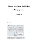
Apoptosis is induced by two main pathways (Fig. 80-5). The extrinsic
pathway of apoptosis is activated by cross-linking members of the tumor necrosis
factor (TNF) receptor superfamily, such as CD95 (Fas) and death receptors DR4
and DR5, by their receptors, Fas ligand or TRAIL (TNF-related apoptosis-
inducing ligand), respectively. This induces the association of FADD (Fas-
associated death domain) and procaspase-8 to death domain motifs of the
receptors. Caspase-8 is activated and then cleaves and activates effector caspases-
3 and -7, which then target cellular constituents (including caspase-activated
DNAse, cytoskeletal proteins, and a number of regulatory proteins), inducing the
morphologic appearance characteristic of apoptosis. The intrinsic pathway of
apoptosis is initiated by the release of cytochrome c and SMAC (second
mitochondrial activator of caspases) from the mitochondrial intermembrane space
in response to a variety of noxious stimuli, including DNA damage, loss of
adherence to the extracellular matrix (ECM), oncogene-induced proliferation, and
growth factor deprivation. Upon release into the cytoplasm, cytochrome c
associates with dATP, procaspase-9, and the adaptor protein APAF-1, leading to
the sequential activation of caspase-9 and effector caspases. SMAC binds to and
blocks the function of inhibitor of apoptosis proteins (IAPs), negative regulators of
caspase activation.
Figure 80-5
Therapeutic strategies to overcome aberrant survival pathways in
cancer cells. 1.
The extrinsic pathway of apoptosis can be selectively induced in
cancer cells by TRAIL (the ligand for
death receptors 4 and 5) or by agonistic
monoclonal antibodies. 2. Inhibition of antiapoptotic Bcl-
2 family members with
antisense oligonucleotides or inhibitors of the BH
3
-
binding pocket will promote
formation of Bak- or Bax-induced pores in the mitochondrial outer membrane. 3.
Epigenetic silencing of APAF-1, caspase-
8, and other proteins can be overcome
using demethylating agents and inhibitors of histone deacetylases. 4.
Inhibitor of
apoptosis proteins (IAP) blocks activation of caspases; small-molecule
inhibitors
of IAP function (mimicking SMAC action) should lower the threshold for
apoptosis. 5.
Signal transduction pathways originating with activation of receptor
tyrosine kinase receptors (RTKs) or cytokine receptors promote survival of cancer
cells by
a number of mechanisms. Inhibiting receptor function with monoclonal
antibodies, such as trastuzumab or cetuximab, or inhibiting kinase activity with
small-molecular inhibitors can block the pathway. 6.
The Akt kinase
phosphorylates many regulators of apop
tosis to promote cell survival; inhibitors of
Akt may render tumor cells more sensitive to apoptosis-
inducing signals;
however, the possibility of toxicity to normal cells may limit the therapeutic value
of these agents. 7 and 8. Activation of the transcri
ption factor NFκB (composed of
p65 and p50 subunits) occurs when its inhibitor, IκB, is phosphorylated by IκB-
kinase (IKK), with subsequent degradation of IκB by the proteasome. Inhibition
of IKK activity should selectively b
lock the activation of NFκB target genes,
many of which promote cell survival. Inhibitors of proteasome function are FDA
approved and may work in part by preventing destruction of IκB, thus blocking
NFκB nuclear localization. NFκB is unlikely to be the onl
y target for proteasome
inhibitors.