Chapter 098. Iron Deficiency and Other Hypoproliferative Anemias (Part 1) pps
Bạn đang xem bản rút gọn của tài liệu. Xem và tải ngay bản đầy đủ của tài liệu tại đây (13.84 KB, 5 trang )
Chapter 098. Iron Deficiency and Other
Hypoproliferative Anemias
(Part 1)
Harrison's Internal Medicine > Chapter 98. Iron Deficiency and Other
Hypoproliferative Anemias
Iron Deficiency and Other Hypoproliferative Anemias: Introduction
Anemias associated with normocytic and normochromic red cells and an
inappropriately low reticulocyte response (reticulocyte index <2.0–2.5) are
hypoproliferative anemias. This category includes early iron deficiency (before
hypochromic microcytic red cells develop), acute and chronic inflammation
(including many malignancies), renal disease, hypometabolic states such as
protein malnutrition and endocrine deficiencies, and anemias from marrow
damage. Marrow damage states are discussed in Chap. 102.
Hypoproliferative anemias are the most common anemias, and anemia
associated with acute and chronic inflammation is the most common of these. The
anemia of inflammation, like iron deficiency, is related in part to abnormal iron
metabolism. The anemias associated with renal disease, inflammation, cancer, and
hypometabolic states are characterized by an abnormal erythropoietin response to
the anemia.
Iron Metabolism
Iron is a critical element in the function of all cells, although the amount of
iron required by individual tissues varies during development. At the same time,
the body must protect itself from free iron, which is highly toxic in that it
participates in chemical reactions that generate free radicals such as singlet O
2
or
OH
–
. Consequently, elaborate mechanisms have evolved that allow iron to be
made available for physiologic functions while at the same time conserving this
element and handling it in such a way that toxicity is avoided.
The major role of iron in mammals is to carry O
2
as part of hemoglobin. O
2
is also bound by myoglobin in muscle. Iron is a critical element in iron-containing
enzymes, including the cytochrome system in mitochondria. Iron distribution in
the body is shown in Table 98-1. Without iron, cells lose their capacity for
electron transport and energy metabolism. In erythroid cells, hemoglobin synthesis
is impaired, resulting in anemia and reduced O
2
delivery to tissue.
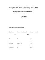
Table 98-1 Body Iron Distribution
Iron Content, mg
Adult Male, 80 kg
Adult Female, 60 kg
Hemoglobin 2500
1700
Myoglobin/e
nzymes
500
300
Transferrin iron 3 3
Iron stores 600–1000
0–300
The Iron Cycle in Humans
Figure 98-1 outlines the major pathways of internal iron exchange in
humans. Iron absorbed from the diet or released from stores circulates in the
plasma bound to transferrin, the iron transport protein. Transferrin is a bilobed
glycoprotein with two iron binding sites. Transferrin that carries iron exists in two
forms—monoferric (one iron atom) or diferric (two iron atoms). The turnover
(half-clearance time) of transferrin-bound iron is very rapid—typically 60–90 min.
Because almost all of the iron transported by transferrin is delivered to the
erythroid marrow, the clearance time of transferrin-bound iron from the circulation
is affected most by the plasma iron level and the erythroid marrow activity. When
erythropoiesis is markedly stimulated, the pool of erythroid cells requiring iron
increases and the clearance time of iron from the circulation decreases. The half-
clearance time of iron in the presence of iron deficiency is as short as 10–15 min.
With suppression of erythropoiesis, the plasma iron level typically increases and
the half-clearance time may be prolonged to several hours. Normally, the iron
bound to transferrin turns over 10–20 times per day. Assuming a normal plasma
iron level of 80–100 µg/dL, the amount of iron passing through the transferrin
pool is 20–24 mg/d.