Chapter 098. Iron Deficiency and Other Hypoproliferative Anemias (Part 2) pot
Bạn đang xem bản rút gọn của tài liệu. Xem và tải ngay bản đầy đủ của tài liệu tại đây (33.44 KB, 5 trang )
Chapter 098. Iron Deficiency and Other
Hypoproliferative Anemias
(Part 2)
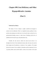
Figure 98-1
Internal iron exchange.
Normally about 80% of iron passing through the
plasma transferrin pool is
recycled from broken-
down red cells. Absorption of about 1 mg/d is required from
the diet in men, 1.4 mg/d in women to maintain homeostasis.
As long as transferrin saturation is maintained between 20–
60% and
erythropoiesis is no
t increased, iron stores are not required. However, in the event
of blood loss, dietary iron deficiency, or inadequate iron absorption, up to 40 mg/d
of iron can be mobilized from stores. RE, reticuloendothelial.
The iron-transferrin complex circulates in the plasma until it interacts with
specific transferrin receptors on the surface of marrow erythroid cells. Diferric
transferrin has the highest affinity for transferrin receptors; apotransferrin
(transferrin not carrying iron) has very little affinity. While transferrin receptors
are found on cells in many tissues within the body—and all cells at some time
during development will display transferrin receptors—the cell having the greatest
number of receptors (300,000 to 400,000/cell) is the developing erythroblast.
Once the iron-bearing transferrin interacts with its receptor, the complex is
internalized via clathrin-coated pits and transported to an acidic endosome, where
the iron is released at the low pH. The iron is then made available for heme
synthesis while the transferrin-receptor complex is recycled to the surface of the
cell, where the bulk of the transferrin is released back into circulation and the
transferrin receptor reanchors into the cell membrane. At this point a certain
amount of the transferrin receptor protein may be released into circulation and can
be measured as soluble transferrin receptor protein. Within the erythroid cell, iron
in excess of the amount needed for hemoglobin synthesis binds to a storage
protein, apoferritin, forming ferritin. This mechanism of iron exchange also takes
place in other cells of the body expressing transferrin receptors, especially liver
parenchymal cells where the iron can be incorporated into heme-containing
enzymes or stored. The iron incorporated into hemoglobin subsequently enters the
circulation as new red cells are released from the bone marrow. The iron is then
part of the red cell mass and will not become available for reutilization until the
red cell dies.
In a normal individual, the average red cell life span is 120 days. Thus, 0.8–
1.0% of red cells turn over each day. At the end of its life span, the red cell is
recognized as senescent by the cells of the reticuloendothelial (RE) system, and
the cell undergoes phagocytosis. Once within the RE cell, the hemoglobin from
the ingested red cell is broken down, the globin and other proteins are returned to
the amino acid pool, and the iron is shuttled back to the surface of the RE cell,
where it is presented to circulating transferrin. It is the efficient and highly
conserved recycling of iron from senescent red cells that supports steady state (and
even mildly accelerated) erythropoiesis.
Since each milliliter of red cells contains 1 mg of elemental iron, the
amount of iron needed to replace those red cells lost through senescence amounts
to 16–20 mg/d (assuming an adult with a red cell mass of 2 L). Any additional iron
required for daily red cell production comes from the diet. Normally, an adult
male will need to absorb at least 1 mg of elemental iron daily to meet needs, while
females in the childbearing years will need to absorb an average of 1.4 mg/d.
However, to achieve a maximum proliferative erythroid marrow response to
anemia, additional iron must be available. With markedly stimulated
erythropoiesis, demands for iron are increased by as much as six- to eightfold.
With extravascular hemolytic anemia, the rate of red cell destruction is increased,
but the iron recovered from the red cells is efficiently reutilized for hemoglobin
synthesis. In contrast, with intravascular hemolysis or blood loss anemia, the rate
of red cell production is limited by the amount of iron that can be mobilized from
stores. Typically, the rate of mobilization under these circumstances will not
support red cell production more than 2.5 times normal. If the delivery of iron to
the stimulated marrow is suboptimal, the marrow's proliferative response is
blunted, and hemoglobin synthesis is impaired. The result is a hypoproliferative
marrow accompanied by microcytic, hypochromic anemia.
While blood loss or hemolysis places a demand on the iron supply,
conditions associated with inflammation interfere with iron release from stores and
can result in a rapid decrease in the serum iron (see below).