Chapter 098. Iron Deficiency and Other Hypoproliferative Anemias (Part 4) docx
Bạn đang xem bản rút gọn của tài liệu. Xem và tải ngay bản đầy đủ của tài liệu tại đây (39 KB, 5 trang )
Chapter 098. Iron Deficiency and Other
Hypoproliferative Anemias
(Part 4)
Stages of Iron Deficiency
Iron-deficiency anemia is the condition in which there is anemia and clear
evidence of iron lack. The progression to iron deficiency can be divided into three
stages (Fig. 98-2). The first stage is negative iron balance, in which the demands
for (or losses of) iron exceed the body's ability to absorb iron from the diet. This
stage results from a number of physiologic mechanisms, including blood loss,
pregnancy (in which the demands for red cell production by the fetus outstrip the
mother's ability to provide iron), rapid growth spurts in the adolescent, or
inadequate dietary iron intake. Blood loss in excess of 10–20 mL of red cells per
day is greater than the amount of iron that the gut can absorb from a normal diet.
Under these circumstances the iron deficit must be made up by mobilization of
iron from RE storage sites. During this period, iron stores—reflected by the serum
ferritin level or the appearance of stainable iron on bone marrow aspirations—
decrease. As long as iron stores are present and can be mobilized, the serum iron,
total iron-binding capacity (TIBC), and red cell protoporphyrin levels remain
within normal limits. At this stage, red cell morphology and indices are normal.
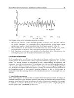
Figure 98-2
Laboratory studies in the evolution of iron deficiency.
Measurements of
marrow iron stores, serum ferritin, and total iron-
binding capacity (TIBC) are
sensitive to early iron-store depletion. Iron-
deficient erythropoiesis is recognized
from additional abnormalities in the serum iron (SI), percent transferrin saturation,
the pattern of marrow sideroblasts, and the red cell
protoporphyrin level. Patients
with iron-
deficiency anemia demonstrate all the same abnormalities plus
hypochromic microcytic anemia. (From Hillman and Finch, with permission.)
When iron stores become depleted, the serum iron begins to fall. Gradually,
the TIBC increases, as do red cell protoporphyrin levels. By definition, marrow
iron stores are absent when the serum ferritin level is <15 µg/L. As long as the
serum iron remains within the normal range, hemoglobin synthesis is unaffected
despite the dwindling iron stores. Once the transferrin saturation falls to 15–20%,
hemoglobin synthesis becomes impaired. This is a period of iron-deficient
erythropoiesis . Careful evaluation of the peripheral blood smear reveals the first
appearance of microcytic cells, and if the laboratory technology is available, one
finds hypochromic reticulocytes in circulation. Gradually, the hemoglobin and
hematocrit begin to fall, reflecting iron-deficiency anemia. The transferrin
saturation at this point is 10–15%.
When moderate anemia is present (hemoglobin 10–13 g/dL), the bone
marrow remains hypoproliferative. With more severe anemia (hemoglobin 7–8
g/dL), hypochromia and microcytosis become more prominent, target cells and
misshapen red cells (poikilocytes) appear on the blood smear as cigar- or pencil-
shaped forms, and the erythroid marrow becomes increasingly ineffective.
Consequently, with severe prolonged iron-deficiency anemia, erythroid
hyperplasia of the marrow develops, rather than hypoproliferation.
Causes of Iron Deficiency
Conditions that increase demand for iron, increase iron loss, or decrease
iron intake or absorption can produce iron deficiency (Table 98-2).
Table 98-2 Causes of Iron Deficiency
Increased demand for iron and/or hematopoiesis
rapid growth in infancy or adolescence
pregnancy
erythropoietin therapy
Increased iron loss
chronic blood loss
menses
acute blood loss
blood donation
phlebotomy as treatment for polycythemia vera
Decreased iron intake or absorption
inadequate diet
malabsorption from disease (sprue, Crohn's disease)
malabsorption from surgery (post-gastrectomy)
acute or chronic inflammation