Hepatocellular Carcinoma: Targeted Therapy and Multidisciplinary P3 pot
Bạn đang xem bản rút gọn của tài liệu. Xem và tải ngay bản đầy đủ của tài liệu tại đây (187.07 KB, 10 trang )
1 Epidemiology and Pathogenesis of Hepatocellular Carcinoma 5
is responsible for the first-pass metabolism of ethanol in the stomach, is signifi-
cantly lower in women than in men, which implies that large amounts of alcohol
will be metabolized by hepatic alcohol dehydrogenase [41, 42]. It is also possi-
ble that genetic variations in carcinogen metabolism, inflammatory response, DNA
repair, and cell cycle regulation play a role in determining individual susceptibil-
ity to alcohol carcinogenesis, which may partially explain variations in HCC risk
by sex.
Seroepidemiological studies have demonstrated a high frequency of anti-HCV
and HCV RNA in alcohol users and those among them who develop alcoholic
liver diseases [43]. Despite this close relationship, there is little understanding of
how HCV and alcohol may interact in the development of HCC. In most stud-
ies, anti-HCV in alcoholics was found to be closely associated with the presence
of HCV RNA in serum, a marker of HCV replication [44], which may suggest
that immunosuppression associated with chronic alcohol consumption may enhance
HCV replication.
Smoking
Cigarette smoking is significantly associated with HCC development [45]. A meta-
analysis on the association between smoking and liver cancer [46] concluded
an overall OR of 1.6 (95% CI, 1.3–1.9) for current smokers and 1.5 (95% CI,
1.1–2.1) for former smokers. The recently released report by IARC had confirmed
that smoking is considered a risk factor for liver cancer [47]. Despite evidence
sufficient to judge the positive association between active smoking and liver can-
cer, smoking–HCC relationship in men and women separately has not been widely
addressed. A US study suggested that smoking is more likely associated with HCC
in men and not women [38]. Moreover, synergistic interactions between cigarette
smoking and alcohol consumption, HBV, or HCV infection were reported by dif-
ferent studies [38, 48, 49]. Despite the significant association between cigarette
smoking and the risk of HCC, passive smoking exposure is not associated with
HCC development [38]. The use of chewing tobacco and snuff was also not related
to HCC development in general or in nonsmokers [38].
The exact mechanism of tobacco hepatocarcinogenesis is unknown; however,
of approximately 4,000 components identified in tobacco smoke, at least 55 are
known carcinogens. The major chemical carcinogens include polycyclic aromatic
hydrocarbons, such as benzo[a]pyrene; aromatic amines, such as 4-aminobiphenyl;
and nitrosamines, such as 4-(methylnitrosamine)-1-(3-pyridyl)-1-butanone. A case–
control study demonstrated that 4-aminobiphenyl DNA adducts contained in
tobacco smoke is a liver carcinogen [50]. In addition, tobacco smoke contains
volatile compounds (e.g., benzene), radioactive elements (e.g., polonium-210), and
free radicals that may also play a role in hepatocarcinogenicity [51, 52]. Substantial
evidence supports the notion that oxidative stress has been linked to tobacco use.
In vitro studies demonstrated that the gas phase of cigarette smoke caused lipid per-
oxidation of human plasma, which was preventable by the addition of ascorbic acid
6 M.M. Hassan and A.O. Kaseb
[53, 54]. This may support t he smoking synergism with alcohol consumption and
chronic viral hepatitis on HCC development.
Aflatoxin Exposure
Aflatoxins (AFs) are toxic secondary fungal metabolites (mycotoxins) produced by
Aspergillus flavus and A. parasiticus. There are four AF compounds: B
1
,B
2
,G
1
,
and G
2
[55]. The most common and most toxic AF is AFB
1
, and the most important
target organ is the liver, where the toxicity can lead to liver necrosis and bile duct
proliferation [55].
In order for AFB
1
to exert its toxic effects, it must be converted to its highly
reactive 8,9-epoxide metabolite by the action of the mixed function monooxy-
genase enzyme systems in the liver (CYP450 dependent) [56, 57]. Therefore,
the development of AF biomarkers is based on detection of the AFB
1
active
metabolites, which can covalently interact with cellular molecules, including DNA,
RNA, and protein. Epidemiologic research has documented a significant risk for
HCC development among individuals who consumed highly AF-contaminated diets
[58, 59].
Hormonal Intake
The use of oral contraceptive pills and risk for HCC development is inconclusive.
A recent review of 12 case–control studies that included 739 HCC cases and 5,223
controls [60] yielded an overall adjusted OR of 1.6 (0.9–2.5); however, six stud-
ies, included in the analysis, showed a significant increase in HCC risk with longer
duration of exposure of oral contraceptives (>5 years). The observed association
between liver cancer and oral contraceptive in animals is believed to be related to
the proliferative effect of estrogen on hepatocytes where estrogen receptors exist
and are highly expressed in HCC [61]. On the other hand, a protective effect of
hormonal replacement therapy on liver cancer was determined by some studies
[62, 63].
Occupational Exposures
Meta-analyses of epidemiological studies indicated a slightly increased risk of
HCC with high level of occupation exposure to vinyl chloride [64]. However,
such risk elevation can be a function of disease misclassification bias, since HCC
was not analyzed separately from other liver tumors. Reviewing the epidemiolog-
ical and experimental studies for the association between vinyl chloride and HCC
indicated no evidence of biological plausibility for the risk of vinyl chloride on
HCC [65].
1 Epidemiology and Pathogenesis of Hepatocellular Carcinoma 7
Chronic Medical Conditions
Diabetes Mellitus
Because the liver plays a crucial role in glucose metabolism, it is not surprising
that diabetes mellitus is an epiphenomenon of many chronic liver diseases such
as chronic hepatitis, fatty liver, liver failure, and cirrhosis. A recent systematic
review of several cohort and case–control studies concluded that diabetes mellitus
is significantly associated with HCC [66].
There are several lines of evidence suggesting that diabetes is in fact an inde-
pendent risk factor for HCC development. This evidence includes (1) results from
review and meta-analysis reports concluding that diabetes is a risk factor of HCC
[66–69]; (2) findings that the positive association between diabetes and HCC is
independent from underlying cirrhosis and chronic liver diseases [70, 71]; (3) find-
ings that the association is positively correlated with disease duration [72–74];
(4) demonstration of the synergistic interaction between diabetes and other HCC
risk factors [72, 75, 76]; (5) findings of HCC recurrence after liver resection and
transplantation among patients with diabetes [77, 78]; (6) suggestion of a biological
plausibility that underlies the association between diabetes and HCC [67, 68, 79];
and (7) t he observation of risk of HCC development among patients with type 1
diabetes mellitus [76].
The key mechanism for liver cell damage induced by type 2 diabetes mellitus
involves insulin resistance and hyperinsulinemia [69, 80]. HCC development related
to hyperinsulinemia can be mediated through inflammation, cellular proliferation,
inhibition of apoptosis, and mutation of tumor suppressor genes [69]. Increased
insulin levels lead to reduced liver synthesis and blood levels of insulin growth fac-
tor binding protein-1 (IGFBP-1), which may contribute to increased bioavailability
of insulin-like growth factor-1 (IGF-1), the promotion of cellular proliferation, and
the inhibition of apoptosis [81]. Insulin also binds to the insulin receptor and acti-
vates its intrinsic tyrosine kinase, leading to phosphorylation of insulin receptor
substrate-1 (IRS-1) [82]. HCC tumor cells have been shown to overexpress both
IGF-1 and IRS-1 [83]. Overexpression of IRS-1 has been associated with the pre-
vention of apoptosis mediated by transforming growth factor-β [84]. In addition,
insulin is associated with lipid peroxidation and increased oxidative stress and the
generation of ROS, which may contribute to DNA mutation [85].
Obesity
It is well established that obesity is significantly associated with a wide spectrum
of hepatobiliary diseases, including fatty liver diseases, steatosis, and cryptogenic
cirrhosis [68, 86]. Once steatosis has developed, cellular adaptations may occur to
allow the cell to survive in the new stressful environment and enhance vulnerability
to a second hit, or genetic and environmental factors, leading to necroinflammatory
changes (non-alcoholic steatohepatitis) or non-alcoholic steatohepatitis (NASH)
where different mediators are involved in such pathogenesis [87]. However, there
8 M.M. Hassan and A.O. Kaseb
is little information regarding the association between obesity and HCC. A recent
meta-analysis 11 cohort studies reported a summary relative risks (95% CI) of 1.17
(1.02–1.34) and 1.89 (1.51–2.36) for overweight and obese individuals, respectively
[88]. Nevertheless, the study did not separate HCC from other primary tumors of
the liver nor control for the confounding effect of HCV, HBV, diabetes, and heavy
alcohol consumption on HCC development.
Lipid peroxidation and free oxygen radicals may play a central role in NASH dur-
ing which the initiation stage of HCC mechanism takes place. Proliferation of oval
cells (the cells of origin for several types of liver cancer) and mutation of P53 tumor
suppressor gene can also be potentiated. It is then suggested that the second stage
(promotion) takes place as a result of balance in apoptotic and antiapoptotic factors;
disturbance in growth factors such as TNF and TGF may facilitate oval cell pro-
liferation [89]. Progression to HCC (stage 3) is suggested to be mediated through
cyclooxygenase-2 (COX-2) gene expression by peroxisome proliferator-activated
receptor (PPAR-β) nuclear receptors implicated in fatty acid oxidation, cell differ-
entiation, inflammation, cell motility, and cell growth [90, 91]. It was suggested that
PPAR-β promotes human HCC cell growth through induction of COX-2 expres-
sion and prostaglandins (PGE
2
) synthesis. The produced PGE
2
phosphorylates and
activates cytosolic phospholipase A
2
α (cPLA
2
α), releasing arachidonic acid for fur-
ther PPAR-β activation and PGE
2
synthesis via COX-2. This positive-forward loop
between PPAR-β and PG pathway likely plays role in the regulation of human cell
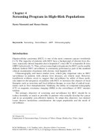
growth and HCC development (Fig. 1.2).
Oxidative stress
Initiation
Lipid peroxidation
Cell proliferation
P53 mutation
Promotion
Antiapoptotic factors
Cell proliferation
TNF-
TGF-
Progression
PPAR
Arachidonic acid
Prostaglandin
COX-2
Hepatocarcinogenesis Pathway
Environmental Exposures
HCV
HBV
Alcohol
Smoking
Obesity
Fig. 1.2 Steps in hepatocarcinogenesis, modified from Xu et al. [90] and Bensinger and
Tontonoz [91]
On the other hand, the association between obesity and HCC is hammered by
the following obstacles: (1) categorizing HCC among patients with primary liver
cancer, (2) inappropriate adjustment for the confounding effect of HCC risk fac-
tors specially type 2 diabetes mellitus, and (3) misclassification of obesity definition
among patients with HCC. Relying on baseline body weight to estimate body mass
index (BMI) at the time of HCC diagnosis could have led to patient misclassification
because most HCC is associated with ascites, which can affect the BMI calcula-
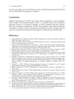
tion and definition of obesity. Results from an ongoing case–control s tudy indicated
1 Epidemiology and Pathogenesis of Hepatocellular Carcinoma 9
BMI
Age prior to diagnosis or enrollment
10
20 30 40 50 60
20
21
22
23
24
25
26
27
28
29
30
HCC Cases
Controls
P< 0.001
Fig. 1.3 Difference in BMI means between cases and controls at different age periods prior to
HCC diagnosis or control enrolment: US case–control study (Hassan unpublished data)
means of BMIs at different age periods prior to HCC development were significantly
larger for HCC patients as compared to healthy controls (Hassan, unpublished data)
(Fig. 1.3).
Thyroid Diseases
Thyroid hormones play an essential role in lipid mobilization, lipid degrada-
tion, and fatty acid oxidation [92]. Patients with hypothyroidism may experience
15–30% weight gain [93] and insulin resistance [94, 95], which are significant fac-
tors of NASH. A recent study [96] reported that the prevalence of hypothyroidism in
patients with NASH was significantly higher than in controls (15% vs 7.2%, respec-
tively; p = 0.001). Such findings were later supported by Reddy and colleagues
[97] from Mayo Clinic who assessed the association between hypothyroidism and
HCC among 54 HCC patients of unknown etiology and 116 HCC patients related
to HCV and alcohol. The study reported OR of 6.8 (95% CI, 1.1–42.1) for HCC
development after adjusting for several confounding factors. Our recently published
case–control study reported positive association between hypothyroidism and HCC
among women [98].
Whether and why hypothyroidism causes HCC is not clear. However, the asso-
ciation between hypothyroidism and NASH can be explained by the underlying
hyperlipidemia, decreased fatty acid oxidation, insulin resistance, and lipid per-
oxidation in patients with hypothyroidism. All of these conditions may enhance
the susceptibility to chronic inflammation, DNA damage, and HCC develop-
ment. Moreover, concurrent thyroid dysfunction among diabetic patients may
exacerbate the coexisting diabetes-induced dyslipidemia and may explain our
observation of HCC risk modification among patients with hypothyroidism and
diabetes [98].
10 M.M. Hassan and A.O. Kaseb
Obesity and hyperinsulinemia may increase the level of insulin-like growth
factor-1, which in turn may reduce hepatic synthesis and blood concentration of
sex hormone-binding globulin (SHBG) [99, 100], a glycoprotein produced in the
liver with high-binding affinity for testosterone and lower affinity for estradiol.
Independent of obesity, there is sufficient evidence that thyroid hormones have a
positive effect on hepatic SHBG synthesis and that patients with hypothyroidism
may experience a lower level of SHBG [101]. Thus, a decreased level of SHBG may
lead to increased plasma testosterone and estradiol, both of which may promote cel-
lular proliferation and inhibit apoptosis. Elevated levels of serum testosterone and
testosterone to estradiol ratio have been proposed to be predictive of HCC develop-
ment in Japanese men with cirrhosis [102]. Nevertheless, the fact that the association
between hypothyroidism and HCC continued to be significant after adjustment for
prior history of obesity suggested that other mechanisms of hepatocarcinogenesis
were involved, especially among women.
Cholelithiasis (Gallbladder Stones)
The prevalence of gallstones in patients with cirrhosis is significantly higher than
in the general population [103, 104]. This is partially attributed to the metabolic
changes such as increased unconjugated bilirubin in bile secondary to hyper-
splenism, decreased cholesterol secretion, and decreased in apolipoprotein (apo)
A-1 and AoA-II sections [105, 106]. A recent study reported significant associa-
tion between gallbladder stones and HCC; the estimated OR (95% CI) was 14.75
(13.14–16.56) [107]. Nevertheless, the association between gallstones and HCC
is difficult to assess from epidemiological studies due to recall bias among HCC
patients and due to the subsequent cholecystectomy procedure with liver resection
in patients with HCC. Therefore, it is not clear whether cholelithiasis is a risk fac-
tor for HCC or a consequence of the underlying chronic liver diseases in patients
with HCC.
Dietary Factors
Most of the epidemiological evidence on diet and liver cancer is based on case–
control studies and retrospective analysis. This type of assessment is subjective
to recall bias due to the fact that patients with chronic liver diseases or cirrho-
sis may change their diet after being diagnosed with liver diseases. An exam-
ple of the association between diet and HCC is HCC risk reduction (25–75%)
among coffee drinkers who consume two to four cups of coffee per day as com-
pared to non-coffee drinkers [ 108–110]. HCC risk reduction was also observed
for the intake of eggs, milk, yogurt, vegetables, white meat, and fruits [111].
Moreover, the intake of dietary antioxidants, especially selenium and retinoic acid,
showed a protective effect for HCC development in HBV carriers and cigarette
smokers [112].
1 Epidemiology and Pathogenesis of Hepatocellular Carcinoma 11
Genetic Risk Factors
Familial Aggregation
Familial aggregation of liver cancer has been reported. However, most of these stud-
ies were conducted among Asians, particularly in China [113–117]. Given the high
prevalence of chronic infection with HBV and that vertical transmission of HBV
is the major source for viral transmission among Asians, the reported association
between a family history of liver cancer and HCC could be explained by clustering
of HBV infection among members of the same family [118]. To avoid this obsta-
cle, Yu et al. [117] matched 553 patients with HCC and 4,684 controls according
to HBV infection status. They reported an OR of 2.4 (95% CI, 1.5–3.9) for HCC
development in subjects with HBV and a family history of HCC as compared to
subjects with HBV but no family history of HCC. A later study by the same inves-
tigators showed that familial segregation of HCC in HBsAg carriers is associated
with familial clustering of liver cirrhosis [119].
A segregation analysis of Chinese HCC patients suggested that a Mendelian auto-
somal recessive major gene might also play role in HCC etiology [114]. In addition,
first-degree family history of liver cancer in American and European populations
is likely to be associated with HCC development independent of chronic infection
with HBV and HCV [120]. Synergism between HBV/HCV and a family history
of liver cancer was also noted by Hassan et al. [120] among Italian and American
individuals.
Inherited Diseases
Hereditary Hemochromatosis
Hereditary hemochromatosis (HHC) is an autosomal recessive genetic disorder of
iron metabolism t hat causes excessive intestinal absorption of dietary iron and
deposition of iron in organs including the liver [121]. Recently, a major histocompat-
ibility complex class I gene named HLA-H or HFE was cloned. Two mutations were
described: Cys282Tyr (C282Y) and His63Asp (H63D)[122]. The C282Y mutation is
more frequent in HHC [123]. There is growing evidence that even mildly increased
amounts of iron in the liver can be damaging, especially when combined with other
hepatotoxic factors such as alcohol consumption and chronic viral hepatitis. Iron
enhances the pathogenicity of microorganisms, adversely affects the function of
macrophages and lymphocytes, and enhances fibrogenic pathways [124, 125], all
of which may increase hepatic injury caused by iron alone or by iron and other
factors such as chronic HCV infection.
Indeed, a synergistic relationship between HCV and iron overload from
hemochromatosis has been suggested [126]. In a study by Hayashi et al., iron deple-
tion improved liver function tests in HCV- infected individuals [127]. In a study by
Mazzella and colleague response of chronic HCV to interferon was shown to be
related to hepatic iron concentration [128].
12 M.M. Hassan and A.O. Kaseb
Possible factors contributing to the actions of iron in chronic viral hepatitis
include enhancement of oxidative stress and lipid peroxidation, exacerbation of
immune-mediated tissue inflammation, enhancement of the rate of viral replication,
enhancement of the rate of viral mutation, possible impairment of cellular immunity
or humoral immunity, and possible impairment of T-lymphocyte proliferation and
maturation [129].
α
1
Antitrypsin Deficiency
α
1
antitrypsin deficiency (AATD) is an autosomal dominant genetic disorder char-
acterized by a deficiency in a major serum protease inhibitor (Pi) [130]. AATD is
caused by a mutation in the 12.2 kb α
1
antitrypsin gene on chromosome 14 [130].
Over 75 different Pi alleles have been identified, most of which not associated with
disease [131]. A relationship exists between Pi phenotypes and serum concentra-
tions of α
1
antitrypsin. Thus, the MM phenotype (normal) is associated with a serum
concentration of 100%, MZ 60%, SS 60%, FZ 60%, M 50%, PS 40%, SZ 42.5%,
ZZ 15%, and Z 0 to 10%. The most common deficiency variant, PiZ, in its homozy-
gote state is often associated with liver cirrhosis and liver cancer [132]. The role
of the heterozygous PiZ state in the development of primary liver cancer is con-
troversial [133–135]. However, there is increasing evidence suggesting that chronic
liver disease develops only when another factor such as HCV infection is present
and acts as a promoter for the liver damage process. α
1
antitrypsin is an acute-phase
reactant whose major role is to inhibit the actions of neutrophil elastase, proteases,
and cathepsin G [136]. Any condition triggering the acute-phase response would be
expected to stimulate the production of α
1
antitrypsin by the liver.
Therefore, it is suggested that chronic HCV infection could constantly stimulate
the hepatocytes to produce the mutant α
1
antitrypsin, leading to more liver dam-
age [137]. Other less frequent inherited disorders such as glycogen storage disorder
disease type I (von Gierke’s disease) [138], Porphyria Cutanea Tarda [139], and
Wilson’s disease [140] have been found to be complicated to HCC. However, the
interactions between these diseases and other established risk factors such as HCV
or HBV have not been studied.
References
1. Parkin DM, Bray F, Ferlay J, Pisani P (2005) Global cancer statistics, 2002. CA Cancer J
Clin 55:74–108
2. Parkin DM, Bray F, Ferlay J, Pisani P (2001) Estimating the world cancer burden: Globocan
2000. Int J Cancer 94:153–156
3. Thomas MB, Zhu AX (2005) Hepatocellular carcinoma: the need for progress. J Clin Oncol
23:2892–2899
4. Altekruse SF, McGlynn KA, Reichman ME (2009) Hepatocellular carcinoma incidence,
mortality, and survival trends in the United States from 1975 to 2005. J Clin Oncol 27:
1485–1491
5. McGlynn KA, London WT (2005) Epidemiology and natural history of hepatocellular
carcinoma. Best Pract Res Clin Gastroenterol 19:3–23
1 Epidemiology and Pathogenesis of Hepatocellular Carcinoma 13
6. El-Serag HB, Rudolph KL (2007) Hepatocellular carcinoma: epidemiology and molecular
carcinogenesis. Gastroenterology 132:2557–2576
7. International Agency for Research on Cancer (IARC) (1994) Monographs on the evaluation
of carcinogenic risks to humans. Hepatitis Viruses 59:182–221
8. Kew MC, Welschinger R, Viana R (2008) Occult hepatitis B virus infection in
Southern African blacks with hepatocellular carcinoma. J Gastroenterol Hepatol 23:
1426–1430
9. Zanetti AR, Van DP, Shouval D (2008) The global impact of vaccination against hepatitis B:
a historical overview. Vaccine 26:6266–6273
10. Brechot C (1987) Hepatitis B virus (HBV) and hepatocellular carcinoma. HBV DNA status
and its implications. J Hepatol 4:269–279
11. Brechot C, Pourcel C, Louise A, Rain B, Tiollais P (1980) Presence of integrated hepatitis
B virus DNA sequences in cellular DNA of human hepatocellular carcinoma. Nature 286:
533–535
12. Rossner MT (1992) Review: hepatitis B virus X-gene product: a promiscuous transcriptional
activator. J Med Virol 36:101–117
13. Simonetti RG, Camma C, Fiorello F, Cottone M, Rapicetta M, Marino L et al (1992)
Hepatitis C virus infection as a risk factor for hepatocellular carcinoma in patients with
cirrhosis. A case-control study. Ann Intern Med 116:97–102
14. Okamoto H, Tsuda F, Sakugawa H, Sastrosoewignjo RI, Imai M, Miyakawa Y et al (1988)
Typing hepatitis B virus by homology in nucleotide sequence: comparison of surface antigen
subtypes. J Gen Virol 69(Pt 10):2575–2583
15. Kidd-Ljunggren K, Miyakawa Y, Kidd AH (2002) Genetic variability in hepatitis B viruses.
J Gen Virol 83:1267–1280
16. Kramvis A, Kew M, Francois G (2005) Hepatitis B virus genotypes. Vaccine 23:2409–2423
17. Alexopoulou A, Dourakis SP (2005) Genetic heterogeneity of hepatitis viruses and its
clinical significance. Curr Drug Targets Inflamm Allergy 4:47–55
18. Choo QL, Richman KH, Han JH, Berger K, Lee C, Dong C et al (1991) Genetic organization
and diversity of the hepatitis C virus. Proc Natl Acad Sci USA 88:2451–2455
19. Alter MJ, Kruszon-Moran D, Nainan OV, McQuillan GM, Gao F, Moyer LA et al (1999)
The prevalence of hepatitis C virus infection in the United States, 1988 through 1994. N
Engl J Med 341:556–562
20. Yoshizawa H (2002) Hepatocellular carcinoma associated with hepatitis C virus infection in
Japan: projection to other countries in the foreseeable future. Oncology 62(Suppl 1):8–17
21. Donato F, Boffetta P, Puoti M (1998) A meta-analysis of epidemiological studies on the
combined effect of hepatitis B and C virus infections in causing hepatocellular carcinoma.
Int J Cancer 75:347–354
22. Freeman AJ, Dore GJ, Law MG, Thorpe M, Von OJ, Lloyd AR et al (2001)
Estimating progression to cirrhosis in chronic hepatitis C virus infection. Hepatology 34:
809–816
23. Parola M, Robino G (2001) Oxidative stress-related molecules and liver fibrosis. J Hepatol
35:297–306
24. Cerutti PA (1994) Oxy-radicals and cancer. Lancet 344:862–863
25. Wiseman H, Halliwell B (1996) Damage to DNA by reactive oxygen and nitrogen species:
role in inflammatory disease and progression to cancer. Biochem J 313(Pt 1):17–29
26. Marra F (1999) Hepatic stellate cells and the regulation of liver inflammation. J Hepatol
31:1120–1130
27. Poli G (2000) Pathogenesis of liver fibrosis: role of oxidative stress. Mol Aspects Med
21:49–98
28. Simmonds P (1995) Variability of hepatitis C virus. Hepatology 21:570–583
29. Dusheiko G, Schmilovitz-Weiss H, Brown D, McOmish F, Yap PL, Sherlock S et al (1994)
Hepatitis C virus genotypes: an investigation of type-specific differences in geographic
origin and disease. Hepatology 19:13–18
14 M.M. Hassan and A.O. Kaseb
30. Nousbaum JB, Pol S, Nalpas B, Landais P, Berthelot P, Brechot C (1995) Hepatitis C virus
type 1b (II) infection in France and Italy. Collaborative Study Group. Ann Intern Med
122:161–168
31. Silini E, Bono F, Cividini A, Cerino A, Bruno S, Rossi S et al (1995) Differential distribution
of hepatitis C virus genotypes in patients with and without liver function abnormalities.
Hepatology 21:285–290
32. De Mitri MS, Poussin K, Baccarini P, Pontisso P, D’Errico A, Simon N et al (1995) HCV-
associated liver cancer without cirrhosis. Lancet 345:413–415
33. International Agency for Research on Cancer (IARC) (1988) Monographs on the evaluation
of carcinogenic risks to humans. Alcohol Drinking 44(44):207–215
34. Batey RG, Burns T, Benson RJ, Byth K (1992) Alcohol consumption and the risk of
cirrhosis. Med J Aust 156:413–416
35. Brechot C, Nalpas B, Feitelson MA (1996) Interactions between alcohol and hepatitis
viruses in the liver. Clin Lab Med 16:273–287
36. Morgan TR, Mandayam S, Jamal MM (2004) Alcohol and hepatocellular carcinoma.
Gastroenterology 127:S87–S96
37. Stewart S, Jones D, Day CP (2001) Alcoholic liver disease: new insights into mechanisms
and preventative strategies. Trends Mol Med 7:408–413
38. Hassan MM, Spitz MR, Thomas MB, El-Deeb AS, Glover KY, Nguyen NT et al (2008)
Effect of different types of smoking and synergism with hepatitis C virus on risk of
hepatocellular carcinoma in American men and women: case-control study. Int J Cancer
123:1883–1891
39. Mancinelli R, Binetti R, Ceccanti M (2007) Woman, alcohol and environment: emerging
risks for health. Neurosci Biobehav Rev 31:246–253
40. Ely M, Hardy R, Longford NT, Wadsworth ME (1999) Gender differences in the relationship
between alcohol consumption and drink problems are largely accounted for by body water.
Alcohol Alcohol 34:894–902
41. Baraona E, Abittan CS, Dohmen K, Moretti M, Pozzato G, Chayes ZW et al (2001) Gender
differences in pharmacokinetics of alcohol. Alcohol Clin Exp Res 25:502–507
42. Frezza M, di PC, Pozzato G, Terpin M, Baraona E, Lieber CS (1990) High blood alcohol
levels in women. The role of decreased gastric alcohol dehydrogenase activity and first-pass
metabolism. N Engl J Med 322:95–99
43. Oshita M, Hayashi N, Kasahara A, Hagiwara H, Mita E, Naito M et al (1994) Increased
serum hepatitis C virus RNA levels among alcoholic patients with chronic hepatitis C.
Hepatology 20:1115–1120
44. Paronetto F (1993) Immunologic reactions in alcoholic liver disease. Semin Liver Dis
13:183–195
45. International Agency for Research on Cancer (2004) (IARC) Monographs on the evaluation
of carcinogenic risks to humans. Tobacco Smoke and Involuntary Smoking 83:161–176
46. Gandini S, Botteri E, Iodice S, Boniol M, Lowenfels AB, Maisonneuve P et al (2008)
Tobacco smoking and cancer: a meta-analysis. Int J Cancer 122:155–164
47. International Agency for Research on Cancer (IARC) (2004) Monographs on the eval-
uation of carcinogenic risks to humans. Tobacco Smoke and Involuntary Smoking 83:
161–176.
48. Franceschi S, Montella M, Polesel J, La VC, Crispo A, Dal ML et al (2006) Hepatitis viruses,
alcohol, and tobacco in the etiology of hepatocellular carcinoma in Italy. Cancer Epidemiol
Biomarkers Prev 15:683–689
49. Mori M, Hara M, Wada I, Hara T, Yamamoto K, Honda M et al (2000) Prospective study of
hepatitis B and C viral infections, cigarette smoking, alcohol consumption, and other factors
associated with hepatocellular carcinoma risk in Japan. Am J Epidemiol 151:131–139
50. Wang LY, Chen CJ, Zhang YJ, Tsai WY, Lee PH, Feitelson MA et al (1998) 4-
Aminobiphenyl DNA damage in liver tissue of hepatocellular carcinoma patients and
controls. Am J Epidemiol 147:315–323